Embed Size (px)
Citation preview
• Five special senses:
• Olfaction
• Taste
• Visual system
• Hearing and balance

• Epithelium of nasal cavity is called olfactory epithelium, located in the roof of nasal cavity
• Olfactory receptor cells present in olfactory epithelium are bipolar neurons with enlarged ends (olfactory vesicles) and contains cilia called olfactory hairs
• Airborne molecules enter nasal cavity and stimulate cilia of olfactory receptors bathed by a layer of mucus
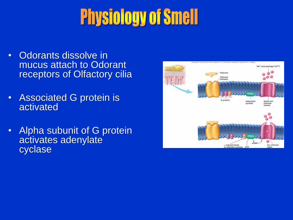
• Odorants dissolve in mucus attach to Odorant receptors of Olfactory cilia
• Associated G protein is activated
• Alpha subunit of G protein activates adenylate cyclase

• And convert ATP to cAMP
• cAMP act as second
messenger opens Na+ and
Ca2+ channels
• Causes depolarization of cilia
• And initiate action potentials in
olfactory neurons

• One receptor may respond to more than one type of odor
• Olfactory epithelium is replaced as it wears down
• Olfactory neurons are replaced by basal cells every two months
• Unique: most neurons are permanent cells (aren't replaced if they die)

• Axons of olfactory neurons (bipolar) in the olfactory epithelium pass through cribiform plate to olfactory bulbs
• Then project through the olfactory tract to olfactory cortex of frontal lobe
• 3 regions in frontal lobe affect conscious perception of smell & interact with limbic system
– Lateral olfactory area: Involved in conscious perception of smell
– Medial olfactory area: Responsible for visceral & emotional reactions to odors
– Intermediate olfactory area: Receives
input from L & M olfactory area
– Axons from L olfactory area project to olfactory tract to olfactory bulb
– In olfactory bulb Sensory information is modulated
•
•

• 10,000 or more taste buds are found on the tongue
• Taste buds are found in papillae of the tongue mucosa
• Types of papillae
– Vallate: Largest, least numerous
– 8-12 in V along border between anterior and posterior parts of the tongue
– Have taste buds
– Fungiform: Mushroom-shaped
– Scattered irregularly over the superior surface of tongue
– Have taste buds

– Foliate: Leaf-shaped
– In folds on the sides of the tongue
– Contain most sensitive taste buds
– Decrease in number with age
– Filiform: Filament-shaped
– Most numerous
– No taste buds

• Taste bud consists of three major cell types
– Supporting cells – insulate the receptor
– Basal cells – dynamic stem cells
– Gustatory cells – taste cells
– Taste cells have microvilli
– (gustatory hairs) extending into taste pores
– Replaced about every 10 days

• There are five basic taste sensations
– Sweet – sugars, saccharin, alcohol, and
some amino acids
– Salt – metal ions
– Sour – hydrogen ions
– Bitter – alkaloids such as quinine and
nicotine
– Umami – (savory) elicited by the amino acid
glutamate

• Activation of Taste Receptors:
• In order to be tasted, a chemical:
– Must be dissolved in saliva
– Must contact gustatory hairs of gustatory cells
– Which contain synaptic vesicles
• Binding of the food chemical:
– To the receptors in the taste cell membrane depolarizes,
releases neurotransmitter from synaptic vesicles
– Binding of neurotransmitter to associated sensory dendrites
initiates an action potential

• Mechanism of Taste
Transduction:
• The stimulus energy of taste is
converted into a nerve impulse
by:
– Na+ influx in salty tastes
– H+ in sour tastes (by directly
entering the cell, by opening
cation channels, or by
blockade of K+ channels)
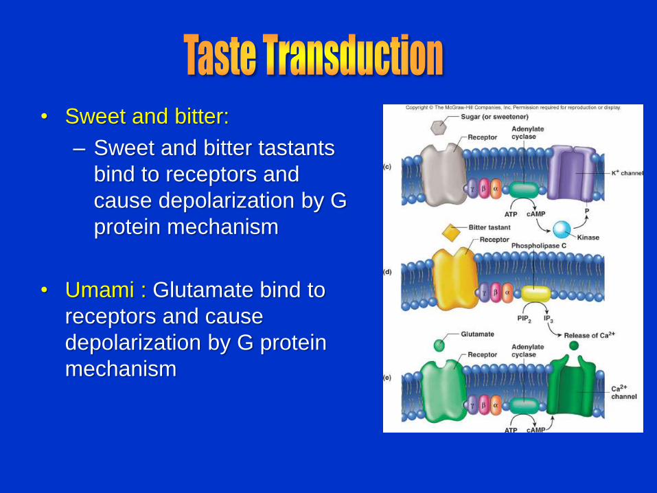
• Sweet and bitter:
– Sweet and bitter tastants
bind to receptors and
cause depolarization by G
protein mechanism
• Umami : Glutamate bind to
receptors and cause
depolarization by G protein
mechanism

• Taste is 80% smell
• Thermoreceptors, mechanoreceptors,
nociceptors also influence tastes
• Temperature and texture enhance or
detract from taste

• Cranial Nerves VII and IX
carry impulses from taste buds
to the nucleus of tractus
solitarius of the medulla
• Where decussation takes
place and these impulses then
travel to the thalamus
• Then thalamus to taste area of
cortex ( inferior end of
postcentral gyrus)
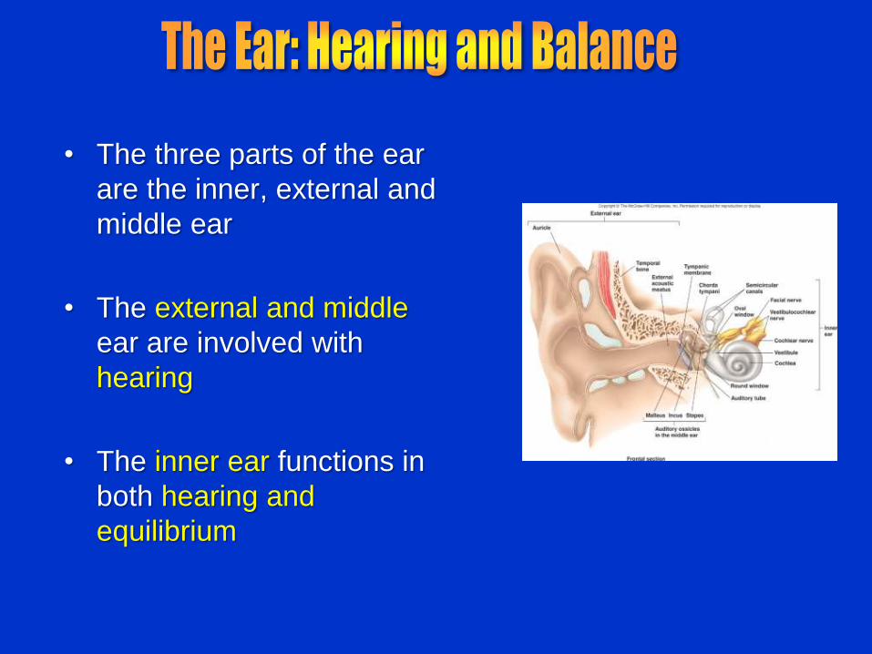
• The three parts of the ear
are the inner, external and
middle ear
• The external and middle
ear are involved with
hearing
• The inner ear functions in
both hearing and
equilibrium

• The auricle (pinna) is composed of:
– The helix (rim)
– The lobule (earlobe)
• External auditory canal
– Short, curved tube lined with hairs & ceruminous glands
– Produce cerumen wax
– Prevent foreign objects
• Tympanic membrane (eardrum)
– Thin connective tissue membrane that vibrates in response to sound
– Transfers sound energy to the middle ear ossicles
– Boundary between external and middle ears
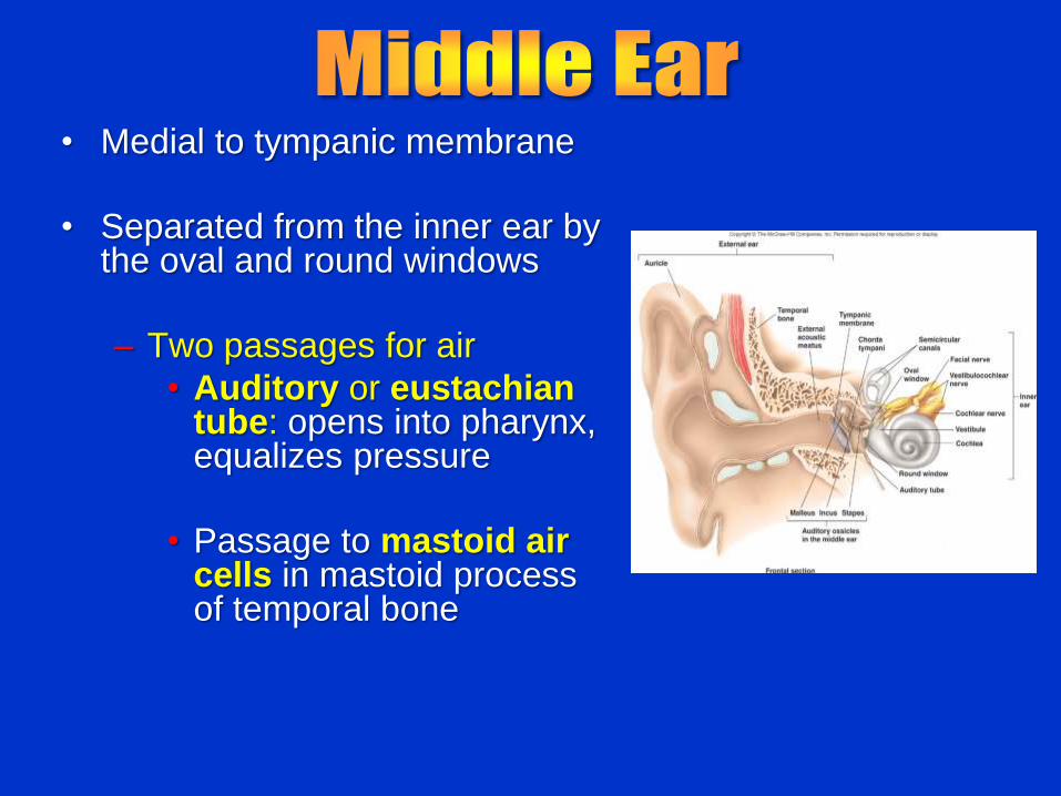
• Medial to tympanic membrane
• Separated from the inner ear by the oval and round windows
– Two passages for air
• Auditory or eustachian tube: opens into pharynx, equalizes pressure
• Passage to mastoid air cells in mastoid process of temporal bone

• Ossicles:
– malleus, incus, stapes:
transmit vibrations from
eardrum to oval window
• Oval window:
– Connection between middle
and inner ear
– Foot of the stapes rests here
and is held in place by
annular ligament

• Consists of :
• Bony labyrinth
– Tunnels and chambers in the temporal bone
– Divided into three regions:
• Cochlea: hearing
• Vestibule: balance
• Semicircular canals: balance
• Membranous labyrinth
– Series of membranous sacs within the bony labyrinth
• Lymphs
– Endolymph: in membranous labyrinth, high conc. of K+
– Perilymph: similar to CSF, space between membranous labyrinth and bony labyrinth

• Is the central egg-shaped cavity of the bony labyrinth
• Suspended in its perilymph are two membranous labyrinth sacs: the saccule and utricle
• The saccule extends into the cochlea
• The utricle extends into the semicircular canals
• These sacs:
– House balance receptors called maculae
– Respond to gravity and changes in the position of the head
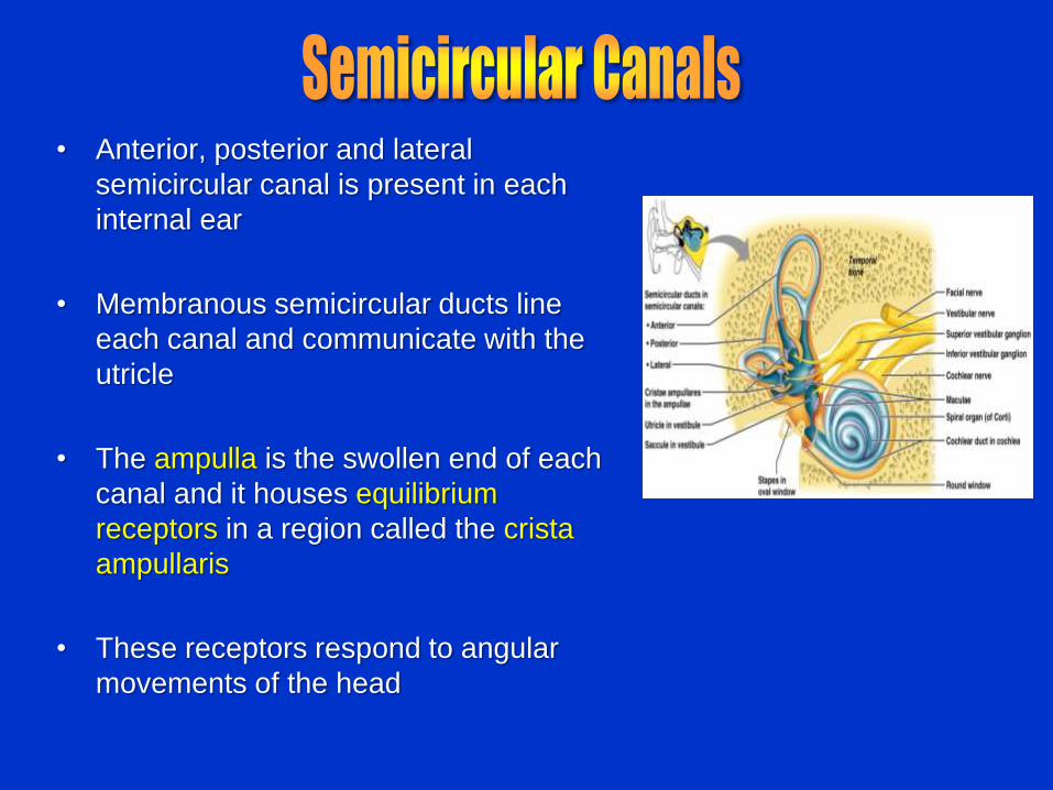
• Anterior, posterior and lateral
semicircular canal is present in each
internal ear
• Membranous semicircular ducts line
each canal and communicate with the
utricle
• The ampulla is the swollen end of each
canal and it houses equilibrium
receptors in a region called the crista
ampullaris
• These receptors respond to angular
movements of the head
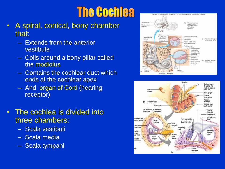
• A spiral, conical, bony chamber that: – Extends from the anterior
vestibule
– Coils around a bony pillar called the modiolus
– Contains the cochlear duct which ends at the cochlear apex
– And organ of Corti (hearing receptor)
• The cochlea is divided into three chambers: – Scala vestibuli
– Scala media
– Scala tympani

• The scala tympani terminates at the round window
• The scalas tympani and vestibuli:
– Are filled with perilymph
– Are continuous with each other via the helicotrema at cochlear apex
• The scala media is filled with endolymph

• The “floor” of the cochlear duct
is composed of:
– The bony spiral lamina
– The basilar membrane,
which supports the organ of
Corti
• The cochlear branch of nerve
VIII runs from the organ of
Corti to the brain
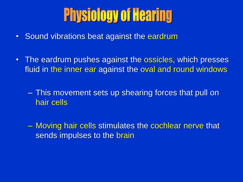
• Sound vibrations beat against the eardrum
• The eardrum pushes against the ossicles, which presses
fluid in the inner ear against the oval and round windows
– This movement sets up shearing forces that pull on
hair cells
– Moving hair cells stimulates the cochlear nerve that
sends impulses to the brain

• Properties of Sound:
• Sound is: – A pressure disturbance (alternating
areas of high and low pressure) originating from a vibrating object
– Composed of areas of rarefaction and compression
– Represented by a S-shaped curve or sine wave in wavelength, frequency, and amplitude
– Crests – compressed area
– troughs – rarefied area

• Properties of Sound:
• Frequency – the number of waves that pass a given point in a given time
• Pitch – perception of different frequencies (we hear from 20–20,000 Hz)
• Higher frequency – higher pitch
• Amplitude – intensity of a sound or height of sine wave crest measured in decibels (dB)
• Loudness – subjective interpretation of sound intensity
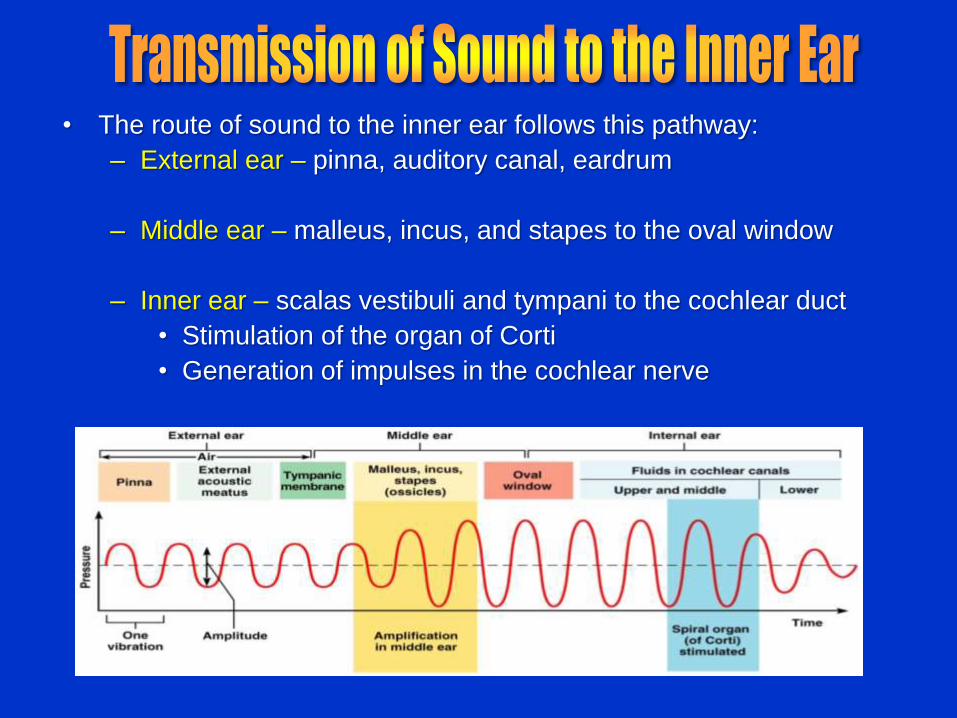
• The route of sound to the inner ear follows this pathway:
– External ear – pinna, auditory canal, eardrum
– Middle ear – malleus, incus, and stapes to the oval window
– Inner ear – scalas vestibuli and tympani to the cochlear duct
• Stimulation of the organ of Corti
• Generation of impulses in the cochlear nerve
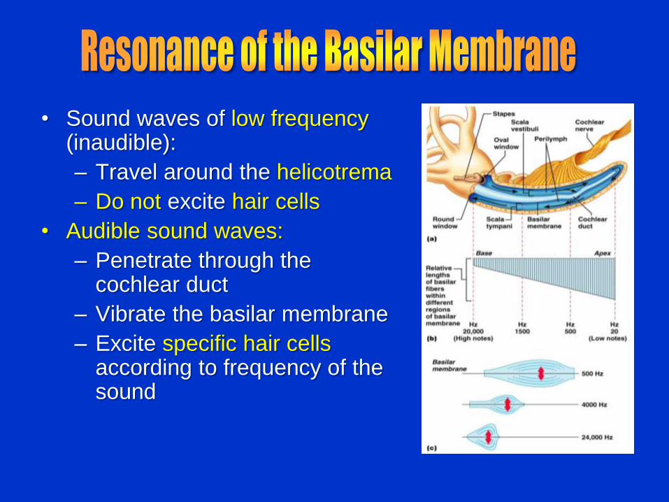
• Sound waves of low frequency (inaudible):
– Travel around the helicotrema
– Do not excite hair cells
• Audible sound waves:
– Penetrate through the cochlear duct
– Vibrate the basilar membrane
– Excite specific hair cells according to frequency of the sound

• Rest on basilar membrane
• And composed of supporting cells and outer and inner hair cells
• Afferent fibers of the cochlear nerve attach to the base of hair cells
• Hair cells have numerous stereocilia (hair)

• Bending of sterocilia:
– Opens mechanically gated K+ ion
channels
– Causes a graded potential and
the release of a neurotransmitter
(probably glutamate)
• The neurotransmitter causes
cochlear fibers to transmit impulses
to the brain, where sound is
perceived

• Impulses from the cochlea pass
via the spiral ganglion of cochlear
nerve to the cochlear nuclei of
medulla
• From there, impulses are sent to
the:
– Superior olivary nucleus
– Inferior colliculus (auditory
reflex center)
• From there, impulses pass to the
auditory cortex in temporal lobe

• Receptors for balance is present in the semicircular
canals and vestibule called Vestibular apparatus
– Maintains our orientation and balance in space
• Two kinds of balance Receptors:
– Vestibular receptors monitor static labyrinth
– Semicircular canal receptors monitor kinetic labyrinth

• Maculae are the sensory receptors for static labyrinth
– Contain supporting cells and hair cells
– Each hair cell has stereocilia and kinocilium embedded in the otolithic membrane
• Otolithic membrane – jellylike mass studded with tiny CaCO3 stones called otoliths
• Utricular hairs respond to horizontal movement
• Saccular hairs respond to vertical movement

• Otolithic movement in the direction of the kinocilia:
– Depolarizes vestibular nerve fibers
– Increases the number of action potentials generated
• Movement in the opposite direction:
– Hyperpolarizes vestibular nerve fibers
– Reduces the rate of impulse propagation
• From this information, the brain is informed of the changing position of the head
• Evaluates position of head relative to gravity
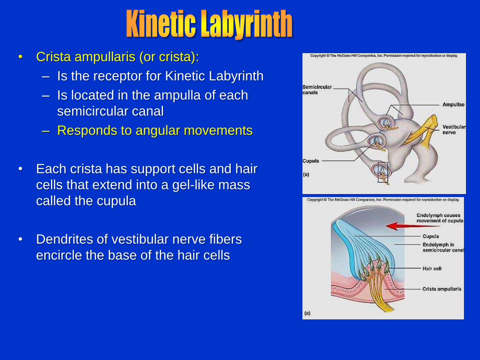
• Crista ampullaris (or crista):
– Is the receptor for Kinetic Labyrinth
– Is located in the ampulla of each
semicircular canal
– Responds to angular movements
• Each crista has support cells and hair
cells that extend into a gel-like mass
called the cupula
• Dendrites of vestibular nerve fibers
encircle the base of the hair cells
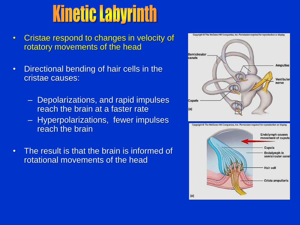
• Cristae respond to changes in velocity of rotatory movements of the head
• Directional bending of hair cells in the cristae causes:
– Depolarizations, and rapid impulses reach the brain at a faster rate
– Hyperpolarizations, fewer impulses reach the brain
• The result is that the brain is informed of rotational movements of the head

• 70% of all sensory receptors are in the eye
• Most of the eye is protected by a cushion
of fat and the bony orbit
• Accessory structures include eyebrows,
eyelids, conjunctiva, lacrimal apparatus,
and extrinsic eye muscles

• Eyebrows:
– Shading the eye
– Preventing perspiration from reaching the eye
• Eyelids (palpebrae):
• Protect the eye from foreign objects
– Palpebral fissure: space between two eyelids
– Canthi: eyelids join at lateral and medial margins of eye
– Medial canthus has lacrimal caruncle with modified sweat and sebaceous glands
– Tarsal or Meibomian glands in eyelids lubricate the eye
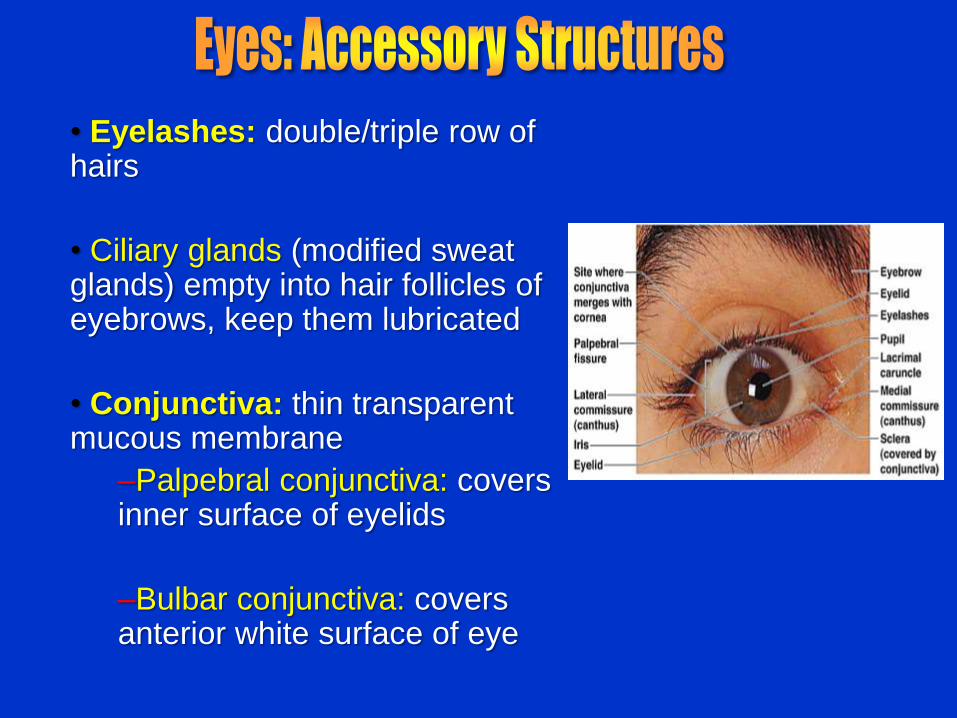
• Eyelashes: double/triple row of hairs
• Ciliary glands (modified sweat glands) empty into hair follicles of eyebrows, keep them lubricated
• Conjunctiva: thin transparent mucous membrane
–Palpebral conjunctiva: covers inner surface of eyelids
–Bulbar conjunctiva: covers anterior white surface of eye

• Lacrimal gland: produces tears to moisten, lubricate, wash
• located above the lateral end of eye
• Tears pass through ducts and then over eye
• Lacrimal canaliculi: collect excess tears through openings called punctum
• Lacrimal sac leads to nasolacrimal duct: opens into nasal cavity beneath the inferior nasal conchae

• Extrinsic eye muscles
– Six muscles attach to
the outer surface of
the eye
– LR6SO4O3
– Produce eye
movements

• The eyeball is composed of three layers:
• Fibrous layer: sclera and cornea
• Vascular layer: choroid, ciliary body, iris
• Nervous layer: retina

• Sclera: white outer layer of eyeball
• Seen anteriorly as the “white of the eye”
• Maintains shape, protects internal structures, provides muscle attachment point
• Sclera is continuous anteriorly with cornea
• Dense collagenous connective tissue with elastic fibers. Collagen fibers are large and opaque.

• Cornea:
• Avascular, transparent, allows light to enter eye; bends and refracts light
• Consists of connective tissue matrix containing collagen, elastic fibers and proteoglycans
• More proteoglycans than sclera, low water content (water would scatter light)

• Has three regions: choroid, ciliary body, and iris
• Choroid region – A dark brown
membrane that forms the posterior portion of the eye
– Light is focused by choroid pigment onto retina thus preventing scattering of the light
– Supplies blood to all eye tunics
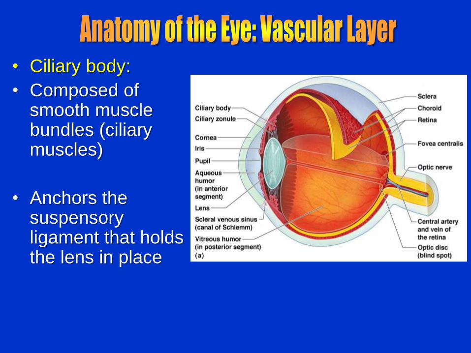
• Ciliary body:
• Composed of smooth muscle bundles (ciliary muscles)
• Anchors the suspensory ligament that holds the lens in place
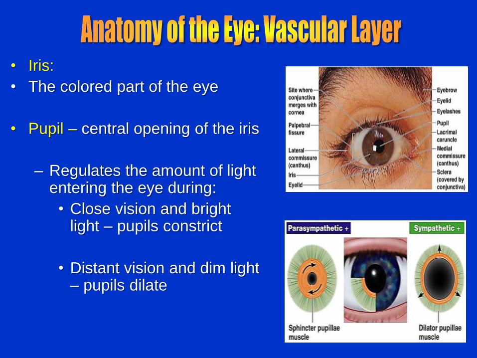
• Iris:
• The colored part of the eye
• Pupil – central opening of the iris
– Regulates the amount of light entering the eye during:
• Close vision and bright light – pupils constrict
• Distant vision and dim light – pupils dilate
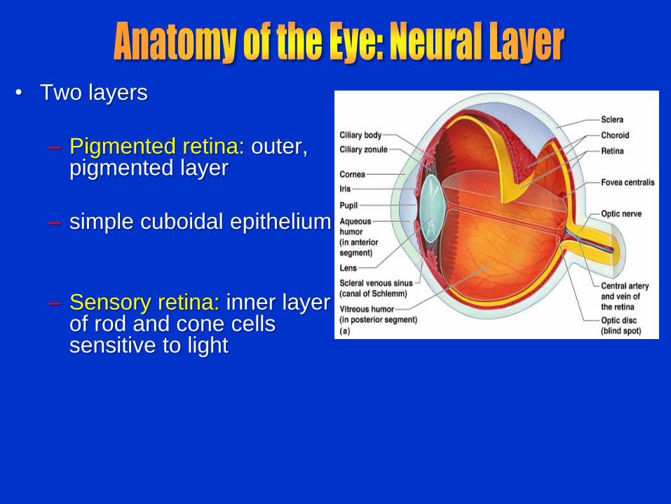
• Two layers
– Pigmented retina: outer, pigmented layer
– simple cuboidal epithelium
– Sensory retina: inner layer of rod and cone cells sensitive to light
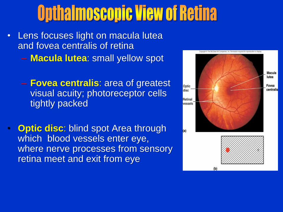
• Lens focuses light on macula lutea and fovea centralis of retina
– Macula lutea: small yellow spot
– Fovea centralis: area of greatest visual acuity; photoreceptor cells tightly packed
• Optic disc: blind spot Area through which blood vessels enter eye, where nerve processes from sensory retina meet and exit from eye
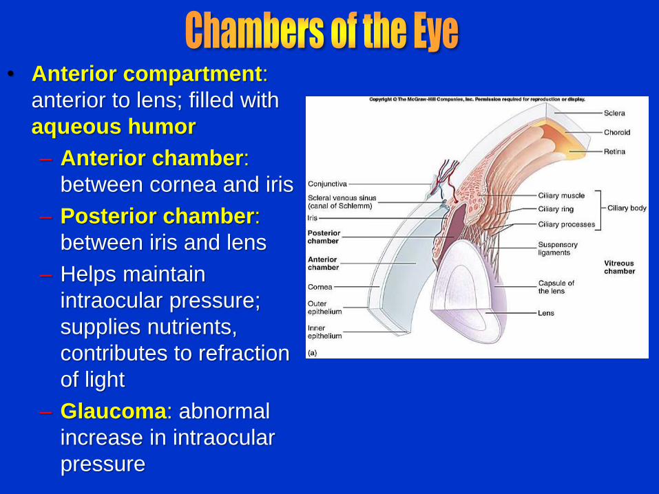
• Anterior compartment:
anterior to lens; filled with
aqueous humor
– Anterior chamber:
between cornea and iris
– Posterior chamber:
between iris and lens
– Helps maintain
intraocular pressure;
supplies nutrients,
contributes to refraction
of light
– Glaucoma: abnormal
increase in intraocular
pressure

•Posterior compartment: posterior to lens
• Filled with jelly-like vitreous humor
• Helps maintain intraocular pressure, holds lens and retina in place, refracts light
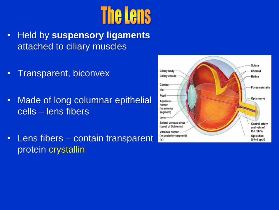
• Held by suspensory ligaments
attached to ciliary muscles
• Transparent, biconvex
• Made of long columnar epithelial
cells – lens fibers
• Lens fibers – contain transparent
protein crystallin

• Electromagnetic radiation –
all energy waves from short
gamma rays to long radio
waves
• Our eyes respond to a small
portion of this spectrum
called the visible light
• Different cones in the retina
respond to different
wavelengths of the visible
spectrum
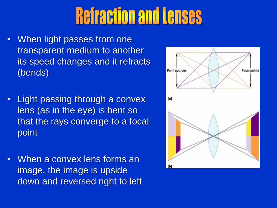
• When light passes from one
transparent medium to another
its speed changes and it refracts
(bends)
• Light passing through a convex
lens (as in the eye) is bent so
that the rays converge to a focal
point
• When a convex lens forms an
image, the image is upside
down and reversed right to left
• Pathway of light entering the eye: cornea, aqueous humor, lens, vitreous humor, and the neural layer of the retina to the photoreceptors
• Light is refracted:
– At the cornea
– Entering the lens
– Leaving the lens
• The lens curvature and shape allow for fine focusing of an image

• Light from a distance needs
little adjustment for proper
focusing
• Far point of vision – the
distance beyond which the
lens does not need to
change shape to focus (20
ft.)
• Ciliary muscle is relaxed
• Lens is flat

• Close vision requires:
– Accommodation – changing the lens shape by ciliary muscles to increase refractory power
– Lens becomes more spherical, greater refraction of light
– Constriction –
– pupillary reflex constricts the pupils
– Pupil diameter is small, depth of focus is greater
– Convergence –
– as objects move close to the eye, eyes are rotated medially

• Emmetropic eye – normal eye with light focused properly
• Myopic eye (nearsighted) – the focal point is in front of the retina
– Corrected with a concave lens
• Hyperopic eye (farsighted) – the focal point is behind the retina
– Corrected with a convex lens

• Sensory retina: three layers of neurons: photoreceptor, bipolar, and ganglionic
• Pigmented retina: single layer of cells; filled with melanin
• With choroid, enhances visual acuity reducing light scattering
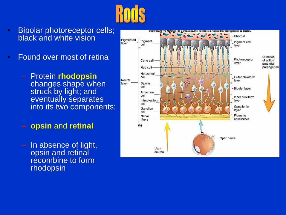
• Bipolar photoreceptor cells; black and white vision
• Found over most of retina
– Protein rhodopsin changes shape when struck by light; and eventually separates into its two components:
– opsin and retinal
– In absence of light, opsin and retinal recombine to form rhodopsin

• Bipolar photoreceptor cells
• Responsible for color vision and visual acuity
– Numerous in fovea and macula lutea; fewer over rest of retina
– As light intensity decreases so does our ability to see color
– Cone cell contain visual pigment - iodopsin: three types that respond to blue, red and green light
– Overlap in response to light, thus interpretations of gradation of color possible: several millions

• Rods and cones synapse with bipolar cells that synapse with ganglion cells
• Axons of retinal ganglion cells form the optic nerve
• Medial fibers of the optic nerve decussate at the optic chiasm
• Most fibers of the optic tracts continue to the lateral geniculate body of the thalamus

• Other optic tract fibers end in superior colliculi (initiating visual reflexes) and pretectal nuclei (involved with pupillary reflexes)
• Optic radiations travel from the thalamus to the visual cortex

• Myopia: Nearsightedness – Focal point too near lens,
image focused in front of retina
• Hyperopia: Farsightedness – Image focused behind retina
• Presbyopia – Degeneration of
accommodation, corrected by reading glasses
• Astigmatism: Cornea or lens not uniformly curved
• Retinal detachment – Can result in complete
blindness
• Glaucoma – Increased intraocular
pressure by aqueous humor buildup
• Cataract – Clouding of lens
• Macular degeneration – Common in older people,
loss in acute vision
• Diabetes – Dysfunction of peripheral
circulation

Effects of Aging on the
Special Senses
• Slight loss in ability to detect odors
• Decreased sense of taste
• Lenses of eyes lose flexibility
• Development of cataracts, macular
degeneration, glaucoma, diabetic
retinopathy
• Decline in visual acuity and color perception