Embed Size (px)
Citation preview


Fish Pathology

Dedication
To my long suffering wife Helen Macgregor. Her friendship, loyalty and support, when times were good and, more importantly,
when they most decidedly were not, will always be appreciated.

Fish PathologyFOURTH EDITION
Edited by
Ronald J. RobertsBVMS, PhD (Glasgow), FRCVS, FRCPath, FSB, FRSE
Commander of the Most Noble Order of the Crown (Thailand)
Hagerman Distinguished Visiting Professor, University of Idaho, USAAdjunct Professor of Fish Pathology, College of Veterinary Medicine,
Washington State University, USATechnical Director, Landcatch Ltd, Scotland,
Emeritus Professor, University of Stirling, Scotland
A John Wiley & Sons, Ltd., Publication
This edition first published 2012 © 2012 by Blackwell Publishing Ltd.Third edition © 2001 Harcourt Publishers Ltd.
Blackwell Publishing was acquired by John Wiley & Sons in February 2007. Blackwell’s publishing program has been merged with Wiley’s global Scientific, Technical and Medical business to form Wiley-Blackwell.
First published 1978Second edition 1989Third edition 2001Fourth Edition 2012Several editions also published in French, Spanish, Italian, German, Japanese, Mandarin and Bahasa Malay.
Registered office: John Wiley & Sons, Ltd, The Atrium, Southern Gate, Chichester, West Sussex, PO19 8SQ, UK
Editorial offices: 9600 Garsington Road, Oxford, OX4 2DQ, UKThe Atrium, Southern Gate, Chichester, West Sussex, PO19 8SQ, UK2121 State Avenue, Ames, Iowa 50014-8300, USA
For details of our global editorial offices, for customer services and for information about how to apply for permission to reuse the copyright material in this book please see our website at www.wiley.com/wiley-blackwell.
The right of Ronald J. Roberts to be identified as the author of this work has been asserted in accordance with the UK Copyright, Designs and Patents Act 1988.
All rights reserved. No part of this publication may be reproduced, stored in a retrieval system, or transmitted, in any form or by any means, electronic, mechanical, photo-
copying, recording or otherwise, except as permitted by the UK Copyright, Designs and Patents Act 1988, without the prior permission of the publisher.
Designations used by companies to distinguish their products are often claimed as trademarks. All brand names and product names used in this book are trade names, service marks, trademarks or registered trademarks of their respective owners. The publisher is not associated with any product or vendor mentioned in this book. This publication is designed to provide accurate and authoritative informa-tion in regard to the subject matter covered. It is sold on the understanding that the publisher is not engaged in rendering professional services. If professional advice or other expert assistance is required, the services of a competent profes-sional should be sought.
Library of Congress Cataloging-in-Publication DataFish pathology / edited by Ronald J. Roberts. – 4th ed. p. cm. Includes bibliographical references and index. ISBN 978-1-4443-3282-7 (hard cover : alk. paper) I. Roberts, Ronald J., 1941– SH171.F58 2012 597–dc23 2011035802
A catalogue record for this book is available from the British Library.
Wiley also publishes its books in a variety of electronic formats. Some content that appears in print may not be available in electronic books.
Set in 9.5/12 pt Times by Toppan Best-set Premedia Limited
1 2012

ContentsContributors viPreface vii
1 TheAquaticEnvironment 1R.J. Roberts
2 TheAnatomyandPhysiologyofTeleosts 17R.J. Roberts and A.E. Ellis
3 ThePathophysiologyandSystematicPathologyofTeleosts 62R.J. Roberts and H.D. Rodger
4 TheImmunologyofTeleosts 144C.J. Secombes and A.E. Ellis
5 NeoplasiaofTeleosts 167R.J. Roberts
6 TheVirologyofTeleosts 186D.A. Smail and E.S. Munro
7 TheParasitologyofTeleosts 292R. Wootten
8 TheBacteriologyofTeleosts 339R.J. Roberts
9 TheMycologyofTeleosts 383R.J. Roberts
10 TheNutritionalPathologyofTeleosts 402R.W. Hardy
11 MiscellaneousNon-infectiousDiseases 425R.J. Roberts
12 LaboratoryMethods 439R.J. Roberts, D.A. Smail and E.S. Munro
References 482Fish Species Index 567Subject Index 571
v

Contributors
A.E. Ellis, MA (Oxon), PhD (Aberdeen)Formerly Senior Principal Scientific OfficerFisheries Research ServiceMarine LaboratoryAberdeen, Scotland
R.W. Hardy, BSc, PhD (Washington)DirectorHagerman Center for Sustainable AquacultureUniversity of IdahoIdaho, United States
E.S. Munro, BSc (RGU, Aberdeen)Group Leader VirologyMarine ScotlandMarine LaboratoryAberdeen, Scotland
R.J. Roberts, BVMS, PhD (Glasgow)FRCVS, FRCPath, FSB, FRSE, Commander of the Most Noble Order of the Crown (Thailand)Hagerman Distinguished Visiting ProfessorUniversity of Idaho, United StatesTechnical DirectorLandcatch LtdOrmsary, Argyll, ScotlandEmeritus Professorformerly Founder DirectorInstitute of AquacultureUniversity of Stirling, Scotland
H.D. Rodger, BVMS (Glasgow), MSc, PhD (Stirling)MRCVSPractice PrincipalVet-Aqua InternationalOranmore, Co.Galway, Ireland
C.J. Secombes, BSc (Leeds), PhD (Hull), DSc (Aberdeen), FSB, FRSEHead, Scottish Fish Immunology Research CentreUniversity of AberdeenScotland
D.A. Smail, BA (Oxon), MSc (Birmingham), PhD (CNAA)Group Leader, Immunology & InfectionMarine ScotlandMarine LaboratoryAberdeen, Scotland
R. Wootten, BSc, PhD (London)Deputy DirectorInstitute of AquacultureUniversity of StirlingStirling, Scotland
In addition, material from previous editions contributed by Dr A.M. Bullock, Dr G.N. Frerichs, Dr P. Tytler and Dr A.L.S. Munro has also been edited, updated and incorpo-rated into the text.
vi

Preface
This is the fourth edition of Fish Pathology. Little did I think when I penned the first few sentences in Nikola Fijan’s Laboratory in Zagreb, in 1976, that this book would survive to a fourth edition and be translated into nine languages. To my surprise and immense pleasure, it is now into its fourth decade and very much accepted as a standard work on its subject.
This edition has been revised and reset, incorporating much new information, and I am very grateful to Wiley-Blackwell for their encouragement to complete what has become a very large text and also for accepting my major demands for colour illustrations. The ability to use such illustrations throughout the text makes the task of writing very much easier, and this volume is illustrated to a degree which would have been unthinkable when planning that first edition, in early 1976.
Much of the revision and editing has once again been carried out in the beautiful and stimulating environment of the Hagerman Laboratories of the University of Idaho. Here, for the third time in my career, I have been presented with the opportunity to develop an international fish disease laboratory from the drawing board, as part of the new facility which Professor Hardy now directs. The facil-ity provides an excellent resource for both cutting-edge research and more contemplative writing of texts such as this. Few can have been so fortunate, and I am most grate-ful to Professor Hardy and the University of Idaho for their generous support.
Teleost fish inhabit what is to most of us an alien world. Their exploitation for food or for sport has, until compara-tively recently, been completely dependent on the hunting of wild stocks. Since the first edition of this book was published in 1978, however, the role of aquaculture in relation to world fish production has been transformed beyond measure. In 1978 aquaculture was responsible for less than 5% of the total fish consumed. Now the figure is nearer 50% by weight and more than 50% in terms of economic value. The blue revolution, no less significant than the green one, which led to self-sufficiency in rice
and grain production in the developing world, is very much underway.
Control of fish diseases has been one of the major factors in allowing this growth in farmed fish production to take place. Our knowledge of fish pathology has expanded dramatically over the past decade, underpinned by the remarkable developments that have taken place in molecular biology and immunopathology. This has made production of a single text, which attempts to encapsulate the main aspects of all of the different disciplines of which the subject is comprised, rather challenging. This will, I suspect, be the last time that anyone will attempt such an all-embracing task. The individual subjects which make up fish pathology have now themselves grown to a size where they each demand their own monographs.
At least, however, I hope that this volume will, for a time, help to ensure that as aquaculture continues to grow, we will still be able to define, to diagnose and ultimately to control, in a sustainable way, the new diseases which will assuredly manifest themselves. The health and welfare of our fish stocks, both wild and farmed, are, I believe, measures of the quality of our entire aquatic environment.
The study of fish diseases requires a wide knowledge, not only of the potential pathogens but also of the envi-ronmental constraints and specialist adaptations which govern the ectothermic, aqueous existence of the teleosts. The inflammatory and immune responses of fishes are greatly modified by the nature of their environment. These, in turn, influence the epizootiology and the clinical char-acteristics of the various conditions and the methods by which they can be controlled.
One of the most heartening advances in the decade since the last edition of Fish Pathology was published has been the further development of sophisticated and efficient vac-cines for many of the most damaging bacterial diseases. The advent of DNA and recombinant vaccines and immune enhancers has contributed greatly to fish welfare. Even more dramatic over the period has been the contribution
vii
viii Preface
of molecular biology to diagnostic advances such as those presented by real-time, or quantitative polymerase chain reaction (PCR), and the advent of breeding programmes that utilise molecular markers for genetic selection for disease resistance.
A very significant challenge presented in writing this book compared to the first three editions has been the advent of molecular techniques for the demonstration of relatedness between microorganisms. As a result of this, the taxonomy of many of the parasite and fungal groups has been completely rewritten. In the past, morphology and other phenotypic characteristics were dominant in deciding to which group a particular pathogen should be assigned. Now this can be decided with much greater certitude based on DNA homologies. While this has con-tributed greatly to precision, it has in many cases com-pletely overturned previous assumptions. The taxonomies found therefore in the chapters on parasitology and mycol-ogy of fishes (Chapters 7 and 9, respectively) will appear somewhat strange to the reader accustomed to these previ-ous certainties. I am very grateful to Dr Rod Wootten for his efforts to present a comprehensible taxonomic basis for the protistan parasites in particular and also to Dr Pieter Van West for his explanation to me of the new groupings of the Oomycetes and the true fungi.
Fish pathology is clearly a multidisciplinary field. The essential purpose of this book, like its predecessors, is to provide a corpus of basic information, drawn from these disciplines, which will be of value to the veterinarian, the microbiologist, the parasitologist, the nutritionist or the hydrologist. There are some 17 000 species of teleosts, all with specific adaptations at the gross, cellular and molecular levels, for their particular niche. In a book such as this, there-fore, rather sweeping generalisations are sometimes neces-sary to render basic information more understandable.
I am very grateful to the authors of the contributed chapters for their forbearance, once again, and for their willingness to accept any editorial liberties that I may have taken in rewriting them for the sake of standardisation. I firmly believe that this is what gives the book its homoge-neity and hope that they consider that their efforts have been justified. I must also acknowledge the assistance of Dr Emmett Shotts, formerly of the Leetown Laboratory, West Virginia and Dr Pieter van West and Professor Monty
Priede of the University of Aberdeen for their willingness to review the bacteriology, mycology and anatomy chap-ters respectively, and Dr Hamish Rodger, formerly of the University of Pennsylvania, for general editorial support. I alone, however, am responsible for all errors and omissions.
Wiley-Blackwell, which also publishes the Journal of Fish Diseases, which I edit with Dr Wootten, has again generously allowed publication of many illustrations which first appeared in that journal. I am also grateful to the plethora of friends throughout the world who have made their illustrations available to me. Their generosity is acknowledged in the text wherever possible, but I apolo-gise in advance for any omissions.
The preparation of this book would have been impos-sible without the drive, help and encouragement of Nigel Balmforth of Wiley-Blackwell, his assistant Carys Williams and the project manager Mirjana Misina. It was also heavily dependent on the encouragement and support of my colleagues Dr Laird Noh, Tracy Brown and Jana Cole at the University of Idaho and Sir William Lithgow, Hugh Currie, Alan Stewart and Neil Manchester at Landcatch Ltd. It is also important to acknowledge once again the determined efforts of Andrew Millar and K.G. Clarke, former colleagues at the University of Stirling, who so wholeheartedly relieved me of administra-tive duties so that I have, as Emeritus Professor, been able to concentrate on the real work of scientific research instead of the tyranny of the bean counter’s spread sheet.
Sadly during the course of the writing of this edition, Dr Tony Ellis, my first PhD student, who went on to estab-lish an outstanding career in fish immunology and had contributed to every edition of this book, succumbed to cancer. He was a free spirit with a very original mind and an excellent scientist and teacher. He will be greatly missed.
Finally I have to thank my wife Helen for providing the line drawings and for her encouragement in what has cer-tainly been a much more demanding task than either of us envisaged.
Ronald J. RobertsHagerman, Idaho
1The Aquatic Environment
INTRODUCTION
Disease, in fishes, is closely linked to environmental stress. In the wild, they generally have some degree of freedom to modify their environment. They can move to more suitable conditions if faced with a negative environ-mental change such as a reduction in oxygen level or increase in temperature. Infected fish will even move to a warmer area to create an enhanced body temperature as an aid to increasing the rate of inflammatory response. In culture conditions, on the other hand, they have limited opportunity to choose their external environmental condi-tions. It is thus important that the conditions under which they are held provide environmental parameters suitable for all of the requirements of the particular species.
The aquatic environment encompasses a wide variety of features, virtually all of which influence the maintenance of homeostasis, essential for growth and reproduction of fishes. If altered beyond acceptable limits, they may pre-dispose to, or actually cause, a wide range of disease processes. Among the most important environmental parameters are physical factors such as the temperature, the intensity and periodicity of light (including shading and background hue), the chemical composition of the water and its biological content, the availability of space and food and the frequency of fright stimuli such as moving shadows (EFSA 2008). Another important factor for wild fish and those farmed in extensive systems is the productivity of the ecosystem which sustains their food supply. Since consideration of ecosystems and the eco-
physiological requirements of fish is beyond the scope of the present text, the reader is referred to Rankin and Jensen (1993), Macan (1974) and Odum (1971).
PHYSICAL AND CHEMICAL ASPECTS OF WATER QUALITY
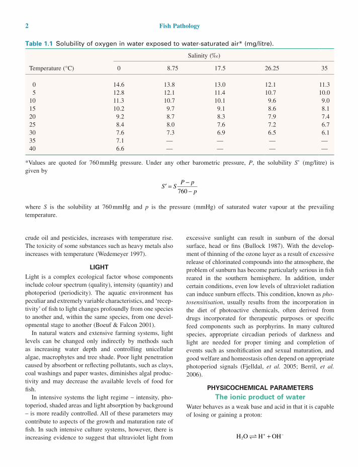
TEMPERATUREFish have upper and lower thermal tolerance limits and optimum temperatures for growth, egg incubation, food conversion and resistance to specific diseases. These optima vary with species and may be different for different parameters such as oxygen tension and water pH. Fresh waters are subject to temperature fluctuations of up to 40°C caused by latitude, season, altitude, time of day, depth and other factors. The range of temperature change of sea-waters is much less, due to water circulation in the seas and oceans and the large volumes of water involved.
Many fish diseases are temperature modulated, with pathogenicity closely related to a specific temperature range. In many host–pathogen systems there is a balance between the host’s defences and the pathogen’s invasive-ness, but this is readily modified by temperature change, especially if it is rapid.
Water temperature also affects other properties of the aquatic environment important for fish health. Dissolved gases generally decrease in solubility with increasing tem-perature (Table 1.1), whereas the solubility of toxic com-pounds, which are only sparingly soluble in water, such as
Fish Pathology, Fourth Edition. Edited by Ronald J. Roberts.© 2012 Blackwell Publishing Ltd. Published 2012 by Blackwell Publishing Ltd.
1
2 Fish Pathology
excessive sunlight can result in sunburn of the dorsal surface, head or fins (Bullock 1987). With the develop-ment of thinning of the ozone layer as a result of excessive release of chlorinated compounds into the atmosphere, the problem of sunburn has become particularly serious in fish reared in the southern hemisphere. In addition, under certain conditions, even low levels of ultraviolet radiation can induce sunburn effects. This condition, known as pho-tosensitisation, usually results from the incorporation in the diet of photoactive chemicals, often derived from drugs incorporated for therapeutic purposes or specific feed components such as porphyrins. In many cultured species, appropriate circadian periods of darkness and light are needed for proper timing and completion of events such as smoltification and sexual maturation, and good welfare and homeostasis often depend on appropriate photoperiod signals (Fjelldal, et al. 2005; Berril, et al. 2006).
PHYSICOCHEMICAL PARAMETERSThe ionic product of water
Water behaves as a weak base and acid in that it is capable of losing or gaining a proton:
H O H OH2
+ −+
crude oil and pesticides, increases with temperature rise. The toxicity of some substances such as heavy metals also increases with temperature (Wedemeyer 1997).
LIGHTLight is a complex ecological factor whose components include colour spectrum (quality), intensity (quantity) and photoperiod (periodicity). The aquatic environment has peculiar and extremely variable characteristics, and ‘recep-tivity’ of fish to light changes profoundly from one species to another and, within the same species, from one devel-opmental stage to another (Boeuf & Falcon 2001).
In natural waters and extensive farming systems, light levels can be changed only indirectly by methods such as increasing water depth and controlling unicellular algae, macrophytes and tree shade. Poor light penetration caused by absorbent or reflecting pollutants, such as clays, coal washings and paper wastes, diminishes algal produc-tivity and may decrease the available levels of food for fish.
In intensive systems the light regime – intensity, pho-toperiod, shaded areas and light absorption by background – is more readily controlled. All of these parameters may contribute to aspects of the growth and maturation rate of fish. In such intensive culture systems, however, there is increasing evidence to suggest that ultraviolet light from
Table 1.1 Solubility of oxygen in water exposed to water-saturated air* (mg/litre).
Temperature (°C)
Salinity (‰)
0 8.75 17.5 26.25 35
0 14.6 13.8 13.0 12.1 11.35 12.8 12.1 11.4 10.7 10.0
10 11.3 10.7 10.1 9.6 9.015 10.2 9.7 9.1 8.6 8.120 9.2 8.7 8.3 7.9 7.425 8.4 8.0 7.6 7.2 6.730 7.6 7.3 6.9 6.5 6.135 7.1 — — — —40 6.6 — — — —
*Values are quoted for 760 mmHg pressure. Under any other barometric pressure, P, the solubility S’ (mg/litre) is given by
′ = −−
S SP p
p760
where S is the solubility at 760 mmHg and p is the pressure (mmHg) of saturated water vapour at the prevailing temperature.

The Aquatic Environment 3
1.2. From this it is apparent that the pH scale varies from 0 to 14.
The pH scale is a negative logarithmic scale, meaning that for a decrease of 1 pH unit there is a 10-fold increase of hydrogen ion concentration. Neutrality on the pH scale is the point where equal amounts of hydrogen and hydroxyl ions exist. This value changes with the salt content and the temperature (Table 1.3). Where hydrogen ions are in excess of hydroxyl ions the solution is said to be acidic, and in the reverse situation it is called alkaline. To calcu-late the actual hydrogen ion concentration at, for example, a pH of 6.6,
pH H= − =+log( ) .6 6
By taking the antilogarithm of both sides,
H anti+ = −( )log .6 6
the number −6.6 can be re-expressed as 0.4–7.0. The antilog of 0.4 = 2.5 and the antilog of −7.0 = 10−7. Therefore,
H g ions/litre+ −= ×2 5 10 7.
and the reverse, to calculate the pH of a solution contain-ing 3.98 × 10⁻⁸ g ions/litre H+:
pH H= − = − ×= − −= − +=
+ −
−
log log .
log . log
.
.
3 98 10
3 98 10
0 6 8
7 4
8
8
The equilibrium constant for the dissociation of water is given by
KC C
Cw
H OH
H O
=×+ −
2
where CH+ and COH– are the respective ion concentrations. Experimental evidence has shown that in pure water at 25°C,
Kw = × −1 00 10 14.
and since both ions are present in equal amounts,
C CH OH g ions/litre+ −= = × −1 10 7
pH and the pH scaleIn dilute aqueous solutions, the pH is defined as the nega-tive logarithm of the hydrogen ion concentration. This is usually written as
pH H= − +log10 C
Using our previous example, the pH of pure water at 25°C is
− × =−log1071 10 7
In dilute aqueous solutions the ion product of water is constant at 1 × 10⁻¹⁴. Additions of hydrogen and hydroxyl ions do not change this constant. The interrelations between hydrogen and hydroxyl ion concentration and their equivalent pH and pOH values are shown in Table
Table 1.2 Interrelationships between pH value and the hydrogen and hydroxyl ion concentrations in a dilute aqueous solution.
Acid solutions Neutral Alkaline solutions
Hydrogen ion (H+) concentration (g/litre)
1 10−1 10−2 10−3 10−4 10−5 10−6 10−7 10−8 10−9 10−10 10−11 10−12 10−13 10−14
Equivalent pH value
0 1 2 3 4 5 6 7 8 9 10 11 12 13 14
Hydroxyl ion (OH−) concentration (g/litre)
10−14 10−13 10−12 10−11 10−10 10−9 10−8 10−7 10−6 10−5 10−4 10−3 10−2 10−1 1
Equivalent pOH value
14 13 12 11 10 9 8 7 6 5 4 3 2 1 0
4 Fish Pathology
terms of pH rather than pOH, it is convenient to use the constant pKAB which relates the dissociation of a weak base to pH rather than pOH. The constant pKAB is derived from pKB by the expression pKAB = 14 − pKB.
Common ion effectThe ionisation of a weak acid or base is greatly reduced by the addition of a compound that dissociates a common ion. For example, the ionisation of ammonium hydroxide is depressed by the addition of sodium hydroxide, which dissociates the common hydroxyl ion, or by the addition of ammonium chloride, which contributes the common ammonium ion. Thus it is evident from inspection of the mass law equation
C C
CK
NH OH
NHB
4
3
+ −×=
that increasing the concentration of hydroxyl ions must reduce the concentration of ammonium ions to maintain the constancy of KB. This explains why, in waters contain-ing ammonium salts, increasing alkalinity causes greater amounts of undissociated ammonia, a point of great sig-nificance in intensive aquaculture.
Buffering actionThe principles of buffer action are best understood in terms of the following:
weak base strong acid neutral salt weak acid+ +
Owing to the common ion effect, the pH of a salt–acid mixture will be higher, and that of a salt–base mixture lower, than the pH of the acid or base alone. The
Ionisation constants of weak acids and bases
Applying the law of mass action to the ionisation of a weak acid,
HA H A
+ −+
we have
C C
CKH A
HAA
+ −×=
where KA is the ionisation or dissociation constant of the weak acid. The stronger the acid, the greater the proportion of ions to undissociated molecules and the larger the value of KA. Because these values are very small, it is usual to express them as negative logarithms and to use the desig-nation pKA, analogous to pH:
p A AK K= log
Weak bases such as ammonium hydroxide behave in a similar manner:
NH H O NH OH
NH OH
NHB
3 2 4
4
3
+ +×
=
+ −
+ −
;
C C
CK
where KB is a measure of the extent to which a weak base dissociates hydroxyl ions. Converting the dissociation constant KB to its negative logarithm yields the constant pKB. The constants KB and pKB of weak bases relate to the hydroxyl ion concentrations and pOH values. Because we customarily express reactions even in alkaline solution in
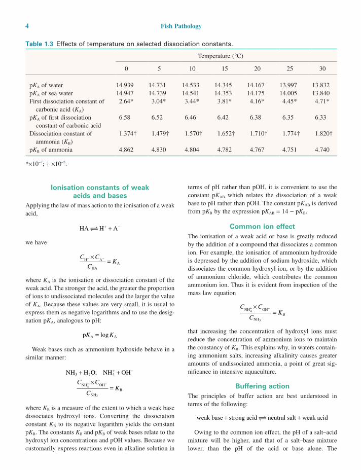
Table 1.3 Effects of temperature on selected dissociation constants.
Temperature (°C)
0 5 10 15 20 25 30
pKA of water 14.939 14.731 14.533 14.345 14.167 13.997 13.832pKA of sea water 14.947 14.739 14.541 14.353 14.175 14.005 13.840First dissociation constant of
carbonic acid (KA)2.64* 3.04* 3.44* 3.81* 4.16* 4.45* 4.71*
pKA of first dissociation constant of carbonic acid
6.58 6.52 6.46 6.42 6.38 6.35 6.33
Dissociation constant of ammonia (KB)
1.374† 1.479† 1.570† 1.652† 1.710† 1.774† 1.820†
pKB of ammonia 4.862 4.830 4.804 4.782 4.767 4.751 4.740
*×10−7; † ×10−5.
The Aquatic Environment 5
Henderson–Hasselbach equation relates the pH to the pro-portion of salt to acid or base:
pH p A or ABsalt
acid or base
= +KC
Clog
This equation states that a weak base binds the hydrogen ions dissociated from the strong acid, thereby buffering (i.e. mitigating) the change in hydrogen ion concentration. The addition of successive increments of strong acid causes only minor changes in pH over the range of effec-tive buffer action. The most effective buffering is exerted within limits of ±1 pH unit on either side of the pKA or pKAB.
By far the commonest source of buffering in fresh and sea water is the carbon dioxide–carbonic acid–bicarbonate–carbonate system represented by
The two dissociation constants are represented by
KC C
CK
KC C
CK
AHCO H
H COA
ACO H
HCO
A
p at
p
1 1
2 2
3
2 3
32
3
6 35 25=×
= °
=×
− +
− +
−
.
== °10 25 25. at
The equilibrium between these different species in fresh water is dependent on the pH, as the following shows:
H CO H HCO
H CO H HCO
HCO
pH
pH
pH
2 391
5 5
39
2 324
7
376
377
%
.
%
% %
%
+ −
+ −
−
+
+
110
32
23H CO+ −+
%
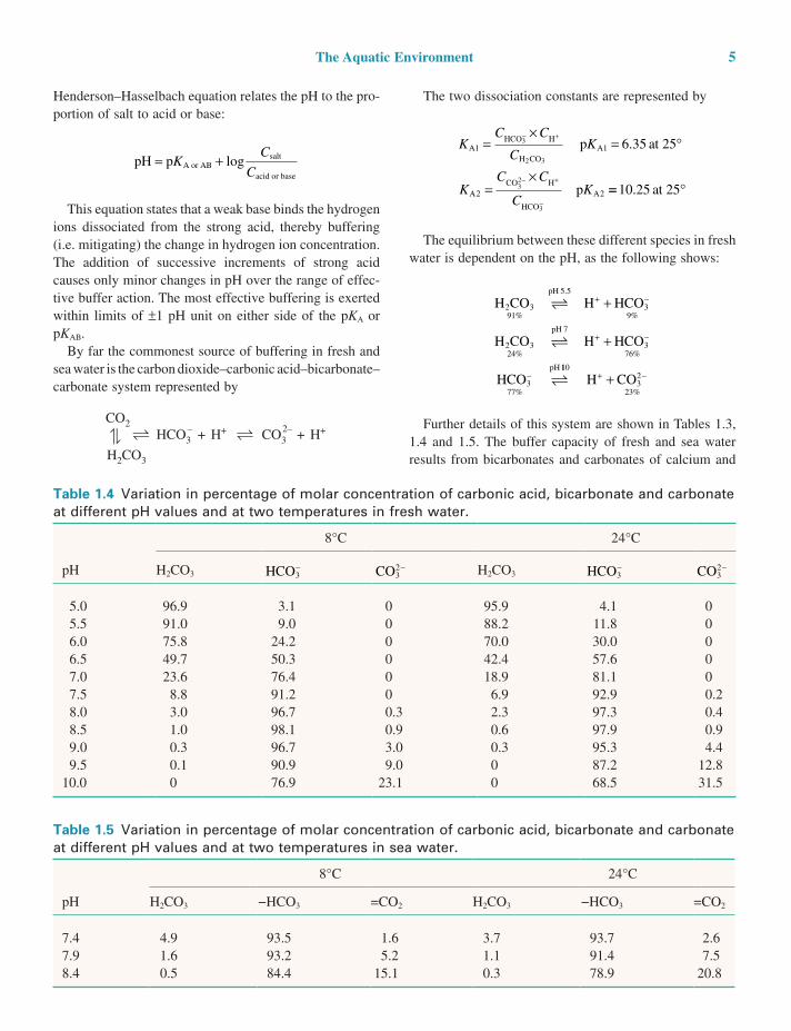
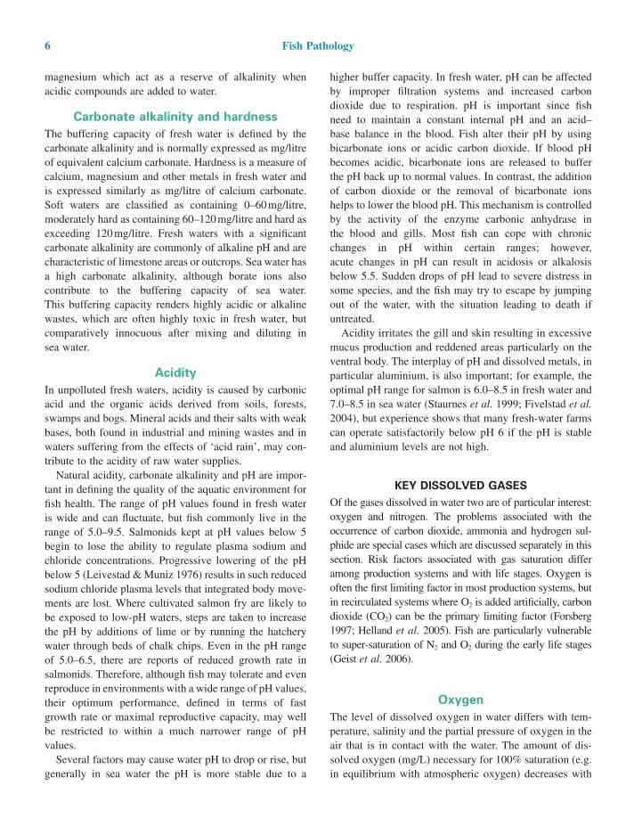
Further details of this system are shown in Tables 1.3, 1.4 and 1.5. The buffer capacity of fresh and sea water results from bicarbonates and carbonates of calcium and
CO2
H2CO3
HCO3 CO3+ H+ + H+2––
Table 1.4 Variation in percentage of molar concentration of carbonic acid, bicarbonate and carbonate at different pH values and at two temperatures in fresh water.
pH
8°C 24°C
H2CO3 HCO3− CO3
2− H2CO3 HCO3− CO3
2−
5.0 96.9 3.1 0 95.9 4.1 05.5 91.0 9.0 0 88.2 11.8 06.0 75.8 24.2 0 70.0 30.0 06.5 49.7 50.3 0 42.4 57.6 07.0 23.6 76.4 0 18.9 81.1 07.5 8.8 91.2 0 6.9 92.9 0.28.0 3.0 96.7 0.3 2.3 97.3 0.48.5 1.0 98.1 0.9 0.6 97.9 0.99.0 0.3 96.7 3.0 0.3 95.3 4.49.5 0.1 90.9 9.0 0 87.2 12.8
10.0 0 76.9 23.1 0 68.5 31.5
Table 1.5 Variation in percentage of molar concentration of carbonic acid, bicarbonate and carbonate at different pH values and at two temperatures in sea water.
pH
8°C 24°C
H2CO3 −HCO3 =CO2 H2CO3 −HCO3 =CO2
7.4 4.9 93.5 1.6 3.7 93.7 2.67.9 1.6 93.2 5.2 1.1 91.4 7.58.4 0.5 84.4 15.1 0.3 78.9 20.8
6 Fish Pathology
higher buffer capacity. In fresh water, pH can be affected by improper filtration systems and increased carbon dioxide due to respiration. pH is important since fish need to maintain a constant internal pH and an acid– base balance in the blood. Fish alter their pH by using bicarbonate ions or acidic carbon dioxide. If blood pH becomes acidic, bicarbonate ions are released to buffer the pH back up to normal values. In contrast, the addition of carbon dioxide or the removal of bicarbonate ions helps to lower the blood pH. This mechanism is controlled by the activity of the enzyme carbonic anhydrase in the blood and gills. Most fish can cope with chronic changes in pH within certain ranges; however, acute changes in pH can result in acidosis or alkalosis below 5.5. Sudden drops of pH lead to severe distress in some species, and the fish may try to escape by jumping out of the water, with the situation leading to death if untreated.
Acidity irritates the gill and skin resulting in excessive mucus production and reddened areas particularly on the ventral body. The interplay of pH and dissolved metals, in particular aluminium, is also important; for example, the optimal pH range for salmon is 6.0–8.5 in fresh water and 7.0–8.5 in sea water (Staurnes et al. 1999; Fivelstad et al. 2004), but experience shows that many fresh-water farms can operate satisfactorily below pH 6 if the pH is stable and aluminium levels are not high.
KEY DISSOLVED GASESOf the gases dissolved in water two are of particular interest: oxygen and nitrogen. The problems associated with the occurrence of carbon dioxide, ammonia and hydrogen sul-phide are special cases which are discussed separately in this section. Risk factors associated with gas saturation differ among production systems and with life stages. Oxygen is often the first limiting factor in most production systems, but in recirculated systems where O2 is added artificially, carbon dioxide (CO2) can be the primary limiting factor (Forsberg 1997; Helland et al. 2005). Fish are particularly vulnerable to super-saturation of N2 and O2 during the early life stages (Geist et al. 2006).
OxygenThe level of dissolved oxygen in water differs with tem-perature, salinity and the partial pressure of oxygen in the air that is in contact with the water. The amount of dis-solved oxygen (mg/L) necessary for 100% saturation (e.g. in equilibrium with atmospheric oxygen) decreases with
magnesium which act as a reserve of alkalinity when acidic compounds are added to water.
Carbonate alkalinity and hardnessThe buffering capacity of fresh water is defined by the carbonate alkalinity and is normally expressed as mg/litre of equivalent calcium carbonate. Hardness is a measure of calcium, magnesium and other metals in fresh water and is expressed similarly as mg/litre of calcium carbonate. Soft waters are classified as containing 0–60 mg/litre, moderately hard as containing 60–120 mg/litre and hard as exceeding 120 mg/litre. Fresh waters with a significant carbonate alkalinity are commonly of alkaline pH and are characteristic of limestone areas or outcrops. Sea water has a high carbonate alkalinity, although borate ions also contribute to the buffering capacity of sea water. This buffering capacity renders highly acidic or alkaline wastes, which are often highly toxic in fresh water, but comparatively innocuous after mixing and diluting in sea water.
AcidityIn unpolluted fresh waters, acidity is caused by carbonic acid and the organic acids derived from soils, forests, swamps and bogs. Mineral acids and their salts with weak bases, both found in industrial and mining wastes and in waters suffering from the effects of ‘acid rain’, may con-tribute to the acidity of raw water supplies.
Natural acidity, carbonate alkalinity and pH are impor-tant in defining the quality of the aquatic environment for fish health. The range of pH values found in fresh water is wide and can fluctuate, but fish commonly live in the range of 5.0–9.5. Salmonids kept at pH values below 5 begin to lose the ability to regulate plasma sodium and chloride concentrations. Progressive lowering of the pH below 5 (Leivestad & Muniz 1976) results in such reduced sodium chloride plasma levels that integrated body move-ments are lost. Where cultivated salmon fry are likely to be exposed to low-pH waters, steps are taken to increase the pH by additions of lime or by running the hatchery water through beds of chalk chips. Even in the pH range of 5.0–6.5, there are reports of reduced growth rate in salmonids. Therefore, although fish may tolerate and even reproduce in environments with a wide range of pH values, their optimum performance, defined in terms of fast growth rate or maximal reproductive capacity, may well be restricted to within a much narrower range of pH values.
Several factors may cause water pH to drop or rise, but generally in sea water the pH is more stable due to a
The Aquatic Environment 7
bility in water exchange (e.g. in fresh-water and sea-water cages).
There is limited knowledge of the impact of short-term variability in O2, but some studies indicate lowered growth and feed conversion ratio in fish subjected to variability in O2 saturation.
In a mixture of gases such as air, each gas dissolves in water according to its solubility. This in turn is controlled by the following:
1. The total air pressure and the partial pressure of the gas in the air mixture in contact with water. In air the partial pressures of nitrogen and oxygen are 0.78 and 0.21, respectively. With pumped water supplies, air and water may be drawn into the pump together so that the air is compressed by the pump, resulting in greater solution of oxygen and nitrogen gases. The water issuing from the pump is described as super-saturated with respect to both gases.
Similarly, in hydroelectric projects water and air may be drawn in together at the intake point and compressed during passage through the turbines, creating super-saturation. In fish held in water super-saturated with oxygen and nitrogen, the condition known as gas-bubble disease may develop.
The ratio of oxygen to nitrogen must be con-sidered as well as the total dissolved gas pressure when assessing possible effects of super-saturation. The maximum safe level is normally 110% total dis-solved gas. A rise from 5°C to 10°C in fresh water (assuming that the water at 5°C is at 100% air saturation) will result in levels of 112% and 113% for nitrogen and oxygen, respectively, at the new temperature unless the water is air equilibrated. A depth of 1 m of water is sufficient hydrostatic pressure to compensate for total gas pressures of 110% satura-tion. Pressures less than 1 atmosphere occur at high altitudes where the values for oxygen solubility given in Table 1.1 are not realised. A correction factor described in Table 1.1 allows the calculation of the oxygen solubility at reduced or increased pressures.
2. Dissolved salt content. As a general rule, oxygen and nitrogen, and indeed most gases, are less soluble in water containing dissolved salts. The effect of increas-ing concentrations of sea-water salts on oxygen solubil-ity is shown in Table 1.1. Exceptions arise where the gas reacts with water or with some other component in solution.
increasing water temperature and salinity (Geist et al. 2006). The relative oxygen consumption (mg O2/kg fish*min) increases with temperature, activity, feed con-sumption and stress level, while it decreases with increas-ing body size (Johansson et al. 2006). The relative oxygen saturation in water (% saturation) is regarded as a key parameter for the physiology of all fishes as it is the rela-tive difference in partial pressure of oxygen that drives the diffusion of oxygen over the gills and into the blood stream (Helland et al. 2005).
The minimum oxygen requirement varies amongst fish species but also varies with size, age, physiolo-gical condition and health (Bickler and Buck 2007). Salmonids, which are cold-water fishes with a high activity level, require at least 40–60% saturation of O2 (this equals around 5 mg per litre depending on water temperature and salinity). Low oxygen levels (hypoxia) can result in poor growth, impaired reproduc-tive and physiological function leading to stress and suppressed immune responses (Bickler and Buck 2007). In young stages, low oxygen may affect egg develop-ment and cause early hatching, deformities in young fish and high mortality (Matschak et al. 1998; Malcolm et al. 2005; Geist et al. 2006). In chronic situations, adult fish of most species will resort to gasping at the surface, increased ventilation leading to increasing heart stroke volume and, ultimately, metabolic acidosis and death.
Specific recommendations for minimum oxygen levels for fish vary widely, and many tropical air-breathing fish can exist in virtually anoxic conditions. Hyperoxygenation of O2 (above 100%) is commonly used in flow-through tanks in intensive culture of salmonids, in order to reduce the water requirement. Several studies have, however, indicated problems in terms of reduced growth, increased stress levels and increased susceptibility to viral diseases following the use of hyperoxygenated water (>150% in inlet water) (Fivelstad et al. 1991; Helland et al. 2005; Sundh, Olsen, Fridell, Gadan, Evansen, Glette, Taranger, Myklebust, & Sundell 2009). However, the negative effects may in part be due to a parallel increase in CO2 and ammonia in the water as a consequence of reduced specific water flow (Helland et al. 2005; Toften et al. 2006). On the other, hand, very high O2 levels may also by themselves have toxic effects on salmon, but more studies are needed to validate this. In flow-through farming systems on river waters, the O2 saturation shows marked variability with time of day and during the season in farming units, due to variability in fish metabolism, algal production and consumption of O2 as well as varia-
8 Fish Pathology
function and plasma chloride levels and elicits a stress response. In chronic situations, this results in poor growth, and very high levels lead to impaired immune function and mortality. Intensive production of smolts commonly results in reduced water quality (high CO2 and low pH) in tanks with restricted water supply, oxygenated water and high fish density (Helland et al. 2005). Growth and physiological measurements in salmonids at the fresh-water stage demon-strate that increasing elevated CO2 concentrations results in corresponding decreases in growth rate and CO2-specific physiological parameters (Danley et al. 2005). High con-centrations of CO2 lead to significantly slower growth and subsequently smaller fish when exposed for 84 days (Danley et al. 2005). CO2-specific changes in haematocrit, plasma cortisol and plasma chloride responses lead to fish suffering stress (Fivelstad et al. 1998; Fivelstad et al. 2003).
It should be borne in mind that CO2 is not likely to be a problem in open production systems without the addition of oxygen. However, CO2 may build up due to inadequate removal in the aerators in recirculated farming systems, and in hyperoxygenated flow through tanks CO2 can also build up due to the low water renewal.
AmmoniaThe undissociated ammonia molecule, NH3, is highly toxic for fish. It is a weak base (Table 1.3), some of whose properties have already been discussed under the ‘Common ion effect’ subsection of this chapter. For example, at pH 7.5 the percentage dissociation is represented by
NH H O NH OHpH
3 2
7 5
4
1 3 98 7
+ → ++ −.
. % . %
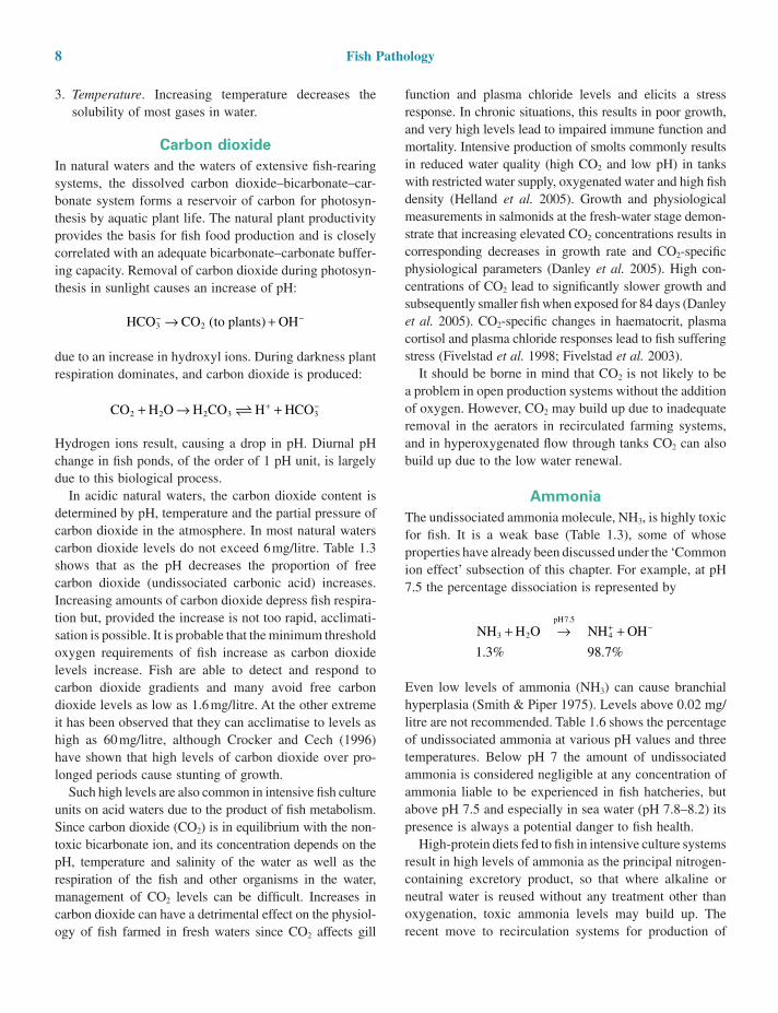
Even low levels of ammonia (NH3) can cause branchial hyperplasia (Smith & Piper 1975). Levels above 0.02 mg/litre are not recommended. Table 1.6 shows the percentage of undissociated ammonia at various pH values and three temperatures. Below pH 7 the amount of undissociated ammonia is considered negligible at any concentration of ammonia liable to be experienced in fish hatcheries, but above pH 7.5 and especially in sea water (pH 7.8–8.2) its presence is always a potential danger to fish health.
High-protein diets fed to fish in intensive culture systems result in high levels of ammonia as the principal nitrogen-containing excretory product, so that where alkaline or neutral water is reused without any treatment other than oxygenation, toxic ammonia levels may build up. The recent move to recirculation systems for production of
3. Temperature. Increasing temperature decreases the solubility of most gases in water.
Carbon dioxideIn natural waters and the waters of extensive fish-rearing systems, the dissolved carbon dioxide–bicarbonate–car-bonate system forms a reservoir of carbon for photosyn-thesis by aquatic plant life. The natural plant productivity provides the basis for fish food production and is closely correlated with an adequate bicarbonate–carbonate buffer-ing capacity. Removal of carbon dioxide during photosyn-thesis in sunlight causes an increase of pH:
HCO CO to plants OH3 2− −→ +( )
due to an increase in hydroxyl ions. During darkness plant respiration dominates, and carbon dioxide is produced:
CO H O H CO H HCO2 2 2 3 3+ → ++ −
Hydrogen ions result, causing a drop in pH. Diurnal pH change in fish ponds, of the order of 1 pH unit, is largely due to this biological process.
In acidic natural waters, the carbon dioxide content is determined by pH, temperature and the partial pressure of carbon dioxide in the atmosphere. In most natural waters carbon dioxide levels do not exceed 6 mg/litre. Table 1.3 shows that as the pH decreases the proportion of free carbon dioxide (undissociated carbonic acid) increases. Increasing amounts of carbon dioxide depress fish respira-tion but, provided the increase is not too rapid, acclimati-sation is possible. It is probable that the minimum threshold oxygen requirements of fish increase as carbon dioxide levels increase. Fish are able to detect and respond to carbon dioxide gradients and many avoid free carbon dioxide levels as low as 1.6 mg/litre. At the other extreme it has been observed that they can acclimatise to levels as high as 60 mg/litre, although Crocker and Cech (1996) have shown that high levels of carbon dioxide over pro-longed periods cause stunting of growth.
Such high levels are also common in intensive fish culture units on acid waters due to the product of fish metabolism. Since carbon dioxide (CO2) is in equilibrium with the non-toxic bicarbonate ion, and its concentration depends on the pH, temperature and salinity of the water as well as the respiration of the fish and other organisms in the water, management of CO2 levels can be difficult. Increases in carbon dioxide can have a detrimental effect on the physiol-ogy of fish farmed in fresh waters since CO2 affects gill
The Aquatic Environment 9
Hydrogen sulphideOther compounds or ions which are toxic for fish may depend on dissociation equilibria to reach toxic levels. Hydrogen sulphide is frequently produced as a result of the organic enrichment of the benthos that takes place immediately beneath cage farms, but it can also occur naturally with the accumulation of natural organic matter in deep sinks in shallow lakes or reservoirs. Benthic anaer-obic deposits of hydrogen sulphide can overturn to produce serious fish losses under certain conditions as it is extremely poisonous for fish as the undissociated molecule. It ionises according to pH, as follows, so the problem is particularly significant in acidic waters.
H S H HS
H S H HS
H S H HS
pH
pH
pH
2
9
2
7
2
7
1
50
99
( %)
( %)
( %)
→ +
→ +
→ +
+ −
+ −
+ −
The maximum acceptable level of undissociated hydrogen sulphide is usually 0.002 mg/litre.
Mineral contentsNaturally occurring fresh waters may vary enormously in mineral content depending on the source and location. Sea and fresh water are clearly distinct, but it is not always appreciated that a continuum of saline content between the two occurs in nature. Two definitions are commonly used to determine the salt content.
Salinity is a measure of the total salts in 1 kg of sea water, when all the carbonate has been converted to oxide, the bromides and iodides have been replaced by chloride
high-value species, such as salmon smolts, requires that ammonia is removed by biological filtration at each cycle. The effects of ammonia toxicity on salmonids in fresh water was reviewed by Knoph and Thorud (1996), and the toxicity of nitrogenous metabolic wastes in marine condi-tions by Handy and Poxton (1993).
The level of unionised ammonia is dependent not only on total ammonia (TAN; NH3 NH4
+) and pH level, but also on temperature and salinity (Fivelstad, Bergheim, & Tyvold 1991; Eddy 2005; Ackerman et al. 2006). Since ammonia toxicity is higher at high pH (e.g. at pH 8 only a level of 5% of total ammonia is in the toxic NH3 form, whereas at pH 9 20% is in the more toxic form) (Fivelstad et al. 1991; Eddy 2005), five times more toxic ammonia is available at 25°C than at 5°C and the proportion of the more toxic NH3 form increases as salinity drops, pH, tem-perature and salinity must all be known to estimate how toxic ammonia will be at any particular level (Ackerman et al. 2006). Ammonia disturbs osmoregulation resulting in higher urine production in fresh water and increased drinking in salt water (Knopf & Olsen 1994; Knopf & Thorud 1996; Eddy 2005).
In general, ammonia toxicity (96 h LC50 for adult Atlantic salmon held in sea water e.g. 0.09–3.35 mg/l NH3) appears to be roughly similar to that for fresh-water salmon (e.g. 0.068–2.0 mg 1/l NH3) (Eddy 2005), but in the marine environment the toxicity of ionised ammonia (NH4
+) should also be considered. The water quality standard for fresh-water salmonids of 21 µg 1(−1) NH3-N is generally con-sidered to be acceptable for most marine fish. During ammonia exposures, whether chronic or episodic, it is younger fish that are most at risk especially if the pH value of the water is decreased (Eddy 2005).
Table 1.6 Variation in percentage of undissociated ammonia in aqueous ammonia solution with temperature and pH.
Temperature (°C)
pH value
6 6.5 7 7.5 8 8.5 9 9.5
0 0.008 0.026 0.083 0.261 0.820 2.55 20.7 45.35 0.013 0.040 0.125 0.400 1.23 3.80 28.3 55.6
10 0.019 0.059 0.186 0.590 1.83 5.56 37.1 65.115 0.027 0.087 0.273 0.860 2.67 7.97 46.4 73.320 0.0400 0.125 0.400 1.24 3.82 11.2 55.7 79.925 0.057 0.180 0.570 1.77 5.38 15.3 64.3 85.130 0.081 0.254 0.800 2.48 7.46 20.3 71.8 89.0
After Emerson et al. (1975). For sea-water values, see Whitefield (1974).
10 Fish Pathology
plies, but ground waters may contain significant quantities of dissolved minerals such as ferrous iron. The toxicity of many heavy metals decreases as the pH increases due to pH-related effects including decreased solubility or increased complexing with other compounds or ions.
Acid rain causing rapid reduction in pH is a particular phenomenon which can greatly affect fish survival in sus-ceptible areas; it is discussed in greater detail in the ‘Acid rain’ subsection of this chapter. It is well established that positively charged aluminium in acidic waters (Dickson 1978) is toxic to fish due to accumulation of Al in fish gills (Muniz and Leivestad 1980a, b; Exley et al. 1991), causing ionoregulatory and/or respiratory failure (Neville 1985; Wood and McDonald 1987; Gensemer and Playle 1999). At relatively low-exposure concentrations, responses are identified at the histological and physiological levels. In fresh waters, Al can be present in different physico-chemical forms varying from simple cations and hydroly-sis products, complexes and polymers, to colloids and particles (Salbu and Oughton 1995), depending on pH, temperature and the concentration of Al complexing ligands present. In anadromous fish, the consequences of exposure to toxic Al at the fresh-water stages may not become apparent until the sea-water stage (Exley et al. 1991).
and the organic matter has oxidised. By definition it is expressed as g/kg or parts per thousand (‰). Salinity measurements can be made directly using a conductivity meter, refractometer or hydrometer. Coastal water com-monly has a variable salinity due to fresh-water runoff.
Chlorinity is a measure of the total halides in a given weight of sea water. It is defined as the mass of silver necessary to precipitate the halogens in 328.523 g of sea water. It is also expressed as parts per thousand (‰). Chlorinity is usually determined by titration.
Oceanic water has a chlorinity of approximately 19‰. The constant relation between salinity and chlorinity is expressed by
S Cl‰ ‰= +0 030 1 8050. .
Biologists are agreed on the need to define salinity or chlorinity ranges, and several classifications have been proposed. One of the more widely accepted classifications is that of Redeke (Table 1.7).
Fish are most at risk when variation in salinity occurs to the extent that the gill and kidney are unable to control the osmolarity of the body fluids.
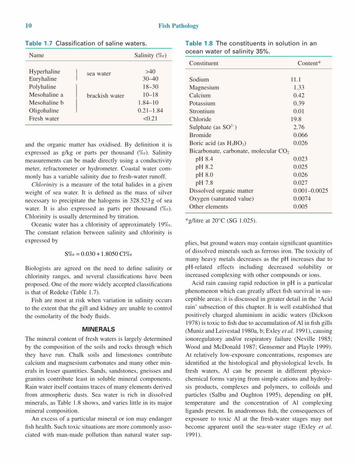
MINERALSThe mineral content of fresh waters is largely determined by the composition of the soils and rocks through which they have run. Chalk soils and limestones contribute calcium and magnesium carbonates and many other min-erals in lesser quantities. Sands, sandstones, gneisses and granites contribute least in soluble mineral components. Rain water itself contains traces of many elements derived from atmospheric dusts. Sea water is rich in dissolved minerals, as Table 1.8 shows, and varies little in its major mineral composition.
An excess of a particular mineral or ion may endanger fish health. Such toxic situations are more commonly asso-ciated with man-made pollution than natural water sup-
Table 1.8 The constituents in solution in an ocean water of salinity 35%.
Constituent Content*
Sodium 11.1Magnesium 1.33Calcium 0.42Potassium 0.39Strontium 0.01Chloride 19.8Sulphate (as SO2−) 2.76Bromide 0.066Boric acid (as H3BO3) 0.026Bicarbonate, carbonate, molecular CO2
pH 8.4 0.023 pH 8.2 0.025 pH 8.0 0.026 pH 7.8 0.027Dissolved organic matter 0.001–0.0025Oxygen (saturated value) 0.0074Other elements 0.005
*g/litre at 20°C (SG 1.025).
Table 1.7 Classification of saline waters.
Name Salinity (‰)
Hyperhaline sea water >40Euryhaline 30–40Polyhaline
brackish water
18–30Mesohaline a 10–18Mesohaline b 1.84–10Oligohaline 0.21–1.84Fresh water <0.21
The Aquatic Environment 11
tures closer to their upper thermal limits than do fish living in temperate waters, an important factor when assessing the significance of thermal pollution in the tropics. Temperature modifies the impact of pollutants. Many are more toxic in warmer waters, and since they are also more soluble at higher temperatures they may also reach higher concentrations.
Heavy metalsThe commonest causes of heavy metal pollution are copper, lead, mercury, zinc, chromium, cadmium, manga-nese and iron. Industrial discharges and seepage from industrial and mining wastes are the commonest sources, although sometimes they occur naturally. Defining maximum safe levels of any particular metal is difficult, as much ancillary information is required, such as the pH, acidity or carbonate alkalinity, temperature, dissolved oxygen content, presence of other metals (they often act synergistically, e.g. cadmium in the presence of zinc or copper) (La Roche 1972), length of exposure, species and age of exposed fish. There are also distinct differences between strains of fish of the same species. Particular strains occur which are adapted to particular levels of, for instance, selenium, that might be toxic to other strains of the same species from a different watershed (Hardy 2009). The pathology of metallic poisoning varies according to the concentration and length of exposure as well as inher-ent susceptibility level, and is not a reliable diagnostic feature unless historical and analytical evidence is also available.
NonmetalsMany non-metals are toxic if present in sufficient quantity. Some of those encountered commonly are ammonia, fluo-rides, cyanides, phosphorus, sulphides, aluminium and beryllium salts, arsenates and halogens, particularly chlo-rine and the chloramines. Many organic compounds used in agriculture and industry are also toxic for fish.
Pesticides are chemicals designed to destroy plant and animal life. The major sources are runoff from treated farmlands, industrial and domestic sewage, spillage and direct application to waterways, such as in herbicide treat-ments and aquatic crop treatments (e.g. rice production).
More recently, however, concern has been expressed about chemicals such as organophosphates and aver-mectins used directly in support of the control of fish para-sites, in particular marine crustacean parasites (Roth et al. 1993). These concerns relate both to possible effects on the environment and also to potential problems due to residues remaining in the fish when it is consumed. For
The effects of high levels of acidity have been best studied in salmonids affected by iatrogenic acid rain in northern latitudes. In Norway, acute mortality of Atlantic salmon has even been described from marine fjord-based fish farms (Bjerknes et al. 2003). Mortality is often related to snowmelt and heavy rainfall in the catchment areas during the winter. Increased fresh-water runoff reduces the surface water salinity from >20 to <10, while water tem-perature is reduced from 8°C to 3°C. Aluminium, trans-ported by acid rivers to the fjords during these episodes, is the cause of the mortality. An increased deposition of aluminium (Al) on the gills of these Atlantic salmon (from <10 to >200 mug g(−1) dry weight) has been demonstrated (Bjerknes et al. 2003). The increases in gill Al were related to increased discharge episodes where acidic, Al-rich fresh water elevated the surface water concentrations of Al from < 20 to >70 mug Al l(−1). Aluminium levels for salmonid-rearing waters should be kept below 20µg/L since this is the critical level that smolts, which are the most sensitive growth stage, can tolerate (Rosseland et al. 2001).
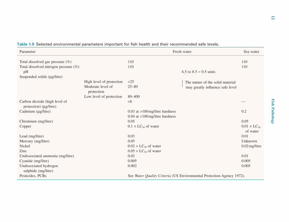
POLLUTANTSThere are many potential pollutants whose occurrence causes a reduction in the quality of the aquatic environ-ment and about which a wealth of short-term toxicity test data has been accumulated. Results are commonly reported as a median lethal concentration (LC50) or median toler-ance limit (TL50). Both indicate the concentration which kills 50% of the test species within a specified time span, usually 96 hours. On no account should LC50 or TL50 levels be viewed as safe levels. Safe levels must allow growth and other normal life processes such as reproduction to continue. Unfortunately, accurate data on safe levels are limited and in many cases the values are empirically derived by dividing the LC50 concentrations by a factor gained from experience. Acceptable concentrations of toxicants to which organisms are to be exposed continually must take into account enhanced concentrations which may be reached occasionally during brief periods, and the long-term effects of cumulative poisons (Sprague 1971) (Table 1.9).
Thermal pollutionIncreased water temperatures may be beneficial for fish culture in that faster growth is achieved as a result of year-round enhancement of the rate of metabolism, but only if the increase is to a level below the thermal limit for the species. Tropical fish often live at environmental tempera-
12 F
ish Pathology
Table 1.9 Selected environmental parameters important for fish health and their recommended safe levels.
Parameter Fresh water Sea water
Total dissolved gas pressure (%) 110 110Total dissolved nitrogen pressure (%) 110 110
pH 6.5 to 8.5 = 0.5 unitsSuspended solids (µg/litre)
High level of protection <25 The nature of the solid material may greatly influence safe levelModerate level of
protection25–80
Low level of protection 80–400Carbon dioxide (high level of
protection) (µg/litre)<6 —
Cadmium (µg/litre) 0.03 at >100 mg/litre hardness 0.20.04 at <100 mg/litre hardness
Chromium (mg/litre) 0.05 0.05Copper 0.1 × LC50 of water 0.01 × LC50
of waterLead (mg/litre) 0.03 0.01Mercury (mg/litre) 0.05 UnknownNickel 0.02 × LC50 of water 0.02 mg/litreZinc 0.05 × LC50 of waterUndissociated ammonia (mg/litre) 0.02 0.01Cyanide (mg/litre) 0.005 0.005Undissociated hydrogen
sulphide (mg/litre)0.002 0.005
Pesticides, PCBs See Water Quality Criteria (US Environmental Protection Agency 1972).
The Aquatic Environment 13
catfishes and sea bass, that the opportunity to directly control tissue contaminant levels has become available. Such considerations are essential in relation to any assess-ment of comparative contaminant levels and their signifi-cance. Comparisons between farmed and wild fish are particularly difficult in this context, and it is essential to compare like with like. Hites, et al. (2004), for example, in an influential but now widely discredited study com-pared farmed Atlantic salmon harvested in mid–reproductive cycle from Atlantic oceanic waters and wild Pacific salmon, of different species, captured in pre-spawning condition from Pacific coastal waters.
EFSA (2005), in a carefully validated peer-reviewed response to such claims, concluded that there was little risk of toxicity from normal consumption of wild or farmed species and that the benefits to human health of such con-sumption far outweighed any minimal risk from organic or inorganic residues.
SewageSewage discharges may reduce water quality, depending on the degree of dilution achieved, the degree of treatment of the original material, its composition and the response of the ecosystem. Oxygen depletion is the most common result of such discharges. It arises from insufficient dilu-tion, and microbial growth on its particulate and soluble organic content. Sewage-derived inorganic nutrients, such as phosphates, ammonia and nitrates, may stimulate exces-sive blooms of algae or attached weed with attendant oxygen depletion and toxin production. Sewage is also a potential source of heavy metals and toxic organic wastes such as PCBs. Although its presence is likely to be short-lived, the highly toxic nitrite ion may also be present in sewage discharges.
Particulate materialsAll natural waters contain some suspended solids. During spates these can rise considerably, but wild fish can nor-mally avoid them. Farmed fish do not have this opportunity, and effects such as gill surface hyperplasia and excessive mucus generation on skin and gills are common. Fish eggs, both in the wild and under farm conditions, are very vulner-able to silt deposits which inhibit respiration through the chorial membrane and encourage microbial growth.
Wastes associated with certain industries, such as quar-rying, sand and gravel extraction, mining, and paper and paint manufacture, and surface disturbance from civil engineering, can introduce large amounts of particulate matter into rivers and their effects on fish health may be observed many miles downstream from its source. As well
these reasons such usages are now closely controlled in most developed countries (Woodward 1996)
A very wide range of compounds is currently in use. They may have direct toxic effects on fish populations and contaminate and accumulate in the food and flesh of wild and cultured fish, or indirect effects due to their influence on invertebrates and plant life. Modern carbonate and organophosphorus pesticides are generally less toxic than highly persistent organochlorine compounds. Many are rapidly inactivated by microbial degradation or absorption on to particulate matter but some, such as DDT or dieldrin, are highly resistant to degradation and are concentrated within the food web, leading to fish and thus to man. The toxicity of individual compounds varies widely and present knowledge of pesticides and their fish toxic pathology is insufficient for reliable diagnosis unless considerable his-torical and analytical data are available. Further details of toxicity are presented in Murty (1986).
Polychlorinated biphenyls (PCB) occur in natural waters from a variety of industrial sources including their use as plasticisers in paints and plastics. They are cumulative toxins for fish. Another industrial toxicant, widely used in the marine environment until recently, was tributyl tin oxide, a potent antifoulant which was extensively used in fish culture to protect nets. It is toxic to many molluscs and its use is now banned in most countries (Fisher et al. 1995).
More recently concerns have been expressed about levels of PCBs and dioxins present in farmed and wild fish tissues and their possible risk for humans consuming them. In all of these discussions, the main weight of emphasis has been on the chemical assessment and putative health risk of consumption of wild and farmed fish by humans, but no consideration was given to the nutritional value of such fish consumption. The importance of fish, and in particular the fatty fishes such as herring, mackerel, tuna and salmon, in the human diet as a source of (omega-3) long-chain n3 polyunsaturated fatty acids (LCn3PUFA), is well recognised. Unfortunately it is also within the lipid component of the fish that lipid-soluble contaminants such as dioxins and PCBs obtained via the food are stored.
Fish is a generic term in the context of human diet, and there are wide differences in both nutritional value and potential contaminant levels, depending not only on the origin and life stage of the fish species, but also on the tissue sampled, the season of harvest and, for farmed fish, the content of the diet. The diet of wild fish is totally beyond human control, and it is only with the development of formulated diets, for farmed species such as salmon,
14 Fish Pathology
of water supplies with calcium-based compounds has been successfully used to treat acidic and aluminium-rich water. Reviews by Driscoll (1985) and Campbell and Stoakes (1985) summarise aluminium and metal toxicity in acid waters. The effects of acidification on fish are covered in reviews by Johnson (1982), Howells (1984) and Exley and Phillips (1988).
TaintsA wide variety of objectionable tastes, odours and colours have been noted in fish flesh. Both natural and industrial causes are implicated. Muddy or earthy tastes in the flesh of pond-reared trout are caused by the activities of soil bacteria of the Actinomycetaceae and certain Cyanobacteria. Industrial wastes implicated in causing taints include oil products, phenolic disinfectants and domestic sewage. Some taints can be removed or reduced by holding fish in clean water for long periods, but taints are more rapidly acquired than eliminated.
ADVERSE BIOLOGICAL FACTORS FOR FISH HEALTH
AQUATIC ANIMALSAll surface waters may contain species of wild fish which can act as reservoirs of infectious disease. Animals other than fish may be reservoirs of infection as well as intermedi-ates in the life cycles of many parasites. If practicable, the removal of molluscs and crustaceans from the inflows and ponds of fish farms will reduce possible infestation of farmed fish. In farming situations, predators and competi-tors for food or space must be excluded from the farm itself and its immediate surroundings if at all possible.
MICROORGANISMSAlgae
Algae may affect fish health through the production of toxins or through mechanical damage to fish gills (e.g. certain diatoms). Toxin-producing algae are found in marine, brackish and fresh waters throughout the world. Under suitable environmental conditions they grow to considerable cell densities (20–100 × 103 cells/ml), called blooms or tides, and during or after these blooms toxins may be produced which are lethal for fish. Toxin-producing algae are confined to three major taxonomic groups, the Pyrophyta, Chrysophyta and Cyanophyta.
as an effect on the gills, which may ultimately lead to high mortalities if such fish are oxygen stressed subsequently, high levels of suspended solids also reduce light penetra-tion into water, resulting in less energy in the food web supporting fish production (European Inland Fisheries Advisory Committee 1965).
Oil pollutionSpills of crude and refined oils can have highly toxic effects in ponds and other enclosed waters where dilution of the water-soluble components is not rapid. Crude oils are relatively less toxic, but use of oil dispersants and their solvents greatly increases the toxicity for fish unless dilu-tion is considerable. Oil spillage at sea or downstream of refineries may make conditions impossible for aquacul-ture. Even when oil levels are minimal and do not affect the fish per se, the resulting taint in the fish flesh makes affected fish totally unmarketable (Goodlad 1996).
Acid rainFish kills associated with the effects of man-made acidifi-cation have been widely recognised in Western Europe and North America for many years. The problems stem from inputs of inorganic acids and particulate metals into the atmosphere from industries burning fossil fuels. The acids are then transported over considerable distances by pre-vailing winds and deposited in areas many miles from the source, leading to the phenomenon of ‘acid rain’. The most vulnerable waters are those in localities with hard, insolu-ble bedrocks where the natural buffering capacity of soils and waters is very low, such as occur in the mountainous areas of Europe and North America. The result is water of an unnaturally low pH, commonly associated with high concentrations of aluminium and other metal ions leached from the substrate of the catchment area by the unnatural acidity, particularly during snowmelt and when heavy rain follows a dry spell. The concentration of aluminium in these waters has been shown to be particularly significant, as aluminium appears to be most toxic to fish at pH 5.0–6.0, which is above the normal toxic threshold for the direct effects of increased acidity at low pH on fish. The toxic thresholds for aluminium are not well established and vary considerably, depending upon the fish species, the stage of the life cycle and other water quality charac-teristics, particularly pH, calcium and the presence of com-plexing ligands (e.g. humic acids). The pathological picture associated with aluminium and acid poisoning is generally nondiagnostic, and analytical and historical data are normally also required for successful diagnosis. Liming
The Aquatic Environment 15
CHARACTERISTICS OF DIFFERENT TYPES OF WATER
SURFACE WATERSRivers and streams
Diurnal temperature fluctuations are considerable in the smaller, shallower streams and rivers in temperate and mountain regions, especially if the waters are without shade from the sun. Great significance must be attached to all temporary, permanent or periodic industrial or agricul-tural activities upstream, which might affect water quality. These will include abstractions, change of use, discharges (including their chemical nature), civil engineering involv-ing changes or disturbances to the bottom of banks, and crop spraying. Other fish farm activities on the same water course may also be an important source of disease.
Lake watersThese are used in aquaculture to provide pumped water supplies or for pen or cage culture. Thermal stratification poses its most serious problems in eutrophic waters, when overturn, or destratification, occurs and the deeper waters, depleted in oxygen or even anoxic, come to the surface. Overturn of stratified waters may occur at any time during stormy periods, but is an annual event after the summer in temperate regions.
GROUND AND SPRING WATERSGround and spring waters are of considerable value for aquaculture because of two major properties: constancy of temperature and the virtual absence of parasites or micro-bial flora. However, fish health may suffer in these waters unless their quality is fully evaluated beforehand by chem-ical analyses and if possible by bioassay (keeping trial batches of fish in the water and observing their growth and development). Spring and ground waters are derived from the same source, namely rain water draining through the surface soil layers until it reaches the water table. The depth of the water table is determined by several factors, including the depth of the impervious layer below it, the rate of rainfall, the porosity of the soil or stone and local topography and geology.
Commonly, ground and spring waters are not saturated with oxygen, but are supersaturated with nitrogen and, if acidic, have high levels of carbon dioxide. Intensive aera-tion may be necessary to add oxygen and remove nitrogen and carbon dioxide. Warm-water springs in areas of vol-canic activity may contain hydrogen sulphide. The water
DinoflagellatesThe Pyrophyta (dinoflagellates) which produce toxin are mostly of marine origin. Massive wild-fish kills resulting from blooms of Gonyaulax tamarensis have been reported from the North Sea and the North Atlantic coast of the United States, from G. catenella in the Northern Pacific from the United States to Japan, and from G. monilata in the Gulf of Mexico. Their incidence is apparently increas-ing and they are responsible for considerable mortality (Dundas et al. 1989; Anderson, et al. 1997). Other species and genera of dinoflagellates produce piscine toxins in laboratory culture, but their ecological significance is unknown. The toxins of most species may well be the same. Khan et al. (1997) have shown that neurotoxins, haemolysins and haemagglutinins may all be present The pharmacology of the neurotoxin in fish is unknown, but in humans and other mammals (in which it is also toxic, causing a disease called paralytic shellfish poisoning) it causes muscular paralysis, with death resulting from res-piratory failure.
Recently major fish kills have been reported in Atlantic salmon culture in North-West Europe in association with ‘red tides’ caused by dinoflagellates such as Gyrodinium aureolum. These are naked dinoflagellates which have a cell-associated toxin that acts on the gills and digestive tract rather than the nervous system (Roberts et al. 1983). Non-toxic dinoflagellates may also cause mortality simply by virtue of their physical effects on the gills (Kent et al. 1995).
PhytoflagellatesThe Chrysophyta are phytoflagellates of brackish and euryhaline waters, and some members produce extracel-lular ichthyotoxins. Prymnesium parvum is the only toxic phytoflagellate of known ecological significance in brackish-water fish kills, with reports of its ichthyotoxic activities coming from Holland, Denmark, England, Belgium and Israel.
Blue-green algaeSome cyanophytes produce toxins which cause mortality to fish and other animals. Several genera, including Microcystis, often grow to form a thick viscous scum 10–20 cm thick on the surface of ponds. The algae cause two direct effects on fish populations: poisoning by excre-tion of ichthyotoxins, and asphyxiation by the rapid deple-tion of oxygen due to algal respiration or the sudden death of a bloom.
16 Fish Pathology
chemistry reflects the chemistry of the rocks with which the water has had contact (often for centuries). Ground waters from strata rich in heavy metals (e.g. lead, copper, chromium and mercury) may be poisonous. Anoxic waters from sandstones may be rich in ferrous salts which on aeration are converted into insoluble ferric hydroxides. These hydroxides must be removed, commonly by mechanical screening. The presence of wild fish popula-tions in spring waters is normally indicative of their suit-ability for aquaculture.
SEA WATERThe major forms of marine fish culture involve the use of floating net pens or cages, net enclosures on the shoreline, lagoons or ponds or pumped sea water in shore-based tanks. Some of the most significant qualities of sea water for fish health are influenced by the proximity of the shore:
1. Temperature. Shallow areas sheltered from water exchange are warmer than the open sea in the tropics and in temperate regions in summer, although in winter the opposite may apply, with ice formation occurring in many temperate regions.
2. Water exchange in all systems except shore-based tanks depends on natural features. Upon this exchange
depends the density at which fish may be kept in indi-vidual units and the overall biomass which any particu-lar area can support. Exchange may be limited if tidal influences are negligible, or erratic if the circulation is wind-driven. If the natural productivity of such waters is high the problems common in eutrophic lakes may occur, such as toxic algal blooms and thermal and oxygen stratification with periodic overturn leading to surface waters low in oxygen.
3. Fresh-water runoff may result in reduced and widely fluctuating salinities, especially in the top 5–10 m. Long sea inlets with large rivers emerging into them are areas most prone to this feature. Fresh-water runoff may contain pollutants toxic for fish and may also be cooler or warmer than sea water.
The natural inhabitants of sea water, particularly fish, will be a possible reservoir of potentially pathogenic microorganisms. Good husbandry can often alleviate if not eliminate the effects of such pathogens. Some shore-based aquaculture facilities and aquaria use sublittoral sandy sea bottoms as filters, placing their intake pipes several metres below the sand surface to ensure water of high clarity and low microbial and parasitic content.
2The Anatomy and Physiology of Teleosts
INTRODUCTION
The purpose of the present chapter is to give a brief outline of the aspects of the anatomy and physiology of teleost fish which are necessary for an appreciation of the patho-logical changes which can occur in fish and for an under-standing of the mechanisms underlying clinical disease manifestations.
Many basic functions of teleosts are similar to those of other vertebrates, and knowledge of mammals is not entirely irrelevant. However, the teleosts must not be regarded as the primitive forebears of mammals; they are advanced, evolutionarily recent and expanding into a mul-titude of niches. There are more species of teleost than any other class of vertebrate (Bone & Moore 2008), so that many generalisations are obviously of dubious value, but the principal exploited species are of a restricted range so that useful statements can still be made. The main empha-sis in this account will be on specific differences from the more familiar mammalian anatomy and physiology.
A major consideration is the aquatic environment and the constraints it imposes on fish. It is against the physical and biological degradative influences of this medium that the milieu interieur of the fish must be maintained and with which necessary exchanges of materials must take place. An overriding factor is the high specific heat of water, which imposes, on most fish, ectothermy (poikilo-thermy; i.e. the body temperature conforms to the environ-mental temperature). No simple physiological constant values can be given (e.g. for heart rate, rate of digestion
or rate of growth); all of these are subject to temperature and this must always be borne in mind when studying teleosts. The same animal is often unrecognisable at dif-ferent temperatures, for example whether adrenaline increases or decreases heart rate in rainbow trout depends on the temperature (Randall 1970).
THE INTEGUMENTARY SYSTEM
The skin is the primary barrier against the environment, allowing normal internal physiological function, so its condition is important in many disease processes. The layers of teleost skin, comprising the cuticle, epidermis, basement membrane, dermis and hypodermis, are indi-cated diagrammatically in Figure 2.1.
CUTICLEThe external layer, the cuticle or glycocalyx, was first described in detail by Whitear (1970) as a mainly muco-polysaccharide layer approximately 1 µm thick. It is nor-mally formed largely from epithelial surface cells rather than by secretion from goblet mucous cells and is a complex of cell protoplasm, sloughed cells and any goblet cell mucus that has been secreted onto the surface (Figure 2.2). The physical consistency of the cuticle varies consid-erably between species, being especially developed in rock pool and benthic species.
The cuticular layer contains specific immunoglobulins and lysozyme (although the amount of the latter varies
Fish Pathology, Fourth Edition. Edited by Ronald J. Roberts.© 2012 Blackwell Publishing Ltd. Published 2012 by Blackwell Publishing Ltd.
17
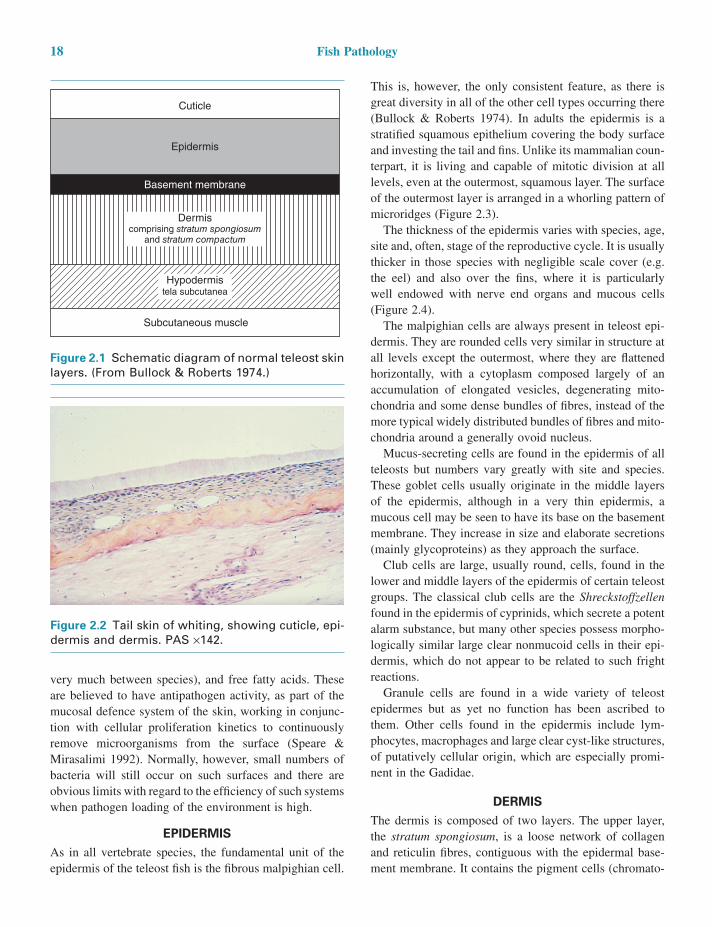
18 Fish Pathology
This is, however, the only consistent feature, as there is great diversity in all of the other cell types occurring there (Bullock & Roberts 1974). In adults the epidermis is a stratified squamous epithelium covering the body surface and investing the tail and fins. Unlike its mammalian coun-terpart, it is living and capable of mitotic division at all levels, even at the outermost, squamous layer. The surface of the outermost layer is arranged in a whorling pattern of microridges (Figure 2.3).
The thickness of the epidermis varies with species, age, site and, often, stage of the reproductive cycle. It is usually thicker in those species with negligible scale cover (e.g. the eel) and also over the fins, where it is particularly well endowed with nerve end organs and mucous cells (Figure 2.4).
The malpighian cells are always present in teleost epi-dermis. They are rounded cells very similar in structure at all levels except the outermost, where they are flattened horizontally, with a cytoplasm composed largely of an accumulation of elongated vesicles, degenerating mito-chondria and some dense bundles of fibres, instead of the more typical widely distributed bundles of fibres and mito-chondria around a generally ovoid nucleus.
Mucus-secreting cells are found in the epidermis of all teleosts but numbers vary greatly with site and species. These goblet cells usually originate in the middle layers of the epidermis, although in a very thin epidermis, a mucous cell may be seen to have its base on the basement membrane. They increase in size and elaborate secretions (mainly glycoproteins) as they approach the surface.
Club cells are large, usually round, cells, found in the lower and middle layers of the epidermis of certain teleost groups. The classical club cells are the Shreckstoffzellen found in the epidermis of cyprinids, which secrete a potent alarm substance, but many other species possess morpho-logically similar large clear nonmucoid cells in their epi-dermis, which do not appear to be related to such fright reactions.
Granule cells are found in a wide variety of teleost epidermes but as yet no function has been ascribed to them. Other cells found in the epidermis include lym-phocytes, macrophages and large clear cyst-like structures, of putatively cellular origin, which are especially promi-nent in the Gadidae.
DERMISThe dermis is composed of two layers. The upper layer, the stratum spongiosum, is a loose network of collagen and reticulin fibres, contiguous with the epidermal base-ment membrane. It contains the pigment cells (chromato-
very much between species), and free fatty acids. These are believed to have antipathogen activity, as part of the mucosal defence system of the skin, working in conjunc-tion with cellular proliferation kinetics to continuously remove microorganisms from the surface (Speare & Mirasalimi 1992). Normally, however, small numbers of bacteria will still occur on such surfaces and there are obvious limits with regard to the efficiency of such systems when pathogen loading of the environment is high.
EPIDERMISAs in all vertebrate species, the fundamental unit of the epidermis of the teleost fish is the fibrous malpighian cell.
Figure 2.1 Schematic diagram of normal teleost skin layers. (From Bullock & Roberts 1974.)
Cuticle
Epidermis
Basement membrane
Hypodermistela subcutanea
Subcutaneous muscle
Dermiscomprising stratum spongiosum
and stratum compactum
Figure 2.2 Tail skin of whiting, showing cuticle, epi-dermis and dermis. PAS ×142.
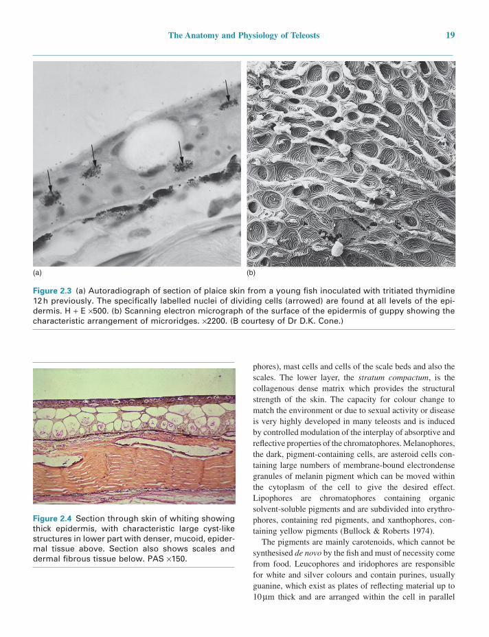
The Anatomy and Physiology of Teleosts 19
Figure 2.4 Section through skin of whiting showing thick epidermis, with characteristic large cyst-like structures in lower part with denser, mucoid, epider-mal tissue above. Section also shows scales and dermal fibrous tissue below. PAS ×150.
Figure 2.3 (a) Autoradiograph of section of plaice skin from a young fish inoculated with tritiated thymidine 12 h previously. The specifically labelled nuclei of dividing cells (arrowed) are found at all levels of the epi-dermis. H + E ×500. (b) Scanning electron micrograph of the surface of the epidermis of guppy showing the characteristic arrangement of microridges. ×2200. (B courtesy of Dr D.K. Cone.)
(b)(a)
phores), mast cells and cells of the scale beds and also the scales. The lower layer, the stratum compactum, is the collagenous dense matrix which provides the structural strength of the skin. The capacity for colour change to match the environment or due to sexual activity or disease is very highly developed in many teleosts and is induced by controlled modulation of the interplay of absorptive and reflective properties of the chromatophores. Melanophores, the dark, pigment-containing cells, are asteroid cells con-taining large numbers of membrane-bound electrondense granules of melanin pigment which can be moved within the cytoplasm of the cell to give the desired effect. Lipophores are chromatophores containing organic solvent-soluble pigments and are subdivided into erythro-phores, containing red pigments, and xanthophores, con-taining yellow pigments (Bullock & Roberts 1974).
The pigments are mainly carotenoids, which cannot be synthesised de novo by the fish and must of necessity come from food. Leucophores and iridophores are responsible for white and silver colours and contain purines, usually guanine, which exist as plates of reflecting material up to 10 µm thick and are arranged within the cell in parallel
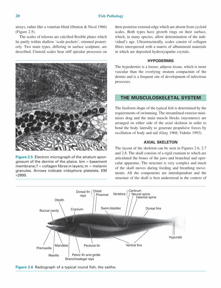
20 Fish Pathology
their posterior external edge which are absent from cycloid scales. Both types have growth rings on their surface, which, in many species, allow determination of the indi-vidual’s age. Ultrastructurally, scales consist of collagen fibres interspersed with a matrix of albuminoid materials in which are deposited hydroxyapatite crystals.
HYPODERMISThe hypodermis is a looser, adipose tissue, which is more vascular than the overlying stratum compactum of the dermis and is a frequent site of development of infectious processes.
THE MUSCULOSKELETAL SYSTEM
The fusiform shape of the typical fish is determined by the requirements of swimming. The streamlined exterior mini-mises drag and the main muscle blocks (myomeres) are arranged on either side of the axial skeleton in order to bend the body laterally to generate propulsive forces by oscillation of body and tail (Gray 1968; Videler 1993).
AXIAL SKELETONThe layout of the skeleton can be seen in Figures 2.6, 2.7 and 2.8. The skull consists of a rigid cranium to which are articulated the bones of the jaws and branchial and oper-cular apparatus. The structure is very complex and much of the skull moves during feeding and breathing move-ments. All the components are interdependent and the structure of the skull is best understood in the context of
Figure 2.6 Radiograph of a typical round fish, the saithe.
Ventral fins
Hypurals
Buccal cavity
Pectoral fin
Pelvic fin and girdle
Dorsal fins
Haemal spine
CentrumVertebra
Swim-bladder
Dorsal finrays
Cranium
MandiblePremaxilla
MaxillaBranchiostegal rays
Neural spine
Otolith
ProximalDistal
Figure 2.5 Electron micrograph of the stratum spon-giosum of the dermis of the plaice. bm = basement membrane; f = collagen fibres in layers; m = melanin granules. Arrows indicate iridophore platelets. EM ×2800.
bmf
f
m
m
arrays, rather like a venetian blind (Denton & Nicol 1966) (Figure 2.5).
The scales of teleosts are calcified flexible plates which lie partly within shallow ‘scale pockets’, oriented posteri-orly. Two main types, differing in surface sculpture, are described. Ctenoid scales bear stiff spicular processes on