Embed Size (px)
Citation preview
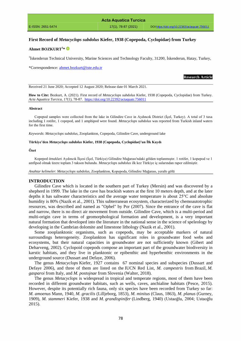
Acta Aquatica Turcica
E-ISSN: 2651-5474 17(1), 78-87 (2021) DOI:https://doi.org/10.22392/actaquatr.756011
78
First Record of Metacyclops subdolus Kiefer, 1938 (Copepoda, Cyclopidae) from Turkey
Ahmet BOZKURT1*
1İskenderun Technical University, Marine Sciences and Technology Faculty, 31200, İskenderun, Hatay, Turkey,
*Correspondence: [email protected]
Research Article
Received 21 June 2020; Accepted 12 August 2020; Release date 01 March 2021.
How to Cite: Bozkurt, A. (2021). First record of Metacyclops subdolus Kiefer, 1938 (Copepoda, Cyclopidae) from Turkey.
Acta Aquatica Turcica, 17(1), 78-87. https://doi.org/10.22392/actaquatr.756011
Abstract
Copepod samples were collected from the lake in Gilindire Cave in Aydıncık District (İçel, Turkey). A total of 3 taxa
including 1 rotifer, 1 copepod, and 1 amphipod were found. Metacyclops subdolus was reported from Turkish inland waters
for the first time.
Keywords: Metacyclops subdolus, Zooplankton, Copepoda, Gilindire Cave, underground lake
Türkiye’den Metacyclops subdolus Kiefer, 1938 (Copepoda, Cyclopidae)’un İlk Kaydı
Özet
Kopepod örnekleri Aydıncık İlçesi (İçel, Türkiye) Gilindire Mağarası'ndaki gölden toplanmıştır. 1 rotifer, 1 kopepod ve 1
amfipod olmak üzere toplam 3 takson bulundu. Metacyclops subdolus ilk kez Türkiye iç sularından rapor edilmiştir.
Anahtar kelimeler: Metacyclops subdolus, Zooplankton, Kopepoda, Gilindire Mağarası, yeraltı gölü
INTRODUCTION
Gilindire Cave which is located in the southern part of Turkey (Mersin) and was discovered by a
shepherd in 1999. The lake in the cave has brackish waters at the first 10 meters depth, and at the later
depths it has saltwater characteristics and the average water temperature is about 25°C and absolute
humidity is 80% (Nazik et al., 2001). This subterranean ecosystem, characterized by chemoautotrophic
resources, was described and named as "Ophel" by Por (2007). Since the entrance of the cave is flat
and narrow, there is no direct air movement from outside. Gilindire Cave, which is a multi-period and
multi-origin cave in terms of geomorphological formation and development, is a very important
natural formation that developed into the literature in the national sense in the science of speleology by
developing in the Cambrian dolomite and limestone lithology (Nazik et al., 2001).
Some zooplanktonic organisms, such as copepods, may be acceptable markers of natural
surroundings heterogeneity. Zooplankton has significant roles in groundwater food webs and
ecosystems, but their natural capacities in groundwater are not sufficiently known (Gibert and
Deharveng, 2002). Cyclopoid copepods compose an important part of the groundwater biodiversity in
karstic habitats, and they live in planktonic or epibenthic and hyperbenthic environments in the
underground source (Dussart and Defaye, 2006).
The genus Metacyclops Kiefer, 1927 contains 67 nominal species and subspecies (Dussart and
Defaye 2006), and three of them are listed on the IUCN Red List, M. campestris from Brazil, M.
gasparoi from Italy, and M. postojnae from Slovenia (Walter, 2018).
The genus Metacyclops is widespread in tropical and temperate regions, most of them have been
recorded in different groundwater habitats, such as wells, caves, anchialine habitats (Pesce, 2015).
However, despite its potentially rich fauna, only six species have been recorded from Turkey so far:
M. amoenus Mann, 1940, M. gracilis (Lilljeborg, 1853), M. minitus (Claus, 1863), M. planus (Gurney,
1909), M. stammeri Kiefer, 1938 and M. grandispinifer (Lindberg, 1940) (Ustaoğlu, 2004; Ustaoğlu
2015).
BOZKURT 2021 ActAquaTr 17(1), 78-87
79
M. subdolus has a European Mediterranean distribution (no record from North Africa); it has first
been reported by Kiefer (1938) from southern Italy (La Zinzulusa, Abysso caves), then from Sardinia
(Lindberg, 1956), Italy (Pesce et al., 1978; Pesce, 1985), Greece (Peloponnesos, Attica, Crete: Pesce,
1978; Pesce and Maggi, 1981, 1983), Mallorca (Can Pastilla: Lescher-Moutoué, 1981), Isreal
(Dimentman and Por, 1991; Defaye and Por, 2010; Spring and Cave), and northern Negev (Defaye
and Dussart, 1995).
A new species of M. subdolous was reported in a limited number of countries and regions, and for
the first time the report has been in Turkey. Some supplementary drawings and descriptions from
Gilindire specimens are provided as a basis for future comparison.
MATERIALS and METHODS
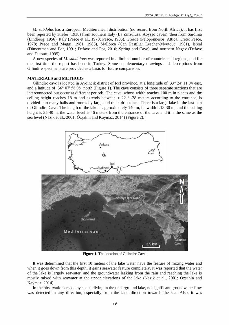
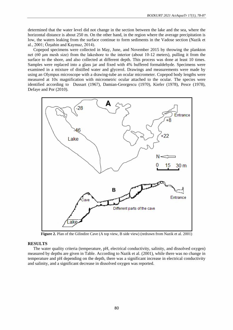
Gilindire cave is located in Aydıncık district of Içel province, at a longitude of 33° 24' 11.04''east,
and a latitude of 36° 07' 58.08'' north (Figure 1). The cave consists of three separate sections that are
interconnected but occur at different periods. The cave, whose width reaches 100 m in places and the
ceiling height reaches 18 m and extends between + 22 / -28 meters according to the entrance, is
divided into many halls and rooms by large and thick dripstones. There is a large lake in the last part
of Gilindire Cave. The length of the lake is approximately 140 m, its width is18-30 m, and the ceiling
height is 35-40 m, the water level is 46 meters from the entrance of the cave and it is the same as the
sea level (Nazik et al., 2001; Özşahin and Kaymaz, 2014) (Figure 2).
Figure 1. The location of Gilindire Cave.
It was determined that the first 10 meters of the lake water have the feature of mixing water and
when it goes down from this depth, it gains seawater feature completely. It was reported that the water
of the lake is largely seawater, and the groundwater leaking from the rain and reaching the lake is
mostly mixed with seawater at the upper elevations of the lake (Nazik et al., 2001; Özşahin and
Kaymaz, 2014).
In the observations made by scuba diving in the underground lake, no significant groundwater flow
was detected in any direction, especially from the land direction towards the sea. Also, it was
BOZKURT 2021 ActAquaTr 17(1), 78-87
80
determined that the water level did not change in the section between the lake and the sea, where the
horizontal distance is about 250 m. On the other hand, in the region where the average precipitation is
low, the waters leaking from the surface continue to form sediments in the Vadose section (Nazik et
al., 2001; Özşahin and Kaymaz, 2014).
Copepod specimens were collected in May, June, and November 2015 by throwing the plankton
net (60 μm mesh size) from the lakeshore to the interior (about 10-12 meters), pulling it from the
surface to the shore, and also collected at different depth. This process was done at least 10 times.
Samples were replaced into a glass jar and fixed with 4% buffered formaldehyde. Specimens were
examined in a mixture of distilled water and glycerol. Drawings and measurements were made by
using an Olympus microscope with a drawing-tube an ocular micrometer. Copepod body lengths were
measured at 10x magnification with micrometric ocular attached to the ocular. The species were
identified according to Dussart (1967), Damian-Georgescu (1970), Kiefer (1978), Pesce (1978),
Defaye and Por (2010).
Figure 2. Plan of the Gilindire Cave (A top view, B side view) (redrawn from Nazik et al. 2001)
RESULTS
The water quality criteria (temperature, pH, electrical conductivity, salinity, and dissolved oxygen)
measured by depths are given in Table. According to Nazik et al. (2001), while there was no change in
temperature and pH depending on the depth, there was a significant increase in electrical conductivity
and salinity, and a significant decrease in dissolved oxygen was reported.
BOZKURT 2021 ActAquaTr 17(1), 78-87
81
Table. Physical-chemical properties of Aynalı Lake in Gilindire Cave (quotation from Nazik et al., 2001).
Depth
(m)
Temp (°C) pH EC (µS/cm) Salinity (ppt) DO (% sat) DO (mg/L)
1.2 21.65 7.29 4334 2.4 86.6 6.81
2.6 21.65 7.25 5106 2.8 84.1 6.60
3.9 21.65 7.27 6397 3.5 79.7 6.22
5.6 21.64 7.26 9002 5.1 75.9 5.87
6.8 21.64 7.27 11131 6.3 74.4 5.71
8.3 21.64 7.29 13426 7.7 71.5 5.44
9.9 21.64 7.30 17012 10.0 69.6 5.22
11.3 21.64 7.31 20691 12.3 61.5 4.55
13.1 21.65 7.30 25844 15.7 53.7 3.89
14.2 21.65 7.31 30923 19.2 46.2 3.28
16.0 21.65 7.30 36708 23.2 43.6 3.02
17.7 21.65 7.32 41700 26.8 46.2 3.13
20.0 21.65 7.33 44831 29.0 48.9 3.27
22.1 21.65 7.34 46796 30.5 47.5 3.15
23.8 21.66 7.35 47899 31.3 44.2 2.92
25.3 21.66 7.35 48294 31.6 42.1 2.77
27.0 21.67 7.33 48419 31.7 43.5 2.87
In this study, 1 rotifer (Philodina sp.), 1 copepod (Metacyclops subdolus), and 1 amphipod (under
review) were identified in the cave.
Taxonomic account:
Order Cyclopoida Burmeister, 1835
Family Cyclopidae Rafinesque, 1815
Subfamily Cyclopinae Rafinesque, 1815
Genus Metacyclops Kiefer, 1927
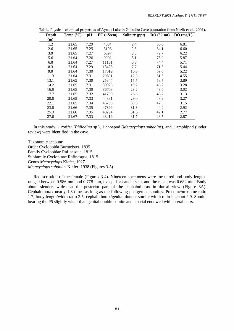
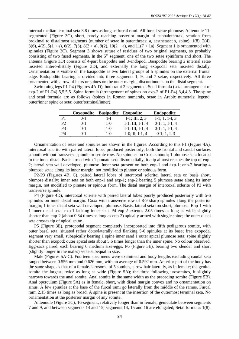
Metacyclops subdolus Kiefer, 1938 (Figures 3-5)
Redescription of the female (Figures 3-4). Nineteen specimens were measured and body lengths
ranged between 0.586 mm and 0.778 mm, except for caudal seta, and the mean was 0.682 mm. Body
about slender, widest at the posterior part of the cephalothorax in dorsal view (Figure 3A).
Cephalothorax nearly 1.8 times as long as the following pedigerous somites. Prosome/urosome ratio
1.7; body length/width ratio 2.5; cephalothorax/genital double-somite width ratio is about 2.9. Somite
bearing the P5 slightly wider than genital double-somite and a serial endowed with lateral hairs.

BOZKURT 2021 ActAquaTr 17(1), 78-87
82
Figure 3. Metacyclops subdolus Female. A) Habitus, dorsal; B) Caudal rami and anal somite, ventral; C)
Antennule; D) Antenna; E) P5 and P6, lateral; F) Genital field, ventral. Scale bars: A 250 μm; B, C, D 100 μm;
E, F 50 μm.
BOZKURT 2021 ActAquaTr 17(1), 78-87
83
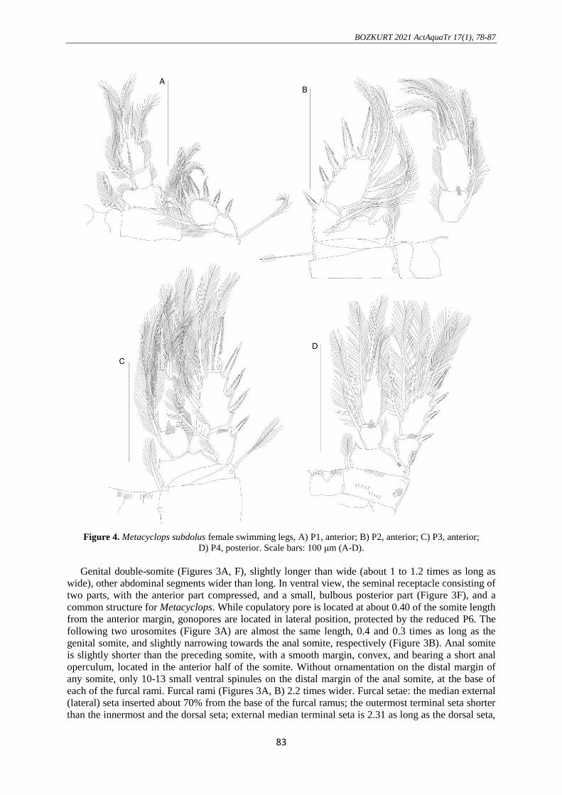
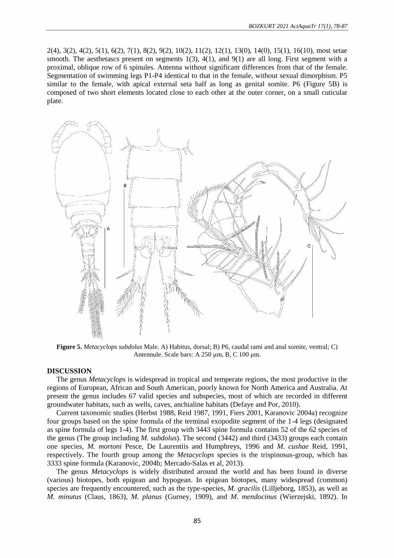
Figure 4. Metacyclops subdolus female swimming legs, A) P1, anterior; B) P2, anterior; C) P3, anterior;
D) P4, posterior. Scale bars: 100 μm (A-D).
Genital double-somite (Figures 3A, F), slightly longer than wide (about 1 to 1.2 times as long as
wide), other abdominal segments wider than long. In ventral view, the seminal receptacle consisting of
two parts, with the anterior part compressed, and a small, bulbous posterior part (Figure 3F), and a
common structure for Metacyclops. While copulatory pore is located at about 0.40 of the somite length
from the anterior margin, gonopores are located in lateral position, protected by the reduced P6. The
following two urosomites (Figure 3A) are almost the same length, 0.4 and 0.3 times as long as the
genital somite, and slightly narrowing towards the anal somite, respectively (Figure 3B). Anal somite
is slightly shorter than the preceding somite, with a smooth margin, convex, and bearing a short anal
operculum, located in the anterior half of the somite. Without ornamentation on the distal margin of
any somite, only 10-13 small ventral spinules on the distal margin of the anal somite, at the base of
each of the furcal rami. Furcal rami (Figures 3A, B) 2.2 times wider. Furcal setae: the median external
(lateral) seta inserted about 70% from the base of the furcal ramus; the outermost terminal seta shorter
than the innermost and the dorsal seta; external median terminal seta is 2.31 as long as the dorsal seta,
BOZKURT 2021 ActAquaTr 17(1), 78-87
84
internal median terminal seta 3.8 times as long as furcal rami. All furcal setae plumose. Antennule 11-
segmented (Figure 3C), short, barely reaching posterior margin of cephalothorax, setation from
proximal to distalmost segments (number of setae in parentheses; a, aesthetasc; s, spine): 1(8), 2(4),
3(6), 4(2), 5(1 + s), 6(2), 7(3), 8(2 + a), 9(2), 10(2 + a), and 11(7 + 1a). Segment 1 is ornamented with
spinules (Figure 3C). Segment 3 shows suture of residues of two original segments, so probably
consisting of two fused segments. In the 5th segment, one of the two setae spiniform and short. The
antenna (Figure 3D) consists of 4-part basipodite and 3-endopod. Basipodite bearing 2 internal setae
inserted antero-distally (Figure 3D), and externally the long exopodal seta inserted distally.
Ornamentation is visible on the basipodite as two lateral groups of 5 spinules on the external frontal
edge. Endopodite bearing is divided into three segments 1, 9, and 7 setae, respectively. All three
ornamented with a row of hairs or spines on the outer margin, discontinuous on the distal segment.
Swimming legs P1-P4 (Figures 4A-D), both rami 2-segmented. Setal formula (setal arrangement of
exp-2 of P1-P4) 5,5,5,5. Spine formula (arrangement of spines on exp-2 of P1-P4) 3,4,4,3. The spine
and setal formula are as follows (spines in Roman numerals, setae in Arabic numerals; legend:
outer/inner spine or seta; outer/terminal/inner).
Coxopodite Basipodite Exopodite Endopodite
P1 0-1 1-I I-1; III, 2, 3 I-1; 1, 1-I, 3
P2 0-1 1-0 I-1; III, I-1, 4 0-1; 1, I-1, 4
P3 0-1 1-0 I-1; III, I-1, 4 0-1; 1, I-1, 4
P4 0-1 1-0 I-0; II, I-1, 4 0-1; 1, I, 3
Ornamentation of setae and spinules are shown in the figures. According to this P1 (Figure 4A),
intercoxal sclerite with paired lateral lobes produced posteriorly, both the frontal and caudal surfaces
smooth without transverse spinule or setule row. No spinules on Coxa smooth; 1 plumose seta located
in the inner distal. Basis armed with 1 pinnate seta distomedially, its tip almost reaches the top of enp-
2; lateral seta well developed, plumose. Inner seta present on both enp-1 and exp-1; enp-2 bearing 4
plumose setae along its inner margin, not modified to pinnate or spinous form.
P2-P3 (Figures 4B, C), paired lateral lobes of intercoxal sclerite; lateral seta on basis short,
plumose distally; inner seta on both enp-1 and exp-1; enp-2 bearing 5 plumose setae along its inner
margin, not modified to pinnate or spinous form. The distal margin of intercoxal sclerite of P3 with
transverse spinule.
P4 (Figure 4D), intercoxal sclerite with paired lateral lobes poorly produced posteriorly with 5-6
spinules on inner distal margin. Coxa with transverse row of 8-9 sharp spinules along the posterior
margin; 1 inner distal seta well developed, plumose. Basis, lateral seta too short, plumose. Enp-1 with
1 inner distal seta; exp-1 lacking inner seta. P4 enp-2 extends 2.05 times as long as wide; slightly
shorter than enp-2 (about 0.84 times as long as enp-2) apically armed with single spine; the outer distal
seta crosses tip of apical spine.
P5 (Figure 3E), protopodal segment completely incorporated into fifth pedigerous somite, with
outer basal seta, situated rather dorsolaterally and flanking 5-6 spinules at its base; free exopodal
segment very small, subapically bearing 1 spine inner sand 1 outer apical plumose seta; spine slightly
shorter than exopod; outer apical seta about 5.6 times longer than the inner spine. No colour observed.
Egg-sacs paired, each bearing 6 medium size-eggs. P6 (Figure 3E), bearing two slender and short
(slightly longer in the males) setae subequal in size.
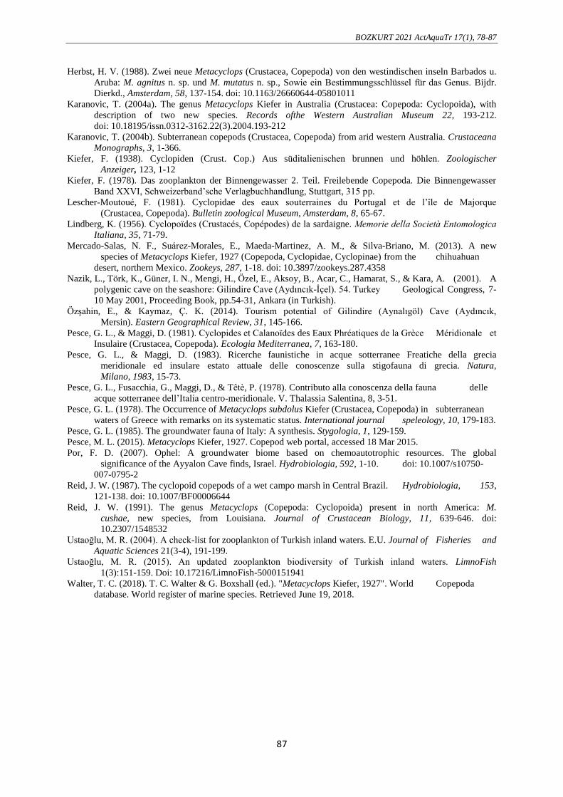
Male (Figures 5A-C). Fourteen specimens were examined and body lengths excluding caudal seta
ranged between 0.556 mm and 0.626 mm, with an average of 0.592 mm. Anterior part of the body has
the same shape as that of a female. Urosome of 5 somites, a row hair laterally, as in female; the genital
somite the largest, twice as long as wide (Figure 5A); the three following urosomites, it slightly
narrows towards the anal somite. Anal somite in the same width as the preceding somite (Figure 5B).
Anal operculum (Figure 5A) as in female, short, with distal margin convex and no ornamentation on
sinus. A few spinules at the base of the furcal rami go laterally from the middle of the ramus. Furcal
rami 2.15 times as long as broad. A spine is present at the insertion of the outermost terminal seta. No
ornamentation at the posterior margin of any somite.
Antennule (Figure 5C), 16-segment, relatively longer than in female; geniculate between segments
7 and 9, and between segments 14 and 15; segments 14, 15 and 16 are elongated; Setal formula: 1(8),
BOZKURT 2021 ActAquaTr 17(1), 78-87
85
2(4), 3(2), 4(2), 5(1), 6(2), 7(1), 8(2), 9(2), 10(2), 11(2), 12(1), 13(0), 14(0), 15(1), 16(10), most setae
smooth. The aesthetascs present on segments 1(3), 4(1), and 9(1) are all long. First segment with a
proximal, oblique row of 6 spinules. Antenna without significant differences from that of the female.
Segmentation of swimming legs P1-P4 identical to that in the female, without sexual dimorphism. P5
similar to the female, with apical external seta half as long as genital somite. P6 (Figure 5B) is
composed of two short elements located close to each other at the outer corner, on a small cuticular
plate.
Figure 5. Metacyclops subdolus Male. A) Habitus, dorsal; B) P6, caudal rami and anal somite, ventral; C)
Antennule. Scale bars: A 250 μm, B, C 100 μm.
DISCUSSION
The genus Metacyclops is widespread in tropical and temperate regions, the most productive in the
regions of European, African and South American, poorly known for North America and Australia. At
present the genus includes 67 valid species and subspecies, most of which are recorded in different
groundwater habitats, such as wells, caves, anchialine habitats (Defaye and Por, 2010).
Current taxonomic studies (Herbst 1988, Reid 1987, 1991, Fiers 2001, Karanovic 2004a) recognize
four groups based on the spine formula of the terminal exopodite segment of the 1-4 legs (designated
as spine formula of legs 1-4). The first group with 3443 spine formula contains 52 of the 62 species of
the genus (The group including M. subdolus). The second (3442) and third (3433) groups each contain
one species, M. mortoni Pesce, De Laurentiis and Humphreys, 1996 and M. cushae Reid, 1991,
respectively. The fourth group among the Metacyclops species is the trispinosus-group, which has
3333 spine formula (Karanovic, 2004b; Mercado-Salas et al, 2013).
The genus Metacyclops is widely distributed around the world and has been found in diverse
(various) biotopes, both epigean and hypogean. In epigean biotopes, many widespread (common)
species are frequently encountered, such as the type-species, M. gracilis (Lilljeborg, 1853), as well as
M. minutus (Claus, 1863), M. planus (Gurney, 1909), and M. mendocinus (Wierzejski, 1892). In

BOZKURT 2021 ActAquaTr 17(1), 78-87
86
hypogean biotopes, we can mention, for example, M. subdolus Kiefer, 1938, recorded from
groundwaters of peri-Mediterranean countries (Defaye and Por, 2010).
Metacyclops is a very ancient genus and has probably colonized fresh waters very early, before the
break-up of Pangaea (Boxshall and Jaume, 2000), many species of the genus are endemic, as the
species recently described from Western Australian groundwaters (Karanovic, 2004a, 2004b). After
the colonization of hypogean fresh waters occurred, it led to the diversification of more or less closely
related taxa on different plates and continents. Further examinations in underground waters and a total
correction of the Metacyclops genus will be important to comprehend the connections between the
species of these commonly conveyed variety (Defaye and Por, 2010).
The occurrence of the species is certainly related to the characteristics of the biotope. The lake of
Gilindire Cave has dual source of water: fresh groundwater mixed saltwater. The M. subdolus
population in the lake consisted of a large number of adults, copepodite and nauplii. The reason for the
abundance of M. subdolus is thought to be the absence of another creature feeding on it.
M. subdolus prefers sulfidic and slightly brackish groundwater (Defaye and Por, 2010), and has a
European Mediterranean distribution (no record from North Africa); it has first been reported by
Kiefer (1938) from southern Italy (La Zinzulusa, Abysso caves), then from Sardinia (Lindberg, 1956),
Italy (Pesce et al., 1978; Pesce, 1985), Greece (Peloponnesos, Attica, Crete: Pesce, 1978; Pesce and
Maggi, 1981, 1983), Mallorca (Can Pastilla: Lescher-Moutoue, 1981). M. subdolus has already been
identified from Israel by Dimentman and Por (1991) from slightly brackish springs near the Dead Sea,
the northern Negev by Defaye and Dussart (1995) and finally by Ayyalon Cave (Defaye and Por,
2010).
The waters where M. subdolus has been found with light brackish water until now are spring water,
caves and wells. Therefore it confirms that M. subdolus is a type of groundwater species that prefers
slight brackish waters.
Minor differences were detected in some characters of M. subdolus in Gilindire cave. İn the
previous definitions, intercoxal scleritis of P1-P4 ornamented with 2 rows of spinules on ventral
margin but in the present, intercoxal scleritis of third and fourth legs ornamented with a row of
spinules on ventral margin, first and second smooth. Two-row spinule present ventral margin of P4
coxa but in previous, coxa smooth. P5: the base of the seta inserted on the somite has a row of
spinules.
Acknowledgments: The author would like to thank Dr. Yavuz Mazlum from Iskenderun Technical
University for critical review of the manuscript, and also thank Dr. Janet W. Reid (Virginia Museum
of Natural History, Martinsville, Virginia, USA) for sending some of the useful literatures and for
confirming the identification of Metacyclops subdolus.
REFERENCES
Boxshall, G. A., & Jaume, D. (2000). Making waves: The repeated colonization of fresh water by copepod
crustaceans. Advances in Ecological Research, 31, 61-79. doi: 10.1016/S0065-2504(00)31007-8
Damian-Georgescu, A. (1970). Fauna Republicii Socialiste Romania, Crustacea. Vol. IV. 11 Copepoda,
Harpacticoida. Bucharest: Academiei Republicii Socialiste Romania 249 pp.
Defaye, D., & Dussart, B. H. (1995). The cyclopid fauna (Crustacea, Copepoda) of inland waters of Israel. 1-
First data from semi-arid and arid regions. Hydrobiologia, 310, 1-10.
Defaye, D., & Por, F. D. (2010). Metacyclops (Copepoda, Cyclopidae) from Ayyalon Cave, Israel. Crustaceana, 83,
399-423. doi: 10.1163/001121610x12627655658320
Dimentman, Ch., & Por, E. D. (1991). The origin of the subterranean fauna of the Jordan-Dead Sea Rift Valley.
Stygologia 6, 155-164.
Dussart, B. (1967). Les Copepodes Des Eaux Continentales D’europe Occidentale, Tale I, Calanoides et
Harpacticoides, N. Boubee et cie, Paris, 500 pp.
Dussart, B. H., & Defaye, D. (2006). World Directory of the Crustacea Copepoda of Inland Waters. II.
Cyclopiformes: 1-354. (Backhuys Publishers, Leiden).
Fiers, F. (2001). Meridiecyclops gen. nov., a new cyclopid genus (Crustacea: Copepoda: Cyclopidae) from
southern Australia. Invertebrate Taxonomy 15, 893-908. doi: 10.1071/IT01003
Gibert, J., & Deharveng, L. (2002). Subterranean ecosystems: a truncated functional
biodiversity. BioScience, 52, 473-481. DOI:10.1641/0006-3568(2002)052 [0473: SEATFB]2.
0.CO;2
BOZKURT 2021 ActAquaTr 17(1), 78-87
87
Herbst, H. V. (1988). Zwei neue Metacyclops (Crustacea, Copepoda) von den westindischen inseln Barbados u.
Aruba: M. agnitus n. sp. und M. mutatus n. sp., Sowie ein Bestimmungsschlüssel für das Genus. Bijdr.
Dierkd., Amsterdam, 58, 137-154. doi: 10.1163/26660644-05801011
Karanovic, T. (2004a). The genus Metacyclops Kiefer in Australia (Crustacea: Copepoda: Cyclopoida), with
description of two new species. Records ofthe Western Australian Museum 22, 193-212.
doi: 10.18195/issn.0312-3162.22(3).2004.193-212
Karanovic, T. (2004b). Subterranean copepods (Crustacea, Copepoda) from arid western Australia. Crustaceana
Monographs, 3, 1-366.
Kiefer, F. (1938). Cyclopiden (Crust. Cop.) Aus süditalienischen brunnen und höhlen. Zoologischer
Anzeiger, 123, 1-12
Kiefer, F. (1978). Das zooplankton der Binnengewasser 2. Teil. Freilebende Copepoda. Die Binnengewasser
Band XXVI, Schweizerband’sche Verlagbuchhandlung, Stuttgart, 315 pp.
Lescher-Moutoué, F. (1981). Cyclopidae des eaux souterraines du Portugal et de l’île de Majorque
(Crustacea, Copepoda). Bulletin zoological Museum, Amsterdam, 8, 65-67.
Lindberg, K. (1956). Cyclopoïdes (Crustacés, Copépodes) de la sardaigne. Memorie della Società Entomologica
Italiana, 35, 71-79.
Mercado-Salas, N. F., Suárez-Morales, E., Maeda-Martinez, A. M., & Silva-Briano, M. (2013). A new
species of Metacyclops Kiefer, 1927 (Copepoda, Cyclopidae, Cyclopinae) from the chihuahuan
desert, northern Mexico. Zookeys, 287, 1-18. doi: 10.3897/zookeys.287.4358
Nazik, L., Törk, K., Güner, I. N., Mengi, H., Özel, E., Aksoy, B., Acar, C., Hamarat, S., & Kara, A. (2001). A
polygenic cave on the seashore: Gilindire Cave (Aydıncık-İçel). 54. Turkey Geological Congress, 7-
10 May 2001, Proceeding Book, pp.54-31, Ankara (in Turkish).
Özşahin, E., & Kaymaz, Ç. K. (2014). Tourism potential of Gilindire (Aynalıgöl) Cave (Aydıncık,
Mersin). Eastern Geographical Review, 31, 145-166.
Pesce, G. L., & Maggi, D. (1981). Cyclopides et Calanoïdes des Eaux Phréatiques de la Grèce Méridionale et
Insulaire (Crustacea, Copepoda). Ecologia Mediterranea, 7, 163-180.
Pesce, G. L., & Maggi, D. (1983). Ricerche faunistiche in acque sotterranee Freatiche della grecia
meridionale ed insulare estato attuale delle conoscenze sulla stigofauna di grecia. Natura,
Milano, 1983, 15-73.
Pesce, G. L., Fusacchia, G., Maggi, D., & Têtè, P. (1978). Contributo alla conoscenza della fauna delle
acque sotterranee dell’Italia centro-meridionale. V. Thalassia Salentina, 8, 3-51.
Pesce, G. L. (1978). The Occurrence of Metacyclops subdolus Kiefer (Crustacea, Copepoda) in subterranean
waters of Greece with remarks on its systematic status. International journal speleology, 10, 179-183.
Pesce, G. L. (1985). The groundwater fauna of Italy: A synthesis. Stygologia, 1, 129-159.
Pesce, M. L. (2015). Metacyclops Kiefer, 1927. Copepod web portal, accessed 18 Mar 2015.
Por, F. D. (2007). Ophel: A groundwater biome based on chemoautotrophic resources. The global
significance of the Ayyalon Cave finds, Israel. Hydrobiologia, 592, 1-10. doi: 10.1007/s10750-
007-0795-2
Reid, J. W. (1987). The cyclopoid copepods of a wet campo marsh in Central Brazil. Hydrobiologia, 153,
121-138. doi: 10.1007/BF00006644
Reid, J. W. (1991). The genus Metacyclops (Copepoda: Cyclopoida) present in north America: M.
cushae, new species, from Louisiana. Journal of Crustacean Biology, 11, 639-646. doi:
10.2307/1548532
Ustaoğlu, M. R. (2004). A check-list for zooplankton of Turkish inland waters. E.U. Journal of Fisheries and
Aquatic Sciences 21(3-4), 191-199.
Ustaoğlu, M. R. (2015). An updated zooplankton biodiversity of Turkish inland waters. LimnoFish
1(3):151-159. Doi: 10.17216/LimnoFish-5000151941
Walter, T. C. (2018). T. C. Walter & G. Boxshall (ed.). "Metacyclops Kiefer, 1927". World Copepoda
database. World register of marine species. Retrieved June 19, 2018.
![PAPERS, [ca. 1861]-1938. GUIDE · PROD Archival FIN ID NYCV85-A452 + LON NYCV85-A452 1 record in AMC Gould, Norman Judd, b. 1877. Papers, [ca.1861]-1938, 1917-1923 (bulk)](https://img.pdfslide.us/doc/110x75/5fd1ffa7284f99717612af18/papers-ca-1861-1938-guide-prod-archival-fin-id-nycv85-a452-lon-nycv85-a452.jpg)


![[Ferenc Kiefer] Hungarian Linguistics](https://img.pdfslide.us/doc/110x75/577cc3f61a28aba71197b501/ferenc-kiefer-hungarian-linguistics.jpg)