Embed Size (px)
Citation preview
Final Report for AOARD Grant FA2386-12-1-4078 “Development of Pulsating Tubules
with Chiral Inversion”
Date 2013-09-21
PI and Co-PI information: Myongsoo Lee; [email protected]; Seoul National
University; Department of Chemistry; Seoul National University, Gwanak-ro 1, Gwanak-gu,
Seoul 151-747, Korea; 82-2-880-4340; 82-2-393-6096.
Period of Performance: 08/16/2012 – 08/15/2013
Abstract: Stimuli responsive nanostructures through rigid-flexible block molecules can hold a
great promise for the fabrication of intelligent nanodevices, nanoelectronics, and
nanobiomaterials. One of the typical features of the rod amphiphiles is its unique anisotropic
molecular shape and strong aggregation tendency through additional π−π stacking interactions,
which have enabled the construction of highly versatile and dynamic nanostructures. Hence,
variations in the molecular structure and the local environment, albeit small, allow the rapid
transformation of equilibrium morphology. In this project, we have studied the development of
stimuli-responsive nanomaterials using on self-assembly of amphiphilic molecules based on
hydrophilic oligo(ethylene glycol) chains and hydrophobic aromatic rods and peptides.
Introduction: Supramolecular nanostructures produced via self-assembling molecules have
attracted interest because the sophisticated structures formed by weak noncovalent interactions
can be triggered by external stimuli leading to dynamic materials. Typical examples of basic
building blocks for self-assembly include lipid molecules, surfactants, supra-amphiphiles, block
molecules, hyperbranched polymers, peptide derivatives and inorganic/organic complexes.
Among them, rigid-flexible block molecules, consisting of aromatic rod and coil segments, are
promising candidates for fabricating self-assembled structures. Aqueous assemblies have great
advantages for the creation of desired materials in terms of biological applications. For the
aqueous self-assembly of amphiphilic rigid-flexible block molecules, the anisotropic orientation
of the stiff rod-like segments and the microsegregation of the incompatible molecular parts are
able to enhance each other in water leading to the formation of thermodynamically stable
supramolecular structures with a rigid hydrophobic core surrounded by flexible hydrophilic
chains. In general, monodisperse rod–coil blocks display highly reproducible and predictable
self-assembly behavior.
Recently, we have studied on the development of diverse aqueous assemblies, such as tubules,
toroids, porous capsules, and helical fibers, by adjusting the relative volume fraction between
hydrophobic and hydrophilic segments of rod–coil molecules consisting of aromatic units and
poly(propylene oxide) (PPO) or poly(ethylene oxide) (PEO) coils. In this project, we focus on
the development of aqueous nanostructure from the self-assembly of rod–coil block molecules.
We also demonstrated the self-assembly of α-helical peptide. Finally, we showed the application
of self-assembled nanostructures into biological system.
Part 1: Supramolecular Switching between Flat Sheets and Helical Tubules Triggered by
Coordination Interaction.
Experiment : We envisioned that the introduction of meta-linked pyridine units at both ends of
Report Documentation Page Form ApprovedOMB No. 0704-0188
Public reporting burden for the collection of information is estimated to average 1 hour per response, including the time for reviewing instructions, searching existing data sources, gathering andmaintaining the data needed, and completing and reviewing the collection of information. Send comments regarding this burden estimate or any other aspect of this collection of information,including suggestions for reducing this burden, to Washington Headquarters Services, Directorate for Information Operations and Reports, 1215 Jefferson Davis Highway, Suite 1204, ArlingtonVA 22202-4302. Respondents should be aware that notwithstanding any other provision of law, no person shall be subject to a penalty for failing to comply with a collection of information if itdoes not display a currently valid OMB control number.
1. REPORT DATE 15 OCT 2013
2. REPORT TYPE Final
3. DATES COVERED 16-08-2012 to 15-08-2013
4. TITLE AND SUBTITLE Development of Pulsating Tubules from Non-Covalent Macrocycles
5a. CONTRACT NUMBER FA23861214078
5b. GRANT NUMBER
5c. PROGRAM ELEMENT NUMBER
6. AUTHOR(S) Myong Soo Lee
5d. PROJECT NUMBER
5e. TASK NUMBER
5f. WORK UNIT NUMBER
7. PERFORMING ORGANIZATION NAME(S) AND ADDRESS(ES) Seoul National University,Seoul,151-747,Seoul,KR,151747
8. PERFORMING ORGANIZATIONREPORT NUMBER N/A
9. SPONSORING/MONITORING AGENCY NAME(S) AND ADDRESS(ES) AOARD, UNIT 45002, APO, AP, 96338-5002
10. SPONSOR/MONITOR’S ACRONYM(S) AOARD
11. SPONSOR/MONITOR’S REPORT NUMBER(S) AOARD-124078
12. DISTRIBUTION/AVAILABILITY STATEMENT Approved for public release; distribution unlimited
13. SUPPLEMENTARY NOTES
14. ABSTRACT Stimuli responsive nanostructures constructed from rigid-flexible block molecules hold great promise forfabrication of intelligent nanodevices, nanoelectronics, and nanobiomaterials. One typical feature of rodamphiphiles is their unique anisotropic molecular shape and strong aggregation tendency throughπ−π stacking interactions, which has enabled construction of highly versatile anddynamic nanostructures. Variations in molecular structure and local environment, albeit small, allow therapid transformation of equilibrium morphology. This research project involved development ofstimuli-responsive nanomaterials using self-assembly of amphiphilic molecules based on hydrophilicoligo(ethylene glycol) chains and hydrophobic aromatic rods and peptides. Pyridine end-substitutedrod?coil block systems were found to self-assemble into flat sheets in dilute aqueous solution, but throughreversible coordination interaction with Ag+1 could change their shape into helical tubules at higher anddiscrete macrocycles at lower concentrations. Metal-containing macrocycles were found to reversibly stackto form helical tubules in response to variation in concentration. The influence of interactions with smallmolecule planer aromatics on 2D structure was examined. Rationally designed macrobicyclic amphiphilesconsisting of a hydrophilic dendron attached to the center of an aromatic plane undergoes self-assemblyinto a 2D structure with nanosized lateral pores through lateral association of amphiphile dimers with auniform diameter of 3.5 nm. The porous sheets efficiently intercalate flat aromatic molecules, such ascoronene, through the conformational inversion of the basal planes of the dimeric micelles. This projectalso examined a novel approach to make short peptides adopt stable α-helical structures throughmacrocyclization of their linear precursors. On incorporation of more hydrophobic amino acid residuesinto the peptide block, the helical structure forces the cyclic molecules to adopt a facially amphiphilicconformation resulting in amphiphilic folding of the cyclic molecule and to formation of undulatednanofibers through directional assembly of discrete micelles. Controlling conformation and stability of thenanofibers tethered by lectin proteins were found to regulate T cell activation. The lengths as well asstability of the protein-coated supramolecular nanofibers could be manipulated by a small variation in therod?coil molecular structure. The unique supramolecular structures with switchable functionality reportedherein might provide new strategies for the design of intelligent materials with simultaneous biological andelectro-optical functions.
15. SUBJECT TERMS Self-Assembly, Stimuli-Responsive Materials, Tubular Nanostructures, Dynamic Response, 2DNanostructures, Proteins, T-Cells, Nanofibers.
16. SECURITY CLASSIFICATION OF: 17. LIMITATIONOF ABSTRACT
Same asReport (SAR)
18. NUMBEROF PAGES
13
19a. NAME OFRESPONSIBLE PERSON a. REPORT
unclassified b. ABSTRACT
unclassified c. THIS PAGE
unclassified
Standard Form 298 (Rev. 8-98) Prescribed by ANSI Std Z39-18
the bent-shaped aromatic segment might form stimuli-responsive 2D structures, since pyridine
units are well-known to interact specifically with Ag(I) ions through reversible coordination
bonds. This recognition event would cause reversible rearrangement of the fully overlapped
zigzag packing arrangement of the bent-shaped rigid segments to maximize the coordination
interactions between pyridine donors and Ag(I) ions at the expense of π–π stacking interactions
between aromatic rod segments. Consequently, this metal-directed rearrangement of the aromatic
segments would endow the 2D structures with dynamic responsive functions. With this direction
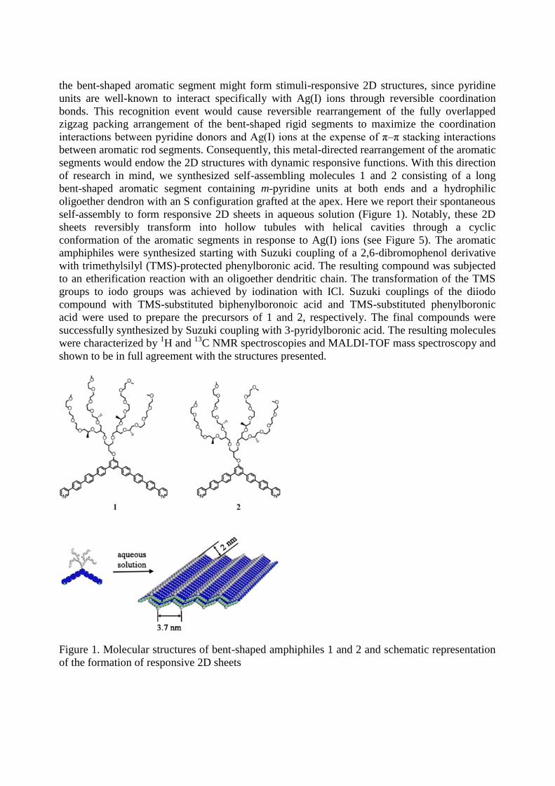
of research in mind, we synthesized self-assembling molecules 1 and 2 consisting of a long
bent-shaped aromatic segment containing m-pyridine units at both ends and a hydrophilic
oligoether dendron with an S configuration grafted at the apex. Here we report their spontaneous
self-assembly to form responsive 2D sheets in aqueous solution (Figure 1). Notably, these 2D
sheets reversibly transform into hollow tubules with helical cavities through a cyclic
conformation of the aromatic segments in response to Ag(I) ions (see Figure 5). The aromatic
amphiphiles were synthesized starting with Suzuki coupling of a 2,6-dibromophenol derivative
with trimethylsilyl (TMS)-protected phenylboronic acid. The resulting compound was subjected
to an etherification reaction with an oligoether dendritic chain. The transformation of the TMS
groups to iodo groups was achieved by iodination with ICl. Suzuki couplings of the diiodo
compound with TMS-substituted biphenylboronoic acid and TMS-substituted phenylboronic
acid were used to prepare the precursors of 1 and 2, respectively. The final compounds were
successfully synthesized by Suzuki coupling with 3-pyridylboronic acid. The resulting molecules
were characterized by 1H and
13C NMR spectroscopies and MALDI-TOF mass spectroscopy and
shown to be in full agreement with the structures presented.
Figure 1. Molecular structures of bent-shaped amphiphiles 1 and 2 and schematic representation
of the formation of responsive 2D sheets
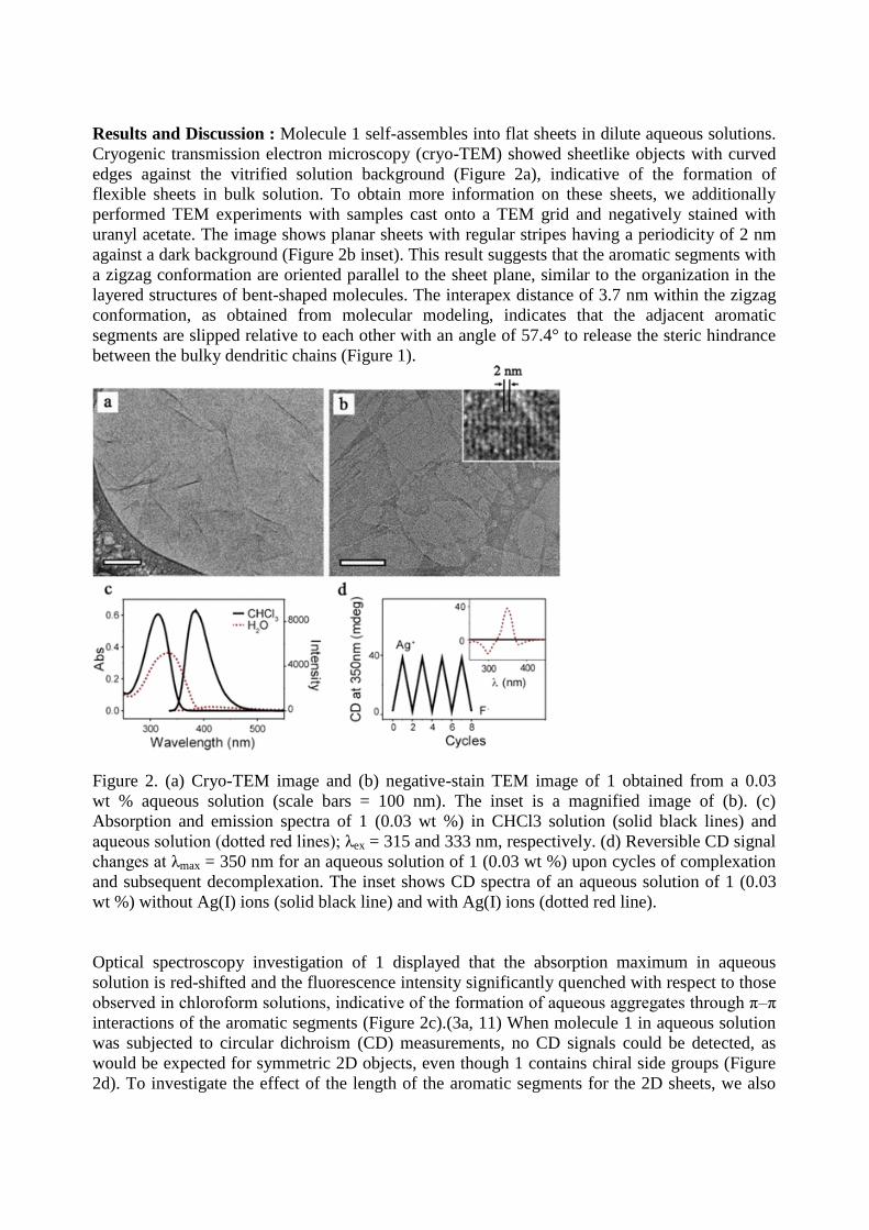
Results and Discussion : Molecule 1 self-assembles into flat sheets in dilute aqueous solutions.
Cryogenic transmission electron microscopy (cryo-TEM) showed sheetlike objects with curved
edges against the vitrified solution background (Figure 2a), indicative of the formation of
flexible sheets in bulk solution. To obtain more information on these sheets, we additionally
performed TEM experiments with samples cast onto a TEM grid and negatively stained with
uranyl acetate. The image shows planar sheets with regular stripes having a periodicity of 2 nm
against a dark background (Figure 2b inset). This result suggests that the aromatic segments with
a zigzag conformation are oriented parallel to the sheet plane, similar to the organization in the
layered structures of bent-shaped molecules. The interapex distance of 3.7 nm within the zigzag
conformation, as obtained from molecular modeling, indicates that the adjacent aromatic
segments are slipped relative to each other with an angle of 57.4° to release the steric hindrance
between the bulky dendritic chains (Figure 1).
Figure 2. (a) Cryo-TEM image and (b) negative-stain TEM image of 1 obtained from a 0.03
wt % aqueous solution (scale bars = 100 nm). The inset is a magnified image of (b). (c)
Absorption and emission spectra of 1 (0.03 wt %) in CHCl3 solution (solid black lines) and
aqueous solution (dotted red lines); λex = 315 and 333 nm, respectively. (d) Reversible CD signal
changes at λmax = 350 nm for an aqueous solution of 1 (0.03 wt %) upon cycles of complexation
and subsequent decomplexation. The inset shows CD spectra of an aqueous solution of 1 (0.03
wt %) without Ag(I) ions (solid black line) and with Ag(I) ions (dotted red line).
Optical spectroscopy investigation of 1 displayed that the absorption maximum in aqueous
solution is red-shifted and the fluorescence intensity significantly quenched with respect to those
observed in chloroform solutions, indicative of the formation of aqueous aggregates through π–π
interactions of the aromatic segments (Figure 2c).(3a, 11) When molecule 1 in aqueous solution
was subjected to circular dichroism (CD) measurements, no CD signals could be detected, as
would be expected for symmetric 2D objects, even though 1 contains chiral side groups (Figure
2d). To investigate the effect of the length of the aromatic segments for the 2D sheets, we also
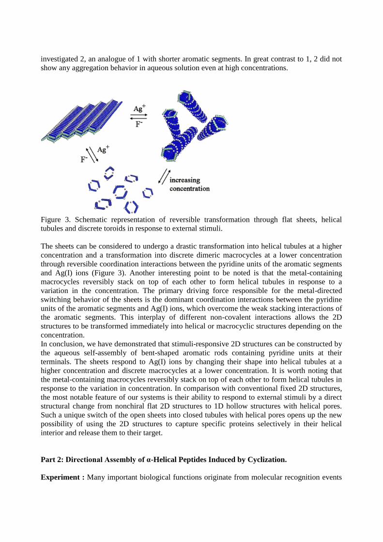
investigated 2, an analogue of 1 with shorter aromatic segments. In great contrast to 1, 2 did not
show any aggregation behavior in aqueous solution even at high concentrations.
Figure 3. Schematic representation of reversible transformation through flat sheets, helical
tubules and discrete toroids in response to external stimuli.
The sheets can be considered to undergo a drastic transformation into helical tubules at a higher
concentration and a transformation into discrete dimeric macrocycles at a lower concentration
through reversible coordination interactions between the pyridine units of the aromatic segments
and Ag(I) ions (Figure 3). Another interesting point to be noted is that the metal-containing
macrocycles reversibly stack on top of each other to form helical tubules in response to a
variation in the concentration. The primary driving force responsible for the metal-directed
switching behavior of the sheets is the dominant coordination interactions between the pyridine
units of the aromatic segments and Ag(I) ions, which overcome the weak stacking interactions of
the aromatic segments. This interplay of different non-covalent interactions allows the 2D
structures to be transformed immediately into helical or macrocyclic structures depending on the
concentration.
In conclusion, we have demonstrated that stimuli-responsive 2D structures can be constructed by
the aqueous self-assembly of bent-shaped aromatic rods containing pyridine units at their
terminals. The sheets respond to Ag(I) ions by changing their shape into helical tubules at a
higher concentration and discrete macrocycles at a lower concentration. It is worth noting that
the metal-containing macrocycles reversibly stack on top of each other to form helical tubules in
response to the variation in concentration. In comparison with conventional fixed 2D structures,
the most notable feature of our systems is their ability to respond to external stimuli by a direct
structural change from nonchiral flat 2D structures to 1D hollow structures with helical pores.
Such a unique switch of the open sheets into closed tubules with helical pores opens up the new
possibility of using the 2D structures to capture specific proteins selectively in their helical
interior and release them to their target.
Part 2: Directional Assembly of α-Helical Peptides Induced by Cyclization.
Experiment : Many important biological functions originate from molecular recognition events
of proteins. For many proteins to bind various biomolecules, well-defined secondary structures
are placed in the recognition domains. The α-helix is a common motif in the secondary structure
of proteins, especially existing vastly in the recognition domains of various protein–protein or
nucleic acid–protein interactions. Inspired by biological systems, many studies have been
focused on the development of stable α-helixes to mimic the interactions between the original
proteins. However, folding of short peptides into an α-helical structure in solution is limited
because stabilizing interactions and the enthalpy gain from hydrogen bonds between amides on
adjacent helical turns are not sufficient to compensate for the entropic cost involved in the
folding of the peptide chain.
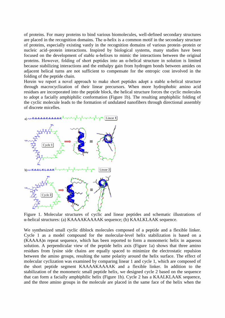
Herein we report a novel approach to make short peptides adopt a stable α-helical structure
through macrocyclization of their linear precursors. When more hydrophobic amino acid
residues are incorporated into the peptide block, the helical structure forces the cyclic molecules
to adopt a facially amphiphilic conformation (Figure 1b). The resulting amphiphilic folding of
the cyclic molecule leads to the formation of undulated nanofibers through directional assembly
of discrete micelles.
Figure 1. Molecular structures of cyclic and linear peptides and schematic illustrations of
α-helical structures: (a) KAAAAKAAAAK sequence; (b) KAALKLAAK sequence.
We synthesized small cyclic diblock molecules composed of a peptide and a flexible linker.
Cycle 1 as a model compound for the molecular-level helix stabilization is based on a
(KAAAA)n repeat sequence, which has been reported to form a monomeric helix in aqueous
solution. A perpendicular view of the peptide helix axis (Figure 1a) shows that three amino
residues from lysine side chains are equally spaced to minimize the electrostatic repulsion
between the amino groups, resulting the same polarity around the helix surface. The effect of
molecular cyclization was examined by comparing linear 1 and cycle 1, which are composed of
the short peptide segment KAAAAKAAAAK and a flexible linker. In addition to the
stabilization of the monomeric small peptide helix, we designed cycle 2 based on the sequence
that can form a facially amphiphilic helix (Figure 1b). Cycle 2 has a KAALKLAAK sequence,
and the three amino groups in the molecule are placed in the same face of the helix when the
helical structure is formed. Moreover, the leucine residues, which are known to interact with
each other through hydrophobic interactions, can be utilized to provide a hydrophobic surface on
one side of the helix. The facial amphiphilicity of this design is easily seen in the view from the
perpendicular helix axis (Figure 1b). An ethylene glycol-based linker segment,
N-(Fmoc-8-amino-3,6-dioxaoctyl)succinamic acid, was introduced as a flexible linker unit. The
cyclization reaction was performed with an on-resin cyclization method to achieve high synthetic
efficiency. Each molecule was characterized by MALDI mass spectroscopy.
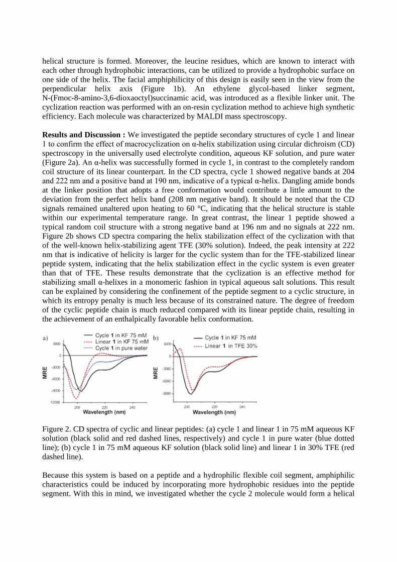
Results and Discussion : We investigated the peptide secondary structures of cycle 1 and linear
1 to confirm the effect of macrocyclization on α-helix stabilization using circular dichroism (CD)
spectroscopy in the universally used electrolyte condition, aqueous KF solution, and pure water
(Figure 2a). An α-helix was successfully formed in cycle 1, in contrast to the completely random
coil structure of its linear counterpart. In the CD spectra, cycle 1 showed negative bands at 204
and 222 nm and a positive band at 190 nm, indicative of a typical α-helix. Dangling amide bonds
at the linker position that adopts a free conformation would contribute a little amount to the
deviation from the perfect helix band (208 nm negative band). It should be noted that the CD
signals remained unaltered upon heating to 60 °C, indicating that the helical structure is stable
within our experimental temperature range. In great contrast, the linear 1 peptide showed a
typical random coil structure with a strong negative band at 196 nm and no signals at 222 nm.
Figure 2b shows CD spectra comparing the helix stabilization effect of the cyclization with that
of the well-known helix-stabilizing agent TFE (30% solution). Indeed, the peak intensity at 222
nm that is indicative of helicity is larger for the cyclic system than for the TFE-stabilized linear
peptide system, indicating that the helix stabilization effect in the cyclic system is even greater
than that of TFE. These results demonstrate that the cyclization is an effective method for
stabilizing small α-helixes in a monomeric fashion in typical aqueous salt solutions. This result
can be explained by considering the confinement of the peptide segment to a cyclic structure, in
which its entropy penalty is much less because of its constrained nature. The degree of freedom
of the cyclic peptide chain is much reduced compared with its linear peptide chain, resulting in
the achievement of an enthalpically favorable helix conformation.
Figure 2. CD spectra of cyclic and linear peptides: (a) cycle 1 and linear 1 in 75 mM aqueous KF
solution (black solid and red dashed lines, respectively) and cycle 1 in pure water (blue dotted
line); (b) cycle 1 in 75 mM aqueous KF solution (black solid line) and linear 1 in 30% TFE (red
dashed line).
Because this system is based on a peptide and a hydrophilic flexible coil segment, amphiphilic
characteristics could be induced by incorporating more hydrophobic residues into the peptide
segment. With this in mind, we investigated whether the cycle 2 molecule would form a helical
structure to induce an amphiphilic feature. Similar to cycle 1, cycle 2 also adopted a helical
conformation different from its linear counterpart (Figure 3a). This was rather surprising because
the three amino groups in cycle 2 are even more closely spaced in the helix. In contrast to its
linear counterpart, which did not show apparent aggregation behavior, the cyclic peptide
self-assembled into a fibrous structure while maintaining an α-helical conformation of the
hydrophobic peptide unit. The transmission electron microscopy (TEM) image (Figure 3c) shows
the formation of unique nanofibers with regular undulation along the fiber axis that have
diameters of 6–7 nm and lengths of a few hundred nanometers. Closer examination of the
samples showed the individual objects along the fiber axis to be oblate rather than spherical
(Figure 3c inset), suggesting that the undulation arises from the micellar stacking.
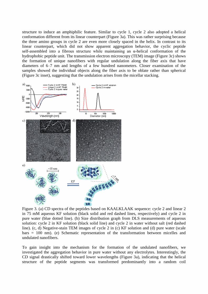
Figure 3. (a) CD spectra of the peptides based on KAALKLAAK sequence: cycle 2 and linear 2
in 75 mM aqueous KF solution (black solid and red dashed lines, respectively) and cycle 2 in
pure water (blue dotted line). (b) Size distribution graph from DLS measurements of aqueous
solution: cycle 2 in KF solution (black solid line) and cycle 2 in water without salt (red dashed
line). (c, d) Negative-stain TEM images of cycle 2 in (c) KF solution and (d) pure water (scale
bars = 100 nm). (e) Schematic representation of the transformation between micelles and
undulated nanofibers.
To gain insight into the mechanism for the formation of the undulated nanofibers, we
investigated the aggregation behavior in pure water without any electrolytes. Interestingly, the
CD signal drastically shifted toward lower wavelengths (Figure 3a), indicating that the helical
structure of the peptide segments was transformed predominantly into a random coil
conformation. This conformational change was accompanied by a structural transformation from
the elongated fibers to spherical micelles. Dynamic light scattering (DLS) analysis showed a
drastic reduction in the hydrodynamic diameter from several hundred nanometers to 10 nm upon
removal of KF salt (Figure 3b). The TEM image shows spherical micelles with an average
diameter of 10 nm, which is larger than the fiber width (Figure 3d). These results suggest that the
leucine–leucine interactions are destabilized in a random coil conformation, resulting in looser
packing between the peptide segments in pure water than in KF solution. Consequently, the
looser packing of the hydrophobic peptide segments with a random coil conformation gives rise
to the spherical micelles with a larger diameter than the fiber width.
From these observations, induction of the α-helical structure of the peptide segment seems to be
the main driving force for the formation of the undulated nanofibers. In the KF solution, which
provides a more hydrophobic environment because of the salting-out effect, peptide chains
would favor being folded into α-helixes. The general effect of salting out in helix stabilization
was observed from cycle 1 folding upon removal of the KF salt (Figure 2a). The helicity of the
peptide decreased, as determined from the size of 222 nm band and the much-shifted negative
minimum. With aid of this salting-out effect, the resulting helical peptides, cycle 2, are oriented
parallel to each other to form oblate micelles in which the hydrophobic leucine residues are
located on the inside and lysine units together with the hydrophilic linkers are on the exterior.
This anisotropic packing arrangement of the helical peptides results in oblate micelles with more
hydrophobic tops and bottoms resulting from the α-helical core. To reduce the exposure of the
hydrophobic parts of the micelles in a water environment, the oblate micelles stack on top of
each other to form undulated nanofibers (Figure 3e).
In conclusion, we have demonstrated that cyclization of block peptides leads to a conformational
transition of the peptide segment from random coil to α-helix, which is important for many
biological applications of small epitopes. When amphiphilicity was introduced into our cyclic
system by elaborate modification of the peptide sequence, the helical conformation of the peptide
forced the molecular cycle to be facially amphiphilic. The resulting facial amphiphiles
self-aggregated into unique undulated nanofibers originating from one-dimensional stacking of
oblate micelles through directional interactions.
Part 3: Switchable Nanoporous Sheets by the Aqueous Self-Assembly of Aromatic
Macrobicycles
Experiment : The self-assembling molecules that form this aggregate consist of a macrobicyclic
aromatic segment and a hydrophilic oligoether dendron grafted at the center of the basal plane
(Figure 1). The synthesis of the flat aromatic amphiphiles began with the Sonogashira coupling
of a dendron-substituted diiodobenzene derivative with 2,6-dibromoethynylbenzene to provide a
tetrabromo building block (see the Supporting Information). Suzuki coupling of the tetrabromo
compound with 3-((triisopropylsilyl)ethynyl)phenylboronic acid ester, followed by silyl-group
deprotection with tetra-n-butylammonium fluoride, then provided a precursor with four terminal
alkyne groups. The final aromatic amphiphiles were synthesized efficiently by intramolecular
Glaser-type coupling of the terminal alkyne groups under dilute reaction conditions (CuCl/CuCl2
in pyridine).9 The resulting molecules were characterized by 1H and
13C NMR spectroscopy and
MALDI-TOF mass spectrometry and were shown to be in full agreement with the structures
presented.
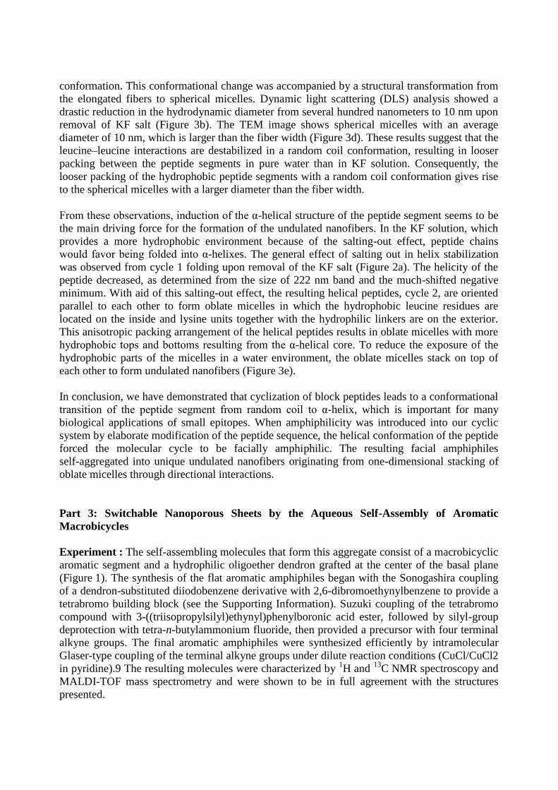
Molecule 1 self-assembles into nanoporous sheets in dilute aqueous solutions. Cryogenic
transmission electron microscopy (cryo-TEM) showed sheetlike objects with curved edges
against the vitrified solution background (Figure 2 a); this observation is indicative of the
formation of flexible sheets in bulk solution. Notably, a high-resolution image revealed that the
sheets contained in-plane nanopores with an average diameter of approximately 4 nm (Figure 2 a,
inset). To obtain more information on these sheets, we also performed TEM experiments with
the samples cast onto a TEM grid (and negatively stained with uranyl acetate). A
low-magnification image showed planar sheets with rugged surfaces against a dark background.
A higher-magnification image showed that the rugged surfaces consisted of uniform micelles and
nanopores (Figure 2 b, inset) and thus suggested that the sheets had formed through a lateral
association of small, discrete micelles. The diameters of the micelles and the pores were
measured to be approximately 3.5 nm and 3–5 nm, respectively. The micellar diameter is about
twice the length of the molecule and thus indicates the presence of dimeric micelles with a
face-to-face stacking of the aromatic basal planes. Further apparent evidence for the formation of
in-plane dimeric micelles was provided by topochemical polymerization of the sheets.
Polymerization of the diacetylene groups by irradiation of the solution with UV light for 6 h
yielded only dimers. This result demonstrates that the primary structure of the 2D sheets consists
of dimeric micelles in which the two aromatic planes within the micellar core face one another in
a slipped π–π stack for efficient dimerization of the diacetylene groups.

Figure 2. a) Cryo-TEM image of a solution of 1 (200 μm; scale bar: 200 nm). b) Negatively
stained TEM image of nanoporous sheets from a 100 μm aqueous solution of 1 (scale bar: 100
nm). The insets in (a) and (b) show magnified images (scale bars: 20 nm) and the line scan
profile along the yellow line. c) Absorption and emission spectra of 1 (100 μm) in CHCl3 (red
line) and aqueous solution (black line); λex=318 nm. d) Molecular modeling of the dimeric
micelle without coronene and the dimeric micelle containing coronene.
The formation of 2D sheets based on pairs of face-to-face-stacked aromatic molecules stimulated
us to investigate whether the planar nanostructure would encapsulate flat aromatic guest
molecules, such as coronene, through π–π stacking interactions in aqueous solution (Figure 2 d).
Indeed, the 2D sheets readily solubilized coronene in aqueous solution with preservation of their
2D structure. Upon the addition of coronene to an aqueous solution of 1, the fluorescence
intensity at 530 nm associated with the coronene emission increased until 0.5 equivalents of
coronene had been added. Upon the further addition of coronene to the solution, the fluorescence
intensity did not change, and precipitation was observed. Therefore, it can be concluded that the
maximum coronene loading per amphiphilic molecule is 0.5 equivalents. This result implies that
the flat conjugated aromatic guest is sandwiched between the two aromatic basal planes of the
dimeric micelles through hydrophobic and π–π stacking interactions. Atomic force microscopy
(AFM) images showed that the layer thickness increased from 2.1 to 2.5 nm upon the addition of
coronene (Figure 3 a,b). Thus, it appears that the coronene molecules, which have a flat
conjugated surface, are effectively intercalated between the aromatic planes of the dimeric
micelles.
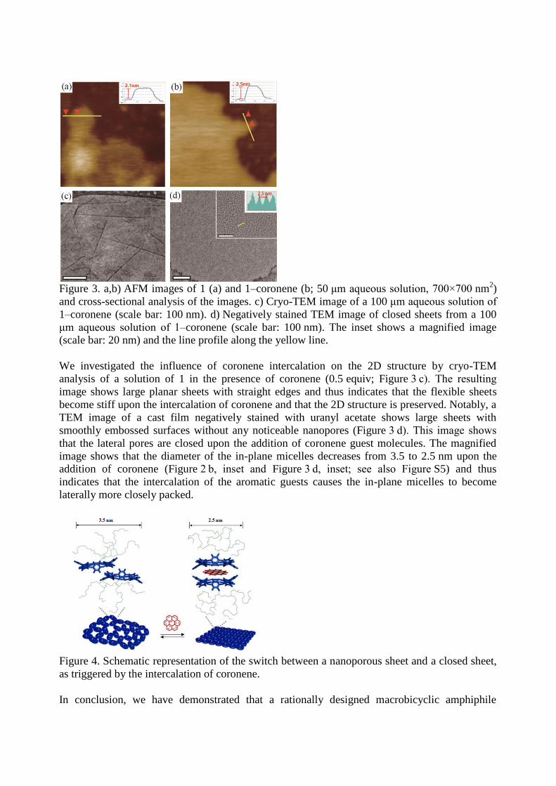
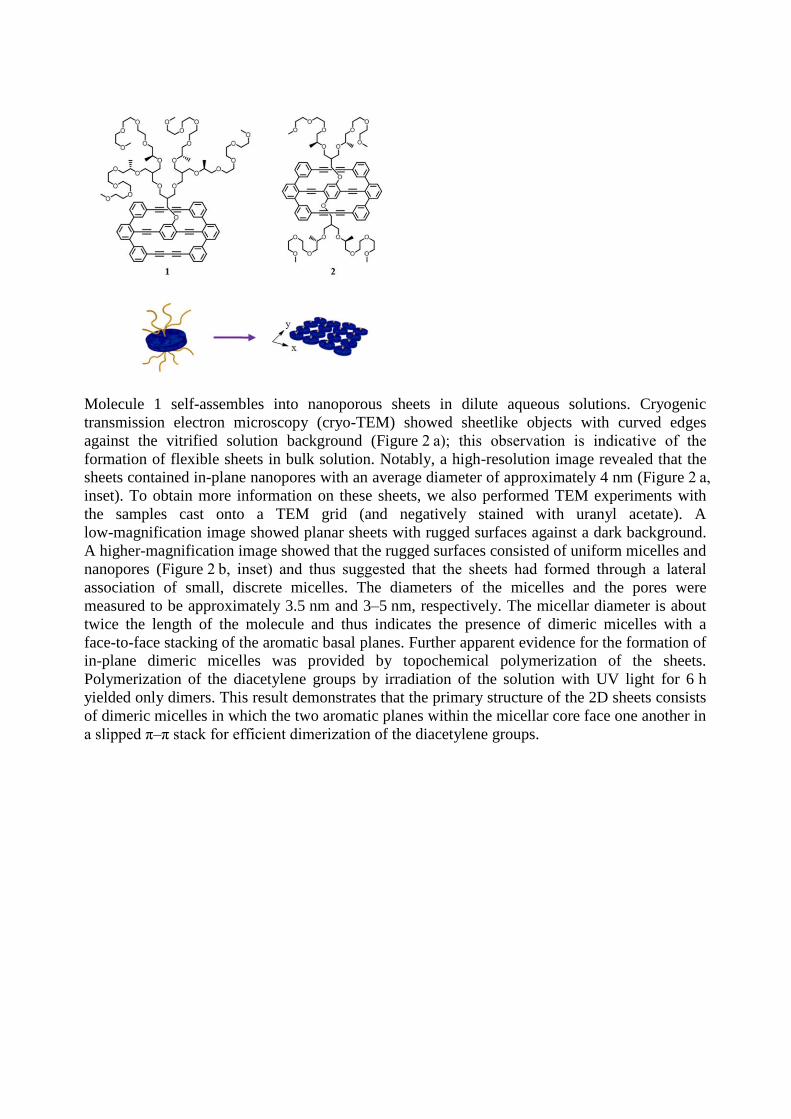
Figure 3. a,b) AFM images of 1 (a) and 1–coronene (b; 50 μm aqueous solution, 700×700 nm
2)
and cross-sectional analysis of the images. c) Cryo-TEM image of a 100 μm aqueous solution of
1–coronene (scale bar: 100 nm). d) Negatively stained TEM image of closed sheets from a 100
μm aqueous solution of 1–coronene (scale bar: 100 nm). The inset shows a magnified image
(scale bar: 20 nm) and the line profile along the yellow line.
We investigated the influence of coronene intercalation on the 2D structure by cryo-TEM
analysis of a solution of 1 in the presence of coronene (0.5 equiv; Figure 3 c). The resulting
image shows large planar sheets with straight edges and thus indicates that the flexible sheets
become stiff upon the intercalation of coronene and that the 2D structure is preserved. Notably, a
TEM image of a cast film negatively stained with uranyl acetate shows large sheets with
smoothly embossed surfaces without any noticeable nanopores (Figure 3 d). This image shows
that the lateral pores are closed upon the addition of coronene guest molecules. The magnified
image shows that the diameter of the in-plane micelles decreases from 3.5 to 2.5 nm upon the
addition of coronene (Figure 2 b, inset and Figure 3 d, inset; see also Figure S5) and thus
indicates that the intercalation of the aromatic guests causes the in-plane micelles to become
laterally more closely packed.
Figure 4. Schematic representation of the switch between a nanoporous sheet and a closed sheet,
as triggered by the intercalation of coronene.
In conclusion, we have demonstrated that a rationally designed macrobicyclic amphiphile
consisting of a hydrophilic dendron attached to the center of an aromatic plane undergoes
self-assembly into a 2D structure with nanosized lateral pores through the lateral association of
amphiphile dimers with a uniform diameter of 3.5 nm. The porous sheets efficiently intercalate
flat aromatic molecules, such as coronene, through the conformational inversion of the basal
planes of the dimeric micelles. Notably, the intercalation of a flat conjugated aromatic guest
causes the reversible transformation of the porous sheets into closed sheets without sacrificing
the 2D structure. This switch is also accompanied by change in the flexibility of the
self-assembled 2D structure from a flexible to a rigid state. This unique supramolecular structure
with switchable functionality might provide a new strategy for the design of intelligent materials
with simultaneous biological and electrooptical functions. In particular, the reversible switching
of the pores opens up the possibility of pumping molecules of interest out of the internal pores of
such 2D systems.
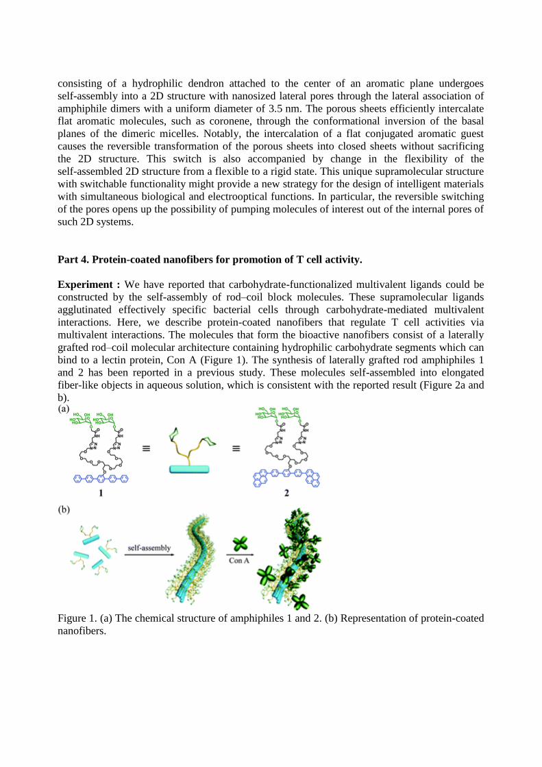
Part 4. Protein-coated nanofibers for promotion of T cell activity.
Experiment : We have reported that carbohydrate-functionalized multivalent ligands could be
constructed by the self-assembly of rod–coil block molecules. These supramolecular ligands
agglutinated effectively specific bacterial cells through carbohydrate-mediated multivalent
interactions. Here, we describe protein-coated nanofibers that regulate T cell activities via
multivalent interactions. The molecules that form the bioactive nanofibers consist of a laterally
grafted rod–coil molecular architecture containing hydrophilic carbohydrate segments which can
bind to a lectin protein, Con A (Figure 1). The synthesis of laterally grafted rod amphiphiles 1
and 2 has been reported in a previous study. These molecules self-assembled into elongated
fiber-like objects in aqueous solution, which is consistent with the reported result (Figure 2a and
b).
Figure 1. (a) The chemical structure of amphiphiles 1 and 2. (b) Representation of protein-coated
nanofibers.
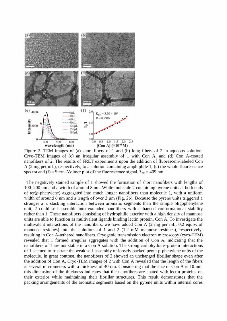
Figure 2. TEM images of (a) short fibers of 1 and (b) long fibers of 2 in aqueous solution.
Cryo-TEM images of (c) an irregular assembly of 1 with Con A, and (d) Con A-coated
nanofibers of 2. The results of FRET experiments upon the addition of fluorescein-labeled Con
A (2 mg per mL), respectively, to a solution containing amphiphile 1; (e) the whole fluorescence
spectra and (f) a Stern–Volmer plot of the fluorescence signal, λex = 409 nm.
The negatively stained sample of 1 showed the formation of short nanofibers with lengths of
100–200 nm and a width of around 8 nm. While molecule 2 containing pyrene units at both ends
of ter(p-phenylene) aggregated into much longer nanofibers than molecule 1, with a uniform
width of around 6 nm and a length of over 2 μm (Fig. 2b). Because the pyrene units triggered a
stronger π–π stacking interaction between aromatic segments than the simple oligophenylene
unit, 2 could self-assemble into extended nanofibers with enhanced conformational stability
rather than 1. These nanofibers consisting of hydrophilic exterior with a high density of mannose
units are able to function as multivalent ligands binding lectin protein, Con A. To investigate the
multivalent interactions of the nanofibers, we have added Con A (2 mg per mL, 0.2 equiv. of
mannose residues) into the solutions of 1 and 2 (1.2 mM mannose residues), respectively,
resulting in Con A-tethered nanofibers. Cryogenic transmission electron microscopy (cyro-TEM)
revealed that 1 formed irregular aggregates with the addition of Con A, indicating that the
nanofibers of 1 are not stable in a Con A solution. The strong carbohydrate–protein interactions
of 1 seemed to frustrate the weak self-assembly of loosely packed penta-p-phenylene units of the
molecule. In great contrast, the nanofibers of 2 showed an unchanged fibrillar shape even after
the addition of Con A. Cryo-TEM images of 2 with Con A revealed that the length of the fibers
is several micrometers with a thickness of 40 nm. Considering that the size of Con A is 10 nm,
this dimension of the thickness indicates that the nanofibers are coated with lectin proteins on
their exterior while maintaining their fibrillar structures. This result demonstrates that the
packing arrangements of the aromatic segments based on the pyrene units within internal cores
play a critical role in forming protein-tethered stable nanostructures.
Fluorescence resonance energy transfer (FRET) could provide one of the good methods to
monitor the formation of soluble Con A–ligand complexes. Subsequently, FRET experiments
were carried out with the mixtures of fluorescein-labeled Con A and amphiphiles 1 and 2,
respectively. As the amount of labeled Con A in a solution containing amphiphile 1 increased,
the intensity of fluorescence emission associated with the pentaphenylene segments decreased
(Fig. 2e). Similar to 1, 2 also showed the fluorescence quenching of the aromatic segments upon
the addition of the fluorescein-labeled Con A. On the basis of the fluorescence titration spectra,
the Stern–Volmer constants (Ksv) of amphiphiles 1 and 2 by Con A were found to be 3.30 × 10⁵
and 3.72 × 10⁵, respectively, predominantly through static quenching (Figure 2). It demonstrated
that the binding affinity was not significantly different between each sample with Con A. This
result indicates that the carbohydrate ligands effectively bind Con A through specific
ligand–protein interactions
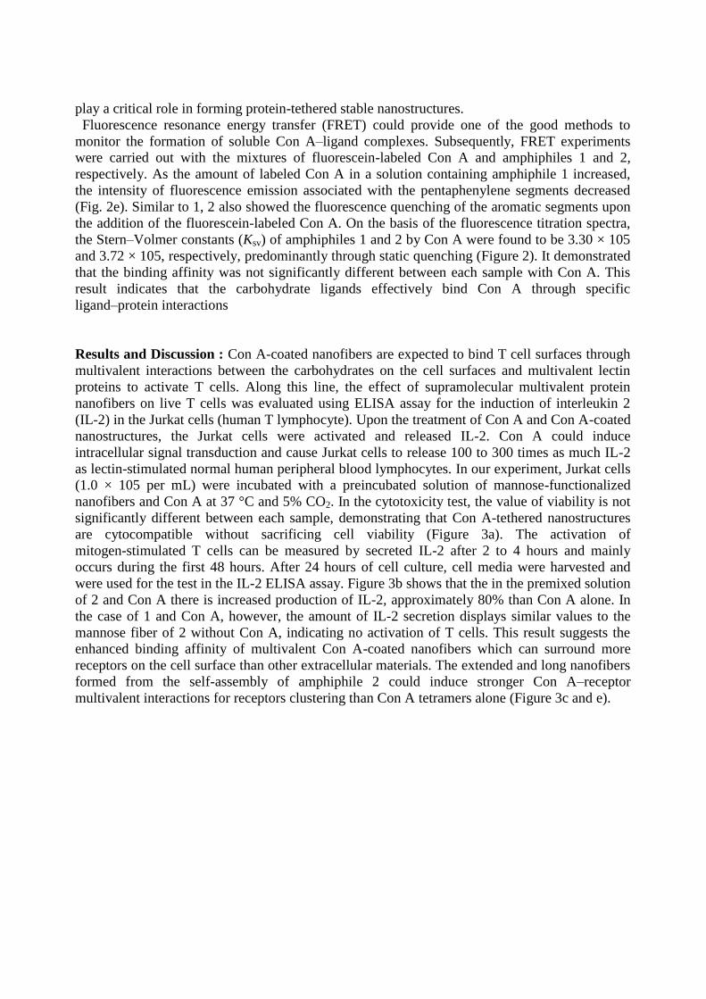
Results and Discussion : Con A-coated nanofibers are expected to bind T cell surfaces through
multivalent interactions between the carbohydrates on the cell surfaces and multivalent lectin
proteins to activate T cells. Along this line, the effect of supramolecular multivalent protein
nanofibers on live T cells was evaluated using ELISA assay for the induction of interleukin 2
(IL-2) in the Jurkat cells (human T lymphocyte). Upon the treatment of Con A and Con A-coated
nanostructures, the Jurkat cells were activated and released IL-2. Con A could induce
intracellular signal transduction and cause Jurkat cells to release 100 to 300 times as much IL-2
as lectin-stimulated normal human peripheral blood lymphocytes. In our experiment, Jurkat cells
(1.0 × 10⁵ per mL) were incubated with a preincubated solution of mannose-functionalized
nanofibers and Con A at 37 °C and 5% CO2. In the cytotoxicity test, the value of viability is not
significantly different between each sample, demonstrating that Con A-tethered nanostructures
are cytocompatible without sacrificing cell viability (Figure 3a). The activation of
mitogen-stimulated T cells can be measured by secreted IL-2 after 2 to 4 hours and mainly
occurs during the first 48 hours. After 24 hours of cell culture, cell media were harvested and
were used for the test in the IL-2 ELISA assay. Figure 3b shows that the in the premixed solution
of 2 and Con A there is increased production of IL-2, approximately 80% than Con A alone. In
the case of 1 and Con A, however, the amount of IL-2 secretion displays similar values to the
mannose fiber of 2 without Con A, indicating no activation of T cells. This result suggests the
enhanced binding affinity of multivalent Con A-coated nanofibers which can surround more
receptors on the cell surface than other extracellular materials. The extended and long nanofibers
formed from the self-assembly of amphiphile 2 could induce stronger Con A–receptor
multivalent interactions for receptors clustering than Con A tetramers alone (Figure 3c and e).
Figure 3. (a) The viability test using the water-soluble tetrazolium salt (WST) method of Jurkat
cells grown with Con A, 1 + Con A and 2 + Con A for one day. (b) Comparison of IL-2
production by Con A and scaffolded Con A. The error bars in (a) and (b) indicate the standard
deviations from three experiments performed in duplicate, and some are smaller than the
symbols. Schematic representation of IL-2 production depending on the fiber length. (c) Con A
alone, (d) 1 + Con A and (e) 2 + Con A.
In summary, we have demonstrated that controlling the conformation and stability of the
nanofibers tethered by lectin proteins can regulate T cell activation. The lengths as well as
stability of the protein-coated supramolecular nanofibers could be manipulated by a small
variation in the rod–coil molecular structure. Elongated nanofibers with stronger aromatic
interactions maintained their fibrillar shape even after tethering Con A, which was not possible
with the short and weakly assembled nanofibers. Notably, extended and long multivalent ligands
promote T cell activation compared with monovalent Con A ligands, observed by the secretion
of IL-2 and the fluorescence emission. These long and flexible supramolecular multivalent
architectures could arrange the desired receptors regularly and be adapted to the mobile
cytoskeleton simultaneously. A self-assembled multivalent architecture based on aromatic
rod–dendritic coil can offer many opportunities for developing biocompatible and bio-responsive
systems.
List of Publications and Significant Collaborations that resulted from your AOARD
supported project:
1) Shin, S.; Lim, S.; Kim, Y.; Kim, T.; Choi, T. L.; Lee, M.: Supramolecular switching between
flat sheets and helical tubules triggered by coordination interaction. J. Am. Chem. Soc., 2013,
135, 2156-2159.
2) Sim, S.; Kim, Y.; Kim, T.; Lim, S.; Lee, M.: Directional assembly of alpha-helical peptides
induced by cyclization. J. Am. Chem. Soc., 2012, 134, 20270-20272.
3) Kim, Y.; Shin, S.; Kim, T.; Lee, D.; Seok, C.; Lee, M.: Switchable nanoporous sheets by the
aqueous self-assembly of aromatic macrobicycles. Angew. Chem. Int. Ed. 2013, 52, 6426-6429.
4) Kim, T.; Lee, H.; Kim, Y.; Nam, J. M.; Lee, M.: Protein-coated nanofibers for promotion of T
cell activity. Chem. Commun. 2013, 49, 3949-3951.
5) Kim, Y.; Li, W.; Shin, S.; Lee, M.: Development of Toroidal Nanostructures by
Self-Assembly: Rational Designs and Applications. Acc. Chem. Res., 2013, ASAP.
6) Li, W.; Kim, Y.; Lee, M.: Intelligent supramolecular assembly of aromatic block molecules in
aqueous solution. Nanoscale 2013, 5, 7711-7723.
7) Kim, Y.; Kim, T.; Lee, M.: From self-assembled toroids to dynamic nanotubules. Polymer
Chemistry 2013, 4, 1300-1308.


![000 Cover€¦ · §”π”¢Õߧ—¡¿’√å [5] ‡À¡ Õπ™â“ßµ“∫Õ¥‡∑’ˬ«‰ª„πªÉ“©–π—Èπé «‘π“ π‘√ÿµ⁄µ‘꓇≥π](https://img.pdfslide.us/doc/110x75/5f232522eb2e571a9f50a057/000-cover-aaaaa-5-a-aaoeaoeaaaaaaaoeaa.jpg)


![Untitled-2 []”π” ”π— ß“π ≥– √√¡ “√ “√»÷ …“¢—Èπæ Èπ∞“π¡’Àπâ“∑’Ë √—∫º ‘¥ Õ∫„π “√‡∑’¬∫«ÿ](https://img.pdfslide.us/doc/110x75/5f2be7ea80b5fd5bee4d40e5/untitled-2-aa-aa-aoe-aa-aa-aoea-aoea-aoea.jpg)
