Embed Size (px)
Citation preview


Fig. 20-12a. Conducting
system of the heart

Fig. 20-13. Sequence of impulse conduction through the heart.
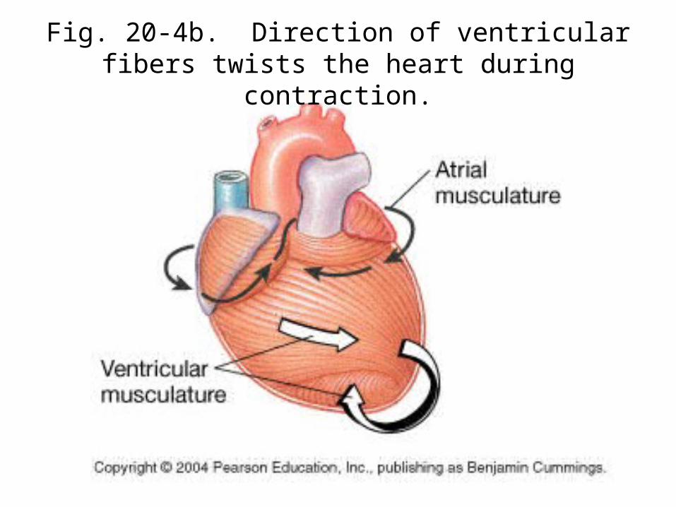
Fig. 20-4b. Direction of ventricular fibers twists the heart during contraction.
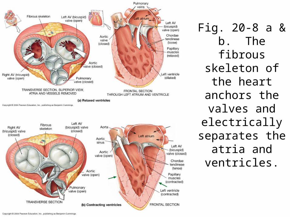
Fig. 20-8 a & b. The fibrous
skeleton of the heart anchors the
valves and electrically
separates the atria and
ventricles.

Fig. 20-8. Layers of the heart wall.

Fig. 21-1. Layers of the walls of blood vessels.

Fig. 21-2. Elastic and muscular arteries

Fig. 21-5. Arterioles and capillary bed

Fig. 21-2. Venules and veins

Fig. 21-4. Continuous and fenestrated capillaries
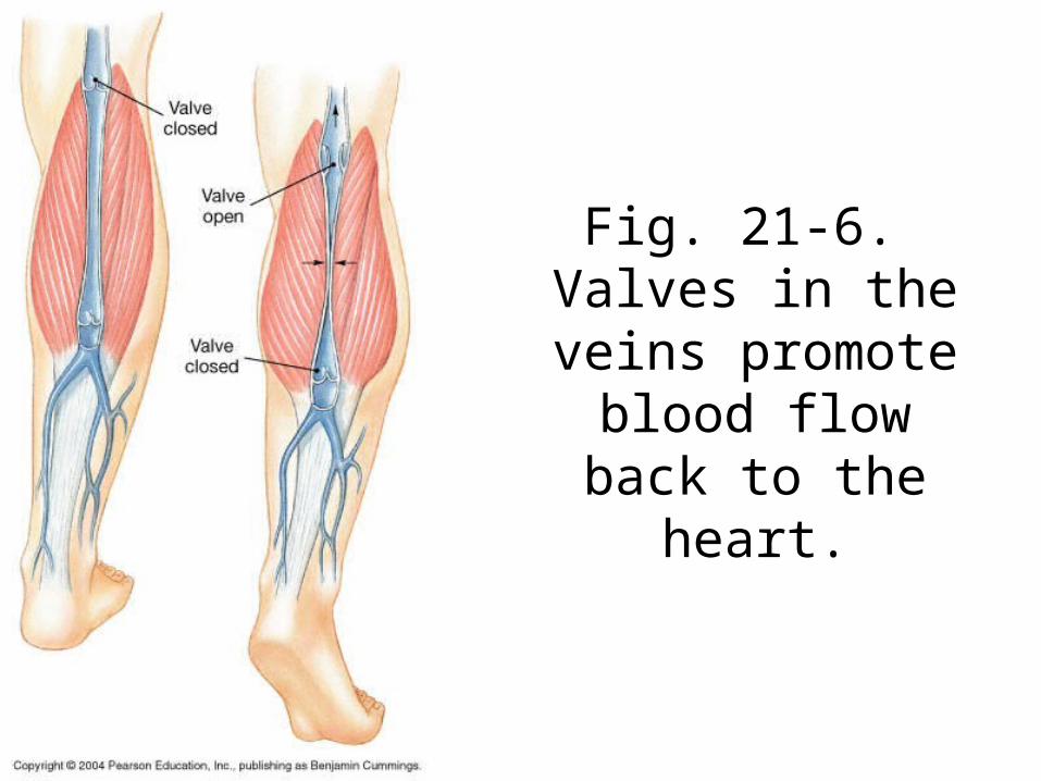
Fig. 21-6. Valves in the veins
promote blood flow back to the heart.