Embed Size (px)
Citation preview

Field Diagnosis of Chickpea Diseases and their Control
Science with a human face
Information Bulletin No. 28(Revised 2012)
© International Crops Research Institute for the Semi-Arid Tropics (ICRISAT), 2012. All rights reserved.
ICRISAT holds the copyright to its publications, but these can be shared and duplicated for non-commercial purposes. Permission to make digital or hard copies of part(s) or all of any publication for non-commercial use is hereby granted as long as ICRISAT is
[email protected]. ICRISAT’s name and logo are registered trademarks and may not be used without permission. You may not alter or remove any trademark, copyright or other notice.
Citation: Nene YL, Reddy MV, Haware MP, Ghanekar AM, Amin KS, Pande S and Sharma M. 2012. Field Diagnosis of Chickpea Diseases and their Control. Information Bulletin No. 28 (revised). Patancheru, A.P. 502 324, India: International Crops Research Institute for the Semi-Arid Tropics. 60 pp. ISBN 92-9066-199-2. Order code: IBE: 028.
Abstract
Chickpea (Cicer arietinum L.) is one of the world’s most important cool season food crops mostly grown in dry lands. The crop suffers from serious diseases that affect it in all growth stages. The pathogens that affect chickpea include fungi, bacteria, viruses, nematodes and mycoplasma, which results in severe economic losses globally. Among these, fungi are the largest and perhaps
pods of chickpea. The handbook is designed to assist agricultural research and extension workers who may have had less formal training in plant pathology to diagnose chickpea diseases. The bulletin provides information on economic importance, distribution,
chickpea diseases, and will be useful to farmers, extension workers,
diseases is also provided.
Cover: (Top L to R) Xylem blackening by fusarium wilt, white mycelial strands of Sclerotium rolfsii on roots, (Bottom L to R)
growth of Botrytis cinerea on pods.

Field Diagnosis of Chickpea Diseases and their Control
YL Nene, MV Reddy, MP Haware, AM Ghanekar, and KS Amin
Patancheru 502 324 Andhra Pradesh, India
Updated and Revised by
Suresh Pande and Mamta Sharma
Information Bulletin No. 28 (revised)
2012

Acknowledgement
We thank NT Vock, University of Queensland, St Lucia 4067, Australia, for contributing Figures 44, and 45, and WJ Kaiser, USDA Regional Plant Introduction Station, 59 Johnson Hall, Washington State University, Pullman, Washington 99164, USA for Figures 46 and 47. We also thank VK Sheila and A Nagavardhini, ICRISAT, for help in the preparation and revision of the bulletin.
About the Authors
YL Nene was Deputy Director General, ICRISAT.
MV Reddy and MP Haware were Sr Plant Pathologists, Legumes Program, ICRISAT.
AM Ghanekar was Plant Pathologist II, Legumes Program, ICRISAT Cooperative Research Station, Haryana Agricultural University, Hisar, Haryana 125 004, India.
KS Amin was Senior Plant Pathologist, National Research Centre for Groundnut (ICAR), Timbawadi, PO, Junagadh, Gujarat 362 015, India.
Suresh Pande is Principal Scientist (Pathology), Grain Legumes Program, ICRISAT, Patancheru, Hyderabad 502324.
Mamta Sharma is Senior Scientist (Pathology), Grain Legumes Program, ICRISAT, Patancheru, Hyderabad 502324.
Contents
Introduction .................................................................1
Fungal diseases affecting aerial plant parts ............2
Ascochyta blight [Ascochyta rabiei (Pass.) Labr.] .................2
Botrytis gray mold [Botrytis cinerea Pers. ex Fr.] ..................8
Alternaria blight [Alternaria alternata (Fr.:Fr.) Kiessler] .......12
Colletotrichum blight [Colletotrichum dematium (Pers. ex Fr.) Grove] ............................................................14
Phoma blight [Phoma medicaginis Malbr. & Roum] ............15
Stemphylium blight [Stemphylium sarciniforme (Cav.) Wilts.] ........................................................................17
Rust [Uromyces ciceris-arietini (Grogn. ) Jacz. & Beyer] ....................................................................17
Powdery mildew [Leveillula taurica (Lev. ) Salmon] ............19
Sclerotinia stem rot [Sclerotinia sclerotiorum (Li b.) de Bary] ....................................................................20
Fungal diseases affecting root/stem base .............22
Fusarium wilt [Fusarium oxysporum Schlechtend. emend. Snyd. et Hans. f. sp. ciceri (Padwick) Matuo et K. Sato] ................................................................22
Verticillium wilt [Verticillium albo-atrum Reinke & Berthier.] ..............................................................25
Collar rot [Sclerotium rolfsii Sacc.] ......................................26
Wet root rot [Rhizoctonia solani Kuhn] ................................28
Dry root rot [Rhizoctonia bataticola (Taub.) Butler] .............29
Black root rot [Fusarium solani (Mart.) Sacc.] .....................30
Phytophthora root rot [Phytophthora medicaginis E. M. Hans & D. P. Maxwell] ................................................32
Pythium root and seed rot [Pythium ultimum Trow] .............33
Foot rot [Operculella padwickii Kheswalla] ..........................35

iv
Bacterial disease ......................................................36
Bacterial blight [Xanthomonas campestris pv. cassiae Kulkarni et al.] ........................................................36
Viral diseases ............................................................37
Stunt [bean (pea) leaf roll virus] ..........................................37
Mosaic [alfalfa mosaic virus] ...............................................40
Proliferation [cucumber mosaic virus] .................................41
Narrow leaf [bean yellow mosaic virus] ...............................42
Necrosis [lettuce necrotic yellows virus] .............................43
Phyllody [mycoplasma] .......................................................43
Control measures .....................................................44
Resistant varieties/lines ......................................................44
Fungicides ..........................................................................47
Cultural practices ................................................................50
Supporting literature ................................................53
Index ..........................................................................55

1
Introduction
Nearly 172 pathogens have been reported so far that infect chickpea (Cicer arietinum L.) in different parts of the world (Nene et al. 1996), but only a few of them have the potential to devastate the crop. Some diseases are persistent problems in chickpea production in wide geographical areas, notably, ascochyta blight, fusarium wilt, dry root rot, stunt [caused by bean (pea) leaf roll virus], botrytis gray mold, collar rot, black root rot, phytophthora root rot, and pythium root and seed rot, while others are sporadic in occurrence or endemic in distribution. Diseases with limited distribution may still be economically important locally. Because of continuous changes in cultural practices, human interventions and climate change, some of the minor diseases may become economically important. One such example is dry root rot (Rhizoctonia bataticola) of chickpea, which is emerging as a potential threat to chickpea cultivation in semi-arid regions because the host plant is predisposed to infection by moisture stress and high temperatures during the flowering to pod filling stage (Sharma et al. 2010).
This bulletin has been prepared to assist in field diagnosis of chickpea diseases. The diseases are grouped into fungal, bacterial and viral diseases. In addition to symptomatology, information about distribution, economic importance, epidemiology and control measures were included to make the bulletin more useful to growers, extension workers, students and scientists. Specific management practices are provided whenever available.
For more detailed information on these diseases, refer to Nene et al. (1978), Nene and Reddy (1987), Pande et al. (2005; 2006 and 2010) and Chen et al. (2011).
2
Fungal diseases affecting aerial plant parts
Ascochyta blight [Ascochyta rabiei (Pass.) Labr.]
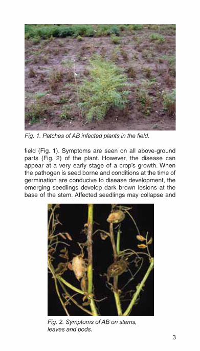
Distribution. Ascochyta blight (AB) is a major disease in west Asia, northern Africa, and southern Europe. The disease usually builds up in February and March in Pakistan and northern India when the crop canopy is very dense and temperatures are favorable to disease development. In West Asia, southern Europe and northern Africa, such conditions usually prevail in March, April and May. In the winter-sown chickpeas of the Mediterranean region, blight symptoms can be found in November and December when the weather is wet and warm. The disease has been reported from 35 countries across 6 continents and as recently seen in Australia and Canada, it can spread rapidly to new areas of chickpea production.
Economic importance. The disease can cause grain yield and quality losses up to 100%.
Epidemiology. AB is a seed borne disease. Diseased debris left over in the fields also serves as a source of primary inoculum. Ascospores were also found to play a role in the initiation of disease epidemics. Secondary spread is through pycnidiospores. Chickpea and its wild relatives are the only confirmed hosts of the fungus A. rabiei. However, Kaiser (1990) reported other hosts of the pathogen, outside the genus Cicer. Cool, cloudy and wet weather favors the disease development. The disease builds up and spreads fast when night temperatures are around 10°C, day temperatures are around 20°C, and rains are accompanied by cloudy days. Excessive canopy development also favors blight development.
Symptoms. The disease is usually seen around flowering and podding time as patches of blighted plants in the
3
field (Fig. 1). Symptoms are seen on all above-ground parts (Fig. 2) of the plant. However, the disease can appear at a very early stage of a crop’s growth. When the pathogen is seed borne and conditions at the time of germination are conducive to disease development, the emerging seedlings develop dark brown lesions at the base of the stem. Affected seedlings may collapse and
Fig. 2. Symptoms of AB on stems, leaves and pods.

4
die (damping-off). Pycnidia may be formed on the lesions. Isolated infected seedlings may not be noticed. But at flowering and podding time, when conditions are usually favorable for disease development, the disease spreads from these isolated seedlings, resulting in patches of blighted plants. When the source of inoculum is airborne conidia or ascospores, the disease initially appears in the form of several small water-soaked necrotic spots on the younger leaves of almost all branches. Under conditions favorable for disease development these spots enlarge rapidly and coalesce, blighting the leaves and buds. Pycnidia are observed on the blighted parts. On a susceptible cultivar, the necrosis progresses from the buds downwards, killing the plant (Fig. 2a). In cases of severe foliar infection, the entire plant dries up suddenly.
If conditions are not favorable for disease development (hot dry weather), the plants do not die and the infection remains in the form of discrete lesions on the leaves, petioles, stems, and pods (Fig. 3). The symptoms on the leaflets are round spots with brown margins and a gray center that contains pycnidia, which are often arranged in concentric rings.
Fig. 2a. AB symptoms on aerial parts of plant.

5
Fig. 3. Lesions caused by AB on
Fig. 4. Elongated lesions on stem and breaking of branches at the point of girdling.
On the stems and petioles, the lesions are obovate or elongate and bear pycnidia (Fig .4). The size of the lesions varies greatly; some may become 3 - 4 cm long on stems and often girdle the affected portion. The stems and petioles usually break at the point of girdling. If blight occurs at the pre-flowering stage and then conditions for its development become unfavorable (hot dry weather)

6
the crop regrows fast but symptoms can still be seen on the older branches. Fully developed lesions on pods are usually round, up to 0.5 cm in diameter, usually with concentric rings of pycnidia (Fig. 5). Several lesions may appear on a single pod and if infection occurs in the early stages of pod development, the pod is blighted and fails to develop any seed (Fig. 6). Late infections result in shriveled and infected seed.
The fungus penetrates the pod and infects the developing seed. Symptoms on the seeds appear as a brown discoloration and often develop into deep, round or irregular cankers, sometimes bearing pycnidia visible to the naked eye (Fig. 7).
Fig. 5. Close up of lesions caused by AB on pod wall with pycnidial bodies arranged in concentric rings.

7
Fig. 6. AB lesions on green pods.
Fig. 7. Lesions caused by AB on kabuli seeds (healthy seeds at left).

8
Botrytis gray mold [Botrytis cinerea Pers. ex Fr.]
Distribution. Botrytis gray mold (BGM) is a serious disease in parts of Bangladesh, India, Nepal, Pakistan, Australia and Argentina. BGM has also been reported from Canada, Chile, Colombia, Hungary, Mexico, Myanmar, Spain, Turkey, the USA and Vietnam.
Economic importance. BGM can cause yield losses up to 100%.
Epidemiology. BGM is a seed borne disease. The fungus has a very wide host range. The disease is usually seen at flowering time when the crop canopy is fully developed. Excessive vegetative growth due to too much irrigation or rain, close spacing, and varieties that have a spreading habit favor disease development. Temperatures between 20 and 25°C and excessive humidity around flowering and podding time favor disease development. As temperatures favorable to BGM are slightly higher than those for ascochyta blight, these diseases may occur one after the other with ascochyta blight appearing first.
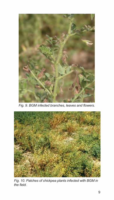
Symptoms. Lack of pod setting is the first indication of the disease (Figs. 8 and 9). Leaves and stems may not show any symptoms. Under weather conditions highly favorable to the disease, foliage shows clear symptoms and plants often die in patches (Fig. 10).
Fig. 8. Plants affected by BGM with no podding.
9
Fig. 10. Patches of chickpea plants infected with BGM in

10
The disease is more severe on portions of the plant hidden under the canopy and is obvious if the canopy is parted to expose the symptoms. Shed flowers and leaves covered with the spore mass can be seen on the ground under the plants.
When humidity is very high, the symptoms appear on stems, leaves, flowers and pods as gray or dark brown lesions covered with moldy sporophores. Lesions on stem are 10 - 30 mm long and girdle the stem completely (Fig. 11). Tender branches break off at the point where the gray mold has caused rotting. Affected leaves and flowers turn into a rotting mass (Fig. 12). Lesions on the
Fig. 11. Mycelial growth and grayish sporulation of Botrytis cinerea on chickpea stem.
Fig. 12. Fungal sporulation on infected stem,

11
Fig. 13. Healthy (left) and BGM infected (right) pods.
Fig. 14. Grayish mycelial growth on infected pods.
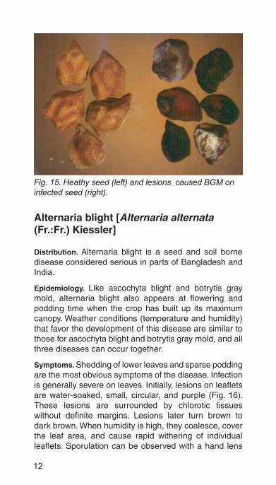
pod are water-soaked and irregular (Fig. 13). On infected plants, the pods contain either small, shriveled seeds or no seeds at all (Fig. 14). Grayish white mycelium may be seen on the infected seeds (Fig. 15). Further details on integrated crop management, with emphasis on botrytis gray mold is found in Pande et al. (2006).
12
Alternaria blight [Alternaria alternata (Fr.:Fr.) Kiessler]
Distribution. Alternaria blight is a seed and soil borne disease considered serious in parts of Bangladesh and India.
Epidemiology. Like ascochyta blight and botrytis gray mold, alternaria blight also appears at flowering and podding time when the crop has built up its maximum canopy. Weather conditions (temperature and humidity) that favor the development of this disease are similar to those for ascochyta blight and botrytis gray mold, and all three diseases can occur together.
Symptoms. Shedding of lower leaves and sparse podding are the most obvious symptoms of the disease. Infection is generally severe on leaves. Initially, lesions on leaflets are water-soaked, small, circular, and purple (Fig. 16). These lesions are surrounded by chlorotic tissues without definite margins. Lesions later turn brown to dark brown. When humidity is high, they coalesce, cover the leaf area, and cause rapid withering of individual leaflets. Sporulation can be observed with a hand lens
Fig. 15. Heathy seed (left) and lesions caused BGM on infected seed (right).

13
Fig. 17. Flowers killed by alternaria blight.
(10X). On the stems, the lesions are elongated and are brown to black. The infected flowers die (Fig. 17). On the pods, the lesions are circular, slightly sunken, and irregularly scattered. Affected pods turn dirty black. On mature pods, the lesions remain as localized, tiny, black superficial flecks. Infected seeds are shriveled. Under favorable weather conditions the entire foliage can die.

14
Colletotrichum blight [Colletotrichum dematium (Pers. ex Fr.) Grove]
Distribution. Colletotrichum blight is a minor seed and soil borne disease reported only from India.
Epidemiology. The disease is generally fatal when the crop is sown early (September) when the temperatures are high (25 – 30°C) and the young crop is caught in the rains during late September or early October. The disease does not normally occur in the postrainy season crop, but if there are unusual rains, the disease can affect the crop.
Symptoms. The disease can kill the plants at any stage of crop growth, depending on the weather conditions and amount of inoculum present. Plants and branches that have dried up and get scattered throughout the field are an indication of colletotrichum blight (Fig. 18). On seedlings, two kinds of symptoms can be observed: (1) elongated, sunken, dark brown spots on the lower part of the stem, extending to the root, and (2) wilting and drying due to severe collar and root infection. In adult plants, lesions are seen on all the above-ground parts. On leaves and pods, lesions are circular to elongate, sunken at the center, and
Fig. 18. A row of dried plants affected by collectotricum blight.

15
with yellow margins. On stems they are elongated and black. The fruiting bodies (acervuli) are scattered within the affected tissues. The fungus penetrates the pod wall and infects the seed (Fig. 19).
Fig. 19. Lesions on pods and seeds.
Phoma blight [Phoma medicaginis Malbr. & Roum]
Distribution. Phoma blight is a minor disease reported from Australia, Bangladesh, India, Syria and USA.
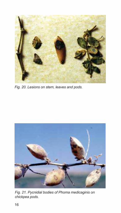
Symptoms. It usually affects the crop in the reproductive phase. The field symptom is the appearance of patches of drying plants. The symptoms are somewhat similar to those of ascochyta blight. Irregular, light brown lesions on the leaves, stems, and pods have dark margins (Fig. 20). Dark, minute, submerged pycnidia are irregularly scattered in the infected tissue (Fig. 21). Seeds from infected pods are discolored and shriveled. The conditions favorable to phoma blight are similar to those that favor ascochyta blight. Further details can be found in Haware and Nene (1981).
16
Fig. 20. Lesions on stem, leaves and pods.
Fig. 21. Pycnidial bodies of Phoma medicaginis on chickpea pods.
17
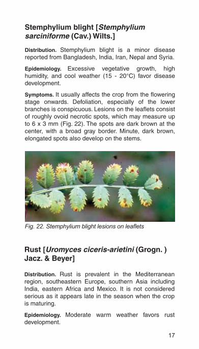
Stemphylium blight [Stemphylium sarciniforme (Cav.) Wilts.]
Distribution. Stemphylium blight is a minor disease reported from Bangladesh, India, Iran, Nepal and Syria.
Epidemiology. Excessive vegetative growth, high humidity, and cool weather (15 - 20°C) favor disease development.
Symptoms. It usually affects the crop from the flowering stage onwards. Defoliation, especially of the lower branches is conspicuous. Lesions on the leaflets consist of roughly ovoid necrotic spots, which may measure up to 6 x 3 mm (Fig. 22). The spots are dark brown at the center, with a broad gray border. Minute, dark brown, elongated spots also develop on the stems.
Fig. 22.
Rust [Uromyces ciceris-arietini (Grogn. ) Jacz. & Beyer]
Distribution. Rust is prevalent in the Mediterranean region, southeastern Europe, southern Asia including India, eastern Africa and Mexico. It is not considered serious as it appears late in the season when the crop is maturing.
Epidemiology. Moderate warm weather favors rust development.

18
Symptoms. The severely infected crop looks rusty because the foliage is coated with rust pustules and urediniospores. The rust appears first mainly on the leaves as small, round or oval, cinnamon brown, powdery pustules (Fig. 23).These pustules tend to coalesce. Sometimes a ring of small pustules can be seen around larger pustules, which occur on both leaf surfaces but more frequently on the lower one (Fig. 24). Occasionally pustules can be seen on stems. Severely infected plants
Fig. 23. Rust pustules on chickpea leaves.

19
may dry up prematurely. Cool and moist weather favors rust buildup; rain does not appear to be essential for the infection to spread.
Powdery mildew [Leveillula taurica (Lev. ) Salmon]
Distribution. Powdery mildew is a minor disease reported from India, Mexico, Morocco, Pakistan, and Sudan.
Epidemiology. Cool and dry weather favors powdery mildew development.
Symptoms. Like rust, powdery mildew appears late in the season when the crop is nearing maturity, except in highly susceptible genotypes, when it appears earlier. Severe infection of powdery mildew can be easily recognized by the white powdery growth on the foliage, which is a characteristic feature of the powdery mildews (Fig. 25). Small patches of white powdery coating initially develop on both surfaces of older leaves. These patches grow and may cover a large area. Affected leaves turn purple and then die. When infection is severe, stems, young leaves and pods are also covered with the powdery coating.
Fig. 25. Powdery mildew symptoms on leaves.

20
Sclerotinia stem rot [Sclerotinia sclerotiorum (Li b.) de Bary]
Distribution. Sclerotinia stem rot is reported from most of the chickpea growing regions of the world. At present, it is a minor disease.
Epidemiology. Excessive vegetative growth, high soil moisture, and cool weather (20°C) favor disease development.
Symptoms. It can affect the crop at any stage. The pathogen has been observed to cause collar rot of seedlings in North African chickpea-growing regions. Otherwise it usually appears after the crop canopy has covered the ground. The disease is characterized by the appearance of chlorotic or drying branches or whole plants scattered in the field. Such drying plants or branches rot at the collar region (Fig. 26) or at any point on the branch. The leaves of affected plants/branches turn yellow or droop while remaining green, then dry up and turn straw colored.
Fig. 26. Rotting of the stems at the basal region.

21
Fig. 28. Brownish sclerotial bodies in a split stem.
A web of white mycelial strands appears at the collar region and above (up to 5 cm) and may cover the base of the branches (Fig. 26). Extended grayish lesions with or without mycelial coating can also be seen on the upper parts of the stems. Whitish or brownish irregular-shaped sclerotia can be seen, occasionally mingled with mycelial strands on branches (Fig. 27), or inside the stem (Fig. 28).
Fig. 27. Extended grayish lesions with mycelial coating.
22
Fungal diseases affecting root/stem base
Fusarium wilt [Fusarium oxysporum Schlechtend. emend. Snyd. et Hans. f. sp. ciceri (Padwick) Matuo et K. Sato]
Distribution. Fusarium wilt disease in now widely spread in most chickpea- growing areas of Asia, Africa, southern Europe and the Americas.
Economic importance. Yearly yield losses are estimated at 10-15% in India and Spain, with losses of 70-100% in years of severe outbreaks of the disease.
Epidemiology. Wilt is a seed and soil borne disease. Wilt incidence is generally higher when chickpea is grown in warmer and drier climates (> 25°C) and when crop rotations are not practiced.
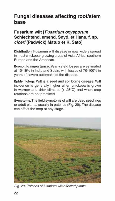
Symptoms. The field symptoms of wilt are dead seedlings or adult plants, usually in patches (Fig. 29). The disease can affect the crop at any stage.
Fig. 29. Patches of fusarium wilt-affected plants.

23
Seedling stage. The disease can be observed within 3 weeks of sowing. Whole seedlings (3 - 5 weeks after sowing) collapse and lie flat on the ground. These seedlings retain their dull green color (Fig. 30). When uprooted, they usually show uneven shrinking of the stem above and below the collar region (soil level). The shrunken portion may be about 2.5 cm or longer.
Affected seedlings do not rot on the stem or root surface. However, when split open vertically from the collar downwards or cut transversely, dark brown to black discoloration of the internal stem tissues is clearly visible (Figs. 31 and 32). In seedlings of
Fig. 31. Internal blackening of the stem caused by fusarium wilt.
Fig. 30. Young plant with dull green leaves killed by fusarium wilt.

24
highly susceptible cultivars, eg, JG 62, which dies within 10 - 15 days after emergence, the black discoloration may not be clearly visible. However, internal browning from root tip upwards is clearly seen.
Adult stage. The affected plants show typical wilting, ie, drooping of the petioles, rachis and leaflets (Fig. 33). Drooping is visible initially in the upper part of the plant but within a day or two, the entire plant droops.
Fig. 32. A transeverse cut of stem showing xylem blackening, caused by fusarium wilt.
Fig. 33. Drooping of fusarium wilt-affected plant.

25
The lower leaves are chlorotic, but most of the other leaves droop while still green. Gradually, however, all the leaves turn yellow and then light brown or straw colored. Dried leaflets of infected plants are not shed at maturity. Affected plants, when uprooted and examined before they are completely dry, show no external rotting, drying, or root discoloration. When the stem is split vertically, internal discoloration can be seen. Around the collar region, above and below, the xylem in the central inner portion (pith and part of the wood) is discolored dark brown or black. In the initial stage of wilting, the discoloration may not be continuous. Discoloration also extends several centimeters above the collar region into the main stem and branches. If the collar region is cut transversely with a sharp razor blade, black discoloration of both pith and xylem can be seen. Sometimes only a few branches are affected, resulting in partial wilt. In certain cultivars (eg, T3), the lower leaves dry up before the plants wilt.
Verticillium wilt [Verticillium albo-atrum Reinke & Berthier.]
Distribution. Verticillium wilt is considered important in Italy, Pakistan and Tunisia, and was recently observed in Syria.
Epidemiology. Verticillium wilt is favored by the similar weather conditions that promote fusarium wilt.
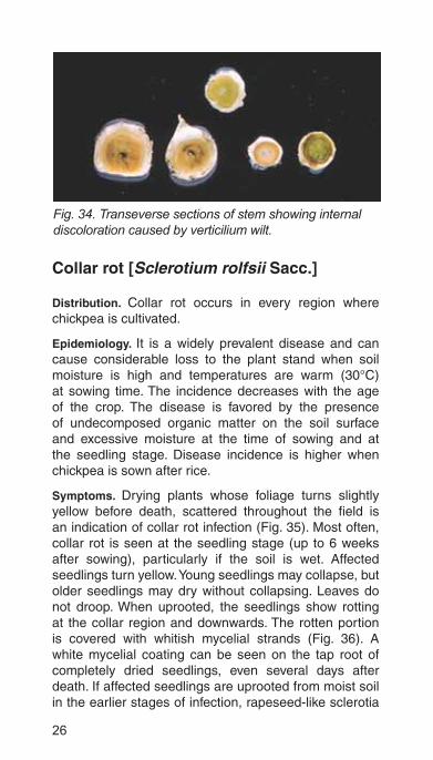
Symptoms. The disease can affect the crop at any stage. The field symptoms of verticillium wilt are similar to those of fusarium wilt .The foliage of affected plants may turn yellow before wilting. The xylem tissue shows a brown discoloration, lighter than that caused by F. oxysporum f. sp. ciceri (Fig. 34). For additional information, see Erwin (1958).
26
Collar rot [Sclerotium rolfsii Sacc.]
Distribution. Collar rot occurs in every region where chickpea is cultivated.
Epidemiology. It is a widely prevalent disease and can cause considerable loss to the plant stand when soil moisture is high and temperatures are warm (30°C) at sowing time. The incidence decreases with the age of the crop. The disease is favored by the presence of undecomposed organic matter on the soil surface and excessive moisture at the time of sowing and at the seedling stage. Disease incidence is higher when chickpea is sown after rice.
Symptoms. Drying plants whose foliage turns slightly yellow before death, scattered throughout the field is an indication of collar rot infection (Fig. 35). Most often, collar rot is seen at the seedling stage (up to 6 weeks after sowing), particularly if the soil is wet. Affected seedlings turn yellow. Young seedlings may collapse, but older seedlings may dry without collapsing. Leaves do not droop. When uprooted, the seedlings show rotting at the collar region and downwards. The rotten portion is covered with whitish mycelial strands (Fig. 36). A white mycelial coating can be seen on the tap root of completely dried seedlings, even several days after death. If affected seedlings are uprooted from moist soil in the earlier stages of infection, rapeseed-like sclerotia
Fig. 34. Transeverse sections of stem showing internal discoloration caused by verticilium wilt.

27
Fig. 36. Yellowing of leaves and constriction at the collar region.
Fig. 37. White mycelial strands of Sclerotium rolfsii on root system.
(1 mm in diameter), attached to mycelial strands around the collar are seen (Fig. 37). The non-affected portion of the root is white inside, as is normal.

28
Wet root rot [Rhizoctonia solani Kuhn]
Distribution. Wet root rot is a minor disease and is reported from several countries.
Epidemiology. No-till or reduced-till conditions favor growth of the pathogen and development of the disease. The pathogen can infect at a wide range of soil temperatures, but cool (11-18°C), wet soil conditions are optimum.
Symptoms. The field symptoms are almost the same as those of collar rot, ie, drying plants scattered throughout the field. Like collar rot, this disease is most often seen at the seedling stage (up to 6 weeks after sowing) in soils with relatively high moisture content. However, in irrigated chickpea, the disease may occur at later stages in the crop growth. Affected seedlings gradually turn yellow and petioles and leaflets droop. Seedlings do not usually collapse. A distinct dark brown lesion appears above the collar region on the main stem and can extend to lower branches in older plants (Fig. 38). The stem and root below the lesion show rotting, frequently with pinkish mycelial growth. Sclerotia are not usually seen.
Fig. 38. Symptoms of wet root rot on the root and lower portions of the branches.

29
by dry root rot.
Dry root rot [Rhizoctonia bataticola (Taub.) Butler]
Distribution. Dry root rot is the most important root rot disease in chickpea particularly in the semi-arid tropics of Ethiopia and in central and southern India. It has also been reported from Australia, Bangladesh, Iran, Kenya, Lebanon, Myanmar, Mexico, Nepal, Pakistan, Spain, Sudan, Turkey and the United States.
Epidemiology. The pathogen is a facultative sporophyte and is both seed borne and soil borne. Maximum ambient temperatures above 30°C, minimum above 20°C, and moisture stress (dry conditions) at the reproductive stages favor disease development.
Symptoms. The disease generally appears around flowering and podding time in the form of scattered dried plants (Figs. 39 and 40). The seedlings can also get infected. The susceptibility of the plant to the disease increases with age.
Drooping of petioles and leaflets is confined to those at the very top of the plant. Sometimes when the rest of the plant is dry, the topmost leaves are chlorotic. The leaves and stems of affected plants are usually straw

30
colored, but in some cases the lower leaves and stems are brown. The lower portion of the tap root usually remains in the soil when plants are uprooted. The tap root is dark, shows signs of rotting, and is devoid of most of its lateral and finer roots (Fig. 41). Dark, minute sclerotial bodies can be seen on the roots exposed or inside the wood.
Fig. 40. Dry root rot affected plant.
Fig. 41. Rotting root system.
Black root rot [Fusarium solani (Mart.) Sacc.]
Distribution. Black root rot is a minor disease reported from Argentina, Chile, India, Mexico, Spain, Syria and USA.
Epidemiology. Excessive moisture and moderately high temperatures (25-30°C) encourage disease development.
Symptoms. The disease can occur at any stage. Affected plants turn yellow and wilt. Dead plants are seen

31
Fig. 42. Scattered dead plants caused by black root rot.
Fig. 43. Rotting of tap root and lateral roots (right).
scattered in the field (Fig. 42). The root system is rotten, most of the finer roots are shed, and the remaining roots turn black (Fig. 43). Affected plants dry prematurely but may go on producing new roots if sufficient moisture is available. Further details can be found in Nene and Reddy (1987).
32
Phytophthora root rot [Phytophthora medicaginis E. M. Hans & D. P. Maxwell]
Distribution. Phytophthora root rot is an important disease in Australia. It is also reported from Argentina, India, Pakistan and Spain.
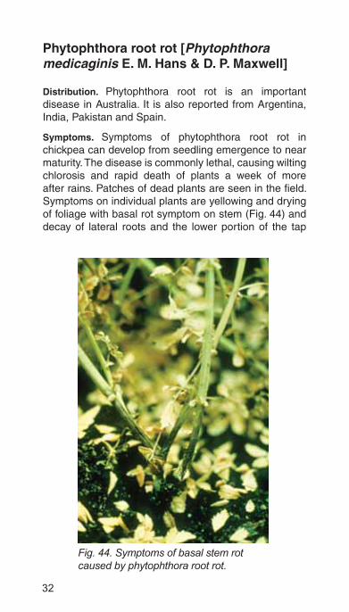
Symptoms. Symptoms of phytophthora root rot in chickpea can develop from seedling emergence to near maturity. The disease is commonly lethal, causing wilting chlorosis and rapid death of plants a week of more after rains. Patches of dead plants are seen in the field. Symptoms on individual plants are yellowing and drying of foliage with basal rot symptom on stem (Fig. 44) and decay of lateral roots and the lower portion of the tap
Fig. 44. Symptoms of basal stem rot caused by phytophthora root rot.

33
root (Fig. 45). On the upper portion of the tap root, dark brown to black lesions are seen, which in some cases extend to the stem base. The advancing margins of these lesions are often reddish brown. These symptoms can be easily confused with those of wet root rot. The disease incidence is high in low lying areas where water stagnates. For additional information, refer to Vock et al. (1980).
Fig. 45. Symptoms of phyophthora root rot.
Pythium root and seed rot [Pythium ultimum Trow]
Distribution. It is a minor disease reported from Brazil, Canada, Spain, India, Iran, Turkey, and USA. The large-seeded kabuli types of chickpea are much more susceptible than the dark-seeded, small, irregularly shaped desi types that have thick seed coats.

34
Symptoms. Emergence is poor because seeds are rotten (Figs. 46 and 47). Affected seedlings are stunted. The larger roots are necrotic, discolored and devoid of rootlets. Stunted plants often die before they flower. For additional information, refer to Kaiser and Hannan (1983).
Fig. 46. Poor emergence of seedlings in certain genotypes due to Pythium seed rot.
Fig. 47. Section of rotting seeds (bottom row) caused by Pythium ultimum in naturally infested soil and healthy seedling at right.

35
Foot rot [Operculella padwickii Kheswalla]
Distribution. Foot rot is a minor disease reported only from India, Bangladesh, and Nepal.
Symptoms. Dead plants are seen in patches or are seen scattered in the field. The symptoms of this disease on the above-ground plant parts are similar to those of fusarium wilt, particularly the drooping of petioles and rachis. Rotting from the collar region downwards is distinct, but there is no visible mycelial growth (Fig. 48). The stem above the rotten portion is discolored, but this discoloration is brown and does not involve the pith, unlike the brown-to-black discoloration in verticillium and fusarium wilts. The disease appears when the soils are wet.
Fig. 48. Foot rot showing rotting of root and drooping leaf.
36
Bacterial disease
Bacterial blight [Xanthomonas campestris pv. cassiae Kulkarni et al.]
Distribution. Bacterial blight is a minor disease reported only from India.
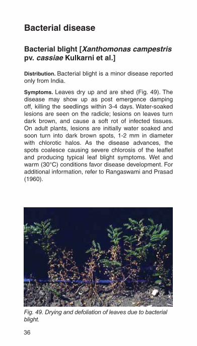
Symptoms. Leaves dry up and are shed (Fig. 49). The disease may show up as post emergence damping off, killing the seedlings within 3-4 days. Water-soaked lesions are seen on the radicle; lesions on leaves turn dark brown, and cause a soft rot of infected tissues. On adult plants, lesions are initially water soaked and soon turn into dark brown spots, 1-2 mm in diameter with chlorotic halos. As the disease advances, the spots coalesce causing severe chlorosis of the leaflet and producing typical leaf blight symptoms. Wet and warm (30°C) conditions favor disease development. For additional information, refer to Rangaswami and Prasad (1960).
Fig. 49. Drying and defoliation of leaves due to bacterial blight.
37
Viral diseases
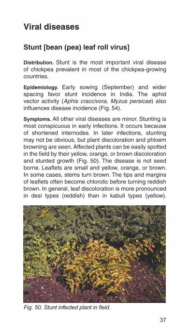
Stunt [bean (pea) leaf roll virus]
Distribution. Stunt is the most important viral disease of chickpea prevalent in most of the chickpea-growing countries.
Epidemiology. Early sowing (September) and wider spacing favor stunt incidence in India. The aphid vector activity (Aphis craccivora, Myzus persicae) also influences disease incidence (Fig. 54).
Symptoms. All other viral diseases are minor. Stunting is most conspicuous in early infections. It occurs because of shortened internodes. In later infections, stunting may not be obvious, but plant discoloration and phloem browning are seen. Affected plants can be easily spotted in the field by their yellow, orange, or brown discoloration and stunted growth (Fig. 50). The disease is not seed borne. Leaflets are small and yellow, orange, or brown. In some cases, stems turn brown. The tips and margins of leaflets often become chlorotic before turning reddish brown. In general, leaf discoloration is more pronounced in desi types (reddish) than in kabuli types (yellow).
Fig. 50.

38
The stems and leaves of diseased plants are stiffer and thicker than normal.
The most characteristic symptom of stunt is phloem browning (Fig. 51). It becomes obvious if the bark is removed at the collar region (by cutting a thin slice length-ways). A transverse cut (Fig. 52) reveals a brown ring or a split through the collar region and reveals brown streaks of discolored phloem vessels (Fig. 53). The interior wood of the root is white, as is normal, without xylem discoloration. If the plants survive up to the podding stage, pod set is sparse. Many plants dry up prematurely. Sometimes
Fig. 51. Phloem browning at the collar region caused by stunt.
Fig. 52. A tranverse section of a stunt-infected plant showing brown phloem ring at the collar region.

39
stunt and fusarium wilt infection occur together. In such cases, xylem discoloration, which is typical of fusarium wilt, is also seen. The wilting is the result of combined infection. Mechanical damage to the phloem by chewing insects, which attack the plant at the collar region (Fig. 54), can also result in leaf discoloration and stunting similar to stunt. But there will be no phloem browning.
Fig. 54. Colonies of Aphis crassivora on the stem.
Fig. 53. A vertical section of stunt infected chikpea plant showing the phloem browing at collar region.

40
Mosaic [alfalfa mosaic virus]
Distribution. Mosaic is a minor disease reported from Algeria, India, Iran, Morocco, New Zealand, and USA.
Symptoms. The first visible symptoms of mosaic in the field are chlorosis of the terminal bud and twisting, followed by necrosis, and the subsequent proliferation of secondary branches. Such new secondary branches are stiff and erect, with smaller leaflets that show a mild mottle. Mosaic is clearly seen in kabuli types, which have larger leaflets (Fig. 55). Very few pods are produced. Premature drying is common. Terminal bud necrosis can also be caused by iron deficiency, but proliferation of branches is not seen in iron deficient plants. Wilting is seen when cultivars such as NEC-10 are inoculated at the seedling stage. It is possible, therefore, that seedlings that wilt without internal or external discoloration may be affected by the mosaic virus.
stiff and erect branches.

41
Proliferation [cucumber mosaic virus]
Distribution. Proliferation is a minor disease reported from Bulgaria, Colombia, India, Iran, Morocco, USA and USSR.
Symptoms. Characteristic symptoms are bushy and stunted plants (Fig. 56). Similar symptoms are produced by a poty virus. Diseased plants produce few flowers and pods, and many die prematurely. Isolated stands of chickpea surrounded by other legumes that attract the aphid vectors (A. craccivora, M. persicae) show a higher incidence of the disease. For additional information, see Kaiser and Danesh (1971) and Chalam (1982).
Fig. 56. Proliferation symptoms caused by cucumber mosaic virus.
42
Narrow leaf [bean yellow mosaic virus]
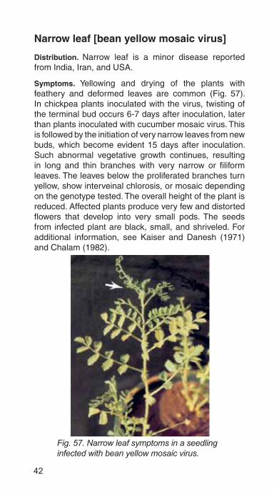
Distribution. Narrow leaf is a minor disease reported from India, Iran, and USA.
Symptoms. Yellowing and drying of the plants with feathery and deformed leaves are common (Fig. 57). In chickpea plants inoculated with the virus, twisting of the terminal bud occurs 6-7 days after inoculation, later than plants inoculated with cucumber mosaic virus. This is followed by the initiation of very narrow leaves from new buds, which become evident 15 days after inoculation. Such abnormal vegetative growth continues, resulting in long and thin branches with very narrow or filiform leaves. The leaves below the proliferated branches turn yellow, show interveinal chlorosis, or mosaic depending on the genotype tested. The overall height of the plant is reduced. Affected plants produce very few and distorted flowers that develop into very small pods. The seeds from infected plant are black, small, and shriveled. For additional information, see Kaiser and Danesh (1971) and Chalam (1982).
Fig. 57. Narrow leaf symptoms in a seedling infected with bean yellow mosaic virus.

43
Necrosis [lettuce necrotic yellows virus]
Distribution. Necrosis is a minor disease reported only from Australia.
Symptoms. Field symptoms of the disease are most prominent on the terminals of the main and axillary shoots, which are usually twisted. The newest leaves show a bleached, necrotic tip burn. The stem and bases of the larger, older leaves develop reddish brown patches, particularly at the nodes. Symptoms on leaflets begin as yellow flecks on veins but the flecks enlarge to produce general chlorosis and kill the leaflet. Infected plants wilt and die.
Phyllody [mycoplasma]
Distribution. Phyllody is a minor disease reported from Ethiopia, India and Myanmar. Characteristic symptoms are excessive proliferation of branches with smaller leaf-lets, giving a bushy appearance to the plant. Diseased plants are scattered in the field and are more easily spotted at flowering and podding time. The flowers are converted into leafy structures (Fig. 58). At the time of crop maturity, when the healthy plants are drying, the diseased plants in the field will be conspicuously green. For additional information, see Ghanekar et al. (1988).
Fig. 58. Twig (left) showing phyllody symptoms (normal twig at right).

44
Co
ntr
ol m
easu
res
Res
ista
nt
vari
etie
s/lin
es
Co
un
try
Dis
ease
R
esis
tan
t cu
ltiv
ars
See
d t
ype
Aus
tral
iaP
hyto
phth
ora
blig
htIC
C11
870,
Yor
ker
desi
Ban
glad
esh
Roo
t dis
ease
sS
abou
r 4,
Fat
ehpu
r 1,
Bha
ugor
ade
siB
otry
tis g
ray
mol
dB
aric
hola
5, I
CC
L 87
322
-B
ulga
riaA
scoc
hyta
blig
htP
lovd
iv 0
19, O
braz
tsov
chi
jlik
1 P
lovd
iv 8
desi
ka
buli
Chi
leR
oot r
otC
alifo
rnia
INIA
, Gua
sos
SN
Aka
buli
Cyp
rus
Asc
ochy
ta b
light
ILC
327
9de
siE
thio
pia
Wilt
Che
fede
siIta
lyA
scoc
hyta
blig
htA
li, S
ulta
no, C
aliff
o,de
siIn
dia
Asc
ochy
ta b
light
F8,
C 1
2/34
, C 2
35, G
543
, H 7
5-35
, GG
68
8,G
NG
146
,Gau
rav,
BG
261
, GG
588
, H
ima
chan
a-1,
Gau
rav,
Var
dan,
Sam
rat,
PB
G 1
and
BG
261
desi
de
si
kabu
li

45
Co
un
try
Dis
ease
R
esis
tan
t cu
ltiv
ars
See
d t
ype
Asc
ochy
ta b
light
ICC
V 0
4052
, 045
09, 0
4505
, 045
12, 0
4513
, 04
523,
045
24, 0
4525
, 045
26, 0
4530
, 045
37,
0550
2, 0
5503
, 055
11, 0
5512
, 055
13, 0
5514
, 05
515,
055
23, 0
5530
, 055
31, 0
5546
, 055
51,
0557
1, 9
8811
, 988
16, 9
8818
Wilt
No.
10,
S 2
6, G
24,
C 2
14, B
G 2
44, P
usa
212,
Avr
odhi
, JG
315
, JG
14,
JG
11,
JG
K 2
, K
AK
2, V
ijay,
Vai
bhav
, JG
63,
Birs
a ca
nna-
3,
WR
315
, JG
74,
JA
KI 9
218,
Vih
ar, J
G 1
265,
B
G 1
053,
PD
G 4
, Guj
arat
gra
m 4
, Guj
arat
gr
am 1
, BG
M 4
7, C
OG
29-
1, L
551
desi
Roo
t dis
ease
sIC
CC
32,
GL
769
Bot
rytis
gra
y m
old
BG
276
, GL
9015
9, G
L 91
040,
GL
9107
1 an
d G
L 92
162
-
Mex
ico
Wilt
Sur
utat
o 77
, Sto
. Dom
ingo
, Sen
ora,
UC
15
and
UC
27
kabu
li
Mor
occo
Asc
ochy
ta b
light
Pch
46
kabu
li

46
Co
un
try
Dis
ease
R
esis
tan
t cu
ltiv
ars
See
d t
ype
Nep
alB
otry
tis g
ray
mol
dIC
C 1
4344
-P
akis
tan
Asc
ochy
ta b
light
F8,
C 1
2/34
, C 7
27, C
235
, CM
72,
C 4
4de
si
desi
Asc
ochy
ta b
light
and
wilt
AU
G 4
80W
iltP
unja
b 20
00de
siS
yria
Asc
ochy
ta b
light
ILC
482
, Gha
b 2
kabu
liS
udan
Wilt
ICC
V 2
, UC
15,
FLI
P 8
5-20
C, F
LIP
85-
29C
an
d F
LIP
85-
30C
kabu
li an
d de
si
Spa
inA
scoc
hyta
blig
htFa
rdan
, Zeg
ri, A
lmen
a, A
lcaz
aba
desi
Wilt
FLI
P 8
4-43
, FLI
P 8
5-20
, FLI
P 8
5-29
C, I
LCs
127,
219
, 237
, 267
, 513
and
CA
295
4de
si
Turk
eyA
scoc
hyta
blig
htG
uney
Sar
isi 4
82, I
LC 1
95de
siU
SA
Roo
t rot
Mis
sion
kabu
liU
SS
RA
scoc
hyta
blig
htV
IR 3
2, N
ut Z
imis
toni
inte
rmed
iate
de
si

47
Fu
ng
icid
es
Dis
ease
See
d t
reat
men
t*F
olia
r ap
plic
atio
n#
Asc
ochy
ta b
light
2-h
imm
ersi
on in
mal
achi
te g
reen
5 m
g/l
Wet
tabl
e su
lphu
r
4-h
imm
ersi
on in
form
al in
12
-h im
mer
sion
in p
imai
cin
150
mg/
Lzi
neb
Phe
nthi
uram
@ 2
g/k
g T
hira
m @
2 g
/kg
ferb
am
Azo
xist
robi
n @
1g/
kgm
aneb
cap
tan
Ben
omyl
@ 2
g/k
gca
ptaf
olC
alix
in M
® (
trid
emor
ph +
man
eb)
@ 3
g/k
gch
loro
thal
onil
Cal
ixin
M®
(tr
idem
orph
+ m
aneb
) +
thira
m
(1:1
) @
2-5
g/k
gdi
thia
non
(Dos
age
@ 3
g/l)
48
Dis
ease
See
d t
reat
men
t*F
olia
r ap
plic
atio
n#
Cal
ixin
M®
(tr
idem
orph
+ m
aneb
) +
B
avis
tin®
(ca
rben
dazi
m)
(1:1
)
Bav
istin
® (
carb
enda
zim
) +
thira
m (
1:3)
th
iabe
ndaz
ole
(3 g
/kg)
Bot
rytis
gra
y m
old
carb
enda
zim
+ th
iram
(1:
1)
vinc
lozo
lin, c
arbe
ndaz
im
tria
dim
efon
Dith
ane
M 4
5® (
man
eb)
Tria
dim
enol
thia
bend
azol
e
ipro
dion
e
thira
m
vinc
lozo
lin
carb
enda
zim
@ 1
g/L
+ th
iram
@
2g/L
carb
enda
zim
@1
g/L
capt
an
chlo
roth
alon
il
man
coze
b
thio
phon
ate
met
hyl
(App
ly a
t 50
days
afte
r so
win
g or
at
the
first
sig
n of
sym
ptom
s)

49
Dis
ease
See
d t
reat
men
t*F
olia
r ap
plic
atio
n#
Fus
ariu
m w
iltC
arbe
ndaz
im @
2.5
g/k
g B
enla
te T
® (
beno
myl
+ th
iram
) @
1.5
g/k
g
Dry
roo
t rot
capt
an
thira
m
Wet
roo
t rot
capt
an, t
hira
m, B
enla
te®
Bla
ck r
oot r
otth
iram
+ b
enom
yl
thira
m +
cap
tan
Pyt
hium
roo
t an
d se
ed r
otm
etal
axyl
Col
lar
rot
Riz
olex
®
Vita
vax
200®
*Whe
reve
r no
t spe
cifie
d, th
e do
se is
3 g
/kg
of s
eed.
# 3 g
or
3 m
l per
lite
r of
wat
er

50
Cu
ltu
ral p
ract
ices
Dis
ease
Pra
ctic
esA
scoc
hyta
blig
htS
ow la
te.
Rem
ove
and
dest
roy
dead
pla
nt d
ebris
.R
otat
e cr
ops.
Sow
dee
p (1
5 cm
or
deep
er).
Wid
er r
ow s
paci
ngA
dopt
low
see
ding
rat
eIn
terc
rop
with
whe
at, b
arle
y, m
usta
rd.
Bur
y di
seas
ed d
ebris
10
cm o
r de
eper
.S
ow d
isea
se-f
ree
seed
Bot
rytis
gra
y m
old
Use
dis
ease
-fre
e se
edB
urn
infe
cted
deb
risD
eep
plou
ghin
gA
dopt
late
sow
ing
and
wid
er r
ow s
paci
ng.
Inte
rcro
p w
ith li
nsee
d or
whe
at

51
Dis
ease
Pra
ctic
esA
void
exc
essi
ve v
eget
ativ
e gr
owth
.A
void
exc
essi
ve ir
rigat
ion.
Use
ere
ct a
nd c
ompa
ct v
arie
ties
Alte
rnar
ia b
light
Mai
ntai
n lo
w p
lant
den
sity
A
void
exc
essi
ve ir
rigat
ion
Scl
erot
inia
ste
m r
otD
eep
plow
ing
Avo
id e
xces
sive
veg
etat
ive
grow
th.
Avo
id e
xces
sive
irrig
atio
n.U
se u
prig
ht g
row
th h
abit
culti
vars
Wid
e ro
w s
paci
ngF
usar
ium
wilt
Use
dis
ease
-fre
e se
ed.
Avo
id s
owin
g w
hen
tem
pera
ture
s ar
e hi
gh (
late
sow
ing)
.F
ollo
w 4
-yea
r cr
op r
otat
ions
Soi
l sol
ariz
atio
n du
ring
sum
mer
mon
ths
Dry
roo
t rot
Avo
id d
roug
ht
Sow
on
time
so th
at th
e cr
op e
scap
es h
ot w
eath
er.

52
Dis
ease
Pra
ctic
esW
et r
oot r
otA
void
exc
essi
vely
ric
h so
il.
Avo
id h
igh
soil
moi
stur
e at
sow
ing.
Bla
ck r
oot r
otA
void
hig
h so
il m
oist
ure.
Pyt
hium
roo
t and
see
d ro
tA
dopt
late
pla
ntin
gTr
eat s
eeds
with
Pen
icili
um o
xalic
um, P
ythi
um o
ligan
drum
, Psu
edom
onas
flu
ores
cens
and
Bac
illus
sub
tilis
Col
lar
rot
Avo
id h
igh
soil
moi
stur
e at
sow
ing
and
seed
ling
stag
e.A
dopt
wid
e ro
w s
paci
ngR
emov
e al
l und
ecom
pose
d or
gani
c m
atte
r w
hile
pre
parin
g se
edbe
dS
oil s
olar
izat
ion
durin
g su
mm
er m
onth
sV
ertic
illiu
m w
iltU
se d
isea
se fr
ee s
eeds
Cro
p ro
tatio
n w
ith w
heat
Phy
toph
thor
a bl
ight
Avo
id p
oorly
dra
ined
fiel
dsC
rop
rota
tion
with
whe
atS
tunt
Ado
pt c
lose
spa
cing
.S
ow w
hen
aphi
d ve
ctor
act
ivity
is lo
w.
It co
uld
be e
arly
or
late
sow
ing
depe
ndin
g on
res
earc
h re
sults
for
a lo
catio
n
53
Supporting literatureChalam TV. 1982. Identification and characterization of cucumber mosaic and bean yellow mosaic viruses affecting chickpea (Cicer arietinum L.) in India. PhD thesis, College of Agriculture, Andhra Pradesh Agricultural University, Hyderabad, India. 96 pp.
Chen W, Sharma HC and Muehlbauer FJ. 2011. Compendium of chickpea and lentil diseases and pests. The American Phytopathological Society, Minnesota, USA.
Erwin DC. 1958. Fusarium lateritium F. ciceri, incitant of Fusarium wilt of Cicer arietinum. Phytopathology 48: 498-501.
Ghanekar AM, Manohar SK , Reddy SV and Nene YL. 1988. Association of a mycoplasma-like organism with chickpea phyllody. Indian Phytopathology 41: 462-464.
Haware MP and Nene YL. 1981. Phoma blight: a new disease of chickpea. Plant Disease 65:282.
Kaiser WJ and Danesh D. 1971. Etiology of virus-induced wilt of Cicer arietinum. Phytopathology 61:453-457.
Kaiser WJ and Hannan RM. 1983. Etiology and control of seed decay and pre-emergence damping-off of chickpea by Pythium ultimum. Plant Disease 67:77-81.
Kaiser WJ. 1990. Host range of the ascochyta blight pathogen of chickpea. Phytopathology 80:889-890. (Abs.).
Nene YL, Haware MP and Reddy MV. 1978. Diagnosis of some wilt-like disorders of chickpea (Cicer arietinum L. ). Information Bulletin no. 3, Patancheru, AP 502324, India: International Crops Research Institute for the Semi-Arid Tropics, 44 pp.
Nene YL, Sheila VK and Sharma SB. 1996. A world list of chickpea and pigeonpea pathogens. 5th edition, ICRISAT, Patancheru, India. Pg 27.
Nene YL and Reddy MV. 1987. Chickpea diseases and their control . Pages 233- 270 in The chickpea (Saxena MC and Singh KB, eds).. Wallingford, Oxon, UK: CAB International .
Pande S, Galloway JJ, Gaur PM, Siddique KHM, Tripathi HS, Taylor P, MacLeod MWJ, Basandrai AK, Baker A, Joshi, Krishna Kishore G, Isenegger DA, Narayana Rao J and Sharma M. 2006. Botrytis gray mold of chickpea: a review of biology, epidemiology, and disease management. Aust J Agric Res 57:1137–1150.

54
Pande S, Sharma M, Gaur PM and Gowda CLL. 2010. Host Plant Resistance to Ascochyta Blight of Chickpea. Information Bulletin No. 82. Patancheru 502 324, Andhra Pradesh, India: International Crops Research Institute for the Semi-Arid Tropics. 40 pp. ISBN 978-92-9066-525-0. Order code: IBE 082.
Pande S, Siddique KHM, Kishore GK, Baya B, Gaur PM, Gowda CLL, Bretag T and Crouch JH. 2005. Ascochyta blight of chickpea: biology, pathogenicity, and disease management. Australian Journal of Agricultural Research 56:317-332.
Pande S, Stevenson PC, Rao JN, Neupane RK, Chaudhary RN, Grzywacz D, Baurai VA and Kishore GK. 2005. Reviving chickpea production in Nepal through integrated crop management, with emphasis on Botrytis gray mold. Plant Disease 89:1252–1262.
Rangaswami G and Prasad NN. 1960. A bacterial disease of Cicer arietinum L. Indian Phytopathology 12:172-175.
Sharma M, Mangla UN, Krishnamurthy L, Vadez V and Pande S. 2010. Drought and dry root rot of chickpea.[Abstract] Page 263 in 5th International Food Legumes Research Conference (IFLRC V) and 7th European Conference on Grain Legumes (AEP II) April 26-30, 2010, Antalya, Turkey.
Vock NT, Langton PW and Pegg KG. 1980. Root rot of chickpea caused by Phytophthora megasperma var. sojae in Queensland. Australian Plant Pathology 9:117.
55
Indexalfalfa mosaic virus 40
Alternaria alternata 12
Alternaria blight 12, 51
Aphis craccivora 37
Ascochyta blight 1, 2, 5, 6, 7, 8, 12, 15 44, 45, 46, 47, 50
Ascochyta rabiei 2
Bacterial blight 36
bean (pea) leaf roll virus 1, 37
bean yellow mosaic virus 42
Black root rot 30, 49, 52
Botrytis gray mold 1, 8, 11, 12, 44, 45, 46, 48, 50
Collar rot 1, 20, 26, 28, 49, 52
Colletotrichum blight 14
Colletotrichum dematium 14
cucumber mosaic virus 41, 42
damping-off 4
Dry root rot 1, 29, 30, 49, 51
Foot rot 35
Fusarium oxysporum 22
Fusarium solani 30
Fusarium wilt 1, 22, 23, 24, 25, 35, 39, 49, 51
lettuce necrotic yellows virus 43
Mosaic 40, 42
mycoplasma 43
Myzus persicae 37
Narrow leaf 42
Necrosis 4, 40, 43
Operculella padwickii Kheswalla 35
Phoma blight 15
Phoma medicaginis 15, 16
Phyllody 43
Phytophthora root rot 1, 32
Powdery mildew 19

56
Proliferation 40, 41, 43
Rhizoctonia bataticola 1, 29
Rhizoctonia solani 28
Root rot 29, 44, 46
Rust 17, 18, 19
Sclerotinia sclerotiorum 20
Sclerotinia stem rot 20, 51
Sclerotium rolfsii 26, 27
Stemphylium blight 17
Stemphylium sarciniforme 17
Stunt 1, 37, 38, 39, 52
Uromyces ciceris-arietini 17, 18
Verticillium albo-atrum 25
Verticillium wilt 25, 52
Wet root rot 28, 33, 49, 52
Wilt 1, 22, 25, 30, 40, 43 44, 45, 46


ISBN 92-9066-199-2 Order code: IBE: 028 383-2012
ICRISAT-Patancheru(Headquarters)Patancheru 502 324Andhra Pradesh, IndiaTel +91 40 30713071Fax +91 40 [email protected]
ICRISAT-Liaison
CG Centers BlockNASC ComplexNew Delhi 110 012, IndiaTel +91 11 32472306
to 08 Fax +91 11 25841294
ICRISAT-Nairobi(Regional hub ESA)PO Box 39063, Nairobi, KenyaTel +254 20 7224550Fax +254 20 [email protected]
ICRISAT-Bamako(Regional hub WCA)BP 320Bamako, MaliTel +223 20 709200Fax +223 20 [email protected]
ICRISAT-NiameyBP 12404, Niamey, Niger (Via Paris)Tel +227 20722529,
20722725Fax +227 [email protected]
ICRISAT-BulawayoMatopos Research StationPO Box 776,Bulawayo, ZimbabweTel +263 383 311 to 15Fax +263 383 [email protected]
ICRISAT-LilongweChitedze Agricultural Research StationPO Box 1096Lilongwe, MalawiTel +265 1 707297,
071, 067, 057Fax +265 1 [email protected]
ICRISAT-Maputoc/o IIAM, Av. das FPLM No 2698Caixa Postal 1906Maputo, MozambiqueTel +258 21 461657Fax +258 21 [email protected]
Contact Information
About ICRISAT
www.icrisat.org
The International Crops Research Institute for the Semi-Arid Tropics (ICRISAT)
development in Asia and sub-Saharan Africa with a wide array of partners throughout the world. Covering 6.5 million square kilometers of land in 55 countries, the semi-arid tropics have over 2 billion people, and 644 million of these are the poorest of the poor. ICRISAT and its partners help empower these poor people to overcome poverty, hunger and a degraded environment through better agriculture.ICRISAT is headquartered in Hyderabad, Andhra Pradesh, India, with two regional
of Centers supported by the Consultative Group on International Agricultural Research (CGIAR).