Embed Size (px)
Citation preview

BioMed CentralBMC Developmental Biology
ss
Open AcceResearch articleFGF9 can induce endochondral ossification in cranial mesenchymeVenkatesh Govindarajan*1 and Paul A Overbeek2Address: 1Cancer Center, Creighton University, Omaha, NE 68178, USA and 2Department of Molecular and Cellular Biology, Baylor College of Medicine, Houston, TX 77030, USA
Email: Venkatesh Govindarajan* - [email protected]; Paul A Overbeek - [email protected]
* Corresponding author
AbstractBackground: The flat bones of the skull (i.e., the frontal and parietal bones) normally formthrough intramembranous ossification. At these sites cranial mesenchymal cells directlydifferentiate into osteoblasts without the formation of a cartilage intermediate. This type ofossification is distinct from endochondral ossification, a process that involves initial formation ofcartilage and later replacement by bone.
Results: We have analyzed a line of transgenic mice that expresses FGF9, a member of thefibroblast growth factor family (FGF), in cranial mesenchymal cells. The parietal bones in these miceshow a switch from intramembranous to endochondral ossification. Cranial cartilage precursorsare induced to proliferate, then hypertrophy and are later replaced by bone. These changes areaccompanied by upregulation of Sox9, Ihh, Col2a1, Col10a1 and downregulation of CbfaI andOsteocalcin. Fate mapping studies show that the cranial mesenchymal cells in the parietal region thatshow a switch in cell fate are likely to be derived from the mesoderm.
Conclusion: These results demonstrate that FGF9 expression is sufficient to convert thedifferentiation program of (at least a subset of) mesoderm-derived cranial mesenchyme cells fromintramembranous to endochondral ossification.
BackgroundBone development can occur in two distinct ways: 1)through endochondral ossification where the mesenchy-mal cells differentiate into chondrocytes and lay down acartilaginous template that is later replaced by bone; or 2)through intramembranous ossification where mesenchy-mal cells directly differentiate into osteoblasts without theformation of a cartilage intermediate. During endochon-dral ossification, the transcription factors Sox9, Sox5 and/or Sox6 are expressed and are involved in the induction ofchondrocytes [1]. Chondrocytes in the growth plate aresubsequently induced to exit the cell cycle and commit toterminal differentiation. The prehypertrophic chondro-
cytes mature into hypertrophic chondrocytes, which laydown a matrix rich in Collagen X, and secrete VEGF [2].VEGF promotes the invasion of blood vessels from theperichondrium, bringing in both the bone forming oste-oblasts and the bone resorbing osteoclasts. The hyper-trophic chondrocytes then undergo apoptosis, and arereplaced by trabecular bone and bone marrow. In con-trast, the flat bones of the skull, the frontal and parietalbones, form by intramembranous ossification. Cranialmesenchymal cells directly differentiate into osteoblaststhat initiate mineralization and secrete an extracellularmatrix rich in Collagen I [3]. Growth of these calvarialbones occurs through proliferation and differentiation of
Published: 20 February 2006
BMC Developmental Biology 2006, 6:7 doi:10.1186/1471-213X-6-7
Received: 03 September 2005Accepted: 20 February 2006
This article is available from: http://www.biomedcentral.com/1471-213X/6/7
© 2006 Govindarajan and Overbeek; licensee BioMed Central Ltd. This is an Open Access article distributed under the terms of the Creative Commons Attribution License (http://creativecommons.org/licenses/by/2.0), which permits unrestricted use, distribution, and reproduction in any medium, provided the original work is properly cited.
Page 1 of 14(page number not for citation purposes)

BMC Developmental Biology 2006, 6:7 http://www.biomedcentral.com/1471-213X/6/7
osteoblasts at the margins or sutures. The molecular path-ways that dictate these alternative ossification programsare not yet well defined. In particular, it is not knownwhether intramembranous ossification is prespecified bythe ontogeny of the cranial mesenchyme or is a responseto local environmental signals.
Fibroblast growth factors (FGFs) appear to be importantfor both types of ossification [4]. FGFs comprise a large
family of proteins that includes at least 22 known mem-bers [5]. FGFs bind and signal through low and high affin-ity FGF receptors [5]. The four known high affinityreceptors (FGFR1–4) are structurally similar transmem-brane receptor tyrosine kinases. During intramembranousossification of the flat bones, FGFR1–3 are expressed bythe differentiating osteoblasts at the osteogenic fronts andalso by the adjacent cartilage [6,7]. FGFR1 is expressed incells close to and within the osteoid; FGFR2 is expressed
Abnormal head development in the OVE1070 FGF9 transgenic miceFigure 1Abnormal head development in the OVE1070 FGF9 transgenic mice. Panel A shows a schematic representation of the FGF9 transgene. The coding region of mouse Fgf9 cDNA was inserted between the αA-crystallin promoter (αAp) and an intron and polyadenylation sequence derived from SV40 virus [47]. The microinjection fragment was generated by SstII digestion. The SV40 sequences were used to make a riboprobe for detection of expression of the transgene. (B and C) Three day old non-transgenic (NT) and FGF9 transgenic mice are shown. The heads of the transgenic mice are 'dome' shaped (C). The OVE1070 transgenic mice often show unfused eyelids at birth (C). (D-K) Skeletal preparations of the FGF9 transgenic mice reveal an expansion in the cartilage territory in the head region. Alcian blue and Alizarin red stained E15 (D and E), P3 (F and G), P10 (H and I) and P30 (J and K) heads are shown. Cartilage is stained blue and bone red. The insets in panels D and E are higher mag-nifications of the frontal and parietal bones in the nontransgenic and transgenic mice respectively. The insets in panels F-I are rear views. The insets in panels J and K are top views. An expansion in the cartilage territory is seen in the FGF9 transgenic mice. Parietal bone (p) is affected in the transgenic skulls but the coronal suture is still present (E inset, arrow). Frontal (f) and occipital bone formation appear to be unaffected (E and G insets). A hole forms in the skull of the FGF9 transgenic mice that is visible by P3 (G inset, arrow) and is seen to persist at P10 (I inset, arrow). The red stain visible at the arrow in the inset in panel G is bone at the base of the head, which is visible since the skull and the brain are transparent after staining and clearing. The cartilage territory is replaced by bone by P30 (K and inset). Scale bar: 1 mm in D and E; 1.6 mm in F and G; 2.5 mm in H-K.
Page 2 of 14(page number not for citation purposes)

BMC Developmental Biology 2006, 6:7 http://www.biomedcentral.com/1471-213X/6/7
in the proliferating osteogenic stem cells; FGFR3 expres-sion is seen in the thin layer of cartilage underlying thelower part of the coronal suture [6]. As FGFR3 null micedo not show defects in calvarial development, it has beenhypothesized that intramembranous bone formation iscontrolled primarily by FGFR1 and FGFR2 [8]. Mutationsin FGFR1, FGFR2 and FGFR3 in humans that affect skele-tal growth are consistent with this hypothesis [9]. Cranio-synostosis is mainly associated with mutations in FGFR1and FGFR2 [9]. Mutations that affect the growth of longbones resulting in syndromes such as Achondroplasia(Ach) and Thanatophoric dysplasia (TD) are mainly local-ized to FGFR3. These autosomal dominant disorders arebelieved to reflect either an enhancement of receptoractivity or a neomorphic gain-of-function effect [9-11].During long bone development, FGF receptors areexpressed in the epiphyseal growth plates: FGFR3 isexpressed in the proliferating chondrocytes; FGFR1 isexpressed in the hypertrophic chondrocytes; FGFR1 andFGFR2 are expressed in the perichondrium [4,12]. FGFR2is expressed in early mesenchymal condensates and in theperiosteal collar around the cartilage models [8]. Targeteddeletion of FGFR2IIIc suggests that FGFR2IIIc is a positiveregulator of ossification in both the osteoblasts andchondrocytic lineages [13]. Targeted deletion of FGFR3results in mice that show overgrowth of the long bonesand abnormal proliferation of chondrocytes suggestingthat FGFR3 stimulation inhibits chondrocyte prolifera-tion [14]. These studies describe roles for FGF receptor-mediated signaling during differentiation/maturation ofchondrocytes.
The roles of FGF ligands in skeletal development areunclear. During calvarial development, Fgf8 is expressedin the osteoblasts; Fgf2 and Fgf4 are expressed in thesutural mesenchyme; Fgf18 is initially expressed in thecranial mesenchymal cells and later, in the differentiatingosteoblasts [8]. Fgf9 is expressed in the sutural mesen-chyme and is upregulated in the endocranial portions ofthe mesenchyme and is downregulated during postnataldevelopment [15][16]. The specific in vivo functions ofthese different FGFs remain undefined. In vitro, cephalicneural crest cells from quail embryos have been shown torespond to exogenous FGF-2 in a dose dependant man-ner; lower doses induce proliferation and higher dosesinduce cartilage differentiation [17]. During long bonedevelopment, at the time of initiation of endochondraldifferentiation, Fgf9 is expressed in the condensing mes-enchyme [18]. Fgf2, Fgf5, Fgf6 and Fgf7 are expressed inloose mesenchyme outside the condensation [19-23].However, mice lacking these Fgfs show no apparentdefects in skeletal development [24-28]. Functionalredundancy between these FGFs may, in part, account forthe lack of phenotype. Therefore, roles for these Fgfs in theearly stages of chondrogenesis have not yet been defined.
In this study, we have analyzed transgenic mice thatexpress FGF9 in their cranial mesenchymal cells. Thesemice show abnormal head development and are bornwith a pronounced bulge in their skulls. Skeletal prepara-tions of these mice revealed dramatic changes in parietalbone formation. In the region where the parietal bonesnormally form, cranial mesenchymal cells are induced todifferentiate into chondrocytes that proliferate, hypertro-phy and subsequently differentiate as bone. Correlativechanges in expression of marker genes Sox9, Col2a1,Col10a1, Ihh, CbfaI and Osteocalcin occur in conjunctionwith the altered differentiation program. Thus the parietalbones in these mice form by endochondral ossificationrather than by the usual intramembranous ossificationroute. Fate mapping studies indicate that the ectopic carti-lagenous precursors in these mice are derived from themesoderm. Based on these results we suggest that cranialmesenchymal cells are competent to initiate endochon-dral ossification, and can be switched to this alternativedevelopmental program by ectopic expression, or overex-pression, of FGF9.
ResultsChanges in cranial morphology in the FGF9 transgenic miceTransgenic mice were generated by microinjection of aconstruct with the αA-crystallin promoter linked to amouse Fgf9 cDNA (Fig. 1A) [29]. Eleven founders carryingthe transgene were identified. Five of them had cataracts(data not shown). In the transgenic line OVE1070, inaddition to lens defects, there were defects in the develop-ment of the skull (Fig. 1B, C). Mice in this family that areheterozygous for the transgene are born with 'dome-shaped' heads (Fig. 1C). This phenotype is even more pro-nounced in homozygous transgenic mice (data notshown).
Calvarial development in FGF9 transgenic miceThe skeletal defects were examined by Alcian blue and Ali-zarin red staining of E15, P3, P10 and P30 mice (Fig. 1D–K). The skeletal preparations show a dramatic expansionof cartilage in the head region by E15 (compare Fig. 1E to1D). Parietal bone development was particularly affected(Fig. 1E and inset). At P3, the phenotypic differencesbetween the nontransgenic and FGF9 transgenic micewere still pronounced. At this age, the calvarial skull innontransgenic mice is mostly ossified except at the sutures(Fig. 1F). In contrast, there is an expanded cartilage terri-tory (blue) in the FGF9 transgenic mice (Fig. 1G). Thetransgenic mice also show a central hole in the skull wherethere is neither cartilage nor bone (Fig. 1G, inset, arrow).Development of the occipital bones appeared normal(Fig. 1G and inset). At P10, the Alcian blue stained tissuesin the transgenic cranium were interspersed with Alizarinred stained regions suggesting that the cartilage territory
Page 3 of 14(page number not for citation purposes)

BMC Developmental Biology 2006, 6:7 http://www.biomedcentral.com/1471-213X/6/7
was being replaced by bony structures (Fig. 1I). By 1month of age, the cartilage territory was completelyreplaced by bone (Fig. 1K and inset).
The alterations in skeletal development in the FGF9 trans-genic mice were examined at the histological level by anal-yses of hematoxylin and eosin stained sections of thecranium. No distinctive differences in morphology wereseen at E11 (data not shown). By E13, more cranial mes-enchymal cells were seen in the skull region of the FGF9transgenic mice (Fig. 2B, B') and these cells were organ-ized differently from nontransgenic mice (compare Fig.2A, B). By E15, the cranial mesenchymal cells in the non-transgenic mice had initiated intramembranous ossifica-tion (Fig. 2C, C', arrow). In contrast, the mesenchymalcells in the FGF9 transgenic skulls differentiated into cellsresembling chondrocytes (Fig. 2D, D'). Within the next
few days these cells increased in size and became hyper-trophic (Fig. 2F, F', H, H'). By P7, perichondrial cells(including the blood vessels) had begun to invade the car-tilage territory (Fig. 2J, J'). By P18, the cartilage territorywas transformed into trabecular bone and bone marrow(Fig. 2L, L'). These results considered together suggest thatthe cranial mesenchymal cells in the parietal region inthese mice recapitulate the sequence of events that occursduring endochondral ossification.
Ectopic expression of the FGF9 transgeneTo test if the alterations seen in the skulls of the OVE1070mice were due to ectopic expression of the transgene, insitu hybridizations were performed using an S35-labelledriboprobe that recognizes the SV40 portion of the trans-gene (Fig. 1A). Transgene expression was seen in the lens[29], the dorsal portion of the retinal pigmented epithe-
Cranial mesenchyme in the FGF9 transgenic mice differentiates into chondrocytesFigure 2Cranial mesenchyme in the FGF9 transgenic mice differentiates into chondrocytes. Heads of nontransgenic (A, A', C, C', E, E', G, G', I, I', K and K') and FGF9 transgenic mice (B, B', D, D', F, F', H, H', J, J', L and L') were sectioned and stained with hema-toxylin and eosin. Panels A'-L' are higher magnifications of the boxed regions in panels A-L respectively. In nontransgenic mice, the mesenchymal cells of the skull form a skeletogenic membrane (arrows in A', C', E' and G') within which the bones form. In the FGF9 transgenic mice the cranial mesenchymal cells (cm) differentiate into chondrocytes (ch) that initially form a structure resembling the hyaline cartilage (D, D', F and F') and later hypertrophy (hyp) (H, H', J and J'). Perichondrial cells, including blood cells (J', arrows) invade and replace the hypertophic chondrocytes forming bone and bone marrow (L and L'). Other abbrevia-tions: b, brain; s, skin. The folding seen in the section in panel G is an artifact of the histology procedure. Note that the skin was removed from P7 and P18 mice to facilitate fixation and histological processing. Scale bar: 50 µm in A'-L'; 100 µm in A, B, C, D, E, H, I and K; 200 µm in F, G, J and L.
Page 4 of 14(page number not for citation purposes)

BMC Developmental Biology 2006, 6:7 http://www.biomedcentral.com/1471-213X/6/7
lium [30] and also in the cranial mesenchymal cells begin-ning at E11 (Fig. 3B', D', F'). Expression levels peak by E13and start to decrease by E15 (Fig. 3D', F'). Transgeneexpression is extinguished by E17 (data not shown). Sec-tion and whole mount in situ hybridizations show thatthe extraocular transgene expression is restricted to themesenchymal cells overlying the future mid and hindbrain regions (Fig. 3G, arrows; 3H-M). These results arguethat the defects in skeletal development in the OVE1070line are attributable to ectopic and transient expression ofthe FGF9 transgene.
Expression of chondrocyte specific markersThe different stages of endochondral ossification are char-acterized by expression of different marker genes. Expres-sion of Sox9, a member of the Sox family of transcriptionfactors, has been shown to be essential for chondrocytecondensation [31]. In particular, Sox9 is required for theexpression of cartilage-specific extracellular matrix com-ponents such as Collagen II (Col2a1) [32]. In the longbone growth plates, Col2a1 is expressed in the resting andproliferating chondrocytes, Ihh in prehypertropicchondrocytes and Col10a1 in hypertropic chondrocytes
Expression pattern of the transgeneFigure 3Expression pattern of the transgene. In situ hybridizations were done on sections of heads from nontransgenic (NT) and FGF9 transgenic mice using a [35S]-labeled SV40 riboprobe. Panels A-F show bright-field images and panels A'-F' show the corre-sponding dark field images. Transgene expression can be seen in the cranial mesenchymal cells at E11 (B'), E13 (D') and E15 (F'). At later stages, expression of the transgene in the cranium was not detected (data not shown). Transgene expression was restricted to the mesenchymal cells overlying the mid (m) to hind brain regions (h) but not the forebrain (f) (G, arrows). Scale bar: 100 µm in A-F'. Whole mount in situ hybridizations on E12.5 heads using a digoxygenin-labelled SV40 riboprobe show expression of the transgene (purple color) in the cranial regions (H-K). Panels H and I are rear views and panels J and K are top views. Scale bar: 1 mm in H, I and K.
Page 5 of 14(page number not for citation purposes)

BMC Developmental Biology 2006, 6:7 http://www.biomedcentral.com/1471-213X/6/7
[3]. Expression of the runt domain transcription factorCbfaI has been shown to be essential for differentiation ofthe cells in the osteoblastic lineage [33]. Osteocalcin, one
of the downstream targets of CbfaI, is expressed exclu-sively in the osteoblasts [34]. Expression of these sixmarker genes was examined by in situ hybridization using
Expression patterns of Sox9, CbfaI and OsteocalcinFigure 4Expression patterns of Sox9, CbfaI and Osteocalcin. In situ hybridizations were done on sections of heads from nontransgenic (NT) and FGF9 transgenic mice using [35S]-labeled riboprobes for Sox9 (A-F'), CbfaI (G-H') and osteocalcin (I-J'). Panels A-J are bright field images and panels A'-J' are the corresponding dark-field images. Expression of Sox9 is induced in the cranial mesen-chymal cells of the FGF9 transgenic mice by E13 (D'). Sox9 expression is detected in the ventricular regions of the brain at E13 in both the transgenic and nontransgenic mice (C', D'). CbfaI and osteocalcin are expressed in the differentiating calvarial oste-oblasts (os) of nontransgenic mice at E15 (G', I') but not in the skulls of the FGF9 transgenic mice (H', J'). Scale bar: 50 µm in G-J'; 100 µm in A-F'.
Page 6 of 14(page number not for citation purposes)
BMC Developmental Biology 2006, 6:7 http://www.biomedcentral.com/1471-213X/6/7
Page 7 of 14(page number not for citation purposes)
Expression patterns of Col2a1, Col10a1 and IhhFigure 5Expression patterns of Col2a1, Col10a1 and Ihh. In situ hybridizations were done on sections of heads from nontransgenic (NT) and FGF9 transgenic mice using [35S]-labeled riboprobes for Col2a (A-H'), Col10a1 (I-J') and Ihh (K-L'). Panels A-L are bright field images and panels A'-L' are the corresponding dark-field images. Expression of Col2a1 is enhanced in the cranial mesenchy-mal cells of the FGF9 transgenic mice by E15 (B') and is decreased in some cells by P7 (H'). Hybridizations performed on adja-cent sections show expression of Col10a1 in these hypertropic (hyp) chondrocytes (J'). Ihh expression was detected in the differentiated chondrocytes by P7 (L'). Abbreviations: b, brain; ch, chondrocytes; hyp, hypertropic chondrocytes; os, osteob-lasts; s, skin. Scale bar: 100 µm in A-L'.

BMC Developmental Biology 2006, 6:7 http://www.biomedcentral.com/1471-213X/6/7
Page 8 of 14(page number not for citation purposes)
Expression patterns of Fgf9, Fgfr2 and Fgfr3Figure 6Expression patterns of Fgf9, Fgfr2 and Fgfr3. In situ hybridizations were done on sections of heads from nontransgenic (NT) and FGF9 transgenic mice using [35S]-labeled riboprobes for Fgf9 (A-D'), Fgfr2 (E-H') and Fgfr3 (I-L'). Panels A-L are bright field images and panels A'-L' are the corresponding dark-field images. Expression of Fgf9 is not seen in the cranial mesenchymal (cm) cells of the nontransgenic mice at E11 (A') or E13 (C') in contrast to age-matched sections of FGF9 transgenic heads (B' and D'). Modest Fgfr2 expression is detected in the cranial mesenchymal cells at E11 in both the transgenic and nontransgenic mice (E' and F') and persists in the expanded cranial mesenchyme at E13 (H'). Fgfr3 expression is initially not seen in the cranial mes-enchymal (cm) cells at E11 (J') but low level expression can be detected in the transgenic mesenchyme at E13 (L'). Scale bar: 100 µm in A-L'.

BMC Developmental Biology 2006, 6:7 http://www.biomedcentral.com/1471-213X/6/7
S35-labelled riboprobes (Figs. 4 and 5). No appreciabledifferences in expression of Sox9 between nontransgenicand FGF9 transgenic mice were seen at E11 (Fig. 4A, A', B,B'). However, significant induction of Sox9 expression wasseen in the cranial mesenchymal cells of the transgenicmice by E13 (Fig. 4C, C', D and 4D'). Sox9 expression wasseen to persist in the developing chondrocytes (Fig. 4F,F'). CbfaI and Osteocalcin are expressed during the normalprogram of intramembranous ossification in nontrans-genic mice, but their expression was not seen in the corre-sponding regions of the embryonic transgenic heads (Fig.4G–J').
Col2a1 expression was seen in the developing chondro-cytes in the transgenic cranium but not the wildtype, atE15, E17 and P3 (Fig. 5A–F'). By P3 expression levels werefound to be reduced in transgenic hypertropic chondro-cytes (Fig. 5E–H'). In situ hybridizations at P7 showedthat Col10a1 and Ihh are expressed in the hypertropicchondrocytes (Fig. 5I–L'). Expression of Col10a1 was notseen in the chondrocytes at P3 or before (data notshown). These results demonstrate that the transgenic cra-nial mesenchymal cells undergo endochondral ossifica-tion instead of intramembranous ossification.
Expression of Fgf9, Fgfr2 and Fgfr3Fgf9 has been reported to be expressed in cranial mesen-chymal cells as well as in the dural mesoderm duringembryonic development [16]. Expression levels of the
Fgf9 transgene and endogenous Fgf9 were compared by insitu hybridizations using an [35S]-labelled Fgf9 riboprobe(Fig. 6A–D'). At E11 and E13, Fgf9 was not expressed atdetectable levels in the cranial mesenchymal cells in non-transgenic mice. In contrast, Fgf9 expression was clearlypresent in the transgenic mice (Fig. 6A–D').
As FGF9 binds to and signals through FGFR2 and FGFR3,in situ hybridizations were performed to examine theirexpression levels (Fig. 6E-L'). Low levels of Fgfr2 expres-sion can be seen in cranial mesenchymal cells at E11 inboth the nontransgenic and transgenic mice (Fig. 6E, E', Fand 6F'). Fgfr2 expression is maintained in the inducedchondrocytes in the transgenic mice (Fig. 6H, H'). In con-trast, Fgfr3 expression was not detected at E11 either intransgenic or in nontransgenic cranial mesenchyme (Fig.6I, I', J, J'). Fgfr3 was expressed at low levels in the differ-entiating chondrocytes of the FGF9 transgenic mice (Fig.6L, L'). In addition, crossing the OVE1070 mice withFGFR3 null mice [14] does not alter the "dome-head"phenotype (data not shown). These results taken togethersuggest that the developmental switch in the transgenicmice may initially be mediated by ectopic activation ofFGFR2.
FGF9 induces proliferation of the cranial mesenchymal cellsTo test if the cranial mesenchymal cells proliferate inresponse to FGF9 expression, sections of E11 and E13
BrdU incorporation and cell proliferationFigure 7BrdU incorporation and cell proliferation. BrdU incorporation (brown stain) was detected by immunohistochemistry (A-D). A significant increase in BrdU positive (brown stained nuclei) cells was detected in the parietal region of the FGF9 transgenic mice at E11 and at E13 (E). Scale bar: 100 µm in A-D.
Page 9 of 14(page number not for citation purposes)
BMC Developmental Biology 2006, 6:7 http://www.biomedcentral.com/1471-213X/6/7
skulls were assayed for BrdU incorporation (Fig. 7). AtE11 and at 13, the cranial mesenchymal cells in the pari-etal region of FGF9 transgenic mice show a significantincrease in BrdU incorporation (Fig. 7A–D). These resultssuggest that FGF9 expression in the cranial mesenchymalcells results in increased proliferation.
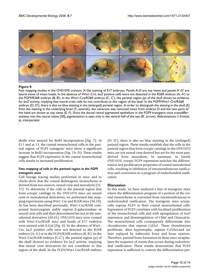
Fate mapping of cells in the parietal region in the FGF9 transgenic miceCell lineage tracing studies performed in mice and inchicks show that the cranial skeletogenic mesenchyme isderived from two sources, neural crest and mesoderm [35-37]. To determine if the cells in the parietal region thatform ectopic cartilage in the OVE1070 mice are neuralcrest or mesodermal derivatives, we performed fate map-ping experiments using Wnt1-Cre and R26R mice [38,39].As has been described previously, Wnt1-Cre/R26R com-pound heterozygotes stably express β-galactosidase inneural crest cells and their descendants but not in the mes-odermal derivatives [40,41]. OVE1070 mice were crossedwith Wnt1-Cre/R26R mice and heads of E17 embryoswere stained with X-Gal (Fig. 8). In the absence of Wnt1-Cre, lacZ positive cells were not detected in the R26Rembryo (A, A') or in the FGF9/R26R embryo (B, B'). In theWnt1-Cre/R26R embryo (C,C'), the parietal region (p) ofthe skull showed no evidence for lacZ activity, implyingthat neural crest derivatives do not contribute to thisregion of the skull. In the FGF9/Wnt1-Cre/R26R embryo
(D, D'), there is also no blue staining in the (enlarged)parietal region. These results establish that the cells in theparietal region that form ectopic cartilage in the OVE1070mice are not neural crest derived but are for the most part,derived from mesoderm. In summary, in familyOVE1070, ectopic FGF9 expression switches the differen-tiation and proliferation properties of cranial mesodermalcells, resulting in inhibition of intramembranous ossifica-tion and conversion to a program of endochondral ossifi-cation.
DiscussionIn this study, we have analyzed a line of transgenic micewhere the differentiation program of a portion of the cra-nial mesenchyme is converted from intramembranous toendochondral ossification. The transgenic mice ectopi-cally express FGF9 in their cranial mesenchymal cells.Expression of FGF9 correlates with localized proliferationof the mesenchymal cells and with upregulation of Sox9expression and downregulation of CbfaI and Osteocalcin.The mesenchymal cells consequently differentiate intochondrocytes that express Col2a1. These chondrocytesproliferate, then hypertrophy, express Col10a1and arelater replaced by trabecular bone and bone marrow.Therefore, parietal bone formation in these mice recapitu-lates the sequence of events that occurs during endochon-dral ossification. These results demonstrate that FGF9expression is sufficient to convert the differentiation pro-
Fate mapping studies in the OVE1070 craniumFigure 8Fate mapping studies in the OVE1070 cranium. X-Gal staining of E17 embryos. Panels A-D are top views and panels A'-D' are lateral views of intact heads. In the absence of Wnt1-Cre, lacZ positive cells were not detected in the R26R embryo (A, A') or the FGF9/R26R embryo (B, B'). In the Wnt1-Cre/R26R embryo (C, C'), the parietal region (p) of the skull shows no evidence for lacZ activity, implying that neural crest cells do not contribute to this region of the skull. In the FGF9/Wnt1-Cre/R26R embryo (D, D'), there is also no blue staining in the (enlarged) parietal region. In order to distinguish the staining in the skull (E) from the staining in the underlying brain (F, asterisk), the calvarium was removed intact from embryo D and the two parts of the head are shown as top views (E, F). Since the dorsal retinal pigmented epithelium in the FGF9 transgenic mice transdiffer-entiates into the neural retina [30], pigmentation is seen only in the ventral half of the eye (B', arrow). Abbreviations: f, frontal; ip, interparietal.
Page 10 of 14(page number not for citation purposes)

BMC Developmental Biology 2006, 6:7 http://www.biomedcentral.com/1471-213X/6/7
gram of (at least a subset of) the cranial mesenchymalcells from intramembranous to endochondral ossifica-tion.
Ectopic expression of the transgeneThe skeletal defects in this transgenic line (OVE1070)occur in mice that are either heterozygous or homozygousfor the FGF9 transgene. It is, therefore, unlikely that theskull defects in this line of mice are due to disruption of agene essential for membranous ossification of the parietalbones. The transgene expression pattern provides compel-ling evidence that the skull defects in these mice are dueto ectopic expression of the transgene. In situ hybridiza-tion analyses show that the transgene is expressed in anappropriate spatial and temporal pattern to induce thedevelopmental changes. In addition, the transgene is notexpressed in regions where membranous ossification stilloccurs (Fig. 3G). Furthermore, transgene expression pre-cedes Sox9 expression and condensation of the mesenchy-mal cells. Also, our results are consistent with the resultsof other studies. For example, recent studies of a mousemodel for Apert syndrome, with a single amino acidchange in FGFR2, support the notion that enhanced acti-vation of FGFR2 can cause some cranial mesenchyme toconvert to a chondrocyte differentiation program [10]. Inaddition, FGF9 expression in a chondrocytic cell line issufficient to induce expression of Sox9 [42]. Addition ofFGF-2 in vitro to cranial mesenchymal cells from quailembryos can induce cartilage differentiation at high doses[17]. Taken together, these results suggest that initiationof chondrogenesis in the cranial mesenchymal cells in thisline of mice is due to ectopic expression of the FGF9 trans-gene.
In the OVE1070 family, expression of the FGF9 transgeneis not only ectopic but also transient. Expression in thecranial mesenchyme was not detectable after E15. Thisimplies that stimulation by FGF9 is sufficient to induce(competent) cranial mesoderm to switch to a chondro-cytic differentiation program, but sustained expression ofFGF9 is not required for the later stages of differentiation.Therefore, the endochondral ossification program, onceinitiated by FGF9, appears to become autonomous.
The reason for the ectopic Fgf9 expression is not clear atpresent. It is possible the transgene array has integrated inthe neighborhood of an endogenous enhancer that directsexpression to the cranial mesenchymal cells. Some of theintegrated copies of the transgene still retain the αA-crys-tallin promoter since the transgene is still expressed in thelens [30]. The transgene is also expressed in the dorsalmargins of the retinal pigmented epithelium (RPE) andthis expression transforms the RPE into neuroretina [30].
Nature of FGF9 inductionExpression of FGF9 in growth plate chondrocytes usingthe Col2a1 promoter results in reduced proliferation andterminal differentiation of chondrocytes [43]. In addition,targeted deletion of FGFR3, one of the receptors throughwhich FGF9 signals, results in mice with overgrowth ofthe long bones. These results suggest that a primary role ofFGF signaling in the long bones is to act as a negative reg-ulator of chondrocyte proliferation. These results are incontrast to our results that show that ectopic FGF9 expres-sion in embryonic cranial mesenchymal cells inducesoverproliferation followed by endochondral ossification.How can these disparate observations be reconciled? Onepossible explanation is the differential timing of FGF9expression. The Col2a1 promoter is active in the differen-tiated chondrocytes, while our transgenic FGF9 isexpressed in undifferentiated cranial mesenchymal cells.Second, there are differences in the responding tissues.Chondrocytes in the long bones originate from the lateralplate mesoderm while the parietal mesenchyme is derivedfrom cranial mesoderm. Since the stimulated cells havedifferent developmental origins, there is no reason toexpect that the responses will be identical. For example,although lens and corneal epithelial cells are related toeach other, both morphologically and developmentally,they respond differently to FGF stimulation [29,44]. Thereduction in proliferation in the long bones in response toFGF stimulation may be a unique property of the growthplate chondrocytes [4]. In contrast, the FGF9-induced pro-liferation of cranial mesenchymal cells is consistent withthe notion that FGFs, in general, act as mitogens duringnormal development.
Endogenous role for FGF9?Though FGF9 appears to be sufficient to induce chondro-genesis in the skull, the role of FGF9 during normal devel-opment of cartilage in the cranium is unclear. Our in situhybridizations did not show any detectable expression ofFgf9 in the calvarial mesenchyme at E11 or E13. Expres-sion is seen later during embryonic development in theendocranial portions of the mesenchyme and is downreg-ulated during postnatal development [16]. This expres-sion pattern suggests that FGF9 is not important for initialspecification of the cells in the chondrocytic lineage in thecranium. Consistent with this model, targeted deletion ofFGF9 does not result in any visible skeletal abnormalitiesin the skull [24,25]. In contrast, in the transgenic mice, theectopic FGF9 plays an instructive role and initiates chon-drogenesis. FGF9 is known to signal through FGFR2 andFGFR3 [45]. Signaling by FGF9 in the transgenic heads islikely to be mediated initially through FGFR2 rather thanFGFR3 as the parietal fate switch in the FGF9 transgenicmice is not rescued by the loss of FGFR3. After Sox9expression has been induced and the chondrocytic pro-gram has been initiated (by E13), expression of the FGF9
Page 11 of 14(page number not for citation purposes)

BMC Developmental Biology 2006, 6:7 http://www.biomedcentral.com/1471-213X/6/7
transgene is no longer required. In contrast to the normalchondrocytic differentiation program in long bones,ectopic chondrogenesis in the cranium proceeds at amuch slower rate. Though the reasons for this are pres-ently unclear, we speculate that this may be due to themulti-step nature of the endochondral differentiationprogram. Respecification and reprogramming of the pari-etal environment is required, and this is accomplished ina less timely fashion than at the normal sites of differenti-ation.
FGFRs and intramembranous ossificationOur findings are in apparent conflict with some of the cur-rent models for the roles of FGF receptors in intramem-branous ossification. Autosomal dominant mutations inFGFR2 or FGFR3 lead to premature differentiation andfusion of the skull sutures in humans and these effects arethought to be the consequence of enhanced receptor activ-ity [9]. Based upon such a model, our transgenic micewould be predicted to exhibit an analogous phenotype,i.e. craniosynostosis. Inappropriate activation of thereceptors in the cranial mesenchyme would be predictedto lead to premature differentiation. However, our studiesindicate that ligand-mediated activation of FGFR2induces proliferation of embryonic cranial mesenchymalcells and subsequent differentiation into cartilage. Byextrapolation then, our results imply that mutations inthe FGFR2 gene in human patients do not result in ligand-independent constitutive activation of the receptor. Thisprediction is supported by the finding that targeted inacti-vation of FGFR2 in mice causes developmental alterationsin cell types that are not affected by the putative gain-of-function mutations. An alternative explanation is thattransient stimulation of FGFR2 (as seen in our FGF9 trans-genic mice) may lead to a qualitatively different cellularresponse compared to sustained ligand-independentstimulation of FGFR2 (as seen in the gain-of-functionmutations in human patients). In either case, our resultsshow that a single FGF can function as an instructive sig-nal, inducing a specific cell type to switch from one differ-entiation program to an autonomous, alternativedifferentiation pathway.
ConclusionAlthough the mechanistic details of FGF9 stimulation ofthe cranial mesenchymal cells remain to be elucidated,the OVE1070 transgenic mice provide the first demonstra-tion in vivo that an FGF can switch the differentiation pro-gram of immature cranial mesoderm. These results alsodemonstrate that mesoderm-derived cranial mesenchy-mal cells are developmentally flexible and can undergoeither intramembranous or endochondral ossification inresponse to extracellular signals. These transgenic miceprovide a model system in which to elucidate the molec-ular connection between stimulation of an FGFR (FGFR2)
and induction of expression of a cell-fate-determiningtranscription factor (Sox9).
MethodsGeneration of FGF9 transgenic miceThe construction of the FGF9 transgene and the genera-tion of transgenic mice have been described previously[29]. The coding region of mouse Fgf9 (a gift from Dr.David M. Ornitz, Washington University School of Medi-cine, St. Louis) was inserted between the αA-crystallin pro-moter [46] and the SV40 small t intron/polyadenylationsequences of the CPV2 vector [47]. FGF9 transgenic micewere identified by isolating genomic DNA and screeningby PCR, using primers specific to the SV40 portion of thetransgene: 5'-GTGAAGGAACCTTACTTCTGTGGTG-3'(SV40A) and 5'-GTCCTTGGGGTCTTCTACCTTTCTC-3'(SV40B). The PCR cycle conditions were as follows: dena-turation at 94°C for 30 seconds, annealing at 58°C for 30seconds and extension at 72°C for 60 seconds, for 35cycles. A final extension step of 72°C for 10 minutes wasincluded.
Skeletal preparationsSkeletal preparations were performed as described previ-ously [48]. Briefly, embryos were first skinned, and evis-cerated, then fixed in 95% alcohol. After fixation, thesamples were stained with Alcian blue, for 1–2 days. Thesamples were then destained in 95% ethanol for eighthours, and cleared in 2% potassium hydroxide from eighthours to overnight depending on the size of the specimen.After clearing, the samples were stained in Alizarin red/1% potassium hydroxide overnight. The samples werecleared in 1% potassium hydroxide and 20% glycerol/1%potassium hydroxide for 2–3 days. Subsequently, thesamples were allowed to harden in 1:1 95% ethanol/glyc-erol for one day and then transferred to absolute glycerolfor storage and photography.
Histological analysesFor routine histology, embryos were obtained from timedpregnancies using FVB/N females that were mated to het-erozygous FGF9 transgenic males. Embryos were deliv-ered by Caesarean section, fixed in 10% formalin,dehydrated, embedded in paraffin, sectioned (5 µm) andstained with hematoxylin and eosin.
Section in situ hybridizationsTo analyze the expression of different markers, in situhybridizations were performed. Mouse cDNA clones forCol2a1, Col10a1, Ihh and Sox9 were obtained from Dr.Benoit DeCrombrugghe (University of Texas, MD Ander-son Cancer Center, Houston). Mouse cDNA clones forCbfaI and osteocalcin were obtained from Dr. GerardKarsenty (Baylor College of Medicine, Houston). cDNAsclones for Fgf9, Fgfr2 and Fgfr3 were obtained from Dr.
Page 12 of 14(page number not for citation purposes)

BMC Developmental Biology 2006, 6:7 http://www.biomedcentral.com/1471-213X/6/7
David Ornitz (Washington University School of Medi-cine, St. Louis). To analyze expression of the FGF9 trans-gene, a [35S]-UTP-labeled riboprobe specific to the SV40sequences of the transgene was made (see Fig. 1). To assayfor endogenous Sox9 expression, a Sox9 antisense probewas synthesized using a HindIII-digested mouse Sox9cDNA and T7 RNA polymerase (Promega). For Col2a1,the antisense probe was synthesized using EcoRI-digestedDNA and T3 RNA polymerase. The antisense probe forCol10aI was synthesized using a HindIII-digested mouseCol10a1 cDNA and T3 RNA polymerase. The antisenseprobe for Ihh was synthesized using a BamHI-digestedmouse Ihh cDNA and T7 RNA polymerase. The antisenseprobe for Fgf9 was synthesized using a KpnI-digestedmouse Fgf9 cDNA and SP6 RNA polymerase. The anti-sense probe for CbfaI was synthesized using an EcoRI-digested mouse CbfaI cDNA and T7 RNA polymerase. Theantisense probe for osteocalcin was synthesized usingBamHI-digested mouse osteocalcin cDNA and T3 RNApolymerase. The antisense probe for Fgfr2 was synthesizedusing an EcoRI-digested mouse Fgfr2 cDNA and T7 RNApolymerase. The antisense probe for Fgfr3 was synthesizedusing a HindIII-digested mouse Fgfr3 cDNA and T3 RNApolymerase. In situ hybridizations on tissue sections weredone using hybridization and washing conditionsdescribed previously [49]. The hybridized slides weresoaked in Kodak NTB-2 emulsion, dried and exposed for3–5 days at 4°C. Following development and fixation, theslides were lightly counterstained with hematoxylin.
Proliferation assayDNA replication was examined by BrdU incorporation asdescribed previously [50]. Cell proliferation was analyzedby counting the number of BrdU positive nuclei frommore than 100 cells in a defined area in the parietal regionin three serial sections from four (E11) or six (E13) non-transgenic and FGF9 transgenic heads. The counts forBrdU-positive cells in nontransgenic and transgenic micewere compared using the t-test (p < 0.01).
Whole mount in situ hybridizationsFor whole mount in situ hybridizations, tissue sampleswere washed thrice in PBS and fixed in 4% paraformalde-hyde. Hybridizations were performed using digoxygenin-labeled sense or antisense SV40 riboprobes followingstandard procedures [51].
Histochemical detection of β-galactosidase activityEmbryos were obtained from timed pregnancies of R26Rhomozygote females mated to OVE1070/Wnt1cre doubletransgenic males [36]. X-Gal staining was performed asdescribed previously [44]. Briefly, heads of E17 embryoswere collected and fixed for 2 hours at 4°C in 0.1 M phos-phate buffer (pH 7.3) containing 2% paraformaldehyde,0.2% glutaraldehyde. Following fixation, the tissue sam-
ples were rinsed thrice at room temperature in 0.1 Mphosphate buffer (pH 7.3) containing 0.01% sodiumdeoxycholate, 0.02% NP-40, 2 mM MgCl2, then stained inan X-gal substrate solution (0.01% sodium deoxycholate,0.02% NP-40, 2 mM MgCl2, 5 mM potassium ferricya-nide, 5 mM potassium ferrocyanide, 1 mg/ml X-gal in 0.1M phosphate buffer (pH 7.3)).
Authors' contributionsVG designed and performed all the experiments anddrafted the manuscript. PAO originated the idea to gener-ate the CPV2-FGF9 mice, helped in the design of theexperiments and the interpretation of the data and editedthe manuscript. Both authors read and approved the man-uscript.
AcknowledgementsWe would like to thank Dr. David Ornitz for providing the mouse Fgf9 cDNA, Dr. Gerard Karsenty for providing the CbfaI and Osteocalcin cDNA clones, Dr. Benoit DeCrombrugghe for providing the Col2a1, Col10a1, Ihh and Sox9 cDNA clones. We are grateful to Gabriele Schuster for perform-ing the microinjections, Long Vien for assistance in animal husbandry, Bar-bara Harris for help in histological analyses, Dr. Frank Lovicu for making the CPV2-FGF9 construct, Drs. Fred A. Pereira, Bernd Fritzsch and David Nichols for helpful suggestions, and Travis Bailey for help in using Adobe Photoshop. We also thank Drs. Gerard Karsenty and Brendan Lee for crit-ically reading the manuscript and for insightful comments and suggestions. This work was supported by NIH grants EY-10448 and EY-10803 (PAO) and revenue from the Nebraska Tobacco Settlement Biomedical Research Development Fund (VG).
References1. de Crombrugghe B, Lefebvre V, Nakashima K: Regulatory mecha-
nisms in the pathways of cartilage and bone formation. CurrOpin Cell Biol 2001, 13(6):721-728.
2. Gerber HP, Vu TH, Ryan AM, Kowalski J, Werb Z, Ferrara N: VEGFcouples hypertrophic cartilage remodeling, ossification andangiogenesis during endochondral bone formation. Nat Med1999, 5(6):623-628.
3. Olsen BR, Reginato AM, Wang W: Bone development. Annu RevCell Dev Biol 2000, 16:191-220.
4. Naski MC, Ornitz DM: FGF signaling in skeletal development.Front Biosci 1998, 3:D781-94.
5. Ornitz DM, Itoh N: Fibroblast growth factors. Genome Biol 2001,2(3):.
6. Iseki S, Wilkie AO, Morriss-Kay GM: Fgfr1 and Fgfr2 have distinctdifferentiation- and proliferation- related roles in the devel-oping mouse skull vault. Development 1999, 126(24):5611-5620.
7. Rice DP, Aberg T, Chan Y, Tang Z, Kettunen PJ, Pakarinen L, MaxsonRE, Thesleff I: Integration of FGF and TWIST in calvarial boneand suture development. Development 2000, 127(9):1845-1855.
8. Ornitz DM, Marie PJ: FGF signaling pathways in endochondraland intramembranous bone development and humangenetic disease. Genes Dev 2002, 16(12):1446-1465.
9. Kannan K, Givol D: FGF receptor mutations: dimerization syn-dromes, cell growth suppression, and animal models. IUBMBLife 2000, 49(3):197-205.
10. Wang Y, Xiao R, Yang F, Karim BO, Iacovelli AJ, Cai J, Lerner CP,Richtsmeier JT, Leszl JM, Hill CA, Yu K, Ornitz DM, Elisseeff J, HusoDL, Jabs EW: Abnormalities in cartilage and bone develop-ment in the Apert syndrome FGFR2(+/S252W) mouse.Development 2005, 132(15):3537-3548.
11. Yu K, Ornitz DM: Uncoupling fibroblast growth factor recep-tor 2 ligand binding specificity leads to Apert syndrome-likephenotypes. Proc Natl Acad Sci U S A 2001, 98(7):3641-3643.
Page 13 of 14(page number not for citation purposes)

BMC Developmental Biology 2006, 6:7 http://www.biomedcentral.com/1471-213X/6/7
12. Wang Q, Green RP, Zhao G, Ornitz DM: Differential regulationof endochondral bone growth and joint development byFGFR1 and FGFR3 tyrosine kinase domains. Development2001, 128(19):3867-3876.
13. Eswarakumar VP, Monsonego-Ornan E, Pines M, Antonopoulou I,Morriss-Kay GM, Lonai P: The IIIc alternative of Fgfr2 is a posi-tive regulator of bone formation. Development 2002,129(16):3783-3793.
14. Deng C, Wynshaw-Boris A, Zhou F, Kuo A, Leder P: Fibroblastgrowth factor receptor 3 is a negative regulator of bonegrowth. Cell 1996, 84(6):911-921.
15. Ornitz DM: Regulation of chondrocyte growth and differenti-ation by fibroblast growth factor receptor 3. Novartis FoundSymp 2001, 232:63-76.
16. Kim HJ, Rice DP, Kettunen PJ, Thesleff I: FGF-, BMP- and Shh-mediated signalling pathways in the regulation of cranialsuture morphogenesis and calvarial bone development.Development 1998, 125(7):1241-1251.
17. Sarkar S, Petiot A, Copp A, Ferretti P, Thorogood P: FGF2 pro-motes skeletogenic differentiation of cranial neural crestcells. Development 2001, 128(11):2143-2152.
18. Colvin JS, Feldman B, Nadeau JH, Goldfarb M, Ornitz DM: Genomicorganization and embryonic expression of the mouse fibrob-last growth factor 9 gene. Dev Dyn 1999, 216(1):72-88.
19. deLapeyriere O, Ollendorff V, Planche J, Ott MO, Pizette S, CoulierF, Birnbaum D: Expression of the Fgf6 gene is restricted todeveloping skeletal muscle in the mouse embryo. Develop-ment 1993, 118(2):601-611.
20. Finch PW, Cunha GR, Rubin JS, Wong J, Ron D: Pattern of kerati-nocyte growth factor and keratinocyte growth factor recep-tor expression during mouse fetal development suggests arole in mediating morphogenetic mesenchymal-epithelialinteractions. Dev Dyn 1995, 203(2):223-240.
21. Haub O, Goldfarb M: Expression of the fibroblast growth fac-tor-5 gene in the mouse embryo. Development 1991,112(2):397-406.
22. Mason IJ, Fuller-Pace F, Smith R, Dickson C: FGF-7 (keratinocytegrowth factor) expression during mouse development sug-gests roles in myogenesis, forebrain regionalisation and epi-thelial-mesenchymal interactions. Mech Dev 1994, 45(1):15-30.
23. Savage MP, Fallon JF: FGF-2 mRNA and its antisense messageare expressed in a developmentally specific manner in thechick limb bud and mesonephros. Dev Dyn 1995,202(4):343-353.
24. Colvin JS, Green RP, Schmahl J, Capel B, Ornitz DM: Male-to-female sex reversal in mice lacking fibroblast growth factor9. Cell 2001, 104(6):875-889.
25. Colvin JS, White AC, Pratt SJ, Ornitz DM: Lung hypoplasia andneonatal death in Fgf9-null mice identify this gene as anessential regulator of lung mesenchyme. Development 2001,128(11):2095-2106.
26. Fiore F, Planche J, Gibier P, Sebille A, deLapeyriere O, Birnbaum D:Apparent normal phenotype of Fgf6-/- mice. Int J Dev Biol 1997,41(4):639-642.
27. Guo L, Degenstein L, Fuchs E: Keratinocyte growth factor isrequired for hair development but not for wound healing.Genes Dev 1996, 10(2):165-175.
28. Hebert JM, Rosenquist T, Gotz J, Martin GR: FGF5 as a regulatorof the hair growth cycle: evidence from targeted and sponta-neous mutations. Cell 1994, 78(6):1017-1025.
29. Lovicu FJ, Overbeek PA: Overlapping effects of different mem-bers of the FGF family on lens fiber differentiation in trans-genic mice. Development 1998, 125(17):3365-3377.
30. Zhao S, Hung FC, Colvin JS, White A, Dai W, Lovicu FJ, Ornitz DM,Overbeek PA: Patterning the optic neuroepithelium by FGFsignaling and Ras activation. Development 2001,128(24):5051-5060.
31. Bi W, Deng JM, Zhang Z, Behringer RR, de Crombrugghe B: Sox9 isrequired for cartilage formation. Nat Genet 1999, 22(1):85-89.
32. Lefebvre V, Huang W, Harley VR, Goodfellow PN, de CrombruggheB: SOX9 is a potent activator of the chondrocyte-specificenhancer of the pro alpha1(II) collagen gene. Mol Cell Biol1997, 17(4):2336-2346.
33. Ducy P, Zhang R, Geoffroy V, Ridall AL, Karsenty G: Osf2/Cbfa1: atranscriptional activator of osteoblast differentiation. Cell1997, 89(5):747-754.
34. Ducy P, Schinke T, Karsenty G: The osteoblast: a sophisticatedfibroblast under central surveillance. Science 2000,289(5484):1501-1504.
35. Couly GF, Coltey PM, Le Douarin NM: The triple origin of skullin higher vertebrates: a study in quail-chick chimeras. Devel-opment 1993, 117(2):409-429.
36. Jiang X, Iseki S, Maxson RE, Sucov HM, Morriss-Kay GM: Tissue ori-gins and interactions in the mammalian skull vault. Dev Biol2002, 241(1):106-116.
37. Noden DM: Interactions and fates of avian craniofacial mesen-chyme. Development 1988, 103 Suppl:121-140.
38. Danielian PS, Muccino D, Rowitch DH, Michael SK, McMahon AP:Modification of gene activity in mouse embryos in utero by atamoxifen-inducible form of Cre recombinase. Curr Biol 1998,8(24):1323-1326.
39. Soriano P: Generalized lacZ expression with the ROSA26 Crereporter strain. Nat Genet 1999, 21(1):70-71.
40. Chai Y, Jiang X, Ito Y, Bringas PJ, Han J, Rowitch DH, Soriano P,McMahon AP, Sucov HM: Fate of the mammalian cranial neuralcrest during tooth and mandibular morphogenesis. Develop-ment 2000, 127(8):1671-1679.
41. Jiang X, Rowitch DH, Soriano P, McMahon AP, Sucov HM: Fate ofthe mammalian cardiac neural crest. Development 2000,127(8):1607-1616.
42. Murakami S, Kan M, McKeehan WL, de Crombrugghe B: Up-regula-tion of the chondrogenic Sox9 gene by fibroblast growth fac-tors is mediated by the mitogen-activated protein kinasepathway. Proc Natl Acad Sci U S A 2000, 97(3):1113-1118.
43. Garofalo S, Kliger-Spatz M, Cooke JL, Wolstin O, Lunstrum GP,Moshkovitz SM, Horton WA, Yayon A: Skeletal dysplasia anddefective chondrocyte differentiation by targeted overex-pression of fibroblast growth factor 9 in transgenic mice. JBone Miner Res 1999, 14(11):1909-1915.
44. Govindarajan V, Ito M, Makarenkova HP, Lang RA, Overbeek PA:Endogenous and ectopic gland induction by FGF-10. Dev Biol2000, 225(1):188-200.
45. Hecht D, Zimmerman N, Bedford M, Avivi A, Yayon A: Identifica-tion of fibroblast growth factor 9 (FGF9) as a high affinity,heparin dependent ligand for FGF receptors 3 and 2 but notfor FGF receptors 1 and 4. Growth Factors 1995, 12(3):223-233.
46. Overbeek PA, Chepelinsky AB, Khillan JS, Piatigorsky J, Westphal H:Lens-specific expression and developmental regulation ofthe bacterial chloramphenicol acetyltransferase gene drivenby the murine alpha A- crystallin promoter in transgenicmice. Proc Natl Acad Sci U S A 1985, 82(23):7815-7819.
47. Reneker LW, Silversides DW, Patel K, Overbeek PA: TGF alphacan act as a chemoattractant to perioptic mesenchymal cellsin developing mouse eyes. Development 1995, 121(6):1669-1680.
48. Nagy A: Manipulating the mouse embryo : a laboratory man-ual. 3rd edition. Cold Spring Harbor, N.Y. , Cold Spring Harbor Lab-oratory Press; 2003:x, 764 p..
49. Robinson ML, MacMillan-Crow LA, Thompson JA, Overbeek PA:Expression of a truncated FGF receptor results in defectivelens development in transgenic mice. Development 1995,121(12):3959-3967.
50. Govindarajan V, Overbeek PA: Secreted FGFR3, but not FGFR1,inhibits lens fiber differentiation. Development 2001,128(9):1617-1627.
51. Wilkinson DG: In situ hybridization : a practical approach. InThe practical approach series 196 Oxford ; New York , Oxford Univer-sity Press; 1998:xviii, 224 p..
Page 14 of 14(page number not for citation purposes)