Embed Size (px)
Citation preview
4/10/2013
1
FESS:The Required Imaging
AnatomyAnatomy
Vincent Chong, MD MBA FRCR
Professor
Department of Diagnostic Imaging
National University Heath System
Singapore
• Kennedy DW. Functional endoscopic sinus surgery: technique. Arch Otolaryngol Head Neck Surg 1985; 111:643-649
• Stammberger H, Posawetz W. Functional endoscopic sinus surgery. Concept, indications and results of the Messerklinger technique. Eur Arch Otorhinolaryngol. 1990;247(2):63-76.
4/10/2013
2
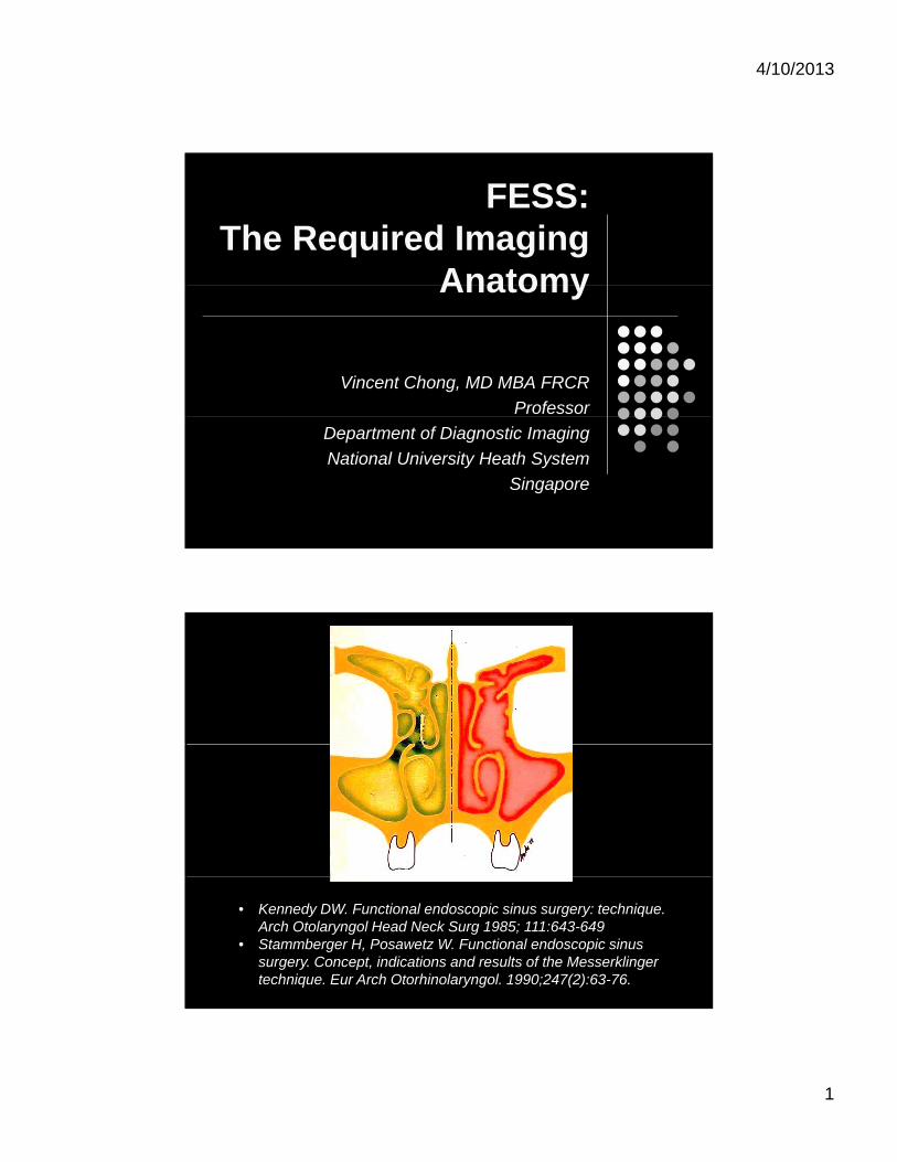
Overview
Normal anatomy General concepts
Specific anatomic structures
Variants
Pathological anatomy Stress normal anatomy Stress normal anatomy
Appropriate imaging modality
Basal Lamella &Drainage System
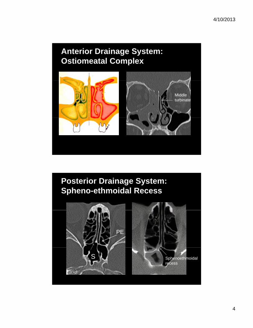
Middle turbinate
Middle turbinate
Basal lamella
4/10/2013
3
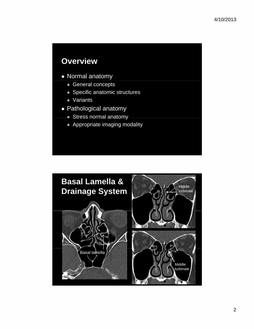
Basal Lamella &Drainage System
Anterior system Ostiomeatal unit
Ethmoid infundibulum
Middle meatusB
Basal Lamella &Drainage System
Posterior system Posterior ethmoid cells
& sphenoid sinus
Sphenoethmoidal recess
Superior meatus SphenoidPE
4/10/2013
4
Anterior Drainage System: Ostiomeatal Complex
Middle turbinate
Posterior Drainage System:Spheno-ethmoidal Recess
PE
S Sphenoethmoidal recess
4/10/2013
5
Spheno-ethmoidal Recess
PE Superiorturbinate Nasal
septum
Identifying the spheno-ethmoidal recess
S
Frontal Recess
Relationships Anterior Agger nasi cell
Posterior Anterior wall of
ethmoid bulla
A
Ethmoid bulla
4/10/2013
6
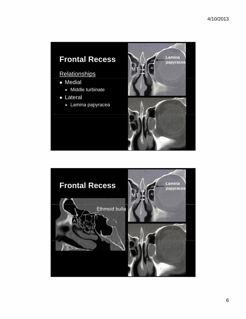
Frontal Recess
RelationshipsA
Lamina papyracea
MT
Medial Middle turbinate
Lateral Lamina papyracea
Frontal RecessA
Lamina papyracea
MT
4/10/2013
7
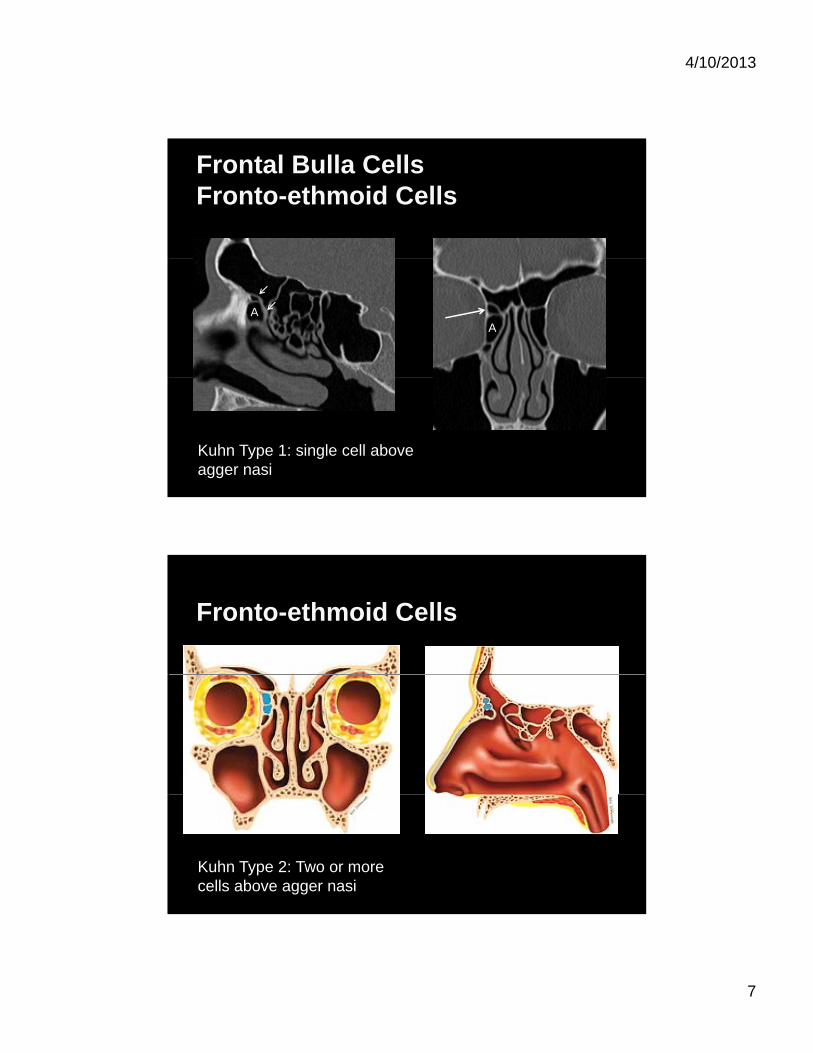
Frontal Bulla CellsFronto-ethmoid Cells
AA
Kuhn Type 1: single cell above agger nasi
Fronto-ethmoid Cells
Kuhn Type 2: Two or more cells above agger nasi
4/10/2013
8
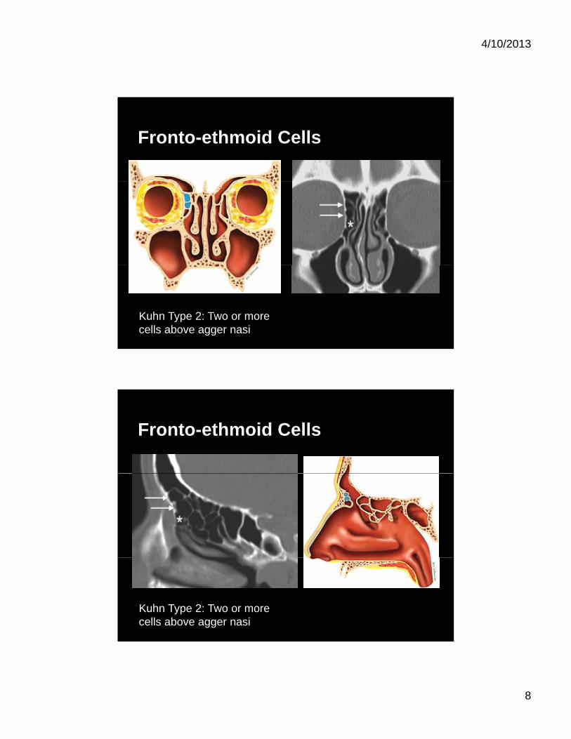
Fronto-ethmoid Cells
Kuhn Type 2: Two or more cells above agger nasi
Fronto-ethmoid Cells
Kuhn Type 2: Two or more cells above agger nasi
4/10/2013
9
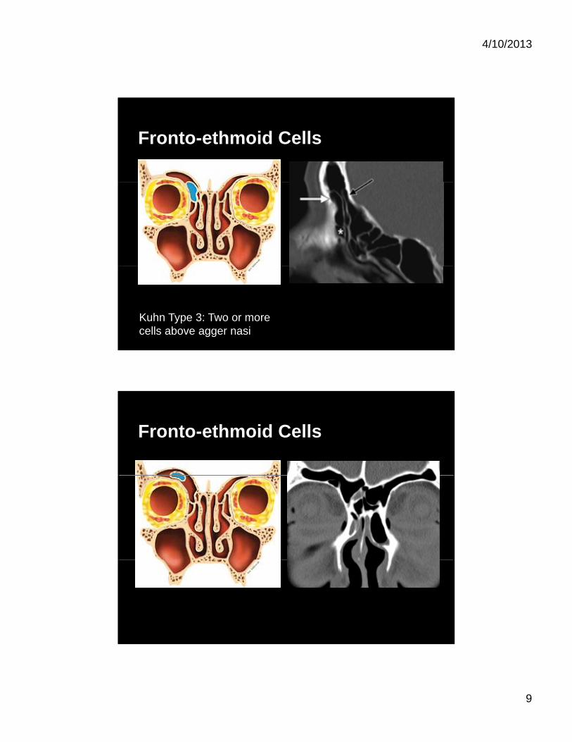
Fronto-ethmoid Cells
Kuhn Type 3: Two or more cells above agger nasi
Fronto-ethmoid Cells
4/10/2013
10
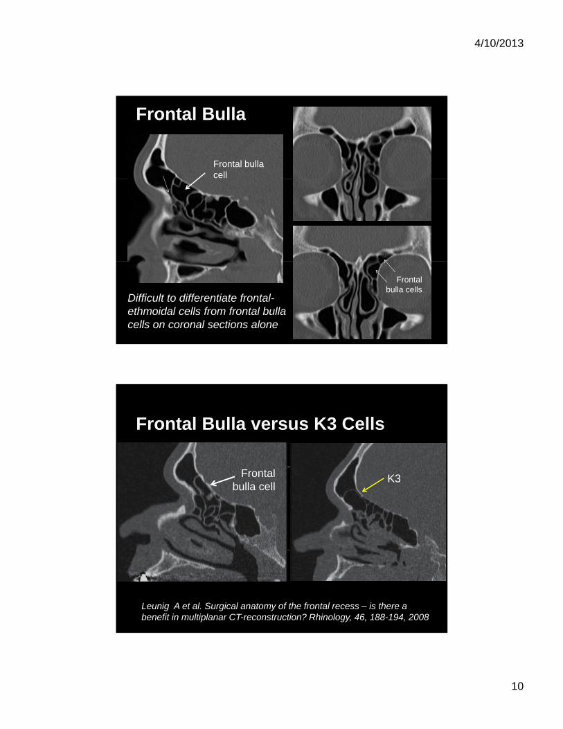
Frontal Bulla
Frontal bulla cell
Difficult to differentiate frontal-ethmoidal cells from frontal bulla cells on coronal sections alone
Frontal bulla cells
Frontal Bulla versus K3 Cells
K3Frontal bulla cell
Leunig A et al. Surgical anatomy of the frontal recess – is there a benefit in multiplanar CT-reconstruction? Rhinology, 46, 188-194, 2008
4/10/2013
11
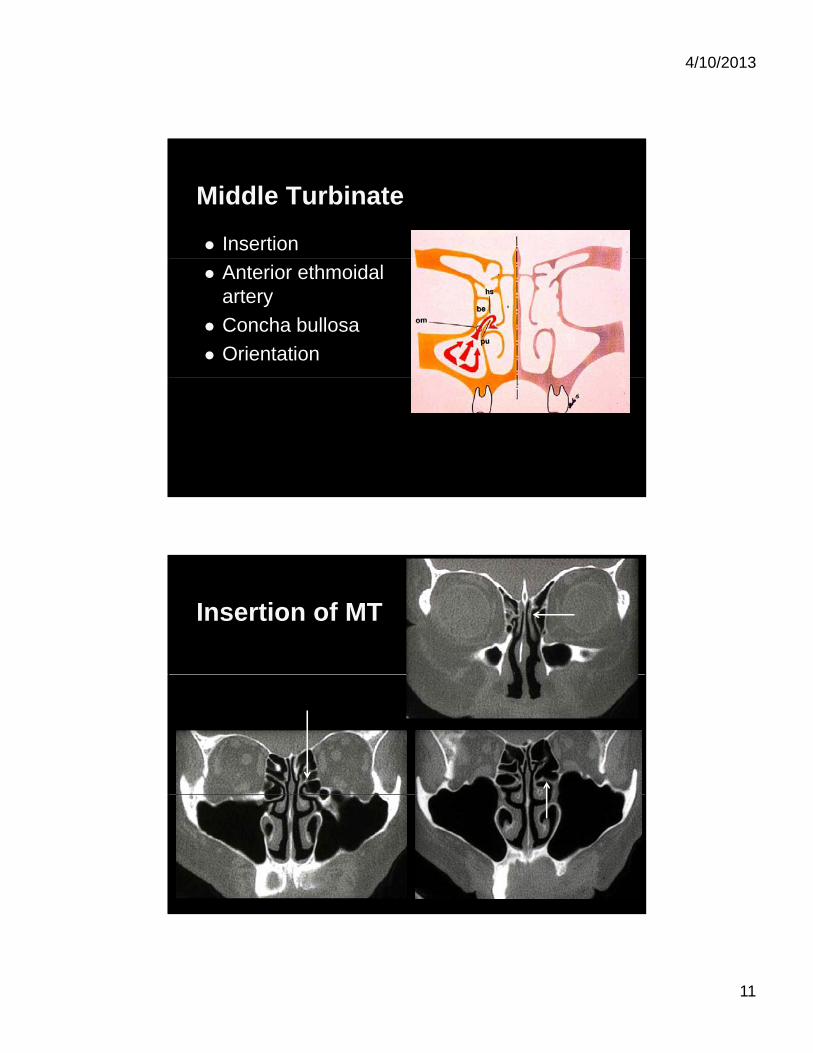
Middle Turbinate
Insertion
Anterior ethmoidal artery
Concha bullosa
Orientation
Insertion of MT
4/10/2013
12
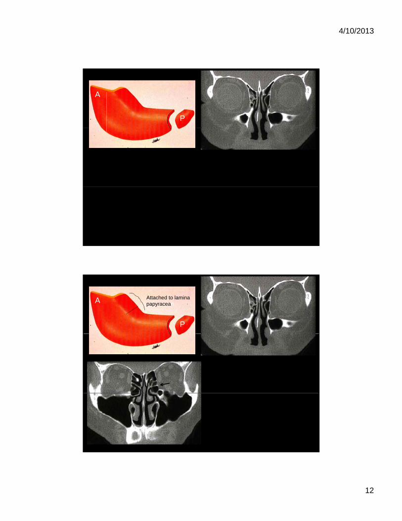
A
P
A
P
Attached to lamina papyracean
4/10/2013
13
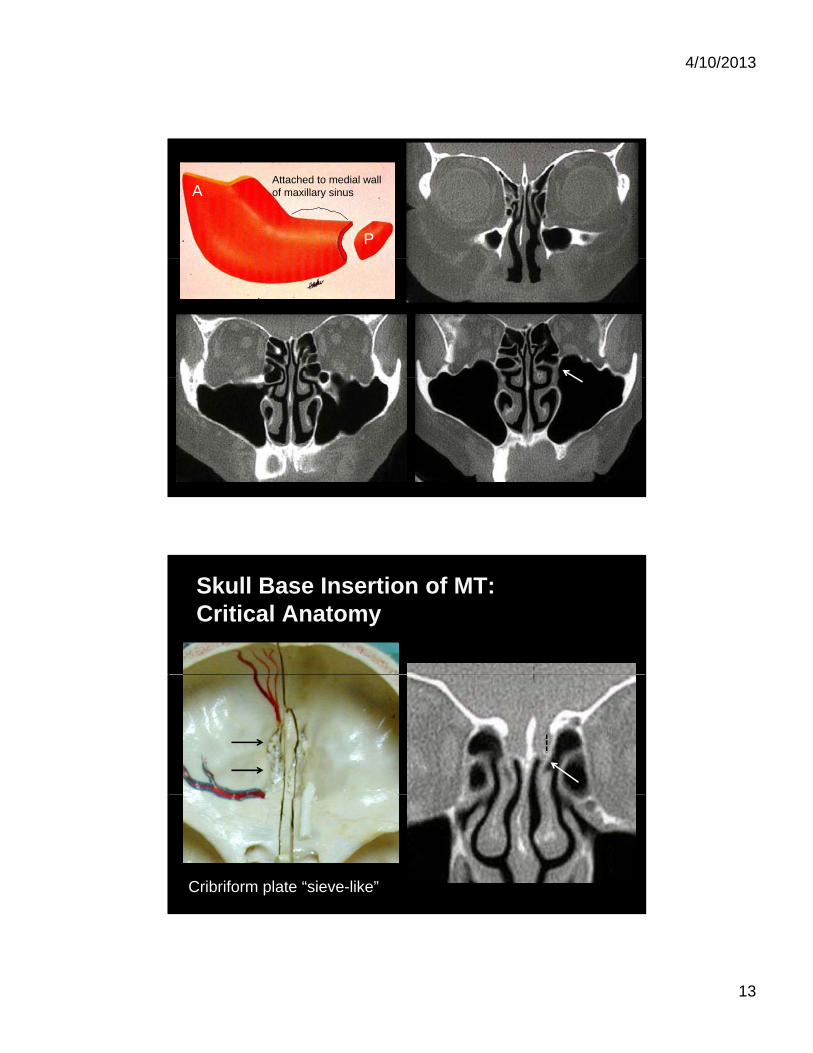
A
P
Attached to medial wall of maxillary sinus
Skull Base Insertion of MT:Critical Anatomy
Cribriform plate “sieve-like”
4/10/2013
14
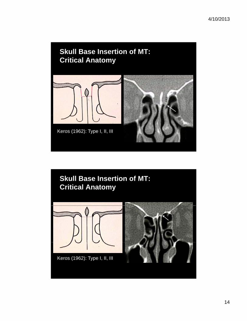
Skull Base Insertion of MT:Critical Anatomy
Keros (1962): Type I, II, III
Skull Base Insertion of MT:Critical Anatomy
Keros (1962): Type I, II, III
4/10/2013
15
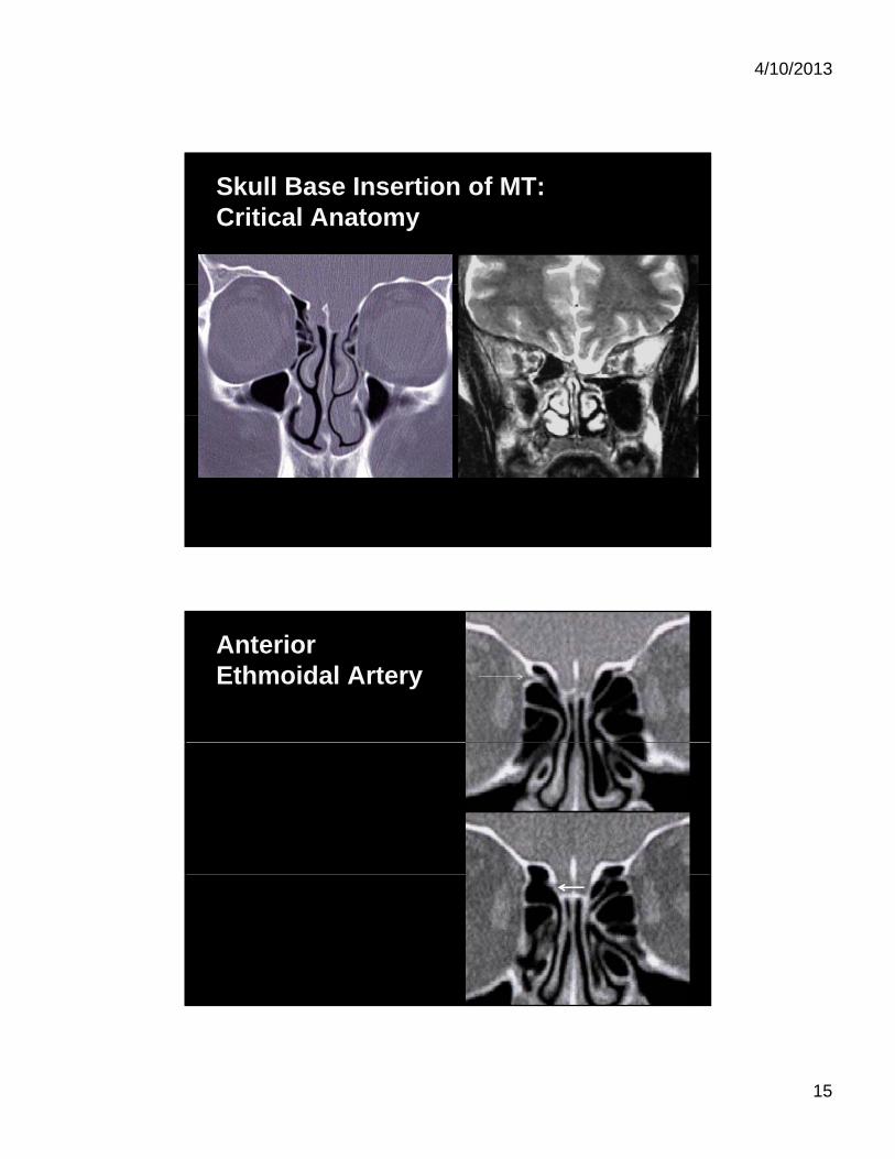
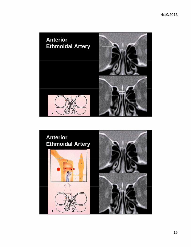
Skull Base Insertion of MT:Critical Anatomy
Anterior Ethmoidal Artery
4/10/2013
16
Anterior Ethmoidal Artery
Anterior Ethmoidal Artery
4/10/2013
17
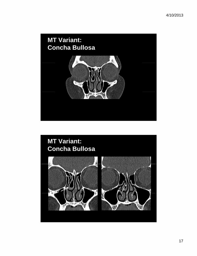
MT Variant:Concha Bullosa
MT Variant:Concha Bullosa
4/10/2013
18
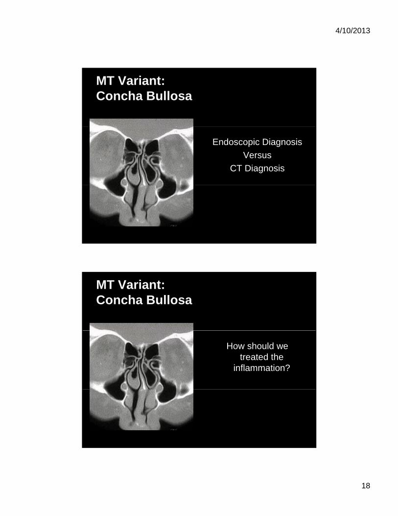
MT Variant:Concha Bullosa
Endoscopic Diagnosis
Versus
CT Diagnosis
MT Variant:Concha Bullosa
How should we treated the
inflammation?
4/10/2013
19
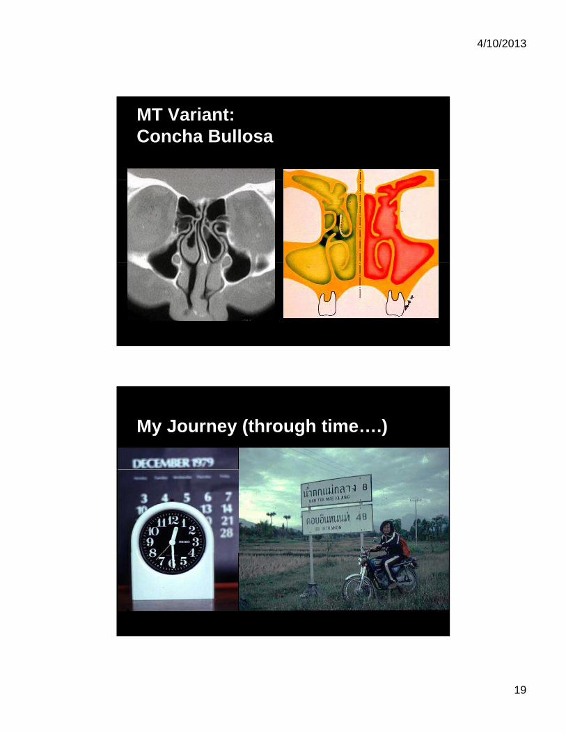
MT Variant:Concha Bullosa
My Journey (through time….)
4/10/2013
20
4/10/2013
21
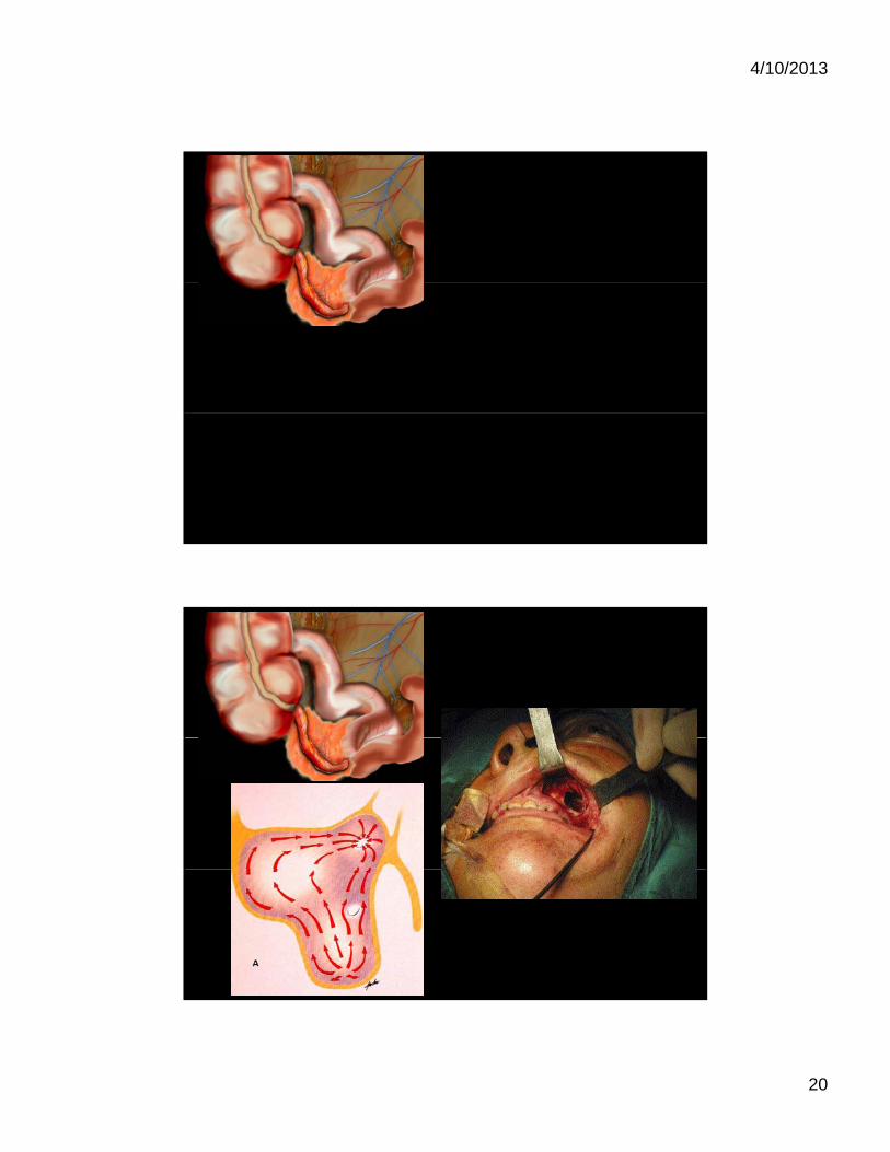
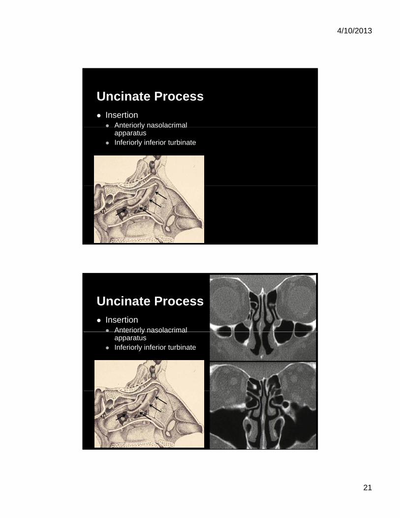
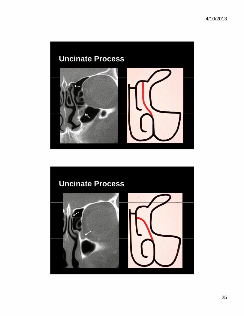
Uncinate Process Insertion Anteriorly nasolacrimal Anteriorly nasolacrimal
apparatus Inferiorly inferior turbinate
Uncinate Process Insertion Anteriorly nasolacrimal Anteriorly nasolacrimal
apparatus Inferiorly inferior turbinate
4/10/2013
22
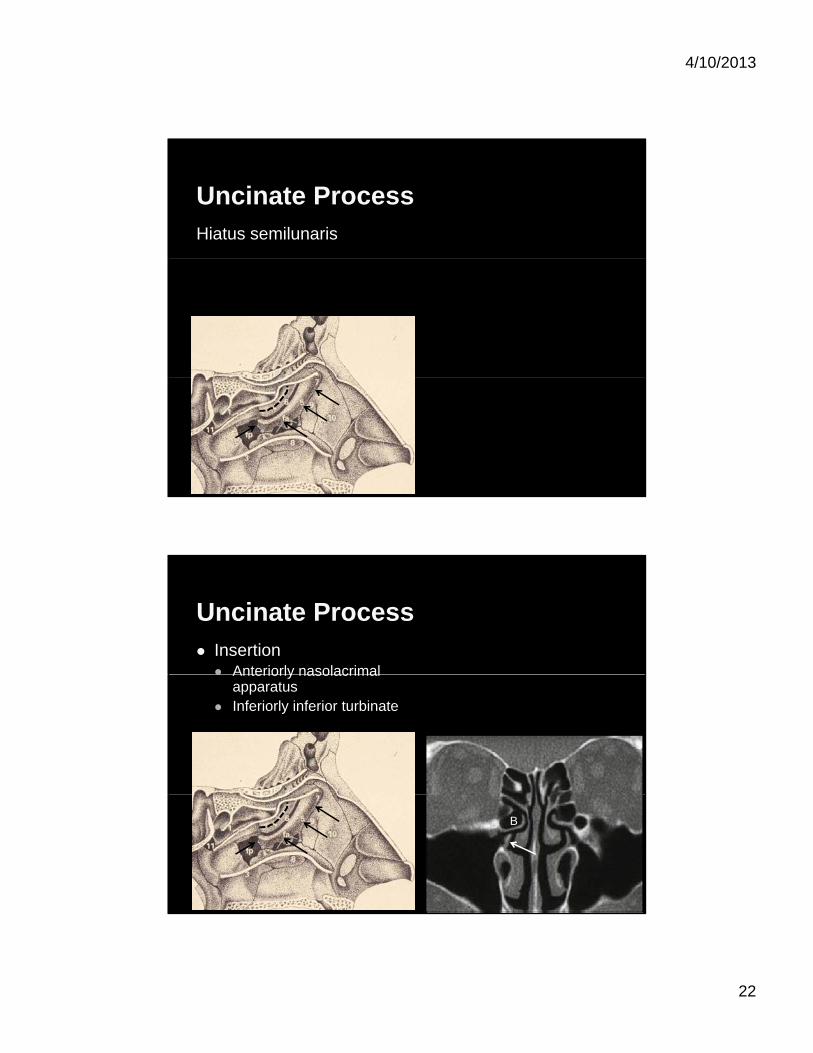
Uncinate ProcessHiatus semilunaris
Uncinate Process Insertion Anteriorly nasolacrimal Anteriorly nasolacrimal
apparatus Inferiorly inferior turbinate
B
4/10/2013
23
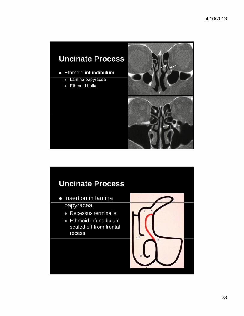
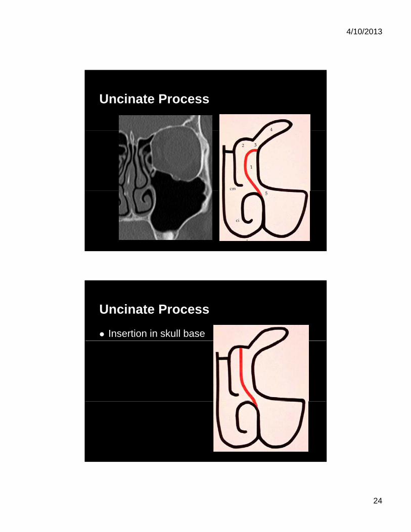
Uncinate Process
Ethmoid infundibulum Lamina papyracea
Ethmoid bulla
Uncinate Process
Insertion in lamina papyracea Recessus terminalis
Ethmoid infundibulum sealed off from frontal recess
4/10/2013
24
Uncinate Process
Uncinate Process
Insertion in skull base
4/10/2013
25
Uncinate Process
Uncinate Process
4/10/2013
26
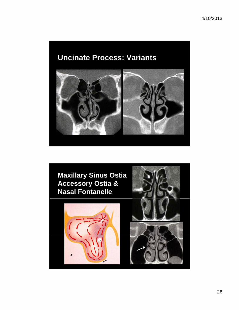
Uncinate Process: Variants
Maxillary Sinus Ostia Accessory Ostia & Nasal Fontanelle
4/10/2013
27
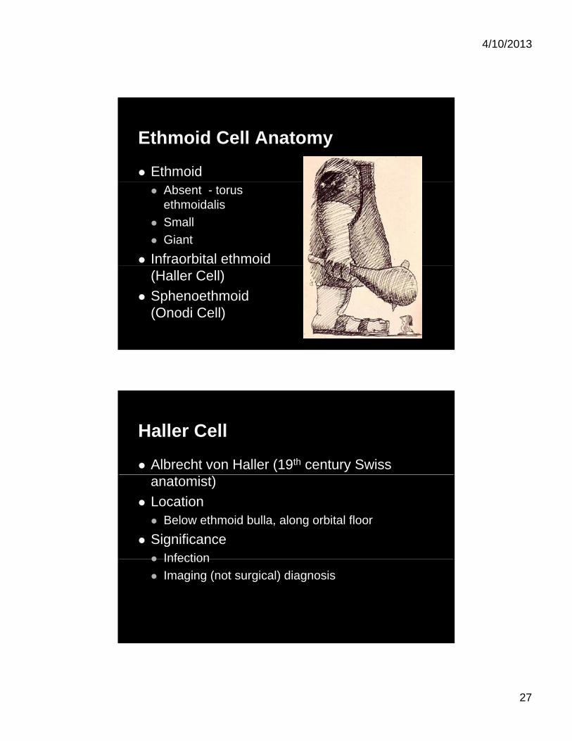
Ethmoid Cell Anatomy
Ethmoid Absent - torus
ethmoidalis
Small
Giant
Infraorbital ethmoid (Haller Cell)
Sphenoethmoid (Onodi Cell)
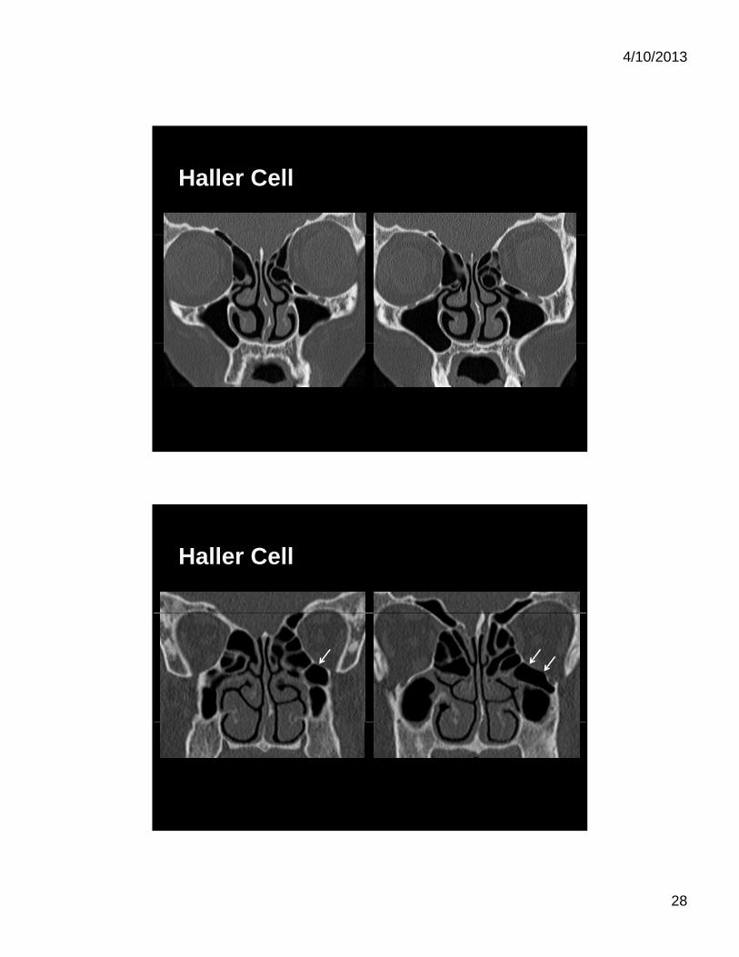
Haller Cell
Albrecht von Haller (19th century Swiss anatomist)
Location Below ethmoid bulla, along orbital floor
Significance Infection Infection
Imaging (not surgical) diagnosis
4/10/2013
28
Haller Cell
Haller Cell
4/10/2013
29
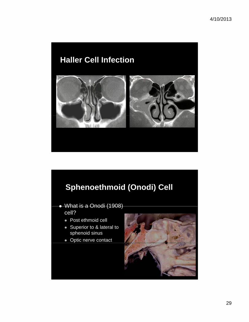
Haller Cell Infection
Sphenoethmoid (Onodi) Cell
What is a Onodi (1908) What is a Onodi (1908) cell? Post ethmoid cell
Superior to & lateral to sphenoid sinus
Optic nerve contact
4/10/2013
30
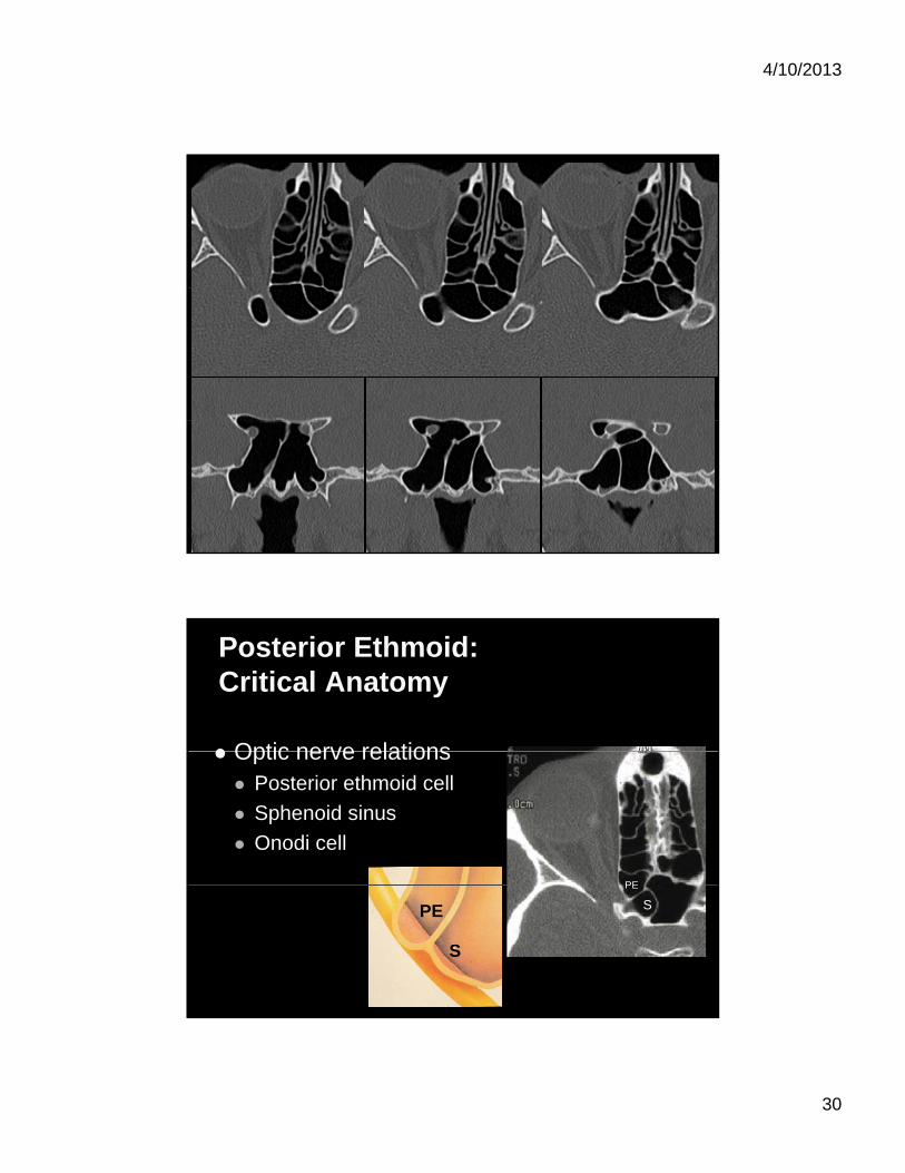
Posterior Ethmoid:Critical Anatomy
Optic nerve relations Optic nerve relations Posterior ethmoid cell
Sphenoid sinus
Onodi cell
PEPE
SPE
S
4/10/2013
31
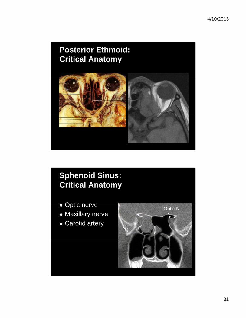
Posterior Ethmoid: Critical Anatomy
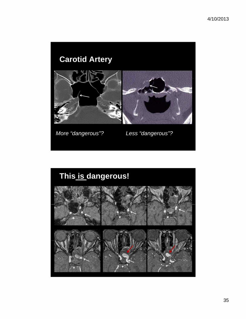
Sphenoid Sinus: Critical Anatomy
Optic nerve Optic nerve
Maxillary nerve
Carotid artery
Optic N
4/10/2013
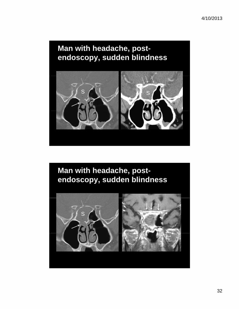
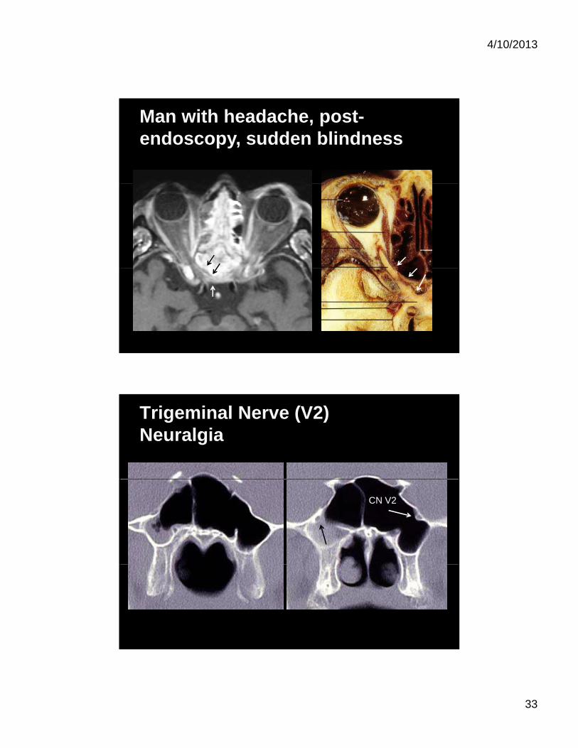
32
Man with headache, post-endoscopy, sudden blindness
SS
Man with headache, post-endoscopy, sudden blindness
S
4/10/2013
33
Man with headache, post-endoscopy, sudden blindness
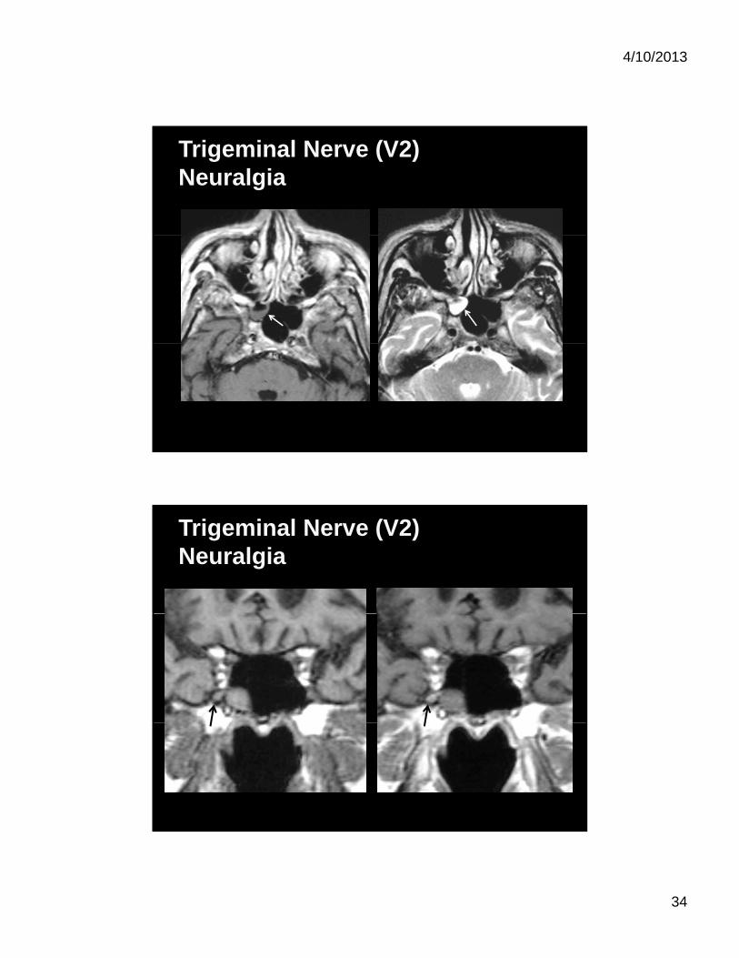
Trigeminal Nerve (V2) Neuralgia
CN V2
4/10/2013
34
Trigeminal Nerve (V2) Neuralgia
Trigeminal Nerve (V2) Neuralgia
4/10/2013
35
Carotid Artery
More “dangerous”? Less “dangerous”?
This is dangerous!
4/10/2013
36
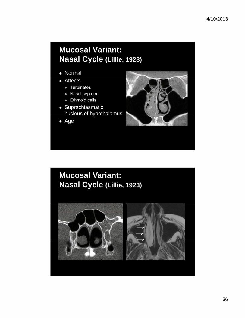
Mucosal Variant:Nasal Cycle (Lillie, 1923)
Normal
Affects Turbinates
Nasal septum
Ethmoid cells
Suprachiasmatic nucleus of hypothalamusnucleus of hypothalamus
Age
Mucosal Variant:Nasal Cycle (Lillie, 1923)
4/10/2013
37
4/10/2013
38
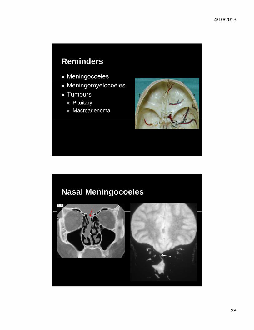
Reminders
Meningocoeles
Meningomyelocoeles
Tumours Pituitary
Macroadenoma
Nasal Meningocoeles
4/10/2013
39
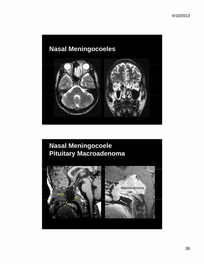
Nasal Meningocoeles
Nasal MeningocoelePituitary Macroadenoma
Macroadenoma

4/10/2013
40
Conclusion:Drainage System
Middle turbinate
Middle turbinate
Basal lamella
Conclusion:
Anterior Ostiomeatal complex
Middle meatus
Posterior Sphenoethmoidal
Superior meatus Superior meatus
4/10/2013
41
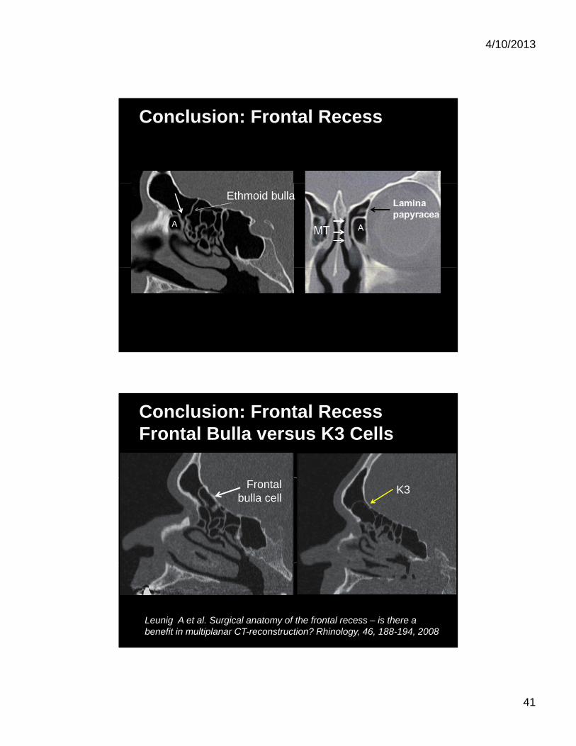
Conclusion: Frontal Recess
A
Ethmoid bulla
Conclusion: Frontal RecessFrontal Bulla versus K3 Cells
K3Frontal bulla cell
Leunig A et al. Surgical anatomy of the frontal recess – is there a benefit in multiplanar CT-reconstruction? Rhinology, 46, 188-194, 2008
4/10/2013
42
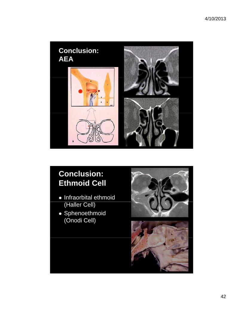
Conclusion: AEA
Conclusion: Ethmoid Cell
Infraorbital ethmoid (Haller Cell)
Sphenoethmoid (Onodi Cell)
4/10/2013
43
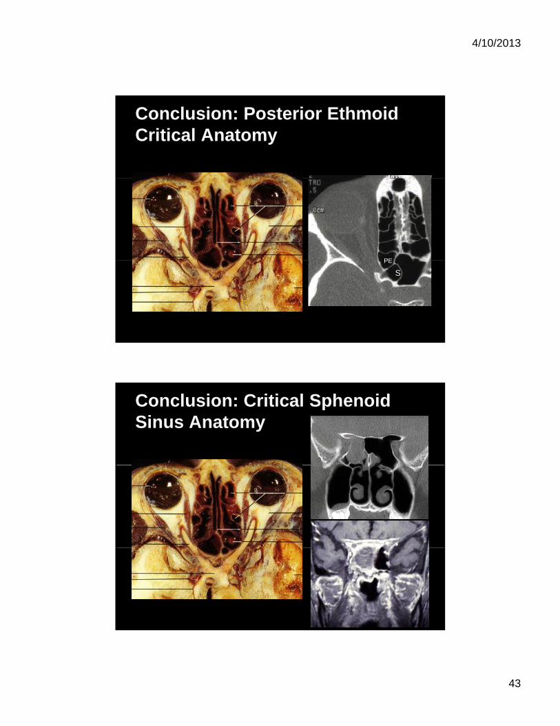
Conclusion: Posterior EthmoidCritical Anatomy
PEPE
S
Conclusion: Critical Sphenoid Sinus Anatomy
4/10/2013
44
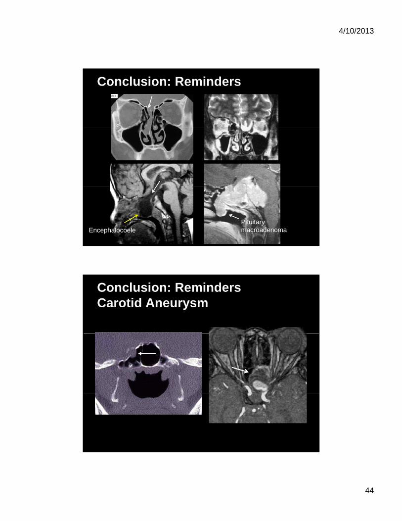
Conclusion: Reminders
EncephalocoelePituitary macroadenoma
Conclusion: RemindersCarotid Aneurysm