Embed Size (px)
Citation preview
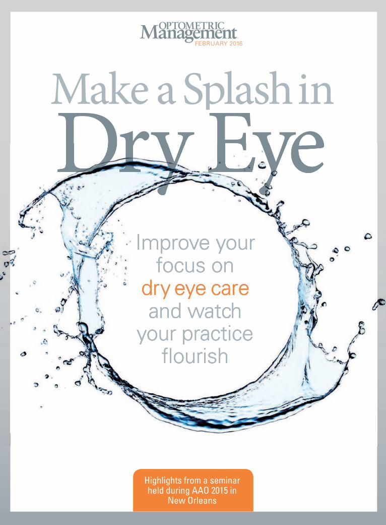
February 2016 FEBRUARY 2016®
Highlights from a seminar held during AAO 2015 in
New Orleans
Improve yourfocus on
dry eye careand watch
your practice �ourish
Make a Splash inDry Eye
Improve yourfocus on
dry eye careand watch
your practice �ourish
Dry Eye
FEBRUARY 2016 | 3
RESTASIS® (Cyclosporine Ophthalmic Emulsion) 0.05%BRIEF SUMMARY—PLEASE SEE THE RESTASIS® PACKAGE INSERT FOR FULL PRESCRIBING INFORMATION.INDICATION AND USAGERESTASIS® ophthalmic emulsion is indicated to increase tear production in patients whose tear production is presumed to be suppressed due to ocular infl ammation associated with keratoconjunctivitis sicca. Increased tear production was not seen in patients currently taking topical anti-infl ammatory drugs or using punctal plugs.CONTRAINDICATIONSRESTASIS® is contraindicated in patients with known or suspected hypersensitivity to any of the ingredients in the formulation.WARNINGS AND PRECAUTIONSPotential for Eye Injury and ContaminationTo avoid the potential for eye injury and contamination, be careful not to touch the vial tip to your eye or other surfaces.Use with Contact LensesRESTASIS® should not be administered while wearing contact lenses. Patients with decreased tear production typically should not wear contact lenses. If contact lenses are worn, they should be removed prior to the administration of the emulsion. Lenses may be reinserted 15 minutes following administration of RESTASIS® ophthalmic emulsion.ADVERSE REACTIONSClinical Trials ExperienceBecause clinical trials are conducted under widely varying conditions, adverse reaction rates observed in the clinical trials of a drug cannot be directly compared to rates in the clinical trials of another drug and may not refl ect the rates observed in practice.In clinical trials, the most common adverse reaction following the use of RESTASIS® was ocular burning (17%).Other reactions reported in 1% to 5% of patients included conjunctival hyperemia, discharge, epiphora, eye pain, foreign body sensation, pruritus, stinging, and visual disturbance (most often blurring).Post-marketing ExperienceThe following adverse reactions have been identifi ed during post approval use of RESTASIS®. Because these reactions are reported voluntarily from a population of uncertain size, it is not always possible to reliably estimate their frequency or establish a causal relationship to drug exposure.Reported reactions have included: hypersensitivity (including eye swelling, urticaria, rare cases of severe angioedema, face swelling, tongue swelling, pharyngeal edema, and dyspnea); and superfi cial injury of the eye (from the vial tip touching the eye during administration).USE IN SPECIFIC POPULATIONSPregnancyTeratogenic Effects: Pregnancy Category CAdverse effects were seen in reproduction studies in rats and rabbits only at dose levels toxic to dams. At toxic doses (rats at 30 mg/kg/day and rabbits at 100 mg/kg/day), cyclosporine oral solution, USP, was embryo- and fetotoxic as indicated by increased pre- and postnatal mortality and reduced fetal weight together with related skeletal retardations. These doses are 5,000 and 32,000 times greater (normalized to body surface area), respectively, than the daily human dose of one drop (approximately 28 mcL) of 0.05% RESTASIS® twice daily into each eye of a 60 kg person (0.001 mg/kg/day), assuming that the entire dose is absorbed. No evidence of embryofetal toxicity was observed in rats or rabbits receiving cyclosporine at oral doses up to 17 mg/kg/day or 30 mg/kg/day, respectively, during organogenesis. These doses in rats and rabbits are approximately 3,000 and 10,000 times greater (normalized to body surface area), respectively, than the daily human dose.Offspring of rats receiving a 45 mg/kg/day oral dose of cyclosporine from Day 15 of pregnancy until Day 21 postpartum, a maternally toxic level, exhibited an increase in postnatal mortality; this dose is 7,000 times greater than the daily human topical dose (0.001 mg/kg/day) normalized to body surface area assuming that the entire dose is absorbed. No adverse events were observed at oral doses up to 15 mg/kg/day (2,000 times greater than the daily human dose).There are no adequate and well-controlled studies of RESTASIS® in pregnant women. RESTASIS® should be administered to a pregnant woman only if clearly needed.Nursing MothersCyclosporine is known to be excreted in human milk following systemic administration, but excretion in human milk after topical treatment has not been investigated. Although blood concentrations are undetectable after topical administration of RESTASIS®
ophthalmic emulsion, caution should be exercised when RESTASIS® is administered to a nursing woman.Pediatric UseThe safety and effi cacy of RESTASIS® ophthalmic emulsion have not been established in pediatric patients below the age of 16.Geriatric UseNo overall difference in safety or effectiveness has been observed between elderly and younger patients.NONCLINICAL TOXICOLOGYCarcinogenesis, Mutagenesis, Impairment of FertilityCarcinogenesis: Systemic carcinogenicity studies were carried out in male and female mice and rats. In the 78-week oral (diet) mouse study, at doses of 1, 4, and 16 mg/kg/day, evidence of a statistically signifi cant trend was found for lymphocytic lymphomas in females, and the incidence of hepatocellular carcinomas in mid-dose males signifi cantly exceeded the control value.In the 24-month oral (diet) rat study, conducted at 0.5, 2, and 8 mg/kg/day, pancreatic islet cell adenomas signifi cantly exceeded the control rate in the low-dose level. The hepatocellular carcinomas and pancreatic islet cell adenomas were not dose related. The low doses in mice and rats are approximately 80 times greater (normalized to body surface area) than the daily human dose of one drop (approximately 28 mcL) of 0.05% RESTASIS® twice daily into each eye of a 60 kg person (0.001 mg/kg/day), assuming that the entire dose is absorbed.Mutagenesis: Cyclosporine has not been found to be mutagenic/genotoxic in the Ames Test, the V79-HGPRT Test, the micronucleus test in mice and Chinese hamsters, the chromosome-aberration tests in Chinese hamster bone-marrow, the mouse dominant lethal assay, and the DNA-repair test in sperm from treated mice. A study analyzing sister chromatid exchange (SCE) induction by cyclosporine using human lymphocytes in vitro gave indication of a positive effect (i.e., induction of SCE).Impairment of Fertility: No impairment in fertility was demonstrated in studies in male and female rats receiving oral doses of cyclosporine up to 15 mg/kg/day (approximately 2,000 times the human daily dose of 0.001 mg/kg/day normalized to body surface area) for 9 weeks (male) and 2 weeks (female) prior to mating.PATIENT COUNSELING INFORMATIONHandling the ContainerAdvise patients to not allow the tip of the vial to touch the eye or any surface, as this may contaminate the emulsion. To avoid the potential for injury to the eye, advise patients to not touch the vial tip to their eye.Use with Contact LensesRESTASIS® should not be administered while wearing contact lenses. Patients with decreased tear production typically should not wear contact lenses. Advise patients that if contact lenses are worn, they should be removed prior to the administration of the emulsion. Lenses may be reinserted 15 minutes following administration of RESTASIS® ophthalmic emulsion.AdministrationAdvise patients that the emulsion from one individual single-use vial is to be used immediately after opening for administration to one or both eyes, and the remaining contents should be discarded immediately after administration.Rx Only
Based on package insert 71876US18 © 2014 Allergan, Inc. Irvine, CA 92612, U.S.A. ® marks owned by Allergan, Inc. APC21XT14Patented. See www.allergan.com/products/patent_noticesMade in the U.S.A.
CHIEF OPTOMETRIC EDITOROptometric Management
Scot Morris, OD, FAAO
SPECIAL PROJECTSDIRECTOR | Angela JacksonMANAGER | Alicia Hoglund
EDITORIAL ASSISTANT | Rachel DanceCONTRIBUTING EDITOR | Desiree I�
DESIGN AND PRODUCTIONPRODUCTION DIRECTOR | Sandra KadenPRODUCTION MANAGER | Bill Hallman
ART DIRECTOR | Bill Pfa�
EDITORIAL AND PRODUCTION OFFICES321 Norristown Road, Suite 150
Ambler, PA 19002Phone: (215) 628-6550
BUSINESS STAFF PUBLISHER | Roger T. Zimmer
SALES | Jacqui DiBianca & Scott SchmidtPROMOTIONAL EVENTS MANAGER
Michelle Kie�er
Copyright 2016, PentaVision LLC.All Rights Reserved.
®
FEBRUARY 2016
Enhancing the Medical Services Side of Your Practice with Dry Eye CareFollow the “Four Ps” to successScot Morris, OD, FAAO
Diagnosing Dry EyeSimple and advanced tools help uncover the signs and symptoms of this common conditionMelissa Barnett, OD, FAAO, FSLS
Dry Eye TherapeuticsThe treatment approach that supercharged my outcomesArthur B. Epstein, OD, FAAO
The Medical Economics of Dry EyeCreate new revenue streams by doing more for your patientsDoug Devries, OD
Contents04
06
09
12
As eye doctors, our job is to solve vision and o c u l ar he a l t h problems. So, if we’re not diagnosing and
treating dry eye, we’re not doing our jobs to our fullest potential. Dry eye care is not only the key to healthier, happier patients, but also, through the provision of medical services, a financial driver for the practice. Approximately 20% of the people who walk through our practice doors have some form of ocular surface disease,1-3
so if we’re not making those medical diagnoses and billing for that medical care every day, we’ve got work to do.
The idea of shaping a practice to conform to a medical model can be intimidating. But, in reality, increasing the medical services portion of a practice by focusing on dry eye care is readily achievable. You don’t even need to attract more patients; you simply need to take better care of the patients you already have. So, how can you enhance the medical side of your practice? I’ve done it in mine, and coached many others through it, by using what I call the “Four Ps”: Plan, Procedure, Product, and Promotion.
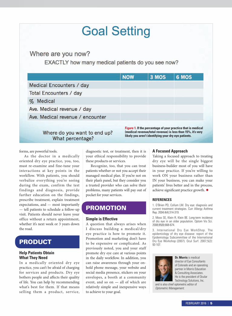
Set Goals, Track Progress�e �rst key to expanding the medical services portion of a practice through dry eye care is to know where you stand. Even if you see only one medical patient each day, that’s the basis for your next step, which is goal setting (Figure 1). You can start small, aiming to double your current daily number from one to two or four to eight, and by
asking every patient just one additional question (e.g., “Are your eyes always that red?”).
No matter what your �rst goals are, track your progress weekly or monthly. If you’re committed to making changes, even your �rst small steps fuel a self-ful�lling cycle that propels you toward improvement. The difference from a �nancial perspective will eventually be tens — if not hundreds — of thousands of dollars added to the bottom line of your practice.
Implementation of the PlanTo reach the goals you have set, you must establish procedures or processes that you and your staff will follow each day. And although your sta� has to accomplish these goals, the effort begins with you.
Think first about your clinical protocols. It helps to write them down. For example, how would you manage meibomian gland disease? What tests would you order? What treatments would you recommend? Next, think about all the different steps that take place from the time a patient enters the o�ce to the time he leaves. What occurs between the patient and the front desk, the technician and the doctor, the doctor and the patient, and everything in between? Each of those points in the work flow is an opportunity to identify the dry eye patients in your practice and educate them so they’ll know that you can take care of their red, uncomfortable, dry eyes and �uctuating vision.
For example, you might have the front desk person ask patients
whether their vision ever fluctuates and if blinking makes it better. Each patient could also be given a dry eye questionnaire. Then, have the technician collect the questionnaire and ask a follow-up question, such as “Do you ‘feel’ your eyes?” If the answer is yes, the technician can let the patient know that doesn’t need to be the case: “We can work on that.”
�ese types of changes in the work flow prompt patients to think about their symptoms, which they may not necessarily associate with problems. Being asked about and educated on their symptoms leads them to realize they need a solution, and makes them aware that you treat medical eye problems.
Once you find the points in the workflow where medically oriented interactions take place, you can work on fine-tuning them. Scripts work wonders for helping everyone in the practice talk with patients about dry eye and treatments in a consistent manner. You can write your own script or use staff and patient education materials created by the companies that have a vested interest in dry eye. Either way, practice using the scripts. You can role play, but there are other ways. I sometimes surreptitiously leave my phone at the front desk and hit record. My sta� does the same to me; then we listen and determine whether we’re all saying what we’re supposed to say.
In addition to de�ning sta� roles so they know what to do and providing the tools and training they need, it’s crucial that they understand WHY you want the practice to be more medically oriented, so they will be motivated to help achieve that goal.
Incentives, which can come in many
Enhance the Medical Side of Your Practice With Dry Eye Care
Follow the “Four Ps” to success
■ By Scot Morris, OD, FAAO
PLAN
PROCEDURE
4 | FEBRUARY 2016
forms, are powerful tools. As the doctor in a medically
oriented dry eye practice, you, too, must re-examine and fine-tune your interactions at key points in the workflow. With patients, you should verbalize everything you’re seeing during the exam, confirm the test f indings and diagnosis, provide further education on the findings, prescribe treatment, explain treatment expectations, and — most importantly — tell patients to schedule a follow-up visit. Patients should never leave your office without a return appointment, whether it’s next week or 3 years down the road.
Help Patients ObtainWhat They NeedIn a medically oriented dry eye practice, you can’t be afraid of charging for services and products. Dry eye bothers people and a�ects their quality of life. You can help by recommending what’s best for them. If that means selling them a product, service,
diagnostic test, or treatment, then it is your ethical responsibility to provide these products or services.
Recognize, too, that you can treat patients whether or not you accept their managed medical plan. If you’re not on their plan’s panel, but they consider you a trusted provider who can solve their problems, many patients will pay out of pocket for your services.
Simple is EffectiveA question that always arises when I discuss building a medical/dry eye practice is how to promote it. Promotion and marketing don’t have to be expensive or complicated. As previously noted, you and your staff promote dry eye care at various points in the daily work�ow. In addition, you can raise awareness through your on-hold phone message, your website and social media presence, stickers on your envelopes, a booth at a community event, and so on — all of which are relatively simple and inexpensive ways to achieve to your goal.
A Focused ApproachTaking a focused approach to treating dry eye will be the single biggest business-builder most of you will have in your practice. If you’re willing to work ON your business rather than IN your business, you can make your patients’ lives better and in the process, achieve signi�cant practice growth. •REFERENCES1. O’Brien PD, Collum LM. Dry eye: diagnosis and current treatment strategies. Curr Allergy Asthma Rep. 2004;4(4):314-319.
2. Moss SE, Klein R, Klein BE. Long-term incidence of dry eye in an older population. Optom Vis Sci. 2008;85(8):668-674.
3. International Dry Eye WorkShop. The epidemiology of dry eye disease: report of the Epidemiology Subcommittee of the International Dry Eye Workshop (2007). Ocul Surf. 2007;5(2):93-107.
Dr. Morris is medical director of Eye Consultants of Colorado and an operating partner in Morris Education & Consulting Associates. He is the president of Ocular Technology Solutions, Inc.
and is also chief optometric editor of Optometric Management.
PROMOTION
PRODUCT
Figure 1. If the percentage of your practice that is medical (medical revenue/total revenue) is less than 15%, it’s very likely you aren’t identifying your dry eye patients.
FEBRUARY 2016 | 5
In my practice at the University o f C a l i f o r n i a , D av i s , a substantial portion of my work involves ocular surface disease, dry eye and fitting specialty
contact lenses. I am extremely aware of the studies demonstrating that patients discontinue contact lens wear, most o�en due to discomfort, at a rate of 13% to 20%.1
As such, I look at treating dry eye and ocular surface disease not only as a way to keep their eyes healthy, but also to keep them in their contact lenses, which, in turn, leads to satisfied patients and contributes to practice revenue.
To identify patients who need dry eye treatment, I use all the available tests and tools — from simple to advanced — an overview of which follows.
Patient Education & QuestionnairesToo o�en, patients aren’t forthcoming
about their symptoms, which is why patient education and clear commun- ication are critical to uncovering problems. In fact, informative bro- chures and questionnaires are the first step of diagnosing dry eye. I’m a firm believer in having informative brochures in the reception area to help start the dry eye conversation. I always have them available, along with a questionnaire, such as the SPEED or OSDI. The paper questionnaires work for me, but for practices that have digital devices for patients in the waiting room, an OSDI app is available.
The questionnaire sets the stage for my second step: asking questions. Many patients are clearly symptomatic, reporting dry, watery, burning eyes, and so on. But others say their eyes feel fine. I always ask those patients with “fine” eyes if they experience blurry or interrupted vision, and more often than not, they say yes. Typically, these patients are the ones
whose vision improves to 20/20 if I instill an arti�cial tear or ask them to blink repeatedly behind the phoropter. In my experience, it is this group of patients in particular who can be kept in contact lenses longer as a result of diagnosing and treating dry eye at an earlier stage.
In addition to asking patients about symptoms, it’s important to ask environmental and lifestyle questions. We know the myriad contributors to dry eye, but it’s important that we convey that knowledge to our patients. For example, I talk with patients about aspects of their home and work environments that may contribute to their symptoms. In some cases, simple changes, such as turning o� the ceiling fan, help significantly. And although patients may not be able to discontinue systemic medications that dry their eyes, they may be able to switch from oral allergy medications to ocular allergy drops or nasal sprays.
Clinical ExaminationWhen I examine a patient, I evaluate:• Adnexa (dermatological
in�ammation, dermatochalasis, rosacea)
• Eyelids and eyelid margins (infectious, in�ammatory, allergic, physiologic [lagophthalmos], blepharitis, meibomian gland dysfunction [MGD], lid-wiper epitheliopathy, giant papillary conjunctivitis)
• Conjunctiva (staining, chemosis, conjuctivochalasis)
• Cornea (topographical, hypoxia, secondary infectious/in�ammatory, dystrophy).A growing amount of research
suggests that MGD is the most frequent cause of dry eye.2 �erefore, it is important to identify MGD or
Diagnosing Dry EyeSimple and advanced tools help uncover the signs and
symptoms of this common condition
■ By Melissa Barnett, OD, FAAO, FSLS
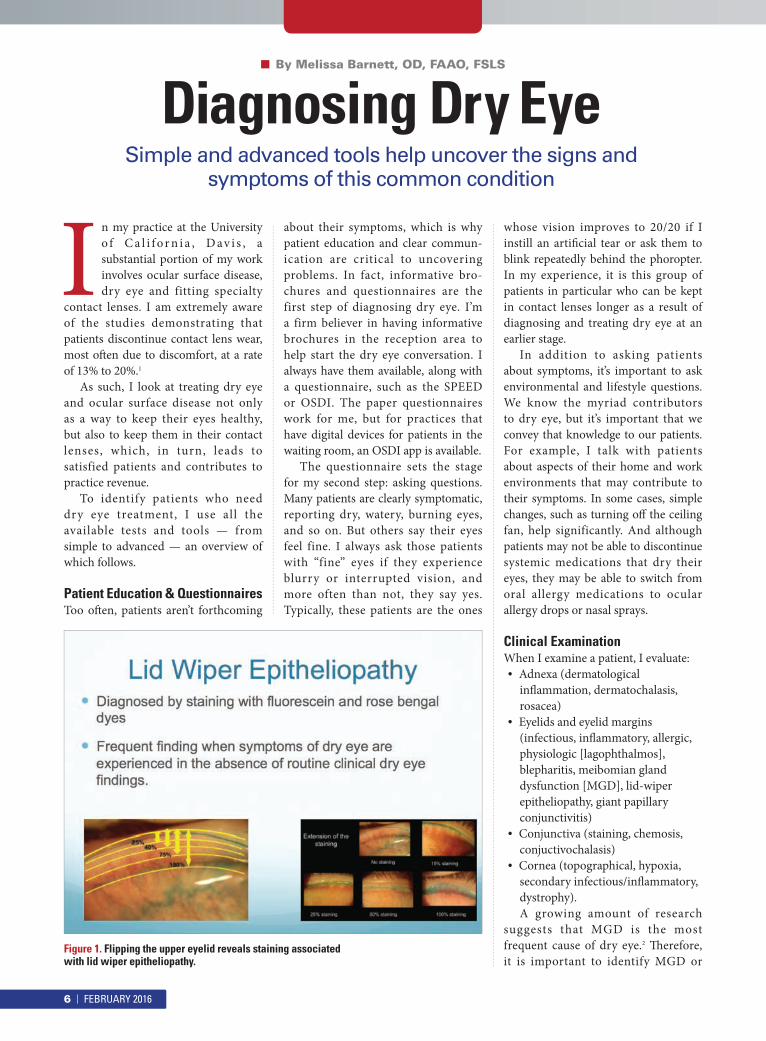
Figure 1. Flipping the upper eyelid reveals staining associated with lid wiper epitheliopathy.
6 | FEBRUARY 2016
rule it out. Non-obvious MGD (NOMGD) may be present, so I always push the lower eyelid gently to express the glands, which often reveals a problem — even in otherwise normal-appearing eyelids.
Clinical Testing■ Schirmer I and II: I sometimes
use the Schirmer test to evaluate aqueous tear production, especially for patients referred by rheumatologists because they usual ly request a Schirmer’s test. In theory, Schirmer I, performed with anesthetic, evaluates baseline secretion, while Schirmer II, performed without anesthetic, measures baseline plus re�ex secretion. More than 10 mm of moisture on the filter paper after 5 minutes is considered a normal test result.
■ C orneal and conjunctival staining: I always have my patients remove their contact lenses so I can use a staining dye. Fluorescein will stain to reveal defects in the corneal and conjunctival epithelium. Rose bengal will stain dead conjunctival cel ls or cel ls unprotected by a normal mucin layer. It also stains the conjunctiva more than the cornea.
The degree of staining correlates well with the degree of aqueous tear de�ciency, tear breakup time (TBUT), and reduced mucus production by conjunctival goblet cell and non-goblet epithelial cells. Lissamine green works by the same mechanism as rose bengal but tends to be less irritating for the patient.
If I see staining in a patient who wants to wear contact lenses for the first time, I treat the ocular surface first, then schedule the contact lens fitting for a later date, especially if multifocal lenses are the goal.
Another helpful aspect of staining in contact lens patients is that it reveals lid wiper epitheliopathy (LWE).3
(�e “lid wiper” is the portion of the upper eyelid marginal conjunctiva that sweeps the ocular surface during blinking.) LWE is a frequent finding when patients have dry eye symptoms without accompanying dry eye signs (Figure 1).
■ Tear breakup time: TBUT is useful to evaluate at every visit when examining the ocular surface. It correlates with both aqueous and evaporative tear de�ciency.
Although TBUT has been criticized for a lack of repeatabi l ity and standardization, I �nd it very useful for monitoring visit-to-visit improvement in a way that is i l lustrative for patients. A TBUT of less than 10 seconds is abnormal, indicating tear film instability. A TBUT of less than 5 seconds is closely associated with dry eye symptoms. It is important to keep in mind that anesthesia decreases TBUT, and �uorescein can destabilize the tear �lm.
■ Point-of-care testing: Point-of-care testing is something many optometrists, including myself, find helpful for making dry eye diagnosis more accurate and e�cient. Two such tests are In�ammaDry (RPS) and the TearLab Osmolarity System.
Inf lammaDr y is based on a quantifiable value of the amount of mat r ix met a l loprote inas e-9 (MMP-9) in the tears. MMPs are proteolytic enzymes produced by stressed epithelial cells on the ocular surface, and MMP-9 is a marker for in�ammation. �e test has been shown to signi�cantly and positively correlate with corneal fluorescein staining scores and abnormal superficial corneal epithelia as seen with confocal microscopy.4 Results are obtained in 10 minutes and easy to interpret. A red line is positive (>40 ng/ml of MMP-9) and a blue line is negative.
The TearLab osmolarity test is similarly quick and easy to use, requiring only nanoliter volumes of tear fluid. It has been shown to be a solid metric for diagnosing and classifying dry eye disease.5 Osmolarity values above 308 mOsms/L are indicative of dry eye. Because the results are a quantitative numerical value, this test is helpful for engaging patients in their care as we work toward improvement.
■ Anterior segment imaging: Recent advances in anterior segment imaging devices, such as corneal
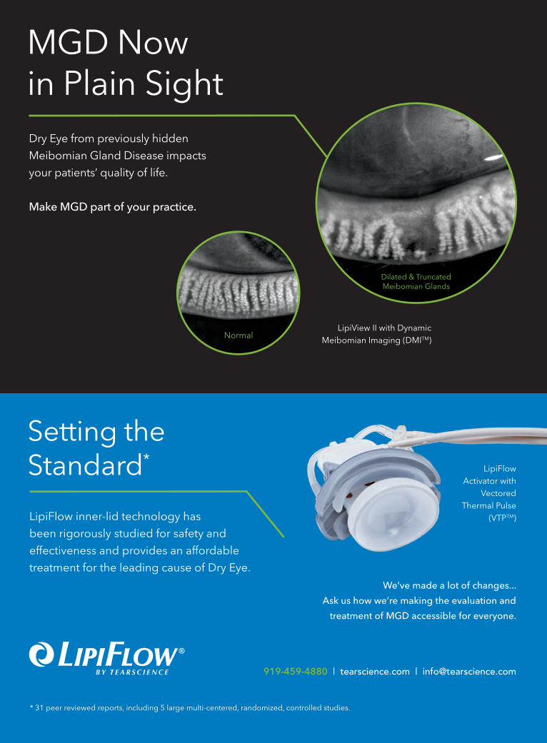
topographers and tomographers, have included ocular surface capabilities, such as tear meniscus assessment. In my practice, I use a Pentacam (Oculus) to evaluate tear �lm regularity. Other available devices include the LipiView II interferometer (TearScience), which measures lipid layer thickness, evaluates blink rate, and enables visualization of meibomian gland structure to aid in earlier detection of MGD. The Medmont topographer is also able to evaluate the tear �lm.
The More Data the BetterDry eye a�ects many patients, not just contact lens wearers. Today’s varied diagnostic tools provide valuable information and, when coupled with an open patient dialogue, will not on ly i mprove d i a g n o s t i c an d treatment abilities, but help boost your bottom line, creating a win-win for everyone involved. •REFERENCES1. Dumbleton K, Caffery B, Dogru M, et al. The TFOS International Workshop on Contact Lens Discomfort: report of the subcommittee on epidemiology. Invest Ophthalmol Vis Sci. 2013;54(11):TFOS20-36.
2. Nichols KK, Foulks GN, Bron AJ, et al. The international workshop on meibomian gland dysfunction: executive summary. Invest Ophthalmol Vis Sci. 2011;52(4):1922-1929.
3. Korb DR, Herman JP, Greiner JV, et al. Lid wiper epitheliopathy and dry eye symptoms. Eye Contact Lens. 2005;31(1):2-8.
4. Chotikavanich S, de Paiva CS, Li DeQ, et al . Production and activity of matr ix metalloproteinase-9 on the ocular surface increase in dysfunctional tear syndrome. Invest Ophthalmol Vis Sci. 2009; 50(7):3203-3209.
5. Lemp MA, Bron AJ, Baudouin C, et al. Tear osmolarity in the diagnosis and management of dry eye disease. Am J Ophthalmol. 2011;151(5):792-798.
Dr. Barnett is a principal optometrist at the University of California, Davis Eye Center in Sacramento, where she specializes in anterior segment disease and specialty contact lenses. She is a Fellow of the
American Academy of Optometry, a Diplomate of the American Board of Certi�cation in Medical Optometry (ABCMO) and serves on the Board of Women of Vision (WOV), Gas Permeable Lens Institute (GPLI), Ocular Surface Society of Optometry (OSSO) and The Scleral Lens Education Society (SLS).
Another helpful aspect of staining in contact lens patients is that it reveals lid wiper epitheliopathy (LWE).3
(�e “lid wiper” is the portion of the upper eyelid marginal conjunctiva that sweeps the ocular surface during blinking.) LWE is a frequent finding when patients have dry eye symptoms without accompanying dry eye signs
optometrists, including myself, find helpful for making dry eye diagnosis more accurate and e�cient. Two such tests are In�ammaDry (RPS) and the
Inf lammaDr y is based on a quantifiable value of the amount of mat r ix met a l loprote inas e-9 (MMP-9) in the tears. MMPs are proteolytic enzymes produced by stressed epithelial cells on the ocular surface, and MMP-9 is a marker for in�ammation. �e test has been shown to signi�cantly and positively correlate with corneal fluorescein staining scores and abnormal superficial corneal epithelia as seen with confocal microscopy.4 Results are obtained in 10 minutes and easy to interpret. A red line is positive (>40 ng/ml of MMP-9) and a blue line is negative.
The TearLab osmolarity test is similarly quick and easy to use, requiring only nanoliter volumes of tear fluid. It has been shown to be a solid metric for diagnosing and classifying dry eye disease.5 Osmolarity values above 308 mOsms/L are indicative of dry eye. Because the results are a quantitative numerical value, this test is helpful for engaging patients in their care as we work toward improvement.
■ Anterior segment imaging: Recent advances in anterior segment imaging devices, such as corneal
contact lens wearers. Today’s varied diagnostic tools provide valuable information and, when coupled with an open patient dialogue, will not on ly i mprove d i a g n o s t i c an d treatment abilities, but help boost your bottom line, creating a win-win for everyone involved. •REFERENCES1. Dumbleton K, Caffery B, Dogru M, et al. The TFOS International Workshop on Contact Lens Discomfort: report of the subcommittee on epidemiology. Invest Ophthalmol Vis Sci. 2013;54(11):TFOS20-36.
2. Nichols KK, Foulks GN, Bron AJ, et al. The international workshop on meibomian gland dysfunction: executive summary. Invest Ophthalmol Vis Sci. 2011;52(4):1922-1929.
3. Korb DR, Herman JP, Greiner JV, et al. Lid wiper epitheliopathy and dry eye symptoms. Eye Contact Lens. 2005;31(1):2-8.
4. Chotikavanich S, de Paiva CS, Li DeQ, et al . Production and activity of matr ix metalloproteinase-9 on the ocular surface increase in dysfunctional tear syndrome. Invest Ophthalmol Vis Sci. 2009; 50(7):3203-3209.
5. Lemp MA, Bron AJ, Baudouin C, et al. Tear osmolarity in the diagnosis and management of dry eye disease. Am J Ophthalmol. 2011;151(5):792-798.
Dr. Barnett is a principal optometrist at the University of California, Davis Eye Center in Sacramento, where she specializes in anterior segment disease and specialty contact lenses. She is a Fellow of the
American Academy of Optometry, a Diplomate of the American Board of Certi�cation in Medical Optometry (ABCMO) and serves on the Board of Women of Vision (WOV), Gas Permeable Lens Institute (GPLI), Ocular Surface Society of Optometry (OSSO) and The Scleral Lens Education Society (SLS).
FEBRUARY 2016 | 7

12 | FEBRUARY 2016
DRY EYE NEVER
FELT SO GOOD.
Artifcial tears aren’t always enough
to alleviate the symptoms of dry eye. Give
your patients a new perspective with
Bio-Tissue’s family of options for facilitating
THE HAPPY EYE COMPANY
quality healing on the ocular surface in
mild to severe cases. Go beyond hydration
to rejuvenation — and see dry eye from
a whole new point of view.
8305 NW 27th Street, Suite 101, Doral, FL 33122 • 888.296.8858 • www.biotissue.com • Bio-Tissue, Inc. 2015. All rights reserved.
For more information please visit:
www.biotissue.com/dryeyeinfo
Approximately 3 years a g o, a f t e r h av i n g pr a c t i c e d i n Ne w York for many years, I relocated to Phoenix
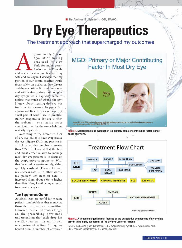
and opened a new practice with my wife and colleague. I decided that my portion of our dream practice would focus solely on ocular surface disease and dry eye. We built it and they came, and with a steady stream of complex dry eye patients, I quickly came to realize that much of what I thought I knew about treating dry eye was fundamentally wrong. In particular, aqueous-deficient dry eye is only a small part of what I see in practice. Rather, evaporative dry eye is often the problem — or at least a major contributor — for the overwhelming majority of patients.
According to the literature, 86% of dry eye patients have evaporative dry eye (Figure 1)1. In my practice in arid Arizona, that number is greater than 90%. I’ve learned that the best and most effective way to manage most dry eye patients is to focus on the evaporative components. With that in mind, a treatment algorithm quickly evolved (Figure 2), and my success rate — in other words, my patient sat isfact ion rate — increased from about 65% to higher than 90%. Here, I outline my essential treatment strategies.
Tear Supplement ChoiceArtificial tears are useful for keeping patients comfortable as they’re moving through the treatment algorithm. However, their effectiveness hinges on the prescr ibing physic ian’s understanding that each drop has specific characteristics and its own mechanism of action. Today, we benefit from a number of advanced
Dry Eye TherapeuticsThe treatment approach that supercharged my outcomes
■ By Arthur B. Epstein, OD, FAAO
Figure 2. A treatment algorithm that focuses on the evaporative components of dry eye has proven to be highly successful at The Dry Eye Center of Arizona.
[MGD = meibomian gland dysfunction; EDE = evaporative dry eye; HOCL = hypochlorous acid; BCL = bandage contact lens; ADE = allergic dry eye]
Figure 1. Meibomian gland dysfunction is a primary or major contributing factor in most cases of dry eye.
FEBRUARY 2016 | 9
formulations. Since most patients have an evaporative component, I usually prescribe a lipid-based product such as Refresh Optive Advanced (Allergan; carboxymethylcellulose [CMC] 0.5% glycerin 1%, polysorbate 80 0.5%), which functionally targets all three layers of the tear �lm.
Understanding how each drop is designed to work allows me to choose the option that best matches each patient’s needs.
Manage Lid FloraWhen p at ients have marg ina l meibomian gland function, stagnant meibum on the lid surfaces creates an environment conducive to bacterial overpopulation — specifically staph species. Staph adhere to the dictum of survival of the �ttest and, as a result, produce pro-in�ammatory exotoxins. Staph additionally elaborates a variety of enzymes such as lipase, which facilitate penetration and infection. Lipase also breaks down the tear lipid layer, causing saponification which destabilizes the tear �lm.
To reduce bacterial overpopulation, inactivate the toxins, and block the enzymes, I prescr ibe pure hypochlorous acid (HOCL), a naturally occurring substance produced by white blood cells to �ght microbial invaders. Pure HOCL is found only in Avenova (NovaBay) and has been shown to have fast-acting, broad-spectrum
activity against overpopulation of microorganisms of the external ocular flora. Avenova is well tolerated and easy for patients to use. They simply spray it on a cotton round or oval; then wipe the upper and lower eyelids.
Although Avenova is effective as part of the regimen for managing blepharitis and meibomian gland dysfunction (MGD), regular use also can prevent these problems from developing by controlling bacterial overpopulation. When I find significant demodex infestation, I recommend Cliradex (Bio-Tissue) moist towelettes for home therapy for 6 to 8 weeks to eradicate the mites.
Nutritional SupportNutritional support is crucial for MGD and dry eye patients. A number of good products are available, including HydroEye (ScienceBased Health). HydroEye contains gamma linolenic acid (GLA) from black currant seed oil, plus the omega-3 fatty acids EPA and DHA from high quality fish oil, and other important nutrients. In a randomized, controlled t r ia l , HydroEye was shown to provide significant dry eye relief, suppress markers of ocular surface inflammation, and maintain corneal surface smoothness.2 Triglyceride-based pure omega-3 products have also been found to be helpful in managing dry eye and MGD.
Address In�ammationNearly every dry eye has concomitant in f l ammat ion , w hich must b e managed. Cyclosporine (Restasis, Allergan) is a time-tested option for addressing in�ammation, and we have other options, too. In my practice, I frequently prescribe topical steroids, and I use doxycycline when I want to ramp up therapy quickly.
Manage ExposureAbout 20% of my patients who are s ignif icant ly symptomatic have nighttime exposure. I can see evidence of this by either direct examination or by observing light from between the lids when I get down beneath the patient and shine a transilluminator on the upper lid sulcus. Rather than asking these patients to use an ocular ointment at night, which can be an exit ramp from therapy, I recommend they wear a silicone eye shield (Onyix, Eye Eco) while they sleep. This creates a moisture barrier and the patients o�en wake without their usual symptoms.
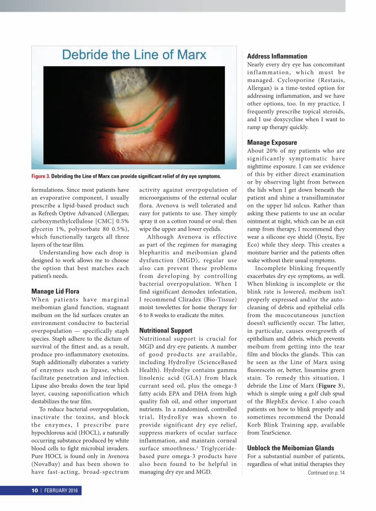
Incomplete blinking frequently exacerbates dry eye symptoms, as well. When blinking is incomplete or the blink rate is lowered, meibum isn’t properly expressed and/or the auto-cleaning of debris and epithelial cells from the mucocutaneous junction doesn’t sufficiently occur. The latter, in particular, causes overgrowth of epithelium and debris, which prevents meibum from getting into the tear film and blocks the glands. This can be seen as the Line of Marx using �uorescein or, better, lissamine green stain. To remedy this situation, I debride the Line of Marx (Figure 3), which is simple using a golf club spud of the BlephEx device. I also coach patients on how to blink properly and sometimes recommend the Donald Korb Blink Training app, available from TearScience.
Unblock the Meibomian GlandsFor a substantial number of patients, regardless of what initial therapies they
Figure 3. Debriding the Line of Marx can provide signi�cant relief of dry eye symptoms.
Continued on p. 14
10 | FEBRUARY 2016
LipiView II with Dynamic
Meibomian Imaging (DMITM)Normal
MGD Now in Plain Sight
Dry Eye from previously hidden
Meibomian Gland Disease impacts
your patients’ quality of life.
Make MGD part of your practice.
Setting the Standard*
LipiFlow inner-lid technology has
been rigorously studied for safety and
effectiveness and provides an affordable
treatment for the leading cause of Dry Eye.
LipiFlow
Activator with
Vectored
Thermal Pulse
(VTPTM)
We’ve made a lot of changes...
Ask us how we’re making the evaluation and
treatment of MGD accessible for everyone.
919-459-4880 | tearscience.com | [email protected]
* 31 peer reviewed reports, including 5 large multi-centered, randomized, controlled studies.
Dilated & Truncated Meibomian Glands
There are many good re a s ons t o d e ve l op a s t r o n g d r y e y e component within your optometric practice: your
patients’ well-being, their perception of you and your practice, the prevention of contact lens dropout, and the referrals that come from treating this chronic, progressive disease. Don’t forget, too, that it can help with the �nancial health of your practice, which is under attack from many angles, including online dispensaries and decreasing vision plan reimbursement.
You really only have two choices for protecting your revenue stream. You can either see more patients or you can do more for the patients you already have.
Dry eye is a great opportunity to do more — to provide much-needed
services for your existing patients. Millions of people su�er from dry eye, including millions of baby boomers, a large percentage of whom are post-menopausal females, who are now joining Medicare, and the majority of eye exams are performed by optometrists. All of this points to the fact that dry eye patients are already in your practice.
In our practice, we’ve created several dry eye-related revenue streams to help us treat our patients, and you can do the same.
Testing and Documentation�e availability of point-of-care testing for dry eye is a positive development. Two notable testing options have been making their way into an increasing number of practices: the TearLab Osmolarity System and In�ammaDry
(RPS). Technicians can perform either of these tests e�ciently as part of the patient workup, and both companies help with training, practice logistics, marketing, and every other aspect of adoption.
For Medicare patients, testing with In�ammaDry (CPT code 83516) and the TearLab Osmolarity System (CPT code 83861), both considered in vitro laboratory devices, is billed under the Clinical Laboratory Fee Schedule, not the Physician Fee Schedule. �erefore, Medicare patient co-payments or deductibles don’t apply; the service is 100% reimbursed. (Coding requirements for commercial carriers may vary.) Optometrists should embrace point-of-care testing as a means to achieve more accurate diagnoses. Not doing so might mean exclusion from insurance panels. Both In�ammaDry and the osmolarity test require a practice to have a Clinical Laboratory Improvement Amendments (CLIA) waiver, which isn’t di�cult to obtain in the vast majority of states.
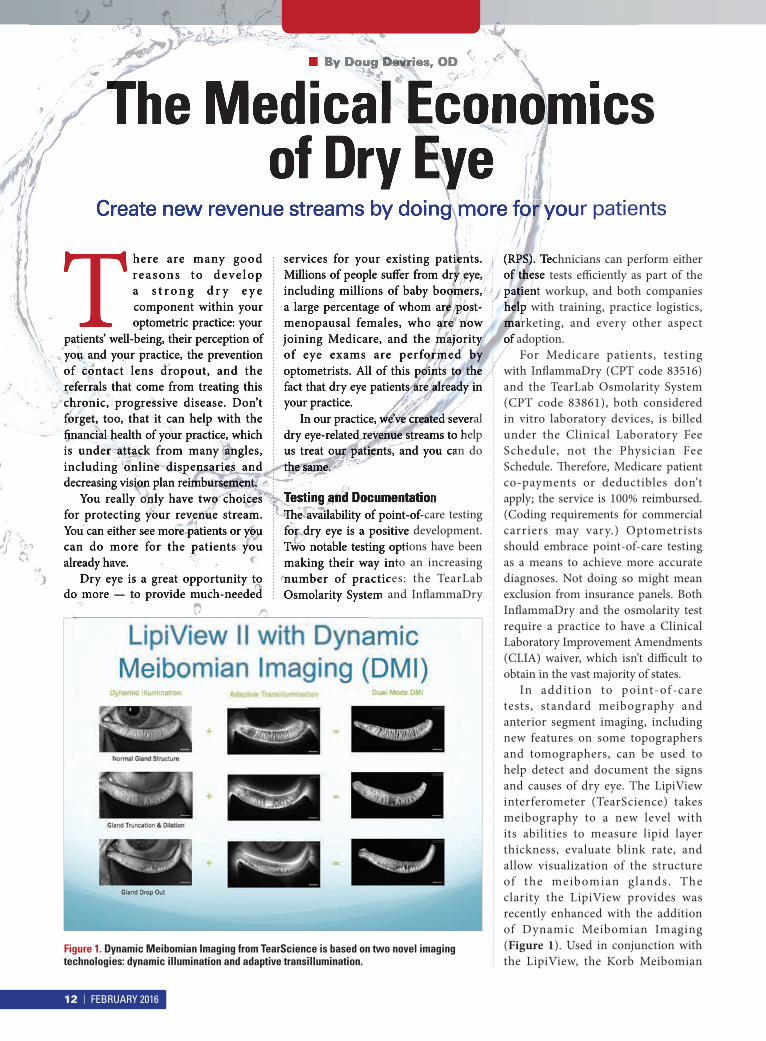
In addit ion to point-of-care tests, standard meibography and anterior segment imaging, including new features on some topographers and tomographers, can be used to help detect and document the signs and causes of dry eye. The LipiView interferometer (TearScience) takes meibography to a new level with its abilities to measure lipid layer thickness, evaluate blink rate, and allow visualization of the structure of the meibomian glands. The clarity the LipiView provides was recently enhanced with the addition of Dynamic Meibomian Imaging (Figure 1). Used in conjunction with the LipiView, the Korb Meibomian
The Medical Economics of Dry Eye
Create new revenue streams by doing more for your patients
■ By Doug Devries, OD
Figure 1. Dynamic Meibomian Imaging from TearScience is based on two novel imaging technologies: dynamic illumination and adaptive transillumination.
There are many good re a s ons t o d e ve l op a s t r o n g d r y e y e component within your optometric practice: your
patients’ well-being, their perception of you and your practice, the prevention of contact lens dropout, and the referrals that come from treating this chronic, progressive disease. Don’t forget, too, that it can help with the �nancial health of your practice, which is under attack from many angles, including online dispensaries and decreasing vision plan reimbursement.
You really only have two choices for protecting your revenue stream. You can either see more patients or you can do more for the patients you already have.
Dry eye is a great opportunity to do more — to provide much-needed
services for your existing patients. Millions of people su�er from dry eye, including millions of baby boomers, a large percentage of whom are post-menopausal females, who are now joining Medicare, and the majority of eye exams are performed by optometrists. All of this points to the fact that dry eye patients are already in your practice.
In our practice, we’ve created several dry eye-related revenue streams to help us treat our patients, and you can do the same.
Testing and Documentation�e availability of point-of-care testing for dry eye is a positive development. Two notable testing options have been making their way into an increasing number of practices: the TearLab Osmolarity System and In�ammaDry
(RPS). Technicians can perform either of these tests e�ciently as part of the patient workup, and both companies help with training, practice logistics, marketing, and every other aspect of adoption.
The Medical Economicsof Dry Eye
Create new revenue streams by doing more for your patients
■ By Doug Devries, OD
12 | FEBRUARY 2016
Gland Evaluator (TearScience) enables standardized, repeatable evaluation of meibomian gland function at the slit lamp.
All of the tests related to dry eye and meibomian gland disease (MGD) are revenue-generators for a practice, some more so than others. They fuel the dry eye segment of your practice by allowing you to uncover more ocular surface disease, schedule more patient visits, and provide more treatments.
Re-Appointment and TreatmentScheduling dr y eye treatments separate from routine exams and dry eye testing — for example, re-appointing patients for subsequent visits — is key to ensuring you’re being paid appropriately for your services. Re-appointments are a staple in most medical care models, in which patients are rarely, if ever, diagnosed and treated on the same day. The subsequent appointments for patients you’re diagnosing and treating for dry eye can be level II, III or IV encounters, depending on the extent of the history, exam, and medical decision-making.
The dry eye treatment armam- entarium has been expanding. In addition to treatments that have been used for years, such as manual meibomian gland expression and punctal occlusion, we can also make use of newer in-office options that take into account the connections between dry eye and lid disease, MGD, and demodex. These include the microblepharoexfoliation BlephEx treatment (Rysurg), the LipiFlow t h e r m a l p u l s a t i o n t r e a t m e n t (TearScience), and the Cliradex Complete eyelid- and eyelash-cleansing treatment (Bio-Tissue). For patients with advanced, chronic, or recurring ocular surface disease, the Prokera biologic corneal bandage (Bio-Tissue), a self-retained, cryopreserved amniotic tissue, can be used to reduce in�ammation and promote healing.
In our practice, we also sell a variety of ancillary products to help patients manage their signs and symptoms, such as eyelid cleansing pads, Cliradex
moist towelettes warm and cool compress gear, moisture chambers, ar t i f ic ia l tears , and HydroEye nutritional supplements (ScienceBased Health). Although our original intent was to bolster patient compliance by making carefully chosen products easily available, we learned that the merchandising adds signi�cantly to our pro�t margin for dry eye-related care.
Real-world NumbersI �nd that most of my dry eye patient visits qualify as level III encounters for insurance purposes. In my state of Nevada, the payment for a level III visit ranges from $74.09-$88.63. And, depending on where a patient is in his treatment plan and what we’re doing at a given visit, I may be billing the third-party carrier or the patient for any of the following in addition to the visit itself (ranges encompass Medicare and private insurers):• Punctal occlusion OU
($231.24-$264.21)• Anterior segment photos
($17.35-$57.78)• Prokera
($1,489.02-$2,532.51)• Osmolarity testing ($12.57-$23.47)• In�ammaDry ($10.57-$19.42)• LipiView ($65-$150)• LipiFlow OU ($950-$2,000) NOTE:
TearScience recently reduced the
price of its single-use activators, and I recently lowered my charge to patients given that I perform a high volume of treatments.)
• Manual meibomian gland expression ($125-$300)
• BlephEx ($150-$250)• Cliradex Complete Demodex
treatment ($125-$200)• Nutritional supplements ($395.40,
or $161 net for a year’s supply)• Lid scrubs, artificial tears, eye
masks, and so on. ($196 per patient net in a year)
These are a few actual examples from my practice, which illustrate the services a patient received and the revenue they generated:■ 4 visits, arti�cial tears, eyelid scrubs,
diagnostics (net $577)■ 4 visits, punctal plugs, artificial
tears, eyelid scrubs, diagnostics (net $768)
■ 4 visits, punctal plugs, artificial tears, eyelid scrubs, diagnostics, BlephEx (net $948)
■ 6 visits, punctal plugs, artificial tears, eyelid scrubs, diagnostics, BlephEx, LipiView, LipiFlow (net $1,556)
■ 6 visits, punctal plugs, artificial tears, eyelid scrubs, diagnostics, BlephEx, LipiView, LipiFlow, 1 Prokera (net $2,464)
■ 6 visits, punctal plugs, artificial tears, eyelid scrubs, diagnostics, BlephEx, LipiView, LipiFlow, 2 Prokera (net $3,272)
Creating ValueAs you can see, the value we can provide to our patients by utilizing the latest dry eye tests and treatments to diagnose and manage them is matched by its significant contribution to the practice bottom line. •
Dr. Devries is a co-founder of Eye Care Associates of Nevada, a statewide medical/surgical practice, where he is the clinical director and director of the Optometric Residency Program.
“You really only have two choices for protecting your revenue stream.
You can either see more patients or you can do
more for the patients you already have. Dry eye is a great opportunity to do
more — to provide much-needed services for your
existing patients.”
FEBRUARY 2016 | 13
receive, MGD is chronic, progressive, and obstructive. For this group, expression of the glands is necessary. Manual expression can be e�ective for some, but it usually needs to be done frequently, which isn’t pleasant for the patient or the doctor. Alternatively, many of my patients opt for LipiFlow. �e in-o�ce LipiFlow treatment uses precisely controlled thermal pulsation applied to the eyelids to unblock the meibomian glands and encourage the natural, normal production of lipids for the tear film. In clinical studies, 79% of patients reported improvement, ranging from 10% to 100%, of their overall dry eye symptoms within 4 weeks of a treatment.3,4
The results my LipiFlow patients achieve range from signi�cant to life-changing. My dry eye practice would be incomplete without it.
Amniotic MembraneFor a pat ient whose cornea is s ignif icant ly compromised due to chronic dry eye and related conditions, amniotic membrane (AM) is a game-changing solution. Prokera (Bio-Tissue) is a proven option that addresses even severe disease. In Prokera, the cryopreserved amniotic membrane wraps around a polycarbonate ring, allowing easy in-office placement onto the cornea. The proprietary cryopreservation method used for Prokera ensures that the HC-HA/PTX3 biologic signaling matrix, growth factors, and proteins responsible for AM’s healing and anti-inflammatory properties remain intact. Prokera is uniquely positioned for possessing both protective and regenerative properties. In some cases, I may use Prokera to heal a patient’s cornea before I begin to address the underlying dry eye issues.
The Science and the ArtWhile my streamlined dr y eye algorithm reflects the treatments I’ve found to be most successful, it re�ects the art in addition to the science. First, I choose natural treatments whenever possible. Pure hypochlorous
acid, nutritional support, amniotic membrane — all occur naturally. Second, I look for synergy. For example, when I prescribe Restasis, I also prescribe a steroid to rapidly ramp up anti-inflammatory activity. I also use doxycycline as an initiator for nutritional supplementation, which may take several weeks to begin to take effect. I believe that looking at the situation holistically has been very helpful for my patients.
Also, I aim to manage my MGD patients much like dentists manage their patients. Since �uoride came into the picture decades ago, they shifted from filling cavities to concentrating on oral health and cosmesis. As a result, dental patients are programmed to have their teeth cleaned every 6 months. They dutifully make those appointments, usually before leaving their current appointment. Similarly, if I have my patients return at regular intervals to address their ocular health issues, I can prevent progressive, chronic MGD from leading to dry eye. That, along with making key products available for sale in the o�ce that bring real bene�t to my patients, is the best way to provide them with what they need while helping to maintain practice viability. •REFERENCES1. Lemp MA, Crews LA, Bron AJ, Foulks GN, Sullivan BD. Distribution of aqueous-de�cient and evaporative dry eye in a clinic-based patient cohort: a retrospective study. Cornea. 2012;31(5):472-478.
2. Sheppard JD, Singh R, McClellan AJ, et al. Long-term supplementation with n-6 and n-3 PUFAs improves moderate-to-severe keratoconjunctivitis sicca: A randomized double-blind clinical trial. Cornea. 2013;32(10):1297-1304.
3. Lane SS, DuBiner HB, Epstein RJ, et al. A new system, the LipiFlow, for the treatment of meibomian gland dysfunction. Cornea. 2012;31(4):396-404.
4. TearScience. Data on �le.
Dr. Epstein is a co-founder of Phoenix Eye Care and The Dry Eye Center of Arizona, where he serves as director of Cornea/ External Disease, Clinical Research and The Dry Eye & Ocular Surface Disease Center.
Continued from p. 10
WHAT HAVEYOU READ
TODAY?
PowerfulInformation
and Real-WorldSolutions
for Vision Care
Professionals
clspectrum.com
eyecarebusiness.com
optometricmanagement.com
ophthalmologymanagement.com
retinalphysician.com
ophthalmicprofessional.com
All Pubs Third_BCI All Pubs Ad FULL.qxd 2/7/14 3:

AV E N O VA . C O M |
� � ��� ���� ��� |
R X O N LY
Daily lid and lash hygiene.
Add Avenova® with NeutroxTM
(Pure Hypochlorous Acid 0.01% as a
preservative), the only Rx lid hygiene
product with pure hypochlorous acid,
to the lid & lash hygiene regimen
for all your patients.
HWbWc�@^eM3Mh�Mc�E64A��O^^cV�����


![Dr.Aghaie-management of dry eye [Read-Only]erc.mums.ac.ir/images/erc/eyecenter/Dr.aghaei.lacri.pdf · Dry Eye : treatment Dry eye /ocular surface disorder is : progressive , life-long](https://img.pdfslide.us/doc/110x75/5c5f186309d3f2515c8d2852/draghaie-management-of-dry-eye-read-onlyercmumsacirimageserceyecenterdr.jpg)