Embed Size (px)
Citation preview
FDG-PET/CT PATTERNS AND FDG-PET/CT PATTERNS AND PREVALENCE OF PERITONEAL PREVALENCE OF PERITONEAL SPREAD IN OVARIAN CANCERSPREAD IN OVARIAN CANCER
Srour SFSrour SF11, Bar-Shalom R, Bar-Shalom R22
11Department of Diagnostic ImagingDepartment of Diagnostic Imaging22Institute of Nuclear MedicineInstitute of Nuclear Medicine
Rambam Health Care CampusRambam Health Care Campus Haifa, IsraelHaifa, Israel
BackgroundBackground
Ovarian cancer (O.C) is the second most common, and the most Ovarian cancer (O.C) is the second most common, and the most common cause for cancer-related death among gynecological common cause for cancer-related death among gynecological tumorstumors
It is responsible for more than half of gynecological mortalityIt is responsible for more than half of gynecological mortality
Most cases revealed at a late stageMost cases revealed at a late stage
The most common secondary spread is to peritoneum and The most common secondary spread is to peritoneum and retroperitoneal L.N.retroperitoneal L.N.
Peritoneal spread (p.s) changes the stage of tumor and affects Peritoneal spread (p.s) changes the stage of tumor and affects treatment strategy and prognosistreatment strategy and prognosis
Imaging MethodsImaging Methods
Conventional imaging methods (US, CT, MRI) are limited Conventional imaging methods (US, CT, MRI) are limited for detecting peritoneal spreadfor detecting peritoneal spread
CT sensitivity for p.s.: 17-54%, depends onCT sensitivity for p.s.: 17-54%, depends on
size, place, morphology, ascitic fluid, diminished size, place, morphology, ascitic fluid, diminished abd. fat volume, and bowel distention with contrastabd. fat volume, and bowel distention with contrast
PET/CT imaging for peritoneal spread is not well PET/CT imaging for peritoneal spread is not well established, but recent studies are encouragingestablished, but recent studies are encouraging
PET/CT patterns of p.s. are variable and not clearly PET/CT patterns of p.s. are variable and not clearly recognizedrecognized

PurposePurpose
To describe and characterize the To describe and characterize the incidence and patterns of peritoneal incidence and patterns of peritoneal
spread of ovarian cancer as spread of ovarian cancer as demonstrated by PET/CT demonstrated by PET/CT
examinationexamination
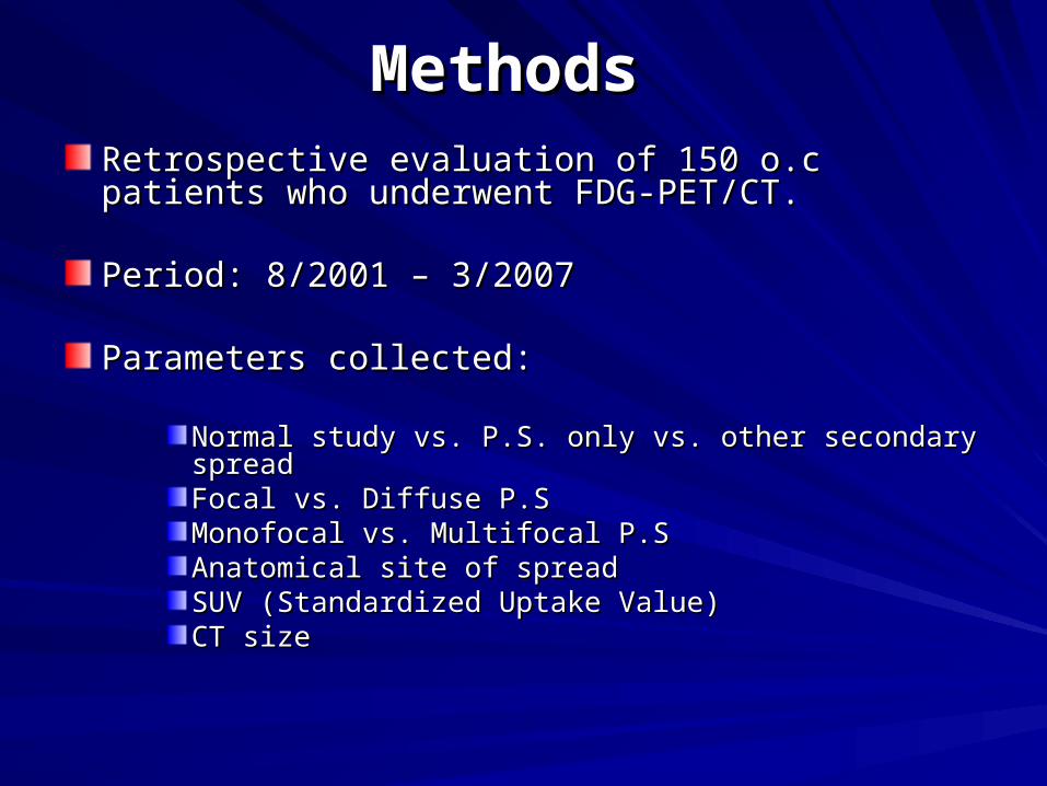
MethodsMethods Retrospective evaluation of 150 o.c patients who Retrospective evaluation of 150 o.c patients who underwent FDG-PET/CT.underwent FDG-PET/CT.
Period: 8/2001 – 3/2007Period: 8/2001 – 3/2007
Parameters collected:Parameters collected:
Normal study vs. P.S. only vs. other secondary spread Normal study vs. P.S. only vs. other secondary spread Focal vs. Diffuse P.SFocal vs. Diffuse P.SMonofocal vs. Multifocal P.SMonofocal vs. Multifocal P.SAnatomical site of spreadAnatomical site of spreadSUV (Standardized Uptake Value)SUV (Standardized Uptake Value)CT sizeCT size
MethodsMethods
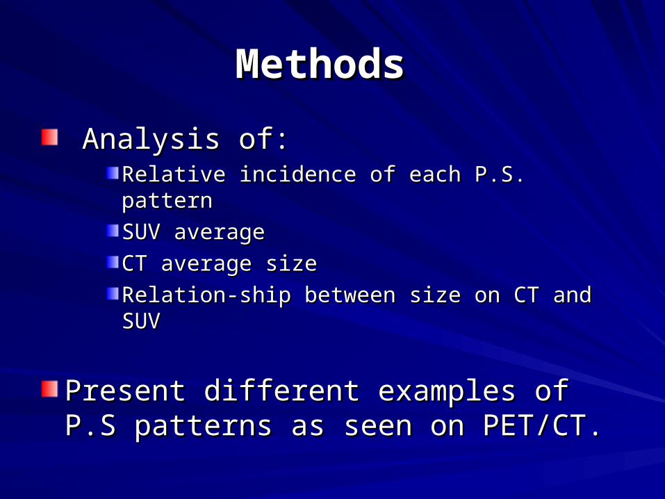
Analysis of:Analysis of:Relative incidence of each P.S. patternRelative incidence of each P.S. pattern
SUV averageSUV average
CT average sizeCT average size
Relation-ship between size on CT and SUVRelation-ship between size on CT and SUV
Present different examples of P.S patterns Present different examples of P.S patterns as seen on PET/CT.as seen on PET/CT.
ResultsResults
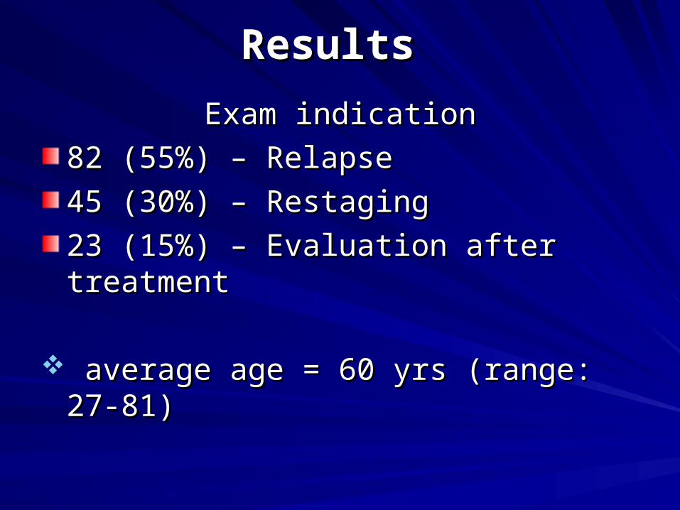
Exam indicationExam indication
82 (55%) – Relapse82 (55%) – Relapse
45 (30%) – Restaging45 (30%) – Restaging
23 (15%) – Evaluation after treatment23 (15%) – Evaluation after treatment
average age = 60 yrs (range: 27-81)average age = 60 yrs (range: 27-81)
Incidence of P.S. PatternsIncidence of P.S. Patterns
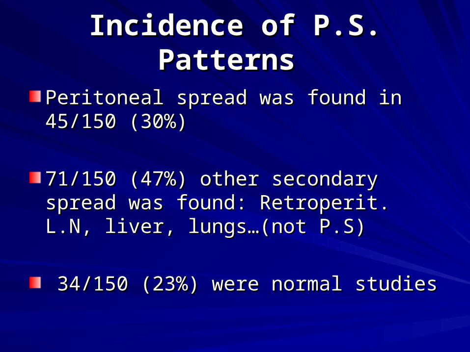
Peritoneal spread was found in 45/150 Peritoneal spread was found in 45/150 (30%)(30%)
71/150 (47%) other secondary spread was 71/150 (47%) other secondary spread was found: Retroperit. L.N, liver, lungs…(not found: Retroperit. L.N, liver, lungs…(not P.S)P.S)
34/150 (23%) were normal studies34/150 (23%) were normal studies
Peritoneal SpreadPeritoneal Spread
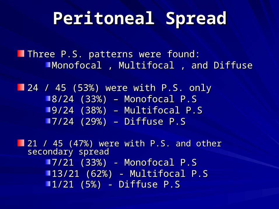
Three P.S. patterns were found:Three P.S. patterns were found:Monofocal , Multifocal , and DiffuseMonofocal , Multifocal , and Diffuse
24 / 45 (53%) were with P.S. only24 / 45 (53%) were with P.S. only
8/24 (33%) – Monofocal P.S8/24 (33%) – Monofocal P.S9/24 (38%) – Multifocal P.S9/24 (38%) – Multifocal P.S7/24 (29%) – Diffuse P.S 7/24 (29%) – Diffuse P.S
21 / 45 (47%) were with P.S. and other secondary spread21 / 45 (47%) were with P.S. and other secondary spread7/21 (33%) - Monofocal P.S7/21 (33%) - Monofocal P.S13/21 (62%) - Multifocal P.S13/21 (62%) - Multifocal P.S1/21 (5%) - Diffuse P.S1/21 (5%) - Diffuse P.S
Peritoneal SpreadPeritoneal Spread
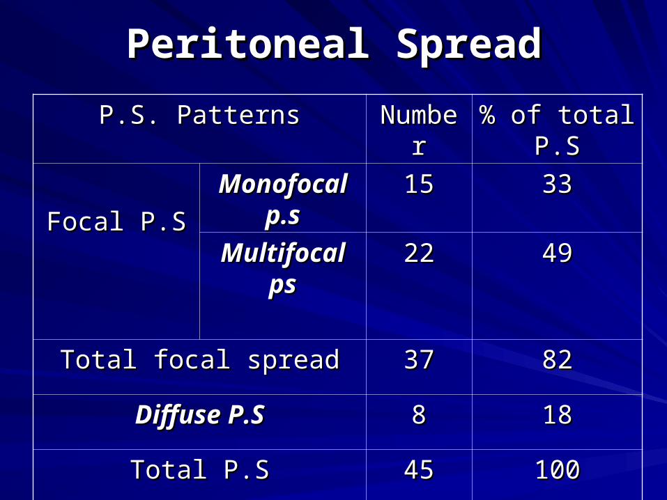
P.S. PatternsP.S. Patterns NumberNumber % of total P.S% of total P.S
Focal P.SFocal P.S
Monofocal Monofocal p.sp.s
1515 3333
Multifocal Multifocal psps
2222 4949
Total focal spreadTotal focal spread 3737 8282
Diffuse P.SDiffuse P.S 88 1818
Total P.STotal P.S 4545 100100
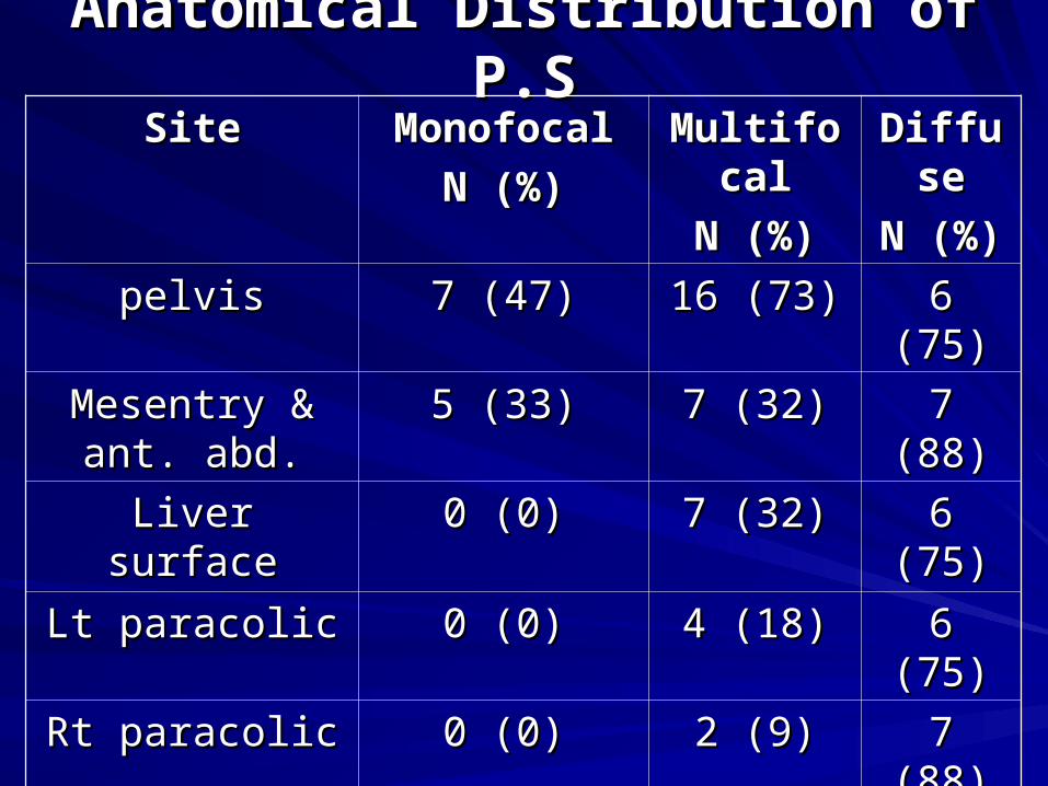
Anatomical Distribution of P.SAnatomical Distribution of P.S
SiteSite MonofocalMonofocal
N (%)N (%)
MultifocalMultifocal
N (%)N (%)
DiffuseDiffuse
N (%)N (%)
pelvispelvis 7 (47)7 (47) 16 (73)16 (73) 6 (75)6 (75)
Mesentry & ant. Mesentry & ant. abd.abd.
5 (33)5 (33) 7 (32)7 (32) 7 (88)7 (88)
Liver surfaceLiver surface 0 (0)0 (0) 7 (32)7 (32) 6 (75)6 (75)
Lt paracolicLt paracolic 0 (0)0 (0) 4 (18)4 (18) 6 (75)6 (75)
Rt paracolicRt paracolic 0 (0)0 (0) 2 (9)2 (9) 7 (88)7 (88)
Inf. Abd.Inf. Abd. 3 (20)3 (20) 0 (0)0 (0) 5 (63)5 (63)
totaltotal 15 (100)15 (100) 22 (100)22 (100) 8 (100)8 (100)
Most Monofocal spread was to pelvis, Most Monofocal spread was to pelvis, mesentry and ant. abd.mesentry and ant. abd.
Most multifocal spread was to pelvis, Most multifocal spread was to pelvis, mesentry and ant. abd.and liver surfacemesentry and ant. abd.and liver surface
Diffuse spread was to all the anatomical Diffuse spread was to all the anatomical sites in the same incidence because of its sites in the same incidence because of its widespread nature.widespread nature.
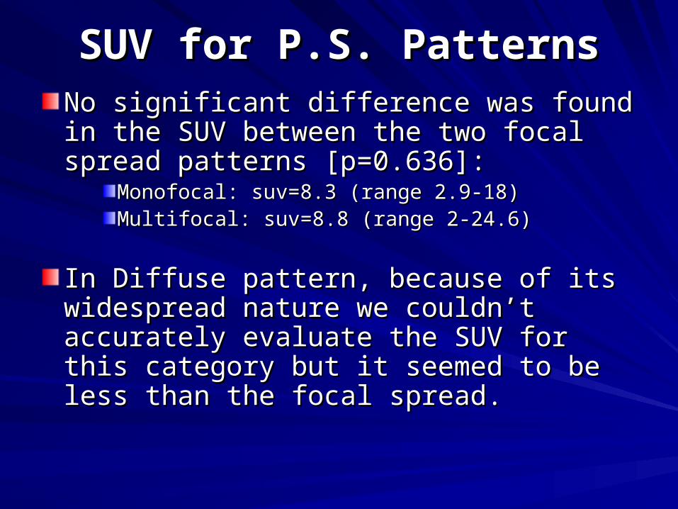
SUV for P.S. PatternsSUV for P.S. PatternsNo significant difference was found in the No significant difference was found in the SUV between the two focal spread SUV between the two focal spread patterns [p=0.636]:patterns [p=0.636]:
Monofocal: suv=8.3 (range 2.9-18) Monofocal: suv=8.3 (range 2.9-18) Multifocal: suv=8.8 (range 2-24.6)Multifocal: suv=8.8 (range 2-24.6)
In Diffuse pattern, because of its In Diffuse pattern, because of its widespread nature we couldn’t accurately widespread nature we couldn’t accurately evaluate the SUV for this category but it evaluate the SUV for this category but it seemed to be less than the focal spread.seemed to be less than the focal spread.

CT size for P.S. PatternsCT size for P.S. Patterns
Average size on CT in the monofocal spread Average size on CT in the monofocal spread was: 1.7cm (range:0.9-3cm)was: 1.7cm (range:0.9-3cm)
Average size on CT in the Multifocal spread Average size on CT in the Multifocal spread was: 2cm (range: 1.1-3.7cm)was: 2cm (range: 1.1-3.7cm)
We found significant relationship between SUV We found significant relationship between SUV of peritoneal spread and average size on CT in of peritoneal spread and average size on CT in the focal spread patterns [p=0.0001-0.004]:the focal spread patterns [p=0.0001-0.004]:
Big size on CT was related to high SUVBig size on CT was related to high SUV
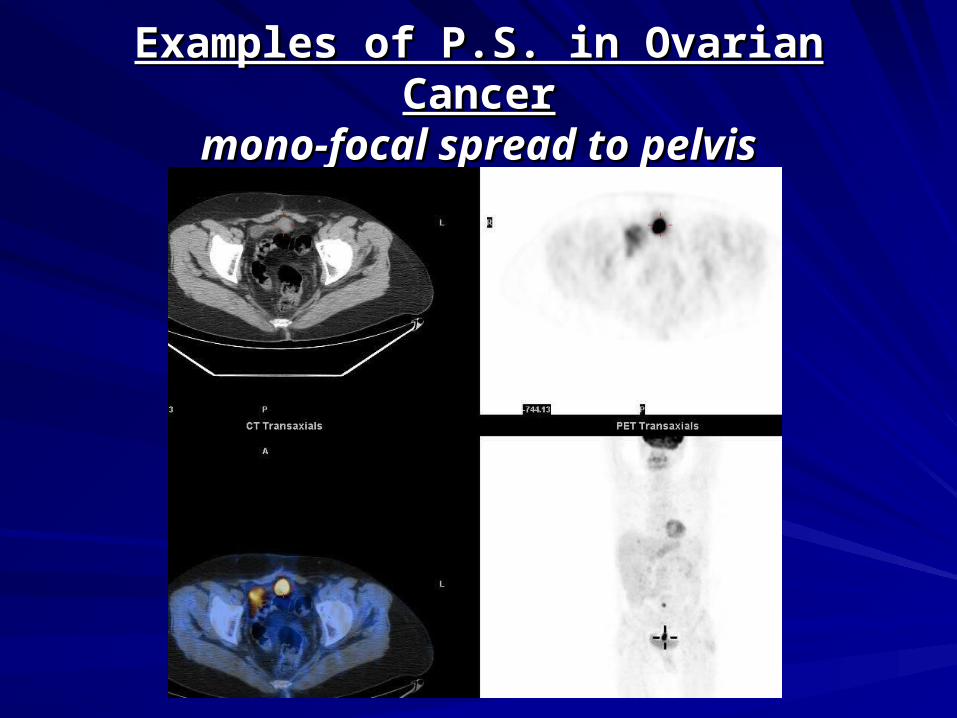
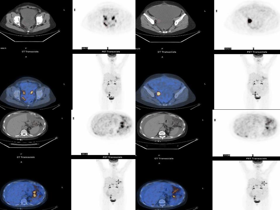
Examples of P.S. in Ovarian CancerExamples of P.S. in Ovarian Cancermono-focal spread to pelvismono-focal spread to pelvis
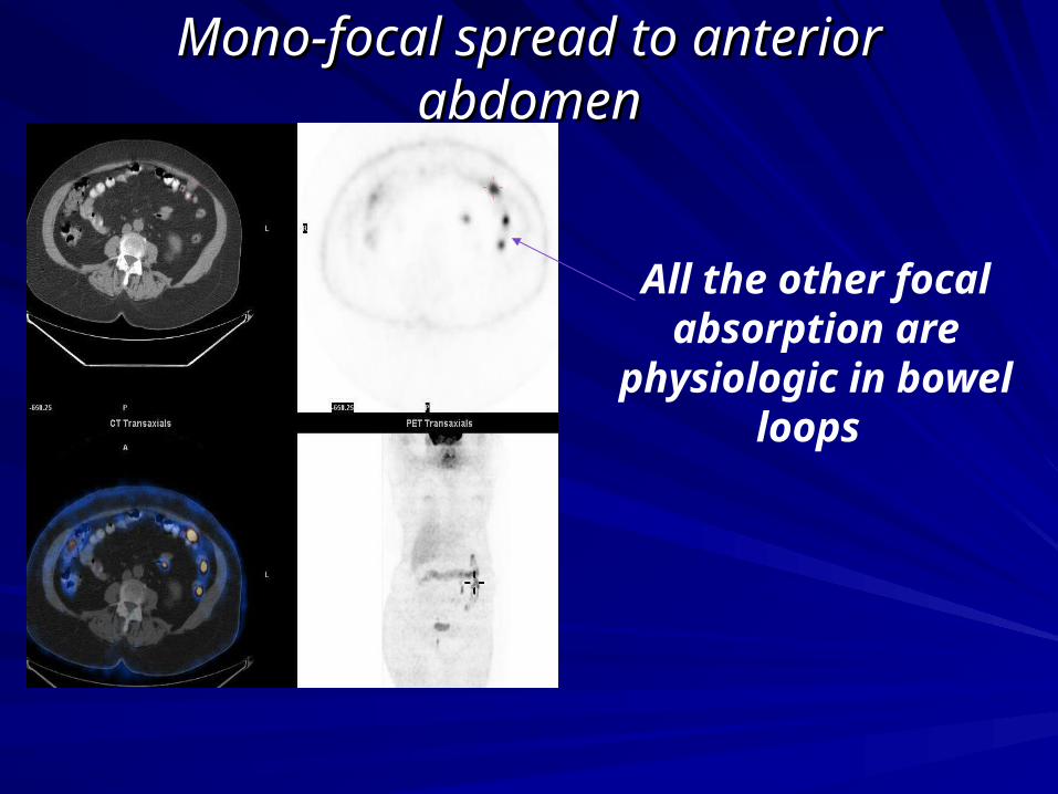
Mono-focal spread to anterior abdomenMono-focal spread to anterior abdomen
All the other focal absorption are physiologic in bowel loops
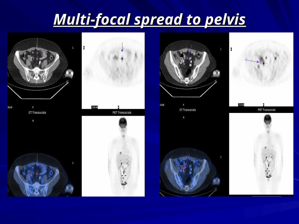
Multi-focal spread to pelvisMulti-focal spread to pelvis
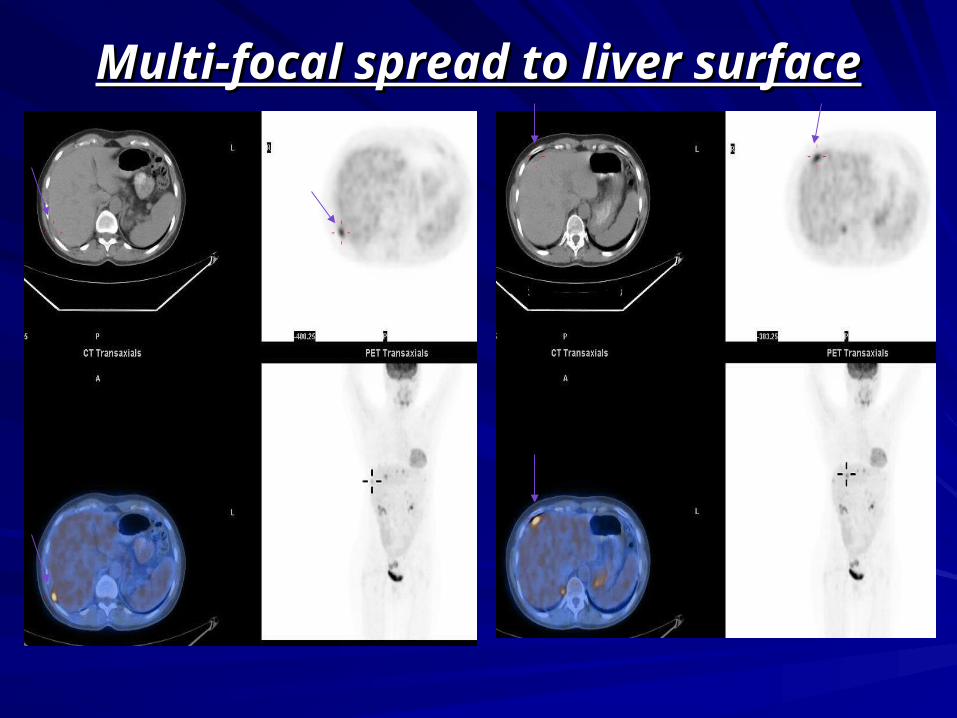
Multi-focal spread to liver surfaceMulti-focal spread to liver surface
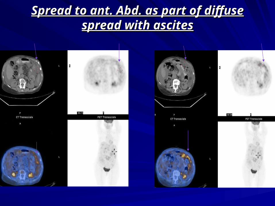
Spread to ant. Abd. as part of diffuse Spread to ant. Abd. as part of diffuse spread with ascitesspread with ascites
Diffuse spread with ascitesDiffuse spread with ascites
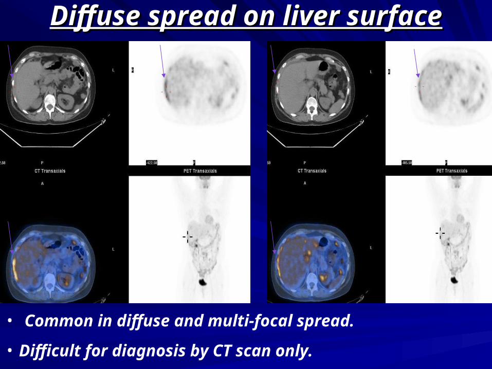
Diffuse spread on liver surfaceDiffuse spread on liver surface
• Common in diffuse and multi-focal spread.
• Difficult for diagnosis by CT scan only.
ConclusionConclusion
Peritoneal spread in ovarian cancer is Peritoneal spread in ovarian cancer is common (30% in our study)common (30% in our study)
As FDG-PET/CT is being an important As FDG-PET/CT is being an important tool for assessing these patients, tool for assessing these patients, familiarity with the variable peritoneal familiarity with the variable peritoneal spread patterns on PET/CT is important spread patterns on PET/CT is important for accurate assessment of disease for accurate assessment of disease status of ovarian cancer patientsstatus of ovarian cancer patients