Embed Size (px)
Citation preview

1
Fatty Alcohols for Wax Esters in Marinobacter aquaeolei VT8: Two Optional 1
Routes in the Wax Biosynthesis Pathway 2
Eric M. Lennemana,b, Janet M. Ohlerta, Nagendra P. Palania and Brett M. Barneya,b,* 3
aDepartment of Bioproducts and Biosystems Engineering and bBiotechnology Institute, 4
University of Minnesota, St. Paul, MN 55108 5
6
Running title: Fatty Alcohol Biosynthesis in Marinobacter 7
Key Words: wax ester, fatty aldehyde reductase, fatty acyl-CoA reductase, fatty acyl-CoA, fatty 8
alcohol 9
*Corresponding author: Brett M. Barney, Department of Bioproducts and Biosystems 10
Engineering, University of Minnesota, 1390 Eckles Avenue, St. Paul, Minnesota, 44108-6130, 11
Ph. 612-626-8751, Fax 612-625-6286, email: [email protected] 12
AEM Accepts, published online ahead of print on 6 September 2013Appl. Environ. Microbiol. doi:10.1128/AEM.02420-13Copyright © 2013, American Society for Microbiology. All Rights Reserved.
on April 4, 2019 by guest
http://aem.asm
.org/D
ownloaded from

2
Abstract 13
The biosynthesis of wax esters in bacteria is accomplished by a unique pathway that 14
combines a fatty alcohol and a fatty acyl-CoA substrate. Previous in vitro enzymatic studies 15
indicate that two different enzymes could be involved in the synthesis of the required fatty 16
alcohol in Marinobacter aquaeolei VT8. In this study, we demonstrate through a series of gene 17
deletions and transcriptional analysis that either enzyme is capable of fulfilling the role of 18
providing the fatty alcohol required for wax ester biosynthesis in vivo, but evolution has clearly 19
selected one of these, a previously characterized fatty aldehyde reductase, as the preferred 20
enzyme to perform this reaction under typical wax ester accumulating conditions. These results 21
complement previous in vitro studies and provide the first glimpse of the role of each enzyme in 22
vivo in the native organism. 23
on April 4, 2019 by guest
http://aem.asm
.org/D
ownloaded from

3
The global cycle of oil is of interest from both the standpoint of energy and the environment, 24
as efforts by humankind to obtain this valuable resource can result in substantial releases of 25
crude oil through incidents such as the Deepwater Horizon oil spill of 2010 in the Gulf of 26
Mexico. It is also noted that crude oil from natural deposits is routinely released into aqueous 27
environments such as the oceans by natural processes where geological reserves meet surface 28
waters. These environments have allowed natural populations of organisms, such as marine 29
bacteria, to evolve to utilize these supplies, rich in reduced carbon, for use as a biological fuel 30
source. A primary focus related to oil degrading marine bacteria is the oxidation of these oils to 31
meet energy requirements of the living cell. Interestingly, for certain marine bacteria found to 32
utilize and degrade oils, these bacteria are also capable of producing natural lipids which have 33
economic values similar to those obtained from harvesting sperm whales prior to the late 20th 34
century, even when grown on simple organic acids or carbohydrates. We have selected the 35
marine bacterium Marinobacter aquaeolei VT8, which was isolated from an oil well off the 36
coast of Vietnam (1), as a model bacterial species to study metabolic processes in an oil 37
metabolizing and neutral lipid accumulating bacteria. In addition to growing on long chain 38
hydrocarbons, M. aquaeolei VT8 also produces a natural hydrocarbon, the wax ester, when 39
grown on simple citric acid cycle intermediates such as succinate or citrate as the sole carbon 40
source (1-3), indicating that all of the precursors required for the biosynthesis of wax ester are 41
indigenous to this strain. 42
Biosynthesis of wax esters is accomplished by the combination of several different enzymes. 43
The wax ester synthase / acyl-coenzyme A:diacylglycerol acyltransferase (WS/DGAT) enzyme 44
catalyzes the reaction of a fatty acyl-CoA substrate with a fatty alcohol (Figure 1). While the 45
fatty acyl-CoA utilized by the WS/DGAT is proposed to come directly from the fatty acyl-CoA 46
on April 4, 2019 by guest
http://aem.asm
.org/D
ownloaded from
4
pool, the fatty alcohol is believed to be produced through the action of several reductase enzymes 47
acting on activated fatty acids or fatty aldehydes. M. aquaeolei VT8 contains at least two 48
enzymes that have been found to produce fatty alcohols from several different substrates in vitro, 49
including fatty aldehydes, fatty acyl-CoAs and fatty acyl-ACPs (4-7). Additionally, both of the 50
enzymes from M. aquaeolei VT8 have been isolated with significantly higher activity than was 51
reported for other enzymes when tested using in vitro assays versus the enzyme isolated from 52
Acinetobacter (4, 6-8). Thus, it was of interest to determine which of the two enzymes found in 53
M. aquaeolei VT8 is responsible for the production of the fatty alcohol in this species under 54
natural wax ester production conditions. To determine the roles of these different enzymes, 55
efforts were undertaken to delete each of the genes using homologous recombination, and also to 56
track the transcription of the genes coding these enzymes during the wax ester accumulation 57
stage of growth. 58
on April 4, 2019 by guest
http://aem.asm
.org/D
ownloaded from

5
Materials and Methods 59
Strains and Reagents. Marinobacter aquaeolei VT8 was obtained from the American Type 60
Culture Collection (ATCC) and cultured aerobically on Miller Lysogeny Broth (LB) at 30 ˚C. 61
Escherichia coli WM3064 (9) was grown on LB supplemented with 20 μg/mL diaminopimelic 62
acid (DAP) at 37 ˚C. For wax ester production, M. aquaeolei VT8 was grown on a minimal 63
medium using either citrate or succinate as a carbon source at a concentration of 7 g per liter (2). 64
When appropriate, the medium was supplemented with kanamycin at 50 μg/mL. A list of 65
reference names for genes and gene products is provided in Table 1 for quick reference. All 66
reagents were obtained from Sigma Aldrich (St. Louis, MO) or Fisher Scientific (Pittsburgh, PA) 67
unless otherwise specified. 68
Conjugation of M. aquaeolei VT8. The M. aquaeolei VT8 conjugation procedure was 69
derived from methods for the conjugation of Psychrobacter arcticus 273-4 and Marinobacter 70
adhaerens (10, 11). Briefly, cultures of the donor cells, E. coli WM3064, containing the specific 71
plasmid and recipient cells, M. aquaeolei VT8, were grown separately on LB plates, then mixed 72
at a ratio of 1:3 donor to recipient cells and spotted onto an LB plate containing DAP, then 73
incubated at 30°C for approximately 24 h. Cells were collected from two spots, washed with LB 74
broth, and resuspended in 100 μL LB, and spread onto LB plates devoid of DAP but containing 75
kanamycin for selection. These plates were then incubated at 30°C for 2 – 4 days at which time 76
colonies were selected and streaked several times to fresh plates prior to PCR verification of 77
deletions using primers flanking the regions of DNA that were manipulated. 78
Single gene deletion experiments in M. aquaeolei VT8. Single gene deletions were 79
accomplished by constructing a plasmid vector containing the mobilization element from 80
pBBR1MCS-2 vector, incorporated into a pUC19 derivative vector pBB053. The regions 81
on April 4, 2019 by guest
http://aem.asm
.org/D
ownloaded from

6
flanking the genes of interest were amplified by PCR and cloned into a separate pUC19 82
derivative vector (pBB053 or pBBTET3) with a different antibiotic marker, and then shuffled to 83
the deletion vector with the mobilization element. Finally, the kanamycin resistance cassette 84
from pBBR1MCS-2 was placed between the two flanking regions to replace the target gene upon 85
double homologous recombination. Specific details of the construction of these vectors are 86
outlined in Table 2 and a list of the primers used to construct these vectors is shown in Table 3. 87
A map of plasmid pPCRWEK29 is shown in Figure 2. Plasmids pPCRWEK29 and 88
pPCRWEK33 were transformed into E. coli strain WM3064, and used to conjugate M. aquaeolei 89
VT8 as described above. 90
Single gene deletion with markerless counter selection. Single gene deletions with 91
markerless selection were constructed as above for double homologous recombinations, with the 92
following exceptions. For counter selections, the sacB gene and promoter from the pSMV3 93
plasmid (9) was cloned into a separate plasmid and multiple restriction sites were removed by 94
site-specific mutagenesis to optimize the gene construct for future use. A vector was then 95
constructed containing the sacB gene, the mobilization element described above, and the 96
kanamycin cassette described above, except that all fragments were inserted into the vector 97
outside of the segment containing the flanking region fragments. This new plasmid is called 98
pPCRWEK50, and a map of the plasmid is shown in Figure 2. Following the conjugation 99
protocol described above, single homologous recombination was used to integrate the entire 100
plasmid into the genome. Once isolated, a counter selection protocol was used by growing M. 101
aquaeolei VT8 in the presence of sucrose. While this procedure has been used successfully in 102
other strains, selection of the markerless gene deletion following a second recombination event 103
was not very efficient, and took several transfers in sucrose containing media before a successful 104
on April 4, 2019 by guest
http://aem.asm
.org/D
ownloaded from

7
gene deletion was obtained by screening multiple colonies grown on LB plates and then 105
identifying those which no longer grew on LB plates supplemented with kanamycin. 106
Wax ester production in gene deletion strains. Once gene deletion strains were confirmed, 107
cultures were grown in a shaker flask in wax ester producing medium, harvested, lyophilized and 108
extracted for wax ester analysis as described previously using a minimal medium with citrate as 109
the carbon source (2). Each gene deletion was grown as three independent cultures and harvested 110
along with a control of the wild-type strain. Cultures were harvested approximately 24 hours 111
after nitrate was depleted in the culture. 112
M. aquaeolei VT8 batch culture experiments for qPCR and wax ester analysis. M. 113
aquaeolei VT8 wild-type was first isolated as a single colony on an LB plate. Batch culture 114
experiments were performed in a nitrogen limited defined medium containing the following per 115
liter; 50 g NaCl, 7 grams sodium citrate, 5 g MgSO4·7H2O, 500 mg K2HPO4, 200 mg 116
CaCl2·2H2O, 15 mg FeSO4·7H2O and 640 mg NaNO3, adjusted to pH 7.3 with NaOH and HCl. 117
A loop full of cells (~50 μL total volume) were scraped from an LB plate containing a fresh lawn 118
of cells, and were transferred to a Celstir flask (Wheaton, Millville, NJ) containing 5 L of the 119
nitrogen limited medium and 100 μL of polypropylene glycol to minimize foaming during the 120
culture. Aeration was provided by a custom aeration bar with three pinholes, with filtered air (0.2 121
μm) provided by a simple aquarium pump. This represented the initial time of the culture 122
experiment, and samples were taken at various time points based on nitrate consumption and cell 123
density. At each time point, a series of samples were drawn, centrifuged and flash frozen for 124
RNA isolation and a separate sample was taken for isolation of cells for quantification of the wax 125
ester fraction. The pH of the culture was adjusted by adding HCl following sampling to maintain 126
the pH below 7.8. Wax esters at each time point were analyzed and quantified versus an external 127
on April 4, 2019 by guest
http://aem.asm
.org/D
ownloaded from

8
standard as described previously (2), and the lipid quantification and dry cell mass obtained from 128
each sampling period were used to categorize the samples as the culture transitioned through 129
three different phases of growth; exponential growth with low wax esters, wax ester production 130
and accumulation stage and wax ester catabolism stage. 131
RNA isolation and RT-qPCR Analysis. RNA was isolated by resuspending frozen cells in 1 132
mL of TRIzol reagent (Invitrogen, Grand Island, NY), then samples were vortexed several 133
minutes until fully dissolved. Following this, 200 μL of chloroform was added, vortexed, and 134
then centrifuged at 12,000 g for 2 minutes. The upper phase was removed, and further purified 135
using the Direct-zol RNA miniprep kit (Zymo Research, Irvine, CA). RNA was eluted and then 136
treated following the manufacturer directions using the RNase-free DNAse Kit (Qiagen, Hilden, 137
Germany) in a total volume of 100 μL for 10 min at room temp, and then suspended in 300 μL 138
of TRIzol and again isolated using the Direct-zol miniprep kit. Isolated RNA quantity was 139
measured using a NanoDrop 2000 spectrophotometer (Thermo Scientific, Waltham, MA), and 140
then 1 μg of total RNA was immediately converted to cDNA using the Improm-II reverse 141
transcriptase kit and random primers (Promega, Madison, WI). Once completed, cDNA was 142
frozen and stored at -20 ˚C. Samples for qPCR were prepared using the SYBR Green Master 143
Mix (Roche, Basel, Switzerland) in a total volume of 400 μL containing 100 ng of cDNA. 144
Samples were prepared in 96 well plates with the addition of specific primer pairs and were 145
analyzed following a standard qPCR protocol on a LightCycler 480 II (Roche, Basel, 146
Switzerland). Primers were designed using primer BLAST with a target PCR product size of 147
approximately 200 bp. 148
All qPCR experiments were performed using cDNA generated from 1.0 μg of isolated RNA 149
based on spectrophotometric quantification. Conditions for qPCR are as follows; an initial 150
on April 4, 2019 by guest
http://aem.asm
.org/D
ownloaded from
9
melting cycle of 95°C for 10 minutes followed by the PCR conditions of 95°C for 10 seconds, 151
58°C for 10 seconds, and 72°C for 15 seconds, repeated 40 times. Data analysis was performed 152
using the crossing point calculation (LightCycler 480 Software Release 1.5.0 SP3, Roche, Basel, 153
Switzerland), and included the reference gene recombinase A (12) in addition to 16S rRNA as 154
reference samples. Data analysis was done by calculating the ΔCp value between each data point 155
and the final time point in the batch culture. Controls were performed for each of the gene targets 156
by comparison of obtained Cp values over a range including a 32 fold decrease in total cDNA 157
using a serial dilution strategy with 6 sample points to confirm a linear relationship based on the 158
exponential function. PCR products were further analyzed by agarose gel electrophoresis to 159
confirm the correct sizes of the products. 160
on April 4, 2019 by guest
http://aem.asm
.org/D
ownloaded from

10
Results and Discussion 161
The marine bacterium Marinobacter aquaeolei VT8 produces wax esters under nutrient 162
limited conditions when grown in the presence of simple carbon sources (such as acetate, citrate 163
or succinate) in a minimal medium (2). Previous studies in our laboratory have characterized 164
several key enzymes that could participate in the wax ester biosynthetic pathway (2, 6, 7). One 165
feature associated with wax ester production in M. aquaeolei VT8 is redundancy of several of the 166
enzymes involved in this pathway (Figure 1). This includes multiple homologs for the wax ester 167
synthase enzyme (2), and two alternative enzymes that have been found to reduce more oxidized 168
pathway intermediates (such as fatty aldehydes or activated fatty acids) to fatty alcohols (2, 4, 6, 169
7). The reasons why M. aquaeolei VT8 has enzyme redundancy within this pathway is unclear, 170
as are the roles that different enzymes play in vivo during wax ester production. This feature of 171
enzyme redundancy differentiates M. aquaeolei VT8 from other model wax ester accumulating 172
organisms such as Acinetobacter calcoaceticus that are reported to have only a single enzyme for 173
each of these roles (2, 4, 6-8, 13). 174
The goal of these experiments was to determine the roles under wax ester accumulating 175
conditions of the two different enzymes reported to reduce fatty acid derived precursors in the 176
wax ester biosynthetic pathway to fatty alcohols in M. aquaeolei VT8 (enzymes 2 and 3 in the 177
pathway shown in Figure 1). Both of these enzymes yield fatty alcohols from fatty aldehydes or 178
activated fatty acids using NADPH as a reductant during in vitro studies with the purified 179
enzymes (4, 6, 7). The genes coding these enzymes do not appear to be part of an operon, and are 180
separated from one another in the genome by approximately 320 kb. Both genes are also 181
significantly distanced from the known wax ester synthase genes (3). To address the question of 182
what role these enzymes play and the function of the enzymes in vivo, two complimentary 183
on April 4, 2019 by guest
http://aem.asm
.org/D
ownloaded from

11
approaches were taken as part of this work. The first consisted of single gene deletions followed 184
by wax ester production analysis, and the second included a gene expression profile for these 185
genes during batch culture in wax ester accumulating medium for the wild-type strain. 186
Single gene deletions for facoar and faldr. To produce single gene deletions, we selected an 187
approach utilizing double homologous recombination and conjugation strategies. A similar 188
approach to what we have taken was recently reported for an alternative strain of Marinobacter 189
(11). We found that pUC19 derived plasmids did not replicate in M. aquaeolei VT8, and could 190
thus serve as a suicide vector for genome integration studies if they contained the proper 191
mobilization element (14), and found the kanamycin cassette from pBBR1MCS-2 (14) to be an 192
ideal selection marker with M. aquaeolei VT8. This strategy was successful in isolating strain 193
Δfacoar containing a single gene deletion of the gene coding for FACoAR (NCBI accession 194
number YP_959769 for the fatty acyl-CoA reductase gene product, or Maqu_2507) and 195
separately strain Δfaldr containing a single gene deletion of the gene coding for FAldR (NCBI 196
accession number YP_959486 for the fatty aldehyde reductase gene product, or Maqu_2220). 197
We will utilize our previous naming scheme of fatty aldehyde reductase (FAldR) for the latter 198
gene product for clarity, though we acknowledge others have reported additional activities with 199
fatty acyl-CoA and fatty acyl-ACP substrates for this gene product (4, 5). 200
Once isolated, each of the individual deletion strains Δfacoar, Δfaldr, and the wild-type M. 201
aquaeolei VT8 were grown under wax ester accumulating conditions using citrate as the primary 202
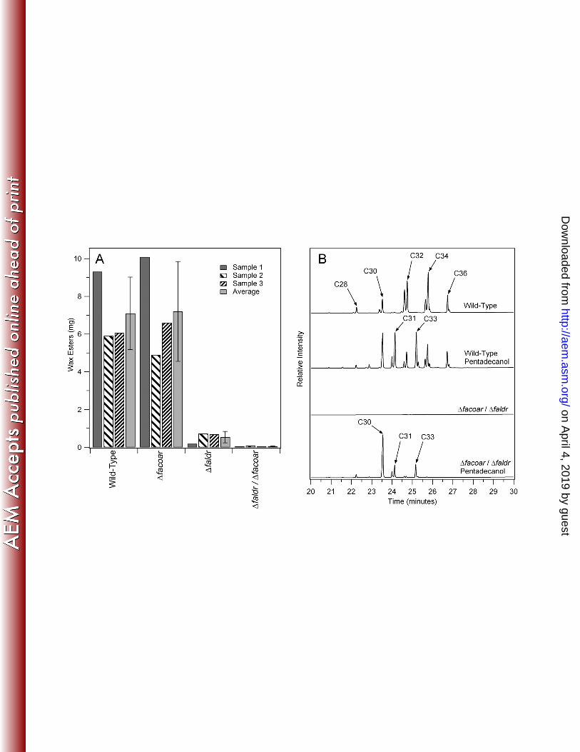
carbon source (2). Figure 3A shows the results of the wax ester analysis from three independent 203
cultures of these strains. Due to differences in peak lipid production (see discussion related to 204
Figure 4 below), there is some degree of variance seen between results, though differences are 205
clear for specific strains. The wild-type and Δfacoar strain yielded similar quantities of wax 206
on April 4, 2019 by guest
http://aem.asm
.org/D
ownloaded from

12
esters (Figure 3A) while the Δfaldr strain resulted in a significant decrease in the amounts of wax 207
esters found. Neither single gene deletion strain resulted in a wax ester deletion phenotype, 208
indicating that both enzymes are capable of fulfilling this role in the wax ester biosynthesis 209
pathway in the absence of the other gene. 210
Double gene deletions of both facoar and faldr. To probe and confirm the specific role of 211
both enzymes, a further effort was initiated to construct a double deletion strain. Here, the first 212
deletion was accomplished by using a single homologous recombination event with a selectable 213
marker for counter selection. We chose to utilize the sacB gene that results in a sucrose sensitive 214
phenotype in certain bacteria (15-17). Following isolation and confirmation of the single 215
homologous recombination based on antibiotic selection, M. aquaeolei VT8 colonies were 216
grown in liquid culture containing sucrose in a minimal medium, and enriched by several 217
subsequent transfers to fresh media before plating and screening for loss of the antibiotic marker. 218
Strains containing the markerless deletion were confirmed by colony PCR. The second gene 219
deletion was then isolated using the double homologous selection method described above for 220
single gene deletions. The toxicity of sacB in M. aquaeolei VT8 was not as potent as was found 221
for controls tested in Escherichia coli, and is best characterized as a screening protocol under the 222
current conditions. We suspect this may be related to poor sugar uptake by M. aquaeolei VT8, 223
which does not grow well on simple sugars. However, utilizing this procedure, a double deletion 224
strain Δfacoar/Δfaldr was obtained. Characterization of wax ester production in the 225
Δfacoar/Δfaldr strain (Figure 3A) revealed only a minimal background of wax esters. This 226
confirms that either enzyme is capable of supporting the wax biosynthetic pathway independent 227
of one another, though the Δfaldr gene product seems to play a greater role in M. aquaeolei VT8 228
under typical wax ester accumulating conditions based on these single gene deletion studies. It 229
on April 4, 2019 by guest
http://aem.asm
.org/D
ownloaded from

13
also supports the proposal that these are the two primary genes capable of producing fatty 230
alcohols, as only trace amounts of wax esters were found in the absence of both genes. 231
Wax ester production phenotype rescue through addition of extraneous alcohols. To 232
confirm that loss of the wax ester production in these deletion strains is based on the lack of this 233
enzymatic step in the pathway (and not a secondary affect related to poor culture health for 234
example), we utilized a strategy taken previously to add foreign fatty alcohols (specifically the 235
odd carbon number pentadecanol that results in unique wax ester products) in an attempt to 236
rescue the wax ester production phenotype in this double deletion strain (2). This results in the 237
production of odd numbered wax esters when provided extraneously to wild type cells (2) as 238
shown in figure 3B. This strategy relies on the fact that waxes found in M. aquaeolei VT8 under 239
the culture conditions utilized here are derived primarily from C16 and C18 fatty acids (2). Thus, 240
addition of pentadecanol to a strain lacking only the enzyme(s) involved in the reduction of fatty 241
acids to fatty alcohols should result in wax esters containing even fatty acids and only 242
pentadecanol as the alcohol. This result is confirmed in Figure 3B (bottom). Two peaks were 243
found at approximately 24 minutes that corresponds to pentadecanol and C16 fatty acid (either 244
C16:1 or C16) and one additional peak was found just after 25 minutes that corresponds to 245
pentadecanol and C18 fatty acid (predominantly C18:1). An additional large peak corresponding 246
to a C30 wax ester results from pentadecanol oxidation within the cell through alternative 247
directions in the pathway (see Figure 1 (18)) resulting in significant amounts of pentadecanol and 248
C15 fatty acid derived wax ester accumulating in the cell as well. This result was further 249
confirmed by treatment of the wax esters from this sample with methanol and acid as described 250
previously (2) followed by characterization of individual components by gas chromatography 251
and mass spectrometry (GC/MS). This analysis found four primary fatty acids; C15, C16:1, C16 252
on April 4, 2019 by guest
http://aem.asm
.org/D
ownloaded from

14
and C18:1 at a ratio of approximately 3.5:0.5:1:1, which agrees well with the wax ester profile of 253
the chromatogram shown in figure 3B (bottom). Importantly, predominant wax esters such as 254
C32 derived from C16 fatty acids and fatty alcohols (at approximately 24.5 minutes) are not 255
prominent in the double deletion strain, and C16 and C18 fatty alcohols were not found during 256
GS/MS analysis in the double deletion strain, while they were present in a wild-type control 257
sample. This confirms that the wax ester biosynthesis pathway can be reconstituted in vivo in the 258
double deletion strain by adding extraneous alcohol (pentadecanol). 259
Transcriptional analysis of facoar and faldr during wax ester accumulation. In addition to 260
the gene deletion studies described above, it was also of interest to investigate the changes in 261
gene transcriptional levels for these two fatty alcohol producing enzymes during a typical batch 262
culture in wild-type M. aquaeolei VT8. To pursue these studies, an approach was taken to grow 263
M. aquaeolei VT8 as a batch culture using a media recipe routinely utilized in our laboratory to 264
induce lipid accumulation. The key feature of this defined medium is that cells exhaust the 265
source of nitrogen prior to reaching maximum cell density for the specific culture conditions, and 266
enter into a nitrogen limited state which results in wax ester accumulation. This is believed to 267
occur because the carbon source needed for energy and cellular building blocks remains 268
plentiful, but the nitrogen required for DNA and protein synthesis is not available for further 269
replication (19). The larger batch culture was selected here so that a thorough sampling of the 270
culture could be made through various stages of the wax ester accumulation and declination 271
process. The sampling strategy adopted included multiple samples for total RNA isolation and 272
the harvest of cells for further drying and wax ester quantification. Lipids were extracted using a 273
previously described protocol that isolates primarily wax esters from dried M. aquaeolei VT8 274
cells, which can be analyzed directly using a gas chromatography method with flame ionization 275
on April 4, 2019 by guest
http://aem.asm
.org/D
ownloaded from

15
detection (GC/FID) to measure specific classes of lipids (2). This method is preferred over 276
indirect methods such as gravimetric approaches, as the specific compound of interest (the wax 277
ester) is separated and specifically quantified using external standards, while polar lipids such as 278
cell membrane components that would contribute to total lipids in certain gravimetric methods 279
are excluded from the measurement by this approach. 280
Figure 3B (Upper Trace) shows a typical GC/FID chromatogram obtained from extraction of 281
dried M. aquaeolei VT8 cells, while Figure 4 shows the typical wax ester accumulation cycle 282
from M. aquaeolei VT8 found when cells were grown as a batch culture. Cells grow 283
exponentially with sufficient amounts of nitrogen to support replication until approximately 40 284
hours when all the available nitrogen in the media is consumed. Wax ester accumulation begins 285
shortly after that, reaching a maximum quantity at about 100 hours (generally about 10% of dry 286
cell mass, as was found for the experiment shown). Following this, the culture enters a final 287
stage where wax esters in the cell begin to decline to very low levels after about 150 hours. The 288
rise and decrease in levels of wax esters in the cell during culture is reproducible (n>5 289
independent experiments), and is responsible for the high sample-to-sample variance for wax 290
concentrations shown in Figure 3A, which is the result of a single time point analysis versus 291
analysis at multiple time points as was done in the batch culture here. The wax ester analysis 292
shown in Figure 4 was arbitrarily fit to a Gaussian curve for the sake of visualization only and is 293
not meant to indicate that the wax ester accumulation cycle behaves precisely in this specific 294
manner (though this works well for most of the batch cultures analyzed in our laboratory). The 295
changes in transcriptional levels (as fold change) of the genes coding for the fatty aldehyde 296
reductase (faldr) and fatty acyl-CoA reductase (facoar) are plotted along with the wax ester 297
analysis in Figure 4. Additionally, several reference genes coding for recombinase (recA), a 298
on April 4, 2019 by guest
http://aem.asm
.org/D
ownloaded from

16
medium alcohol dehydrogenase (madh) and a fatty aldehyde dehydrogenase (falddh) and the 16S 299
rRNA are included for comparison (12, 18, 20). Two of the reference samples (madh, recA) 300
showed only slight changes in transcription levels during this experiment, and 16S rRNA levels 301
were also consistent. The falddh was included as it may be involved in the pathway necessary to 302
oxidize extraneously added pentadecanol to pentadecanoic acid to produce the C30 wax ester 303
found in phenotype rescue experiments (Figure 3B) (2, 18). Transcriptional levels of falddh were 304
100 fold higher during exponential growth, but dropped during wax ester accumulation and 305
remained low during the stage of wax ester depletion. Transcription of facoar was also elevated 306
during exponential growth, but rapidly declined during the initial phase of wax ester 307
accumulation, while only the faldr showed elevated transcriptional levels during the wax ester 308
accumulation phase of the culture (between 40 and 100 hours), dropping only after wax esters 309
had reached their peak concentration during the culture. Relative levels of faldr, facoar and 310
falddh were all within one fold (Cp value within one cycle) of one another at the final time point, 311
though they were approximately eight fold (Cp values within two to three cycles) lower than the 312
madh and recA reference samples. 313
Agreement between RT-qPCR and gene deletion experiments. The results from 314
transcriptional analysis of mRNA for the fatty aldehyde reductase and fatty acyl-CoA reductase 315
correlate well with what was found in the gene deletion studies. The fatty aldehyde reductase 316
transcription appears to be up-regulated during lipid accumulation. The fatty acyl-CoA reductase 317
transcriptional levels are elevated primarily during exponential growth under the conditions 318
utilized here, but dropped substantially once the cell entered into the wax ester accumulation 319
stage. Thus, the deletion of the gene coding the fatty acyl-CoA reductase (Δfacoar) had very 320
little effect on levels of wax esters. However, deletion of the gene coding for the fatty aldehyde 321
on April 4, 2019 by guest
http://aem.asm
.org/D
ownloaded from

17
reductase (Δfaldr) dramatically decreased the levels of wax esters accumulated, but did not 322
completely delete the wax ester accumulation phenotype. 323
The findings from these studies which looked at features of these enzymes in vivo in the 324
indigenous organism contrasts with what has been found previously for these fatty alcohol 325
producing enzymes through in vitro experiments (21). Studies with isolated enzymes indicate 326
that the fatty acyl-CoA reductase is the more active of the two enzymes following purification (6, 327
7). Homologs to the fatty acyl-CoA reductase are also more prevalent in other model wax ester 328
accumulating species (Acinetobacter, Psychrobacter and Rhodococcus), while the fatty aldehyde 329
reductase is found less frequently (7). This was a key motivator to pursue these studies, as M. 330
aquaeolei VT8 provides an ideal test case to analyze which fatty alcohol yielding enzyme 331
evolution has selected to optimize wax ester production when both options exist within the 332
repertoire of enzymes in the cell. As mentioned previously, additional activities toward fatty 333
acyl-CoA and fatty acyl-ACP have been reported for the fatty aldehyde reductase from M. 334
aquaeolei VT8 since the initial characterization of this enzyme (4, 5). Additionally, others have 335
recently demonstrated that the fatty aldehyde reductase (Maqu_2220 or NCBI accession number 336
YP_959486) resulted in improved fatty alcohol yields when heterologously expressed in E. coli 337
versus several other fatty acyl-CoA reductases (21). 338
Summary of findings. These results demonstrate that while M. aquaeolei VT8 has 339
incorporated redundancy within the wax ester production pathway to yield the fatty alcohols 340
required for wax ester production, evolution has selected one specific branch of this pathway as 341
the clear preferred route when producing wax esters from simple organic acids such as succinate 342
or citrate. 343
344
on April 4, 2019 by guest
http://aem.asm
.org/D
ownloaded from

18
TABLE 1. Selected Proteins for mRNA Transcription Analysis 345
Gene Product (gene)a
Protein (or Ribosomal) Product (NCBI Reference)b
KEGG Pathway (or Putative Pathway)c
Protein / Nucleotide Accession Numberd
FACoAR (facoar)
Fatty Acyl-CoA Reductase
Wax Ester Biosynthesis
YP_959769
FAldR (faldr) Fatty Aldehyde Reductasee Wax Ester Biosynthesis YP_959486 FAldDH (falddh) Fatty Aldehyde Dehydrogenase Wax Ester Biosynthesis YP_960668 Medium ADH (madh) Medium Alcohol Dehydrogenase Unknown YP_958650 16S rRNA 16S Ribosomal RNA Ribosome NR_027551 (3) Recombinase (recA)
Recombinase A Homologous Recombination YP_959349
346 a Simple reference for gene product (or gene) used in figures 347 b Full name of the gene product based on NCBI reference number or previous annotations 348 c Potential pathways that gene products are involved in from KEGG or the pathway shown in Figure 1 349 a NCBI gene or protein accession number 350 eWhile annotated here as fatty aldehyde reductase, it is also noted that this gene product has been reported to have 351 fatty acyl-CoA reductase and fatty acyl-ACP reductase activity by others (4, 5) 352
on April 4, 2019 by guest
http://aem.asm
.org/D
ownloaded from

19
TABLE 2. Key parent plasmids and relevant derivatives of these plasmids used for the construction of Marinobacter aquaeolei VT8 manipulated strains. Plasmida
Relevant Genes Cloned or Plasmid Manipulation
Vector
Reference or Source
pBBR1MCS-2 Plasmid containing mobilization element (14) pSMV3 Plasmid containing sacB gene (9) pBB053 Removed NdeI site from pUC19 by silent mutation pUC19 This Study pBB114 Replaced pUC19 Amp resistance with Kan resistance
cassette from pUC4K, then removed NsiI and HindIII sites from cassette by silent mutation
pUC19 pUC4K (22)
pBBTET3 Replaced pUC19 Amp resistance with Tet resistance cassette from pRK415
pUC19 pRK415 (23)
pPCRKAN4 Cloned Kan cassette from pBBR1MCS-2 into pBBTET3
pBBTET3 This Study
pPCRMOB4 Moved mobilization element from pBBR1MCS-2 into
pUC19 pUC19 This Study
pPCRSACB6 The sacB gene from pSMV3 was cloned into pBB053 and then EcoRI, HindIII, XbaI and KpnI sites were removed by site-specific mutagenesis with silent mutations
pBB053 This Study
pPCRSACB7 Moved sacB gene cassette from pPCRSACB6 to pBBTET3
pBBTET3 This Study
pPCRWEK4 Derivative of pBB053 for gene insertions pBB053 This Study pPCRWEK5 Moved mobilization element from pPCRMOB4 into
pPCRWEK4 pBB053 This Study
pPCRWEK12 Cloned gene facoar and flanking regions from M. aquaeolei VT8 genome with primers BBP1477 and BBP1478 into pBBTET3 EcoRI and XbaI sites
pBBTET3 This Study
pPCRWEK14 Performed PCR with primers to remove gene for facoar from pPCRWEK12 leaving flanking regions and adding BamHI site
pBBTET3 This Study
pPCRWEK20 Cloned gene faldr and flanking regions from M. aquaeolei VT8 genome with primers BBP1522 and BBP1523 into pBBTET3 EcoRI and XbaI sites
pBBTET3 This Study
pPCRWEK26 Moved facoar flanking segments fragment from pPCRWEK14 into pPCRWEK5
pBB053 This Study
pPCRWEK27 Performed PCR with primers to remove gene faldr from pPCRWEK20 leaving flanking regions and adding BamHI site
pBBTET3 This Study
pPCRWEK29b,c Moved Kan cassette from pPCRKAN4 into BamHI cut pPCRWEK26
pBB053 This Study
pPCRWEK32 Moved faldr flanking segments fragment from pPCRWEK27 into pPCRWEK5
pBB053 This Study
pPCRWEK33c Moved Kan cassette from pPCRKAN4 into BamHI cut pPCRWEK32
pBB053 This Study
pPCRWEK48 Moved sacB gene cassette from pPCRSACB7 into KpnI and HindIII cut pPCRWEK32
pBB053 This Study
pPCRWEK50b,c Moved Kan cassette cut with BamHI from pPCRKAN4 into BglII cut pPCRWEK48
pBB053 This Study
a Sequences of all plasmids in this study are available upon request 353 b Plasmid maps are also provided in Figure 2 354 c Plasmids shown in bold are completed vectors used to transform M. aquaeolei VT8 355
on April 4, 2019 by guest
http://aem.asm
.org/D
ownloaded from

20
356 TABLE 3. Primers used in this study Primer Designation
Primer Sequencea
Purpose
BBP1477 5’GACATCTA GACTGGATCT TGTCTTCCCG
GGAACCAC3’ facoar gene and flanking region cloning
BBP1478 5’GACAGAAT TCTGGATTTC ACCGGCATCG ATCC3’
facoar gene and flanking region cloning
BBP1479 5’GACAGGAT CCATATGTAC TCCATTCTGC CTGTTGTGTT TTTG3’
facoar gene deletion
BBP1480 5’GACAGGAT CCGATATACT GGTAATCGTC GTTATAAACC AAG3’
facoar gene deletion
BBP1522 5’GNNNGAAT TCGATCGCGC CAGTCTTGCT CGTCATTTG3’
faldr gene and flanking region cloning
BBP1523 5’GNNNTCTA GAAGCTTCGA AGCGTTCAGG ACACCGTCCT CGAAC3’
faldr gene and flanking region cloning
BBP1524 5’GNNNGGAT CCCTTCTCCG GGGCAGGAAA GCGTTTCTG3’
faldr gene deletion
BBP1525 5’GNNNGGAT CCGATAGAAC TCCTTCTCTG AGATCACTAA TGCCG3’
faldr gene deletion
BBP1558 5’CGAGATGC TGAACGTTCA TGTTGGC3’ faldr deletion confirmation BBP1559 5’CACAGAGT GGATCGCACC AATACG3’ faldr deletion confirmation BBP1548 5’GTATTCGC CTGCCTCCGG GTACTTC3’ facoar deletion confirmation BBP1549 5’CACACGCG AAAGACAAGA AGGAAGC3’ facoar deletion confirmation BBP1678 5’GTTCCGTT CCGCATCTAC CG3’ facoar qPCR BBP1679 5’CCAGTGCA TCGACCACGA AA3’ facoar qPCR BBP1409 5’CCGTCTTC GCGAGGCCGA TT3’ faldr qPCR BBP1410 5’TGATGGCC AGCGCCTTGT CG3’ faldr qPCR BBP1403 5’TTTCCGCT GCTGATGGCC GC3’ falddh qPCR BBP1404 5’CGCTTGCT GGTCGCCAAA GC3’ falddh qPCR BBP1352 5’TCCTGCCG TATCCACCGG CT3’ recA qPCR BBP1353 5’AAACCGGG TCCAGAGCGT GC3’ recA qPCR BBP1126 5’GCACGCTC TGGACCCGGT TT3’ recA qPCR BBP1127 5’CCACGTGG CTGTCGCCCA TT3’ recA qPCR BBP1413 5’TTGTTGGC CGGGTTACCG CC3’ madh qPCR BBP1414 5’TAGCCGCC GTGAGTGACC GA3’ madh qPCR BBP1128 5’CCGGCTAA CTCCGTGCCA GC3’ 16S rRNA qPCR BBP1129 5’ACGCATTT CACCGCTACA CAGG3’ 16S rRNA qPCR 357 a Specific restriction enzyme sites added to primers are underlined for clarity 358
on April 4, 2019 by guest
http://aem.asm
.org/D
ownloaded from

21
Acknowledgements 359
This work is supported by a grant from the National Science Foundation to B.M.B. (Award 360
Number 0968781). Further support was provided through generous startup funds through the 361
University of Minnesota. We thank Erin Ray, Robert Willis and Zeyuan Wu who assisted in 362
preliminary studies related to this work. 363
364
on April 4, 2019 by guest
http://aem.asm
.org/D
ownloaded from

22
References 365
1. Huu NB, Denner EBM, Ha DTC, Wanner G, Stan-Lotter H. 1999. Marinobacter 366
aquaeolei sp. nov., a halophilic bacterium isolated from a Vietnamese oil-producing well. 367
Int. J. Syst. Bacteriol. 49:367-375. 368
2. Barney BM, Wahlen BD, Garner E, Wei JS, Seefeldt LC. 2012. Differences in 369
Substrate Specificities of Five Bacterial Wax Ester Synthases. Appl. Environ. Microbiol. 370
78:5734-5745. 371
3. Singer E, Webb EA, Nelson WC, Heidelberg JF, Ivanova N, Pati A, Edwards KJ. 372
2011. Genomic Potential of Marinobacter aquaeolei, a Biogeochemical 373
"Opportunitroph". Appl. Environ. Microbiol. 77:2763-2771. 374
4. Hofvander P, Doan TTP, Hamberg M. 2011. A prokaryotic acyl-CoA reductase 375
performing reduction of fatty acyl-CoA to fatty alcohol. FEBS Letters 585:3538-3543. 376
5. McDaniel R, Behrouzian B, Clark L, Hattendorf D, Valle F. Feb 14, 2013 2013. 377
Production of Fatty Alcohols with Fatty Alcohol Forming Acyl-CoA Reductases (FAR) 378
patent US20130040352. 379
6. Wahlen BD, Oswald WS, Seefeldt LC, Barney BM. 2009. Purification, 380
Characterization, and Potential Bacterial Wax Production Role of an NADPH-Dependent 381
Fatty Aldehyde Reductase from Marinobacter aquaeolei VT8. Appl. Environ. Microbiol. 382
75:2758-2764. 383
7. Willis RM, Wahlen BD, Seefeldt LC, Barney BM. 2011. Characterization of a Fatty 384
Acyl-CoA Reductase from Marinobacter aquaeolei VT8: A Bacterial Enzyme 385
Catalyzing the Reduction of Fatty Acyl-CoA to Fatty Alcohol. Biochemistry 50:10550-386
10558. 387
on April 4, 2019 by guest
http://aem.asm
.org/D
ownloaded from

23
8. Reiser S, Somerville C. 1997. Isolation of mutants of Acinetobacter calcoaceticus 388
deficient in wax ester synthesis and complementation of one mutation with a gene 389
encoding a fatty acyl coenzyme a reductase. J. Bacteriol. 179:2969-2975. 390
9. Saltikov CW, Newman DK. 2003. Genetic identification of a respiratory arsenate 391
reductase. Proc. Natl. Acad. Sci. U. S. A. 100:10983-10988. 392
10. Bakermans C, Sloup RE, Zarka DG, Tiedje JM, Thomashow MF. 2009. 393
Development and use of genetic system to identify genes required for efficient low-394
temperature growth of Psychrobacter arcticus 273-4. Extremophiles 13:21-30. 395
11. Sonnenschein EC, Gärdes A, Seebah S, Torres-Monroy I, Grossart HP, Ullrich MS. 396
2011. Development of a genetic system for Marinobacter adhaerens HP15 involved in 397
marine aggregate formation by interacting with diatom cells. J. Microbiol. Meth. 87:176-398
183. 399
12. Takle GW, Toth IK, Brurberg MB. 2007. Evaluation of reference genes for real-time 400
RT-PCR expression studies in the plant pathogen Pectobacterium atrosepticum. BMC 401
Plant Biol. 7. 402
13. Röttig A, Steinbüchel A. 2013. Acyltransferases in Bacteria. Microbiol. Mol. Biol. R. 403
77:277-321. 404
14. Kovach ME, Elzer PH, Hill DS, Robertson GT, Farris MA, Roop RM, 2nd, Peterson 405
KM. 1995. Four new derivatives of the broad-host-range cloning vector pBBR1MCS, 406
carrying different antibiotic-resistance cassettes. Gene 166:175-176. 407
15. Blomfield IC, Vaughn V, Rest RF, Eisenstein BI. 1991. Allelic Exchange in 408
Escherichia coli Using the Bacillus subtilis sacB Gene and a Temperature-Sensitive 409
Psc101 Replicon. Mol. Microbiol. 5:1447-1457. 410
on April 4, 2019 by guest
http://aem.asm
.org/D
ownloaded from

24
16. Pelicic V, Reyrat JM, Gicquel B. 1996. Generation of unmarked directed mutations in 411
Mycobacteria, using sucrose counter-selectable suicide vectors. Mol. Microbiol. 20:919-412
925. 413
17. van der Geize R, Hessels GI, van Gerwen R, van der Meijden P, Dijkhuizen L. 2001. 414
Unmarked gene deletion mutagenesis of kstD, encoding 3-ketosteroid Delta(1)-415
dehydrogenase, in Rhodococcus erythropolis SQ1 using sacB as counter-selectable 416
marker. FEMS Microbiol. Lett. 205:197-202. 417
18. Ishige T, Tani A, Sakai Y, Kato N. 2000. Long-chain aldehyde dehydrogenase that 418
participates in n-alkane utilization and wax ester synthesis in Acinetobacter sp strain M-419
1. Appl. Environ. Microbiol. 66:3481-3486. 420
19. Wältermann M, Hinz A, Robenek H, Troyer D, Reichelt R, Malkus U, Galla HJ, 421
Kalscheuer R, Stöveken T, von Landenberg P, Steinbüchel A. 2005. Mechanism of 422
lipid-body formation in prokaryotes: how bacteria fatten up. Mol. Microbiol. 55:750-763. 423
20. Wei J, Timler JG, Knutson CM, Barney BM. 2013. Branched-chain 2-keto acid 424
decarboxylases derived from Psychrobacter. FEMS Microbiol. Lett. 346:105-112. 425
21. Liu A, Tan X, Yao L, Lu X. 2013. Fatty alcohol production in engineered E. coli 426
expressing Marinobacter fatty acyl-CoA reductases. Appl. Microbiol. Biotechnol. 427
97:7061-7071. 428
22. Taylor LA, Rose RE. 1988. A correction in the nucleotide sequence of the Tn9O3 429
kanamycin resistance determinant in pUC4K. Nuc. Acids Res. 16:358. 430
23. Mather MW, McReynolds LM, Yu CA. 1995. An Enhanced Broad-Host-Range Vector 431
for Gram-Negative Bacteria - Avoiding Tetracycline Phototoxicity during the Growth of 432
Photosynthetic Bacteria. Gene 156:85-88. 433
on April 4, 2019 by guest
http://aem.asm
.org/D
ownloaded from

26
435
Figure 1. Wax Ester Pathway. Shown are the proteins that comprise the current pathway for 436
wax ester biosynthesis in lipid accumulating bacteria such as M. aquaeolei VT8. Various 437
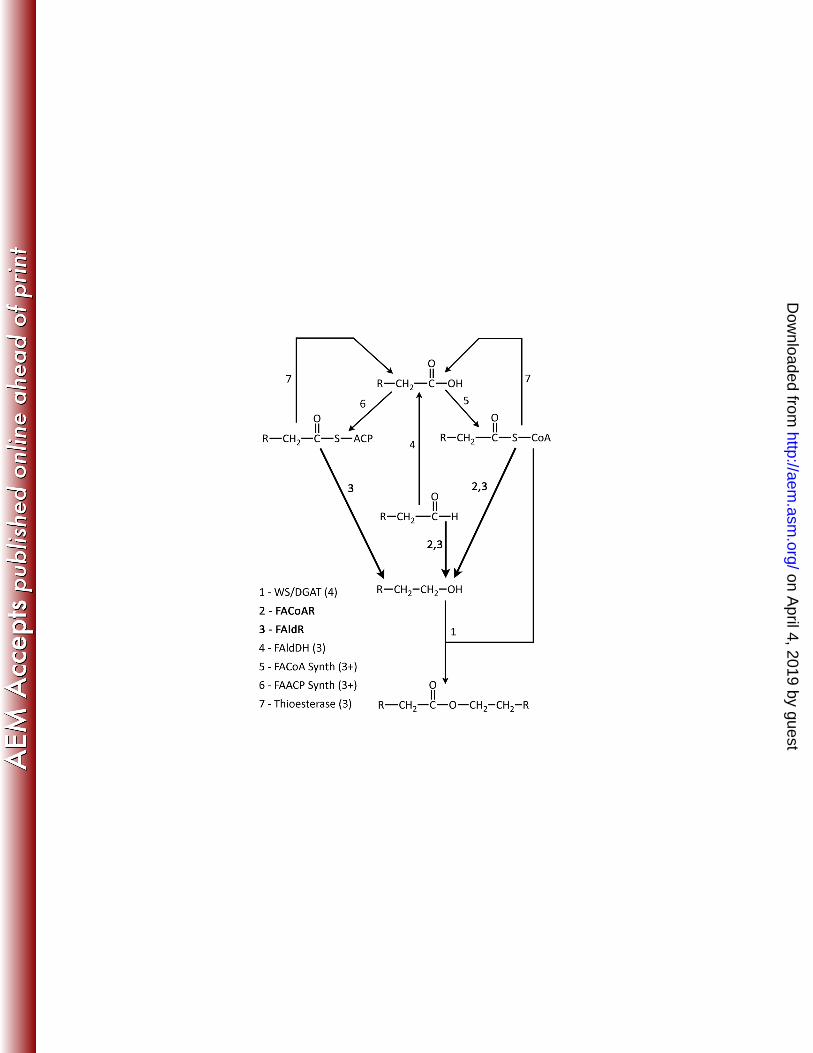
proteins of the pathway are numbered, including the wax synthase (1), fatty acyl-CoA reductase 438
(2), fatty aldehyde reductase (3), fatty aldehyde dehydrogenase (4), fatty acyl-CoA synthetase 439
(5), fatty acyl-ACP synthetase (6) and thioesterase (7). Values shown following specific enzymes 440
in parenthesis indicate the number of known or putative homologs found in M. aquaeolei VT8. 441
Enzymes shown in bold and darker arrows are those highlighted in this study. 442
on April 4, 2019 by guest
http://aem.asm
.org/D
ownloaded from

27
443
Figure 2. Key Plasmids for Gene Deletion Studies. Shown are representations of two of the 444
final plasmid constructs utilized for gene deletion studies. Plasmid pPCRWEK29 was used to 445
perform double homologous recombination by replacing the gene of interest (facoar in this 446
construct) with the antibiotic marker for kanamycin. Plasmid pPCRWEK50 was used to perform 447
a single homologous recombination that was selected using the kanamycin marker. A counter 448
selection following a second recombination event, using the toxicity of the sacB gene product, 449
resulted in markerless deletion of the faldr gene. Images produced using the program pDRAW32 450
(Acaclone Software). 451
on April 4, 2019 by guest
http://aem.asm
.org/D
ownloaded from

28
452
Figure 3. Gene Deletion and Wax Ester Production Rescue Studies. Shown on the left panel 453
are the results of the quantities of wax esters obtained from replicate cultures under wax ester 454
accumulating conditions of wild-type M. aquaeolei VT8 cells under wax ester accumulating 455
conditions versus the two single gene deletion strains and the double deletion strain (A). 200 mg 456
of dried cell mass was extracted in each case. Error bars represent the standard deviation of the 457
three samples. Shown on the right (B) are gas chromatography chromatograms illustrating the 458
results of studies with wild-type cells and the Δfaldr/Δfacoar double deletion strain grown under 459
normal wax accumulation conditions and under the same conditions with added extraneous 460
pentadecanol resulting in the accumulation of odd numbered wax esters (for wild-type) and the 461
rescue of wax ester production based on only these odd alcohols (for the double deletion).462
on April 4, 2019 by guest
http://aem.asm
.org/D
ownloaded from

29
463
Figure 4. Gene Transcription During Batch Culture of M. aquaeolei VT8. Shown are results 464
obtained from an RT-qPCR analysis for a single batch culture of wild-type M. aquaeolei VT8 465
cells grown under wax ester accumulating conditions over the course of several days. 466
Transcriptional levels to determine fold change in mRNA levels were compared against the 467
results obtained for the last time point (normalized to 1 fold, left y-axis) and are plotted on a log 468
scale for simplicity. Results obtained from the wax ester analysis for this culture are fit to a 469
simple Gaussian curve for clarity and shown on the same graph (right y-axis). Where shown, 470
statistics represent the average and standard deviation for three replicates. 471
472
on April 4, 2019 by guest
http://aem.asm
.org/D
ownloaded from