Embed Size (px)
Citation preview
Vol. 27, No. 3INFECTION AND IMMUNITY, Mar. 1980, p. 969-9780019-9567/80/03-0969/10$02.00/0
Fatal Measles Infection in Marmosets Pathogenesis andProphylaxis
PAUL ALBRECHT,`* DOUGLAS LORENZ,2 MICHAEL J. KLUTCH,1 JAMES H. VICKERS,3 ANDFRANCIS A. ENNIS'
Division of Virology,1 Division ofBlood and Blood Products,2 and Division ofPathology,3 Bureau ofBiologics, Food and Drug Administration, Department ofHealth, Education and Welfare, Public Health
Service, Bethesda, Maryland 20205
Moustached marmosets (Saguinus mystax) were infected intranasally witheither of two low-passaged, wildlike strains of measles virus, strain Edmonston orstrain JM. The infection resulted in 25 and 100% mortality, respectively, 12 to 14days after infection. Clinical signs, gross pathological findings, and histologylacked the characteristic features of measles in other primates. A deficientimmune response and widespread gastroenterocolitis appeared to be the maincauses for the fatal outcome. Fluorescent-antibody staining detected largeamounts of measles antigen in lymphatic tissues, the gastrointestinal and respi-ratory tracts, the salivary glands, pancreas, liver, kidney, and other visceraltissues. Live attenuated or inactivated measles vaccine proved equally effective inpreventing fatal measles in marmosets. Challenge with live virus of animals whichwere primed 1 year previously with inactivated alum-absorbed vaccine resultedin a precipitous response, with a 100- to 1,000-fold increase in antibody titers.This vigorous booster response suggests the existence of a primary deficiency inlymphocyte cooperation in marmosets, which upon adequate priming is followedby extensive clonal expansion and antibody synthesis. Marmosets appear to bethe most susceptible primate species to measles infection. They are capable ofdistinguishing differences in virulence of virus strains with a level of sensitivitynot available in other animals.
Measles is a common, frequently serious infec-tion of children or young adults. Death occursin 1 out of 1,000 individuals who contract thedisease (3). Delayed complications of measleswhich occur years later in the form of a chronic,fatal brain disease known as subacute sclerosingpanencephalitis have been recognized (31). Ahighly successful live virus vaccine was devel-oped and licensed in the United States in 1963.A considerable drawback felt over the years
was the lack of a laboratory animal capable ofdistinguishing differences in the pathogenicproperties of natural and attenuated strains ofmeasles virus. This shortcoming has now beenovercome by the observation that marmosets,primates living in parts of Central and SouthAmerica, contract a fatal disease when infectedwith natural measles but not when infected withthe vaccine virus and that mortality after naturalinfection depends on the virus strain used, theroute of inoculation, and possibly other circum-stances.
MATERIALS AND METHODSAnimals. Feral Saguinus mystax from Peru were
used exclusively. The marmosets were apparentlyhealthy, adolescent to adult animals which had been
in captivity for at least 6 months before their use inthese experiments. Some had recovered from experi-mental human hepatitis A virus infection. They hadno aberrant clinical signs at the outset of the presentexperiment.The animals were housed in cages open on one side
and arranged in two tiers. Either a single animal or acompatible male-female pair was housed in each cage.Only one strain of virus was introduced into a room ofmarmosets for a given infectious experiment. Theanimals were maintained on dry, commercial monkeydiet, fruit, and water.
Viruses. Two natural and one attenuated strain ofmeasles virus were used. The low-passage naturalstrain Edmonston was recieved from John Endersthrough Hope Hopps in third passage in human em-bryonic kidney cell cultures. It received a total of nineadditional passages in primary human and monkeykidney cells in our laboratories. Its titer was 103plaque-forming units per ml in Vero cell cultures.Strain JM was isolated in this laboratory from a throatswab of a patient with typical measles rash. The viruswas isolated in Vero cultures and became cytopathicafter seven consecutive cell subcultures. It was iden-tified as measles virus by neutralization with mono-specific anti-serum. The titer of the seventh Vero cellpassage used in this experiment was 105 plaque-form-ing units/ml.A commercial vaccine, strain Moraten (Attenuvax,
Merck Sharp and Dohme, West Point, Pa.), was used969
on Decem
ber 2, 2018 by guesthttp://iai.asm
.org/D
ownloaded from
970 ALBRECHT ET AL.
without further passage as attenuated measles virus.It was derived from the Edmonston strain by 116passages in human and chicken fibroblast cultures(22). The titer was 1038 plaque-forming units/ml inVero cell cultures.
Killed measles vaccine was prepared in 1964 byPfizer as the licensed biologic for human use. Killedmeasles vaccine has not been used in humans since1967 due to hypersensitivity, which developed in somechildren, on reinfection with live virus (18). The viruswas grown in primary monkey kidney cells, inactivatedby Formalin, and concentrated by adsorption to alu-minum hydroxide. It was stored at 2 to 80C. Thevaccine passed the prescribed potency test when eval-uated shortly before use in this study.
Cell cultures and media. Vero cells at passagelevels 170 through 220 were used for virus titration,virus isolation from marmoset tissues, and antibodydeterminations. The cells were grown and maintainedin Eagle minimum essential medium supplementedwith 10% heat-inactivated fetal bovine serum, 0.03%glutamine, 50 ,ug of gentamicin per ml, and 0.4 jig ofFungizone per ml. Overlay medium consisted of Eaglemiminum essential medium containing 5% fetal bovineserum and a final concentration of 0.5% agarose(Seakem agarose, Microbiological Associates, Be-thesda, Md.).Experimental infection and immunization. Un-
diluted live measles virus was administered intramus-cularly in a 0.5-ml volume. For intranasal infection atotal volume of 0.5 ml of undiluted virus was sprayedwith a nebulizer into both nares and into the pharynx.Killed measles vaccine was administered intramuscu-larly, 0.5 ml/dose, three times in weekly intervals.
Virus isolation. Peripheral blood mononuclearcells separated on Ficoll-Hypaque or trypsin-dispersedautopsy tissues were mixed with an equal number ofVero cells and seeded in stationary culture vessels.The cultures were observed for 10 days, and if nocytopathic effect developed they were trypsinized andsubcultured. At this stage, the monolayers were com-posed almost exclusively of Vero cells. In several in-stances second and third subcultures were performedat 3- to 4-day intervals. Isolates were identified in situby the fluorescent-antibody technique.Antibody determinations. Early in the study it
became apparent that the marmosets' antibody re-sponse to measles was poor and that the hemaggluti-nation inhibition test lacked the necessary sensitivity.Therefore, a virus neutralization test was used whichyielded titers on the average 50-fold higher than thehemagglutination inhibition test. Vero cell cultureswere grown in plastic disposable trays containing 24wells 16 mm in diameter. The cell suspension wasseeded at 250,000, 150,000, or 100,000 cells per well in1.0 ml of Eagle minimum essential medium and used1, 2, or 3 days later, respectively.
Heparinized blood was spun at 900 x g, and theplasma was collected and inactivated for 30 min at560C. Fourfold dilutions of plasma in Eagle minimumessential medium containing 10% fetal bovine serumwere mixed with an equal volume of virus diluted toyield 20 to 30 plaque-forming units/well in the controltitration. The virus was the Edmonston strain receivedfrom John Enders and adapted to Vero cells by six
additional passages. The titer of this virus pool was105 plaque-forming units/mL. After the plasma-virusmixtures were incubated for 1 h at 370C, 0.1 ml of themixture was inoculated into each of two wells andadsorbed for 1 h at 360C. The inoculum was thenreplaced with 1.0 ml of agarose overlay, and the cul-tures were incubated in an inverted position in a 5%C02 incubator at 360C. Four days after infection 0.5ml of a second agarose overlay containing neutral red(1:10,000) was added. After a total incubation of 6 daysthe overlays were stripped, and the cell sheets wereair dried. The neutralizing antibody titer was ex-pressed as the highest final dilution ofplasma reducingthe number of virus plaques by 50%. The titer wascalculated by the Karber formula (27).
Fluorescent-antibody technique. Cryostat sec-tions were air dried, fixed with acetone, and stainedby the indirect technique, using serum from a patientwith subacute sclerosing panencephalitis (hemagglu-tination inhibition titer, 1:4,096) or from a hyperim-munized monkey (hemagglutination inhibition titer, 1:2,048) in the first step and a fluorescein-isothiocya-nate-labeled porcine antibody to human gamma glob-ulin (prepared in our laboratory) in the second step.Measles-negative sera from humans or monkeys andtissues from noninfected marmosets served as controls.
Histology. Tissues were fixed in a solution com-posed of formaldehyde, acetic acid, and saturated so-lution of picric acid in water in proportions of 25:5:75(Carnoy solution), embedded in paraffin, sectioned at7 pim, and stained with hematoxylin and eosin.
RESULTSInfection with natural measles virus. In-
fection ofmarmosets with the Edmonston orJMstrain of measles virus resulted in 25 to 100%mortality depending on the route of inoculationand the virus strain used (Table 1). The Edmon-ston strain was highly pathogenic on intramus-cular inoculation but killed only one of fouranimals inoculated intranasally. The JM strain,in contrast, killed all four animals infected bythe intranasal route.None of the animals dying from acute measles
infection showed clinical signs characteristic ofthe disease in humans or in subhuman primates.Notably absent were rash, coryza, and conjunc-tivitis. As a rule, the animals became less active,stopped eating, and were moribund the followingday. Diarrhea was frequent, but the significanceof it was initially overlooked, as diarrhea occurscommonly in marmoset colonies.Morphology of infection with natural
measles virus. Gross findings at autopsy wereunrevealing. Occasionally spleen and lymphnodes were enlarged. Careful examination failedto reveal pneumonitis or encephalitis, the mostcommon fatal complications of measles in otherprimates.The histological changes were predominantly
nonspecific in nature. There was slight to mod-
INFECT. IMMiUN.
on Decem
ber 2, 2018 by guesthttp://iai.asm
.org/D
ownloaded from
MEASLES IN MARMOSETS 971
TABLE 1. Experimental infection ofmarmosets (S. mystax) with natural measles virus
AnimoutfDays from Neutralizing antibody titerAnma Route of in Dasfono. Virus strain oculationa inoculation Preinocula- Postinoculation (day Cause of deathto death tion of bleeding)91 Edmonston i.m. 15 <8 32 (14) Measles'A34 Edmonston i.m. 12 <8 8 (12) MeaslesA91 Edmonston i.m. 12 <8 8 (12) Measles
M17 Edmonston i.n. 15 <8 16 (14) MeaslesM9 Edmonston i.n. 57 <8 215 (35) BronchopneumoniacM37 Edmonston i.n. 32 <8 815 (31) SepticemiacM50 Edmonston i.n. 32 <8 2,640 (31) Chronic nephritisc
61-1 JM i.n. 13 <8 <8 (13) Measles57-4 JM i.n. 13 <8 <8 (13) MeaslesM42 JM i.n. 14 <8 <8 (14) MeaslesM43 JM i.n. 14 <8 <8 (14) Measlesa i.m., Intramuscular; i.n., intranasal.b Determined by virus isolation and detection of measles antigen in tissues by fluorescent-antibody staining.c Measles antigen was not detected nor was virus isolated from tissues taken at autopsy.
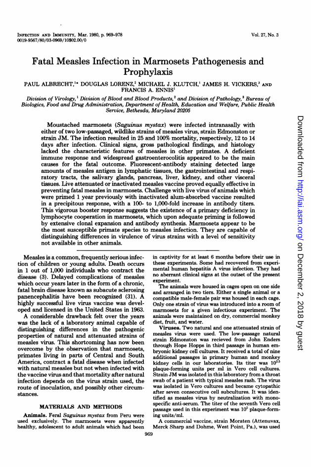
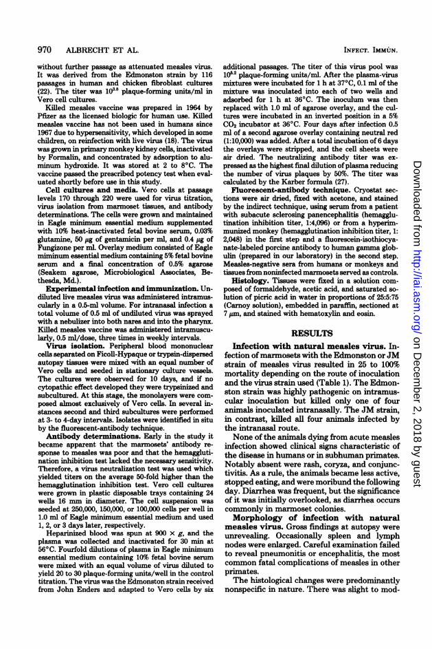
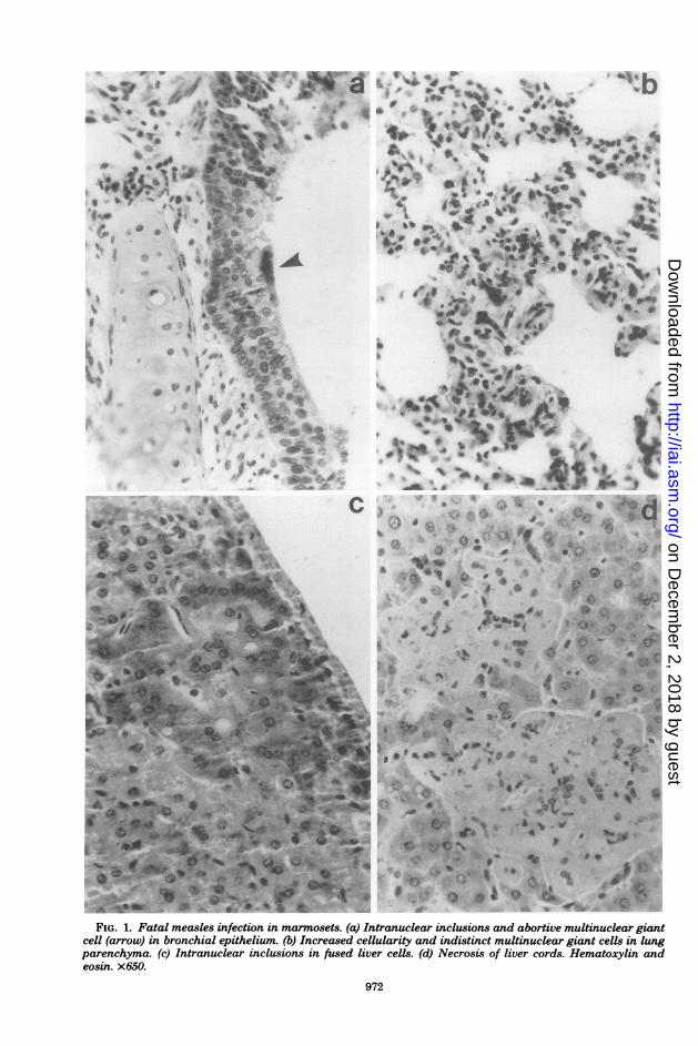
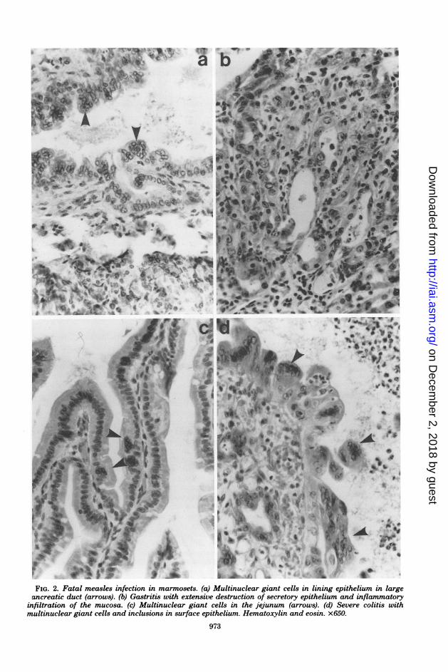
erate bronchitis (Fig. la) and increased cellular-ity in some areas of the lungs (Fig. lb). Thymus,spleen, and lymph nodes showed various degreesof lymphocytic depletion. The most prominentchanges were seen in the stomach and colon inthe form of a focal gastritis and colitis (Fig. 2bthrough d). The kidneys, liver (Fig. lc and d),pancreas, and salivary gland contained rare toinfrequent foci of parenchymal necrosis.
Histological changes characteristic of measlesinfection, namely, multinuclear giant cells andviral inclusions, were rare and seen in only a fewof the animals.. Intranuclear and cytoplasmicinclusions and indistinct giant cells were foundfocally in epithelial cells of the gastrointestinaltract (Figs. 2b, c, and d), in bronchial epithelium(Fig. la), in alveolar cells of the lung (Fig. lb),in the liver parenchyma (Fig. ic), and in thepancreatic ducts (Fig. 2a), in that order of fre-quency. Viral inclusions or giant cells were notseen in the lymphatic tissues.Fluorescent-antibody staining ofmeasles
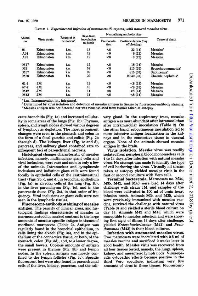
antigen. The paucity of clinical, gross, and his-tological findings characteristic of measles inmarmosets stood in marked contrast to the largeamounts of measles-specific antigen in tissues ofmoribund marmosets (Table 2). Antigen wasregularly found in the bronchial epithelium, incells lining the alveoli (Fig. 3a), and in the epi-thelium or the connective tissue, or both, of thestomach, colon (Fig. 3d), and, to a lesser degree,the small bowels. Copious amounts of antigenwere present in thymus (Fig. 3b) and lymphnodes. In the spleen, antigen was mostly con-fined to the lymph follicles (Fig. 3c). Specificfluorescent foci were also found in parenchymalcells of the liver, kidney, pancreas, and the sali-
vary gland. In the respiratory tract, measlesantigen was more abundant after intranasal thanafter intramuscular inoculation (Table 2). Onthe other hand, subcutaneous inoculation led tomore intensive antigen localization in the kid-neys and in the connective tissue in visceralorgans. None of the animals showed measlesantigen in the brain.Virus isolation. Measles virus was readily
isolated from peripheral blood mononuclear cells4 to 14 days after infection with natural measlesvirus. No attempt was made to identify the typeof cell harboring the virus. Virtually all tissuestaken at autopsy yielded measles virus in thefirst or second coculture with Vero cells.Terminal bacteremia. Marmosets no. M34,
M35, M42, and M43 were bled 14 days afterchallenge with strain JM, and samples of theblood were cultivated in 100 ml of brain heartinfusion broth. Animals M34 and M35, whichwere previously immunized with measles vac-cine, survived the challenge with natural virus(Table 3) and yielded a sterile blood culture onday 14. Animals M42 and M43, which weresusceptible to measles infection and were show-ing first signs of illness 14 days after challenge,yielded Enterobacteriaceae (M42) and Pseu-domonas (M43) in their blood cultures.Infection with attenuated measles virus.
Two marmosets were inoculated with 0.5 ml ofmeasles vaccine and sacrificed 2 weeks later ingood health. Measles virus was recovered fromall four tissues tested, namely, the lungs, spleen,kidney, and mesenteric lymph node. Virus-spe-cific cytopathic effects became positive in thethird Vero coculture, indicating very lbwamounts of virus in these tissues. Fluorescent-
VOL. 27, 1980
on Decem
ber 2, 2018 by guesthttp://iai.asm
.org/D
ownloaded from
v I_
t
a *' !CArPI f
k le '~o
S i
_* *#%t cv
* a , z
4gAA6@ .tPgaf'5.......-t...a. u
% .i i 'Ofztt f
-Fsr ts
II
k.
0*.3
*
j c%- IVt Is.
sfi .ai, C 0AC.
I_0 40jJ 6 (*'4 *1
r
3I ."~*4 *..*,
0
d%_a
baLos
'O .~ 9
ov
w 11 c.,
( 0
Mr 4b
,.)@'.ba >'^o4 tA
( w , ,u~j, .
W W r e e'. SL 04X *0
r.' 'T ') dQ o
FIG. 1. Fatal measles infection in marmosets. (a) Intranuclear inclusions and abortive multinuclear giantcell (arrow) in bronchial epithelium. (b) Increased cellularity and indistinct multinuclear giant cells in lungparenchyma. (c) Intranuclear inclusions in fused liver cells. (d) Necrosis of liver cords. Hematoxylin andeosin. x650.
972
PI -
laim f 4
* -s'9 0a,
At
'
l.koI
t.}IA^ Jb
e :> .&t Ns;. t
'*f % s
p
s-J
!.4.-A
*' O '.
*'CL
a a* ACi 4
t .$r VP' '
'* gW
%-
O.
f
,r: -w
on Decem
ber 2, 2018 by guesthttp://iai.asm
.org/D
ownloaded from
0 . .g m-/ of t ,, g i
as~~~~3*__ .1pbi >V
vFer-*4ia* al
VW.~
-,m.*
0:VIb%
.
-VI0
FIG. 2. Fatal measles infection in marmosets. (a) Multinuclear giant cells in lining epithelium in largepancreatic duct (arrows). (b) Gastritis with extensive destruction of secretary epithelium and inflammatory
infiltration of the mucosa. (c) Multinuclear giant cells in the jejunum (arrows). (d) Severe colitis withmultinuclear giant cells and inclusions in surface epithelium. Hematoxylin and eosin. x650.
973
a b
& f . e.
on Decem
ber 2, 2018 by guesthttp://iai.asm
.org/D
ownloaded from
TABLE 2. Antigen distribution in tissues of marmosets (S. mystax) dying of acute measles infectionMeasles antigen in tissues'
Animal Virus strain Route of Days fromAno.al Virus strain inocula- inoculation Lower Lym- Gastroin- Connec-nlo. tiona to death respira- phatic testinal Liver Kidney tive tis-
tory tract tissues tract sue
91 Edmonston i.m. 15 1+ 2+ ND 0 1+ 2+A34 Edmonston i.m. 12 2+ 3+ ND ± 2+ 2+A91 Edmonston i.m. 12 1+ 3+ ND ± ± 2+
M17 Edmonston i.n. 15 3+ 2+ 3+ ± ± ±
61-1 JM in. 13 2+ 3+ 2+ 2+ 1+ 1+57-4 JM i.n. 13 3+ 3+ ND 1+ ± ±M42 JM i.n. 14 3+ 3+ 3+ 2+ ± ±M43 JM i.n. 14 2+ 2+ 3+ 2+ ± ±a i.m., Intramuscular; i.n., intranasal; ND, not done.b± to 3+ = amount of measles antigen in tissues as detected by fluorescent-antibody staining.
antibody staining of tissues showed rare smallfoci of specific antigen in the thymus, stomach,and colon only.Immunization with inactivated and live
measles vaccine. Five marmosets were immu-nized with inactivated vaccine, 10 animals wereimmunized with live attenuated vaccine, and 10control animals were inoculated with saline (Ta-ble 4). Seven of the 25 animals died within a 2-month period. In none of them was death attrib-utable to measles infection. The 28% nonspecificdeath rate was not too unusual considering therepeated handling of the animals and theirknown susceptibility to stress and bacterial andparasitic infections in captivity (5, 23).The animals responded to immunization with
moderate antibody titers, which dropped to verylow levels 12 months after immunization (Table5). Reinfection with attenuated or natural mea-sles virus triggered a striking anamnestic re-sponse in animals sensitized earlier with inacti-vated vaccine. Antibody titers increased 100- to1,000-fold in the acute stage and remained 10-fold higher 18 months after the booster (Table3).Three months after immunization with live
vaccine two animals were challenged with thehighly virulent JM strain. The animal with neu-tralizing antibody titers of 1:395 was completelyprotected. The animal with a titer of 1:75 becamereinfected but was free of disease. The two sus-ceptible controls died of acute measles 14 daysafter infection (Table 3).
DISCUSSIONThe findings of the present study demonstrate
that infection of the moustached marmoset withnatural measles virus is associated with highmortality. The same appears to be true of other
subspecies ofmarmoset monkeys. Levy and Mir-kovic (28) described a measles epizootic amongSanguinus oedipus, S. fuscicollis, and Cal-lithrixjacchus marmosets that decimated mostof their colony over a 6-month period. A recentoutbreak of disease characterized by gastritisand colitis occurred at the New England PrimateCenter and caused 10% mortality. It was proba-bly caused by measles or a closely related para-myxovirus (10).The different rates of mortality after infection
with the Edmonston and JM strains suggest thatmarmosets may be suitable indicators ofmeaslesvirus virulence. In a study to be published sep-arately, we found that strain JM, but not strainEdmonston, spread readily among marmosetshoused in the same animal room. The possibilityhas been considered that severe measles epidem-ics in human populations resulting in high mor-tality may have been due in part to virus strainsof increased virulence. The question remainedlargely unanswered because of the lack of asuitable animal indicator model.
Parenteral inoculation of marmosets with theEdmonston strain, which in effect bypassed thenatural portal of entry, was appreciably morepathogenic than intranasal inoculation. It wastherefore reassuring to find that the attenuatedvariant of strain Edmonston lacked virulence onintramuscular inoculation. Although it is theo-retically possible that the immunosuppressionwhich accompanies natural and attenuated mea-sles infection (9) contributed to nonspecificdeaths observed in these marmosets, this cannotbe determined from the small sample of animalsin this study. Considering the extreme virulenceof low-passaged measles in marmosets and thelikelihood that introduction of the virus into acolony may go unrecognized for a considerabletime, it is prudent to use preventive immuniza-
974 ALBRECHT ET AL. INFECT. IMMUN.
on Decem
ber 2, 2018 by guesthttp://iai.asm
.org/D
ownloaded from
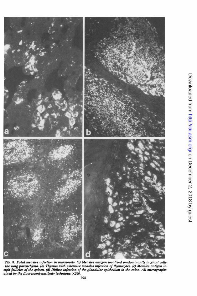
FIG. 3. Fatal measles infection in marmosets. (a) Measles antigen localized predominantly in giant cellsthe lung parenchyma. (b) Thymus with extensive measles infection of thymocytes. (c) Measles antigen inmph follicles of the spleen. (d) Diffuse infection of the glandular epithelium in the colon. All micrographsained by the fluorescent-antibody technique. x260.
975
I I
on Decem
ber 2, 2018 by guesthttp://iai.asm
.org/D
ownloaded from
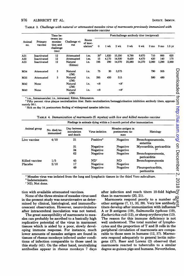
TABLE 3. Challenge with natural or attenuated measles virus of marmosets previously immunized withmeasles vaccine
Time be- Postchallenge antibody titer (reciprocal)tween im- Route
Animal Primary muniza- Challenge vi- of inoc-no. vaccine tion and rus ulationa 0 1 wk 2 wk 3 wk 6 wk 3 mo 9 mo 1.5 yr
challenge(mo)
A21 Inactivated 12 Attenuated i.m. 20b 1,820 15,350 8,790 8,670 710 800 620A22 Inactivated 12 Attenuated i.m. 15 4,170 14,930 8,430 4,470 420 140 170A23 Inactivated 12 Natural i.n. 195 290 14,070 25,580 14,370 2,060 1,290 2,560
(Edmn)
M34 Attenuated 3 Natural i.n. 75 30 3,575 790 505(JM)
M35 Attenuated 3 Natural i.n. 395 450 515 580 480(JM)
M42 None Natural i.n. <8 <8c(JM)
M43 None Natural i.n. <8 c
(JM)a i.m., Intramuscular; i.n., intranasal; Edmn, Edmonston.bFifty percent virus plaque neutralization titer. Ratio neutralization/hemagglutination inhibition antibody titers, approxi-
mately 50:1.c Sick on day 14; postmortem finding of widespread measles infection.
TABLE 4. Immunization ofmarmosets (S. mystax) with live and killed measles vaccineFindings in animals dying within a 2-month period after immunization
Animal group No. died/no. Day between Measles antigen inimmunized inoculation Virus isolation postmortem tis- Histology
and death sues
Live vaccine 4/10 21 Positivea Negative Bronchopneumonia,enteritis
31 Negative Negative Myocarditis, pericarditis34 Negative Negative ?b
62 Negative Negative Bronchopneumonia,pericarditis
Killed vaccine 1/5 45 NDc ND BronchopneumoniaPlacebo 2/10 17 Negative Negative ?
49 Negative Negative Pleuropericarditis,peritonitis
a Measles virus was isolated from the lung and lymphatic tissues in the third Vero subculture.b Indeterminate.'ND, Not done.
tion with available attenuated vaccines.None of the three strains of measles virus used
in the present study was neuroinvasive as deter-mined by clinical, histological, and immunoflu-orescent observation. However, neurovirulenceafter intracerebral inoculation was not tested.The great susceptibility of marmosets to mea-
sles can probably be ascribed to a basically highreplicative potential of the virus in marmosettissues which is aided by a poor, slowly devel-oping immune response. For instance, muchlower amounts of measles antigen are found intissues of rhesus monkeys infected under condi-tions of infection comparable to those used inthis study (41). On the other hand, neutralizingantibodies appear in rhesus monkeys 7 days
after infection and reach titers 10-fold higherthan in marmosets (20, 21).Marmosets respond poorly to a number of
other antigens (7, 11, 33, 39). Very low antibodytiters develop after immunization with influenzaA or B antigens (19), Salmonella typhosa (19),Escherichia coli (12), or sheep erythrocytes (13).The reason for this immune deficiency is notwell understood. The total number of lympho-cytes and the proportion of T and B cells in theperipheral circulation of marmosets are compa-rable to those seen in humans (12, 37). Marmo-sets respond adequately to general lectin mito-gens (37). Baer and Lorenz (2) observed thatmarmosets reacted to tuberculin to a similardegree as guinea pigs and humans. Nevertheless,
976 ALBRECHT ET AL. INFECT. IMMUN.
on Decem
ber 2, 2018 by guesthttp://iai.asm
.org/D
ownloaded from
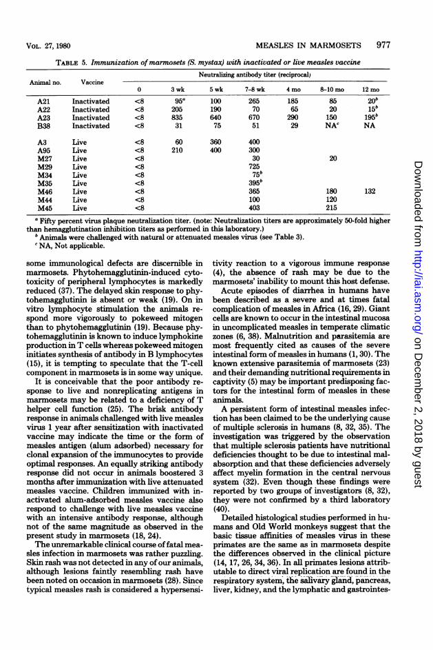
TABLE 5. Immunization of marmosets (S. mystax) with inactivated or live measles vaccineNeutralizing antibody titer (reciprocal)
Animal no. Vaccine0 3wk 5wk 7-8wk 4 mo 8-lOmo 12mo
A21 Inactivated <8 95a 100 265 185 85 20bA22 Inactivated <8 205 190 70 65 20 15bA23 Inactivated <8 835 640 670 290 150 195bB38 Inactivated <8 31 75 51 29 NAC NA
A3 Live <8 60 360 400A95 Live <8 210 400 300M27 Live <8 30 20M29 Live <8 725M34 Live <8 75bM35 Live <8 395bM46 Live <8 365 180 132M44 Live <8 100 120M45 Live <8 403 215
aFifty percent virus plaque neutralization titer. (note: Neutralization titers are approximately 50-fold higherthan hemagglutination inhibition titers as performed in this laboratory.)
b Animals were challenged with natural or attenuated measles virus (see Table 3).c NA, Not applicable.
some immunological defects are discernible inmarmosets. Phytohemagglutinin-induced cyto-toxicity of peripheral lymphocytes is markedlyreduced (37). The delayed skin response to phy-tohemagglutinin is absent or weak (19). On invitro lymphocyte stimulation the animals re-spond more vigorously to pokeweed mitogenthan to phytohemagglutinin (19). Because phy-tohemagglutinin is known to induce lymphokineproduction in T cells whereas pokeweed mitogeninitiates synthesis of antibody in B lymphocytes(15), it is tempting to speculate that the T-cellcomponent in marmosets is in some way unique.
It is conceivable that the poor antibody re-sponse to live and nonreplicating antigens inmarmosets may be related to a deficiency of Thelper cell function (25). The brisk antibodyresponse in animals challenged with live measlesvirus 1 year after sensitization with inactivatedvaccine may indicate the time or the form ofmeasles antigen (alum adsorbed) necessary forclonal expansion of the immunocytes to provideoptimal responses. An equally striking antibodyresponse did not occur in animals boostered 3months after immunization with live attenuatedmeasles vaccine. Children immunized with in-activated alum-adsorbed measles vaccine alsorespond to challenge with live measles vaccinewith an intensive antibody response, althoughnot of the same magnitude as observed in thepresent study in marmosets (18, 24).The unremarkable clinical course of fatal mea-
sles infection in marmosets was rather puzzling.Skin rash was not detected in any of our animals,although lesions faintly resembling rash havebeen noted on occasion in marmosets (28). Sincetypical measles rash is considered a hypersensi-
tivity reaction to a vigorous immune response(4), the absence of rash may be due to themarmosets' inability to mount this host defense.Acute episodes of diarrhea in humans have
been described as a severe and at times fatalcomplication of measles in Africa (16, 29). Giantcells are known to occur in the intestinal mucosain uncomplicated measles in temperate climaticzones (6, 38). Malnutrition and parasitemia aremost frequently cited as causes of the severeintestinal form of measles in humans (1, 30). Theknown extensive parasitemia of marmosets (23)and their demanding nutritional requirements incaptivity (5) may be important predisposing fac-tors for the intestinal form of measles in theseanimals.A persistent form of intestinal measles infec-
tion has been claimed to be the underlying causeof multiple sclerosis in humans (8, 32, 35). Theinvestigation was triggered by the observationthat multiple sclerosis patients have nutritionaldeficiencies thought to be due to intestinal mal-absorption and that these deficiencies adverselyaffect myelin formation in the central nervoussystem (32). Even though these findings werereported by two groups of investigators (8, 32),they were not confirmed by a third laboratory(40).Detailed histological studies performed in hu-
mans and Old World monkeys suggest that thebasic tissue affinities of measles virus in theseprimates are the same as in marmosets despitethe differences observed in the clinical picture(14, 17, 26, 34, 36). In all primates lesions attrib-utable to direct viral replication are found in therespiratory system, the salivary gland, pancreas,liver, kidney, and the lymphatic and gastrointes-
MEASLES IN MARMOSETS 977VOL. 27, 1980
on Decem
ber 2, 2018 by guesthttp://iai.asm
.org/D
ownloaded from
978 ALBRECHT ET AL.
tinal systems (17, 36). It is safe to assume thatthe fatal course of measles in marmosets resultsfrom a combination of immune paralysis andintensive viral damage to selected tissues andorgans. The most severe lesions were seen in thegastrointestinal system and probably led to afunctional breakdown with protein loss, dehy-dration, and terminal enteric bacteremia.
LITERATURE CMD1. Axton, J. H. M. 1975. Measles: a protein-losing enter-
opathy. Br. Med. J. 3:79.2. Baer, H., and D. Lorenz. 1970. The induction of imme-
diate and delayed sensitivity in the marmoset. Proc.Soc. Exp. Biol. Med. 134:410-412.
3. Black, F. L. 1976. Measles, p. 297-316. In A. S. Evans(ed.), Viral infections of humans. Plenum Medical BookCo., New York.
4. Burnet, F. M. 1968. Measles as an index of immunologicalfunction. Lancet ii:610-613.
5. Cicmanec, N. L. 1977. Medical problems encountered ina Callitrichid colony, p. 331-336 In D. G. Kleiman (ed.),The biology and conservation of the CallitrichidaeSmithsonian Institution Press, Washington, D.C.
6. Corbett, E. U. 1945. The visceral lesions in measles. Am.J. Pathol. 21:905-919.
7. Deinhardt, F., D. Peterson, G. Cross, L. Wolfe, andA. W. Holmes. 1975. Hepatitis in marmosets. Am. J.Med. Sci. 270:73-80.
8. Ebina, T., T. Tsukamoto, H. Suzuki, S. Takase, K.Itahara, and N. Ishida. 1979. Measles virus in jejunumof patients with multiple sclerosis. Lancet i:99.
9. Fireman, P., G. Friday, and J. Kumate. 1969. Effect ofmeasles vaccine of immunologic responsiveness. Pedi-atrics 43:264-269.
10. Fraser, C. E. O., L. Chalifoux, P. Seghal, R. D. Hunt,and N. W. King. 1978. A paramyxovirus causing fatalgastroenterocolitis in marmoset monkeys. PrimatesMed. 10:261-270.
11. Gengozian, N., and F. Deinhardt (ed.). 1978. Marmo-sets in experimental medicine. In Primates in medicine,vol. 10. S. Karger, New York.
12. Gengozian, N., J. R. Kateley, and D. A. Nickerson.1978. Marmoset species variation in the humoral anti-body response: in vivo and in vitro studies. Immunology35:549-558.
13. Gengozian, N., B. L. Salter, N. L. Basford, and J. R.Kateley. 1976. Characterization of the antibody re-sponse of the marmoset to sheep red blood cells. Clin.Exp. Immunol. 23:525-535.
14. Giannelli, F., and A. M. Calvi. 1961. The liver functionin measles. Minerva Pediactr. 13:1730-1737.
15. Greaves, M., and G. Janossy. 1972. Elicitation of selec-tive T and B lymphocyte responses by cell surfacebinding ligands. Transplant. Rev. 11:87-92.
16. Guilloset, N. 1979. Measles in Africa. Clin. Pediatr. 18:95-100.
17. Hall, W. C., R. M. Kovatch, P. H. Herman, and J. G.Fox. 1971. Pathology of measles in rhesus monkeys.Vet. Pathol. 8:307-319.
18. Harris, R. W., P. Isacson, and D. T. Karzon. 1969.Vaccine-induced hypersensitivity: reactions to live mea-sles and mumps vaccine in prior recipients of inacti-vated measles vaccine. J. Pediatr. 74:552-563.
19. Harvey, J. S., Jr., P. J. Felsenburg, R. L. Heberling,W. T. Kniker, and S. S. Kalter. 1974. Immunologicalcompetence in non-human primates: differences ob-served in four species. Clin. Exp. Immunol. 16:267-278.
20. Hicks, J. T., and P. Albrecht. 1976. Cytolytic, comple-ment-dependent antibodies to measles virus in rhesusmonkeys after administration of live or killed virus. J.
INFECT. IMMUN-
Infect. Dis. 133:648-654.21. Hicks, J. T., J. L. Sullivan, and P. Albrecht. 1977.
Immune responses during measles infection in immu-nosuppressed rhesus monkeys. J. Immunol. 119:1452 -1456.
22. Hilleman, M. R., E. B. Buynak, R. E. Weibel, J.Stokes, J. E. Whitman, and M. B. Leagus. 1968.Development and evaluation of the Moraten measlesvirus vaccine. J. Am. Med. Assoc. 206:587-590.
23. Hunt, R. D., M. P. Anderson, and L. V. Chalifoux.1978. Spontaneous infectious diseases in marmosets.Primates Med. 10:239-253.
24. Karelitz, S., B. C. Berliner, M. Orange, S. Penbhark-kul A. Ramos, and P. Muenboon. 1963. Inactivatedmeasles virus vaccine. Subsequent challenge with atten-uated live vaccine. J. Am. Med. Assoc. 184:673-679.
25. Katz, D. H., and B. Benacerraf. 1972. The regulatoryinfluence of activated T lymphocytes on B cell re-sponses to antigen. Adv. Immunol. 15:1-94.
26. Kempe, C. H., and V. A. Fulginiti. 1963. The pathogen-esis of measles virus infection. Arch. Gesamte Virus-forsch. 16:103-128.
27. Lennette, E. H. 1969. General principles underlying lab-oratory diagnosis of viral and rickettsial infection, p. 1-65. In E. H. Lennette and N. J. Schmidt (ed.), Diagnos-tic procedures for viral and rickettsial infections, 4th ed.American Public Health Association Inc., New York.
28. Levy, B. M., and R. R. Mirkovic. 1966. An epizootic ofmeasles in a marmoset colony. Lab. Anim. Sci. 21:33-39.
29. Morley, D. 1969. Severe measles in the tropics. Br. Med.J. 1:297-300.
30. Morley, D. C. 1974. Measles in the developing world.Proc. R. Soc. Med. 67:1112-1115.
31. Payne, F. E., J. V. Baublis, and H. H. Itabashi. 1969.Isolation of measles virus from cell cultures of brainfrom a patient with subacute sclerosing panencephalitis.N. Engl. J. Med. 281:585-589.
32. Pertschuk, L. P., A. W. Cook, and T. Gupta. 1976.Measles antigen in multiple sclerosis: identification inthe jejunum by immunofluorescence. Life Sci. 19:1603 -1607.
33. Peterson, D. A., L. G. Wolfe, F. Deinhardt, D. C.Gajdusek, and C. J. Gibbs, Jr. 1973/74. Transmissionof kuru and Creutzfeld-Jakob disease to marmoset mon-keys. Intervirology 2:14-19.
34. Potkay, S., J. R. Ganaway, N. G. Rogers, and R.Kinard. 1966. An epizootic of measles in a colony ofrhesus monkeys (Macacca mulatta). Am. J. Vet. Res.27:331-334.
35. Prasad, I., L. P. Pertschuk, J. D. Broome, J. Gupta,and A. W. Cook. 1977. Recovery of paramyxovirusfrom the jejunum of patients with multiple sclerosis.Lancet i:1117-1119.
36. Sergiev, P. G., N. E. Ryazantseva, and I. G. Shroit.1960. The dynamics of pathological processes in exper-imental measles in monkeys. Acta Virol. 4:265-273.
37. Wallen, W. C., A. P. Claysmith, and J. L. Ciemanec.1978. Lymphocyte functions and subpopulation distri-bution in marmosets. Primates Med. 10:184-192.
38. Watson, A. J., and J. M. Parkin. 1970. Jejunal biopsyfindings during prodromal stage of measles in a childwith coeliac disease. Lancet ii:1134-1135.
39. Wolfe, L. G., and F. Deinhardt. 1978. Overview of viraloncology studies in Saguinus and Callithrix species.Primates Med. 10:96-118.
40. Woyciechowska, J. L., D. L. Madden, and J. L. Sever.1977. Absence of measles virus antigen in jejunum inmultiple sclerosis patients. Lancet ii:1046-1049.
41. Yamanouchi, K., F. Chino, F. Kobune, H. Kodama,and T. Tsuruhara. 1973. Growth of measles virus inthe lymphoid tissue ofmonkeys. J. Infect. Dis. 128:795-799.
on Decem
ber 2, 2018 by guesthttp://iai.asm
.org/D
ownloaded from