Embed Size (px)
Citation preview
Liao et al. Journal of Biomedical Science 2012, 19:61http://www.jbiomedsci.com/content/19/1/61
RESEARCH Open Access
Sufficient virus-neutralizing antibody in thecentral nerve system improves the survival ofrabid ratsPi-Hung Liao1, Hui-Hua Yang1, Ping-Tse Chou2, Ming-Hseng Wang3, Po-Chun Chu4, Hao-Li Liu4
and Li-Kuang Chen1,2,5,6,7*
Abstract
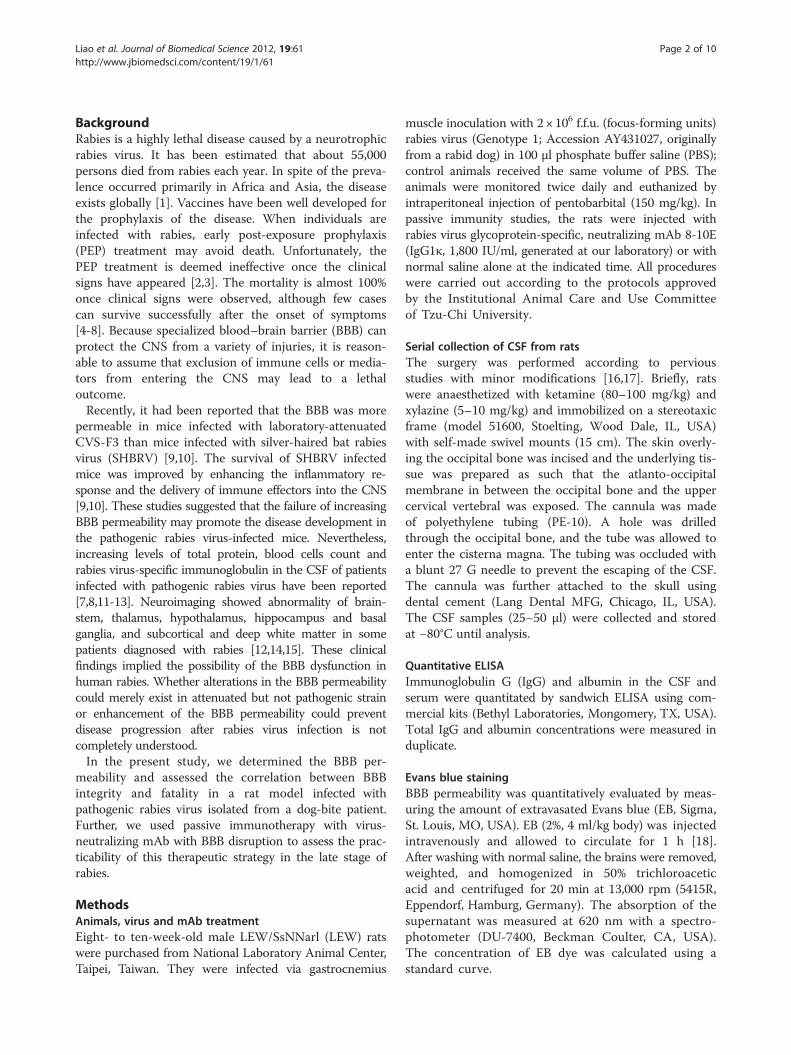
Background: Rabies is known to be lethal in human. Treatment with passive immunity for the rabies is effectiveonly when the patients have not shown the central nerve system (CNS) signs. The blood–brain barrier (BBB) is acomplex functional barrier that may compromise the therapeutic development in neurological diseases. The goal ofthis study is to determine the change of BBB integrity and to assess the therapeutic possibility of enhancing BBBpermeability combined with passive immunity in the late stage of rabies virus infection.
Methods: The integrity of BBB permeability in rats was measured by quantitative ELISA for total IgG and albuminlevels in the cerebrospinal fluid (CSF) and by exogenously applying Evans blue as a tracer. Western blotting ofoccludin and ZO-1, two tight junction proteins, was used to assess the molecular change of BBB structure.The breakdown of BBB with hypertonic arabinose, recombinant tumor necrosis factor-alpha (rTNF-γ), and focusedultrasound (FUS) were used to compare the extent of BBB disruption with rabies virus infection. Specific humoralimmunity was analyzed by immunofluorescent assay and rapid fluorescent focus inhibition test. Virus-neutralizingmonoclonal antibody (mAb) 8-10E was administered to rats with hypertonic breakdown of BBB as a passiveimmunotherapy to prevent the death from rabies.
Results: The BBB permeability was altered on day 7 post-infection. Increased BBB permeability induced by rabiesvirus infection was observed primarily in the cerebellum and spinal cord. Occludin was significantly decreased inboth the cerebral cortex and cerebellum. The rabies virus-specific antibody was not strongly elicited even in thepresence of clinical signs. Disruption of BBB had no direct association with the lethal outcome of rabies. Passiveimmunotherapy with virus-neutralizing mAb 8-10E with the hypertonic breakdown of BBB prolonged the survival ofrabies virus-infected rats.
Conclusions: We demonstrated that the BBB permeability was altered in a rat model with rabies virus inoculation.Delivery of neutralizing mAb to the infected site in brain combined with effective breakdown of BBB could be anaggressive but feasible therapeutic mode in rabies when the CNS infection has been established.
Keywords: Rabies, Blood–brain barrier, Central nerve system, Cerebrospinal fluid, Occludin, Hypertonic breakdown,Virus-neutralizing monoclonal antibody, Passive immunotherapy
* Correspondence: [email protected] of Medical Sciences, Tzu Chi University, Hualien, Taiwan2Department of Emerging Infectious Pathogen Research Laboratory, BuddhistTzu Chi General Hospital, Hualien, TaiwanFull list of author information is available at the end of the article
© 2012 Pi-Hung et al.; licensee BioMed Central Ltd. This is an Open Access article distributed under the terms of the CreativeCommons Attribution License (http://creativecommons.org/licenses/by/2.0), which permits unrestricted use, distribution, andreproduction in any medium, provided the original work is properly cited.
Liao et al. Journal of Biomedical Science 2012, 19:61 Page 2 of 10http://www.jbiomedsci.com/content/19/1/61
BackgroundRabies is a highly lethal disease caused by a neurotrophicrabies virus. It has been estimated that about 55,000persons died from rabies each year. In spite of the preva-lence occurred primarily in Africa and Asia, the diseaseexists globally [1]. Vaccines have been well developed forthe prophylaxis of the disease. When individuals areinfected with rabies, early post-exposure prophylaxis(PEP) treatment may avoid death. Unfortunately, thePEP treatment is deemed ineffective once the clinicalsigns have appeared [2,3]. The mortality is almost 100%once clinical signs were observed, although few casescan survive successfully after the onset of symptoms[4-8]. Because specialized blood–brain barrier (BBB) canprotect the CNS from a variety of injuries, it is reason-able to assume that exclusion of immune cells or media-tors from entering the CNS may lead to a lethaloutcome.Recently, it had been reported that the BBB was more
permeable in mice infected with laboratory-attenuatedCVS-F3 than mice infected with silver-haired bat rabiesvirus (SHBRV) [9,10]. The survival of SHBRV infectedmice was improved by enhancing the inflammatory re-sponse and the delivery of immune effectors into the CNS[9,10]. These studies suggested that the failure of increasingBBB permeability may promote the disease development inthe pathogenic rabies virus-infected mice. Nevertheless,increasing levels of total protein, blood cells count andrabies virus-specific immunoglobulin in the CSF of patientsinfected with pathogenic rabies virus have been reported[7,8,11-13]. Neuroimaging showed abnormality of brain-stem, thalamus, hypothalamus, hippocampus and basalganglia, and subcortical and deep white matter in somepatients diagnosed with rabies [12,14,15]. These clinicalfindings implied the possibility of the BBB dysfunction inhuman rabies. Whether alterations in the BBB permeabilitycould merely exist in attenuated but not pathogenic strainor enhancement of the BBB permeability could preventdisease progression after rabies virus infection is notcompletely understood.In the present study, we determined the BBB per-
meability and assessed the correlation between BBBintegrity and fatality in a rat model infected withpathogenic rabies virus isolated from a dog-bite patient.Further, we used passive immunotherapy with virus-neutralizing mAb with BBB disruption to assess the prac-ticability of this therapeutic strategy in the late stage ofrabies.
MethodsAnimals, virus and mAb treatmentEight- to ten-week-old male LEW/SsNNarl (LEW) ratswere purchased from National Laboratory Animal Center,Taipei, Taiwan. They were infected via gastrocnemius
muscle inoculation with 2 × 10⁶ f.f.u. (focus-forming units)rabies virus (Genotype 1; Accession AY431027, originallyfrom a rabid dog) in 100 μl phosphate buffer saline (PBS);control animals received the same volume of PBS. Theanimals were monitored twice daily and euthanized byintraperitoneal injection of pentobarbital (150 mg/kg). Inpassive immunity studies, the rats were injected withrabies virus glycoprotein-specific, neutralizing mAb 8-10E(IgG1κ, 1,800 IU/ml, generated at our laboratory) or withnormal saline alone at the indicated time. All procedureswere carried out according to the protocols approvedby the Institutional Animal Care and Use Committeeof Tzu-Chi University.
Serial collection of CSF from ratsThe surgery was performed according to perviousstudies with minor modifications [16,17]. Briefly, ratswere anaesthetized with ketamine (80–100 mg/kg) andxylazine (5–10 mg/kg) and immobilized on a stereotaxicframe (model 51600, Stoelting, Wood Dale, IL, USA)with self-made swivel mounts (15 cm). The skin overly-ing the occipital bone was incised and the underlying tis-sue was prepared as such that the atlanto-occipitalmembrane in between the occipital bone and the uppercervical vertebral was exposed. The cannula was madeof polyethylene tubing (PE-10). A hole was drilledthrough the occipital bone, and the tube was allowed toenter the cisterna magna. The tubing was occluded witha blunt 27 G needle to prevent the escaping of the CSF.The cannula was further attached to the skull usingdental cement (Lang Dental MFG, Chicago, IL, USA).The CSF samples (25–50 μl) were collected and storedat −80°C until analysis.
Quantitative ELISAImmunoglobulin G (IgG) and albumin in the CSF andserum were quantitated by sandwich ELISA using com-mercial kits (Bethyl Laboratories, Mongomery, TX, USA).Total IgG and albumin concentrations were measured induplicate.
Evans blue stainingBBB permeability was quantitatively evaluated by meas-uring the amount of extravasated Evans blue (EB, Sigma,St. Louis, MO, USA). EB (2%, 4 ml/kg body) was injectedintravenously and allowed to circulate for 1 h [18].After washing with normal saline, the brains were removed,weighted, and homogenized in 50% trichloroaceticacid and centrifuged for 20 min at 13,000 rpm (5415R,Eppendorf, Hamburg, Germany). The absorption of thesupernatant was measured at 620 nm with a spectro-photometer (DU-7400, Beckman Coulter, CA, USA).The concentration of EB dye was calculated using astandard curve.
Liao et al. Journal of Biomedical Science 2012, 19:61 Page 3 of 10http://www.jbiomedsci.com/content/19/1/61
Western blotting analysisConventional Western blotting was used. Briefly, brainsamples were homogenized in ice-cold radioimmunopre-cipitation (RIPA) buffer, supplemented with proteaseinhibitor cocktail tablets (Roche Diagnostics GmbH,Germany). The homogenates were centrifuged and pro-tein concentrations of the supernatants were determinedusing a Pierce protein assay kit (Pierce, Rockford, IL,USA). Equal amounts of protein (80 μg) were loadedonto 7.5% or 10% sodium dodecyl sulfate-polyacylamidegel, transferred onto polyvinylidene difluoride mem-branes (Millipore, Bedford, MA, USA). The membranewas then blocked with 5% nonfat milk and incubatedwith primary antibodies overnight at 4°C. The primaryantibodies and concentrations used were as follows:rabbit polyclonal anti-occludin (Zymed Laboratories,South San Francisco, CA, USA, 1:200), rabbit polyclonalanti-ZO-1 (Zymed Laboratories, USA, 1:200), and mousemonoclonal anti-β-actin (BD Biosciences, San Jose, CA,USA, 1:5000). Following washing and incubation withthe secondary antibody (Chemicon, Temecula, CA, USA,1:2000) for 60 min at room temperature, the membraneswere then probed with chemiluminescence reagents usinga commercially available kit (ECL Plus Western blottingdetection system, Amersham Biosciences, Little Chalfont,Buckinghamshire, UK) to visualize the signals, followed byexposure to X-ray films (Kodak, Rochester, NY, USA). In-tensities of the band were quantified with a densitometricanalysis system (GS-800 Calibrated Densitometer, Bio-Rad, Hercules, CA, USA), and calculated as the opticaldensity x area of band.
Disruption of BBB permeabilityThree methods were use to disrupt the BBB permeabil-ity. Hyperosmotic solution and TNF-α were performedas described previously [19-21]. Briefly, rats wereanesthetized with ketamine (80–100 mg/kg) and xylazine(5–10 mg/kg), and infused via carotid artery with 1.6 Marabinose (0.12 ml/sec, constant for 30 sec) or 106 IUhuman recombinant TNF-α protein (Peprotech Inc,Rocky Hill, NJ, USA). FUS treatment was described pre-viously [22,23]. SonoVueW SF6-coated ultrasound micro-bubbles (2–5 mm mean diameter, 1.5 mg/kg; BraccoDiagnostics Inc., Princeton, NJ, USA) were administeredintravenously by continuous injection with 0.2 ml salinesolution containing 0.1 ml heparin by a micropump(0.6 ml/min). Moderate ultrasound power (4.3 W, equiva-lent to a pressure of 0.4 MPa) was delivered to the brainwith the center of the focal zone positioned at a penetra-tion depth of 2–3 mm. In order to increase the BBBpermeability of entire half brain, multiple exposures werecarried out to completely cover the hemisphere. Animalstypically underwent 3 sonications, with the spacing be-tween individual adjacent focal positions set to 3 mm. In
each sonication, burst mode ultrasound was delivered withthe following parameters: burst length=10 ms, pulse re-petitive frequency=2 Hz, and sonication duration=1 min.
Measurement of serum and CSF antibody titersLevels of rabies virus-specific total IgG from ratsinfected with rabies virus as well as uninfected controlgroups were measured by indirect immunofluorescentassay (IFA). The titer was defined as the reciprocal ofthe highest dilution factor of test samples in which 50% ormore of the fields examined contained specifically fluores-cing cells. Virus-neutralizing antibody (VNA) titers weredetermined by the rapid fluorescent focus inhibition test(RFFIT) as previously described [24,25]. The VNA titerwas determined as the last dilution of serum that wascapable of reducing the number of FFU by 50%.
Passive immunotherapy with BBB openingExperiments were divided into two parts: (1) for the pas-sive immunotherapy alone, the rabies virus-infected ratswere intraperitoneally administrated 10 mg mAb 8-10E,and (2) for the passive immunotherapy combined withBBB opening (BBBO), mAb 8-10E was administered6 hrs before BBB disruption. The studies were carriedout on day 7 and day 9 post-infection (p.i.). The ratswere continually observed to day 40 p.i. and then eutha-nized for analysis.
Reverse transcription-polymerase chain reaction (RT-PCR)analysisTotal RNA was extracted from the cerebral cortex, cere-bellum, and spinal cord of rabies virus infected andcontrol rats using the RNeasy Mini kit (Qiagen, Hilden,Germany) according to the manufacturer’s instructions.The nucleoprotein (N) gene of rabies virus was amplifiedby one-step RT-PCR. The nucleotide sequences of theprimers used for PCR are forward primer, 5'- CTACAATGGATGCCGAC-3' and reverse primer, 5'-TCATAACGGAGAGATCGCCAC-3'. RT-PCR was per-formed with 4 μl of RNA (0.2-0.4 μg) and the OneStepRT-PCR kit (Qiagen, Germany) according to the manu-facture’s instructions. Each reaction mixture was incu-bated at 50°C for 30 min, followed by 40 cycles at 94°Cfor 30 sec, 55°C for 30 sec, 72°C for 1 min, and 72°Cfor 10 min. Equal amounts of amplified PCR productswere electrophoresed in 1.5% agarose gels containingethidium bromide. The bands were visualized underUV light and photographed.
Statistical analysisAll data were expressed as mean ± SEM. One-wayANOVA was used to compare means between experi-mental groups. Student’s t-test was used to determinethe significance between groups. The survival rate was
Liao et al. Journal of Biomedical Science 2012, 19:61 Page 4 of 10http://www.jbiomedsci.com/content/19/1/61
analyzed by the Kaplan–Meier method and log-rankstatistics were used to test the difference betweengroups. The GraphPad Prism statistical package, version5.0c (GraphPad Software, Inc., San Diego, CA, USA) wasused for comparison. Differences were considered statis-tically significant at p < 0.05.
ResultsRabies viral antigens were detected in the CNS of LEWrats after challengeClinical signs of rabies infection appeared on day 9–10p.i.. Unsteady gait and hunched back were observedin the beginning, swimming movement, slow motion,and limbs paralysis in the late stage. Rats died fromthe infection within 2 to 4 days once the neural signswere observed. The viral genome was detected byIFA, RT-PCR and immunohistochemistry on day 5 p.i.in the brain and spread out within the next 2 days(data not shown).
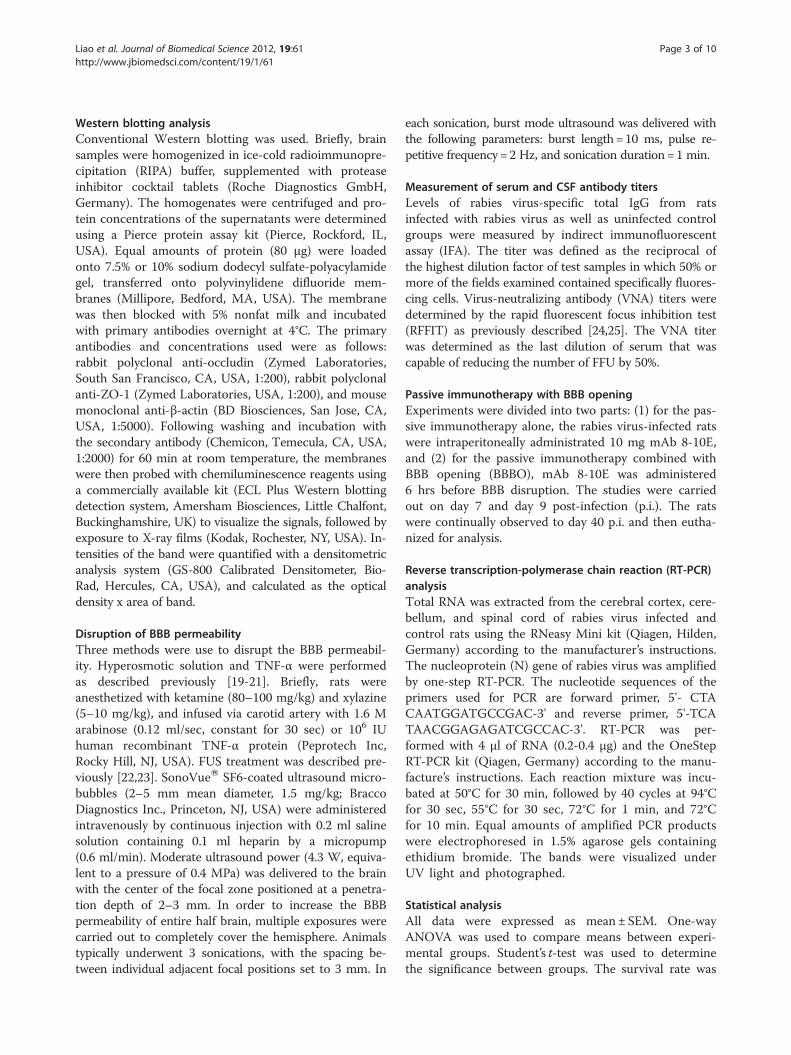
BBB permeability was disrupted after rabies viruschallengeTo determine the possible alteration of BBB permeabil-ity, the CSF were serially sampled and analyzed. Thestatus of the BBB permeability was monitored post-infection every single day by quantifying the albuminand total IgG in the CSF using quantitative ELISA. Theresults showed that the albumin and total IgG levelswere elevated significantly on day 7 and continually
Figure 1 BBB permeability was altered in the CSF of rats infected witalbumin and (B) total IgG from the CSF after rabies virus infection and expdifferences in permeability was calculated using Student’s t-test and are depresented in (C) for albumin and (D) for IgG. Each dot represented one ind
elevated to day 12 p.i. (Figure 1A, B). The time course ofthe changes of the CSF to serum ratios of albumin andtotal IgG in rabies virus-infected rats is shown inFigure 1C and D. There were about 7- and 10-foldincrease in albumin and total IgG, respectively, in theCSF on day 12 p.i. when compared to uninfected rats.The time course of the increase in BBB permeabilityparalleled that of animal illness.
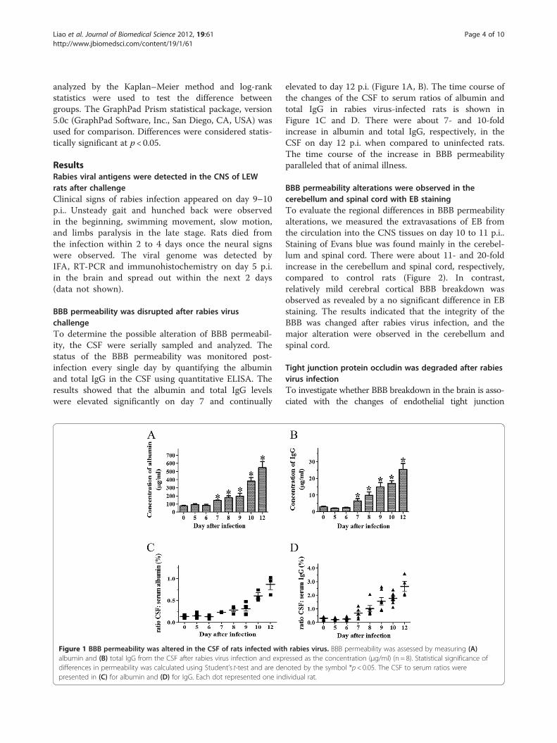
BBB permeability alterations were observed in thecerebellum and spinal cord with EB stainingTo evaluate the regional differences in BBB permeabilityalterations, we measured the extravasations of EB fromthe circulation into the CNS tissues on day 10 to 11 p.i..Staining of Evans blue was found mainly in the cerebel-lum and spinal cord. There were about 11- and 20-foldincrease in the cerebellum and spinal cord, respectively,compared to control rats (Figure 2). In contrast,relatively mild cerebral cortical BBB breakdown wasobserved as revealed by a no significant difference in EBstaining. The results indicated that the integrity of theBBB was changed after rabies virus infection, and themajor alteration were observed in the cerebellum andspinal cord.
Tight junction protein occludin was degraded after rabiesvirus infectionTo investigate whether BBB breakdown in the brain is asso-ciated with the changes of endothelial tight junction
h rabies virus. BBB permeability was assessed by measuring (A)ressed as the concentration (μg/ml) (n = 8). Statistical significance ofnoted by the symbol *p< 0.05. The CSF to serum ratios wereividual rat.
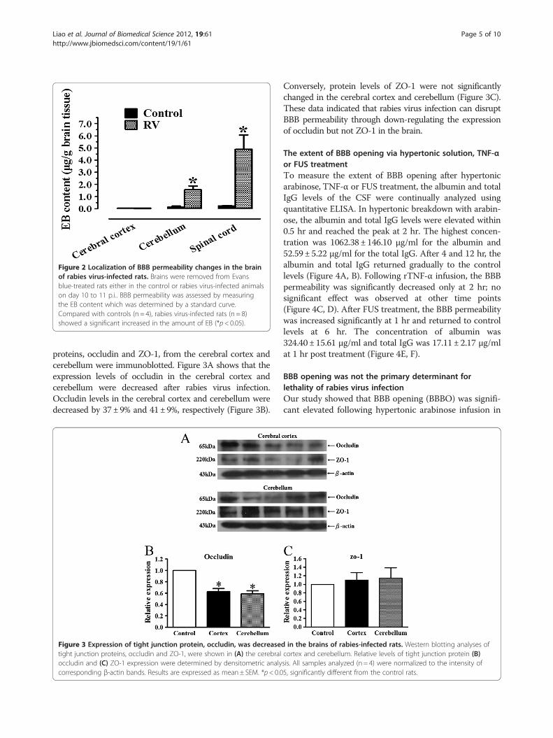
Figure 2 Localization of BBB permeability changes in the brainof rabies virus-infected rats. Brains were removed from Evansblue-treated rats either in the control or rabies virus-infected animalson day 10 to 11 p.i.. BBB permeability was assessed by measuringthe EB content which was determined by a standard curve.Compared with controls (n = 4), rabies virus-infected rats (n = 8)showed a significant increased in the amount of EB (*p< 0.05).
Liao et al. Journal of Biomedical Science 2012, 19:61 Page 5 of 10http://www.jbiomedsci.com/content/19/1/61
proteins, occludin and ZO-1, from the cerebral cortex andcerebellum were immunoblotted. Figure 3A shows that theexpression levels of occludin in the cerebral cortex andcerebellum were decreased after rabies virus infection.Occludin levels in the cerebral cortex and cerebellum weredecreased by 37±9% and 41±9%, respectively (Figure 3B).
Figure 3 Expression of tight junction protein, occludin, was decreasetight junction proteins, occludin and ZO-1, were shown in (A) the cerebraloccludin and (C) ZO-1 expression were determined by densitometric analycorresponding β-actin bands. Results are expressed as mean± SEM. *p< 0.0
Conversely, protein levels of ZO-1 were not significantlychanged in the cerebral cortex and cerebellum (Figure 3C).These data indicated that rabies virus infection can disruptBBB permeability through down-regulating the expressionof occludin but not ZO-1 in the brain.
The extent of BBB opening via hypertonic solution, TNF-αor FUS treatmentTo measure the extent of BBB opening after hypertonicarabinose, TNF-α or FUS treatment, the albumin and totalIgG levels of the CSF were continually analyzed usingquantitative ELISA. In hypertonic breakdown with arabin-ose, the albumin and total IgG levels were elevated within0.5 hr and reached the peak at 2 hr. The highest concen-tration was 1062.38 ± 146.10 μg/ml for the albumin and52.59 ± 5.22 μg/ml for the total IgG. After 4 and 12 hr, thealbumin and total IgG returned gradually to the controllevels (Figure 4A, B). Following rTNF-α infusion, the BBBpermeability was significantly decreased only at 2 hr; nosignificant effect was observed at other time points(Figure 4C, D). After FUS treatment, the BBB permeabilitywas increased significantly at 1 hr and returned to controllevels at 6 hr. The concentration of albumin was324.40± 15.61 μg/ml and total IgG was 17.11 ± 2.17 μg/mlat 1 hr post treatment (Figure 4E, F).
BBB opening was not the primary determinant forlethality of rabies virus infectionOur study showed that BBB opening (BBBO) was signifi-cant elevated following hypertonic arabinose infusion in
d in the brains of rabies-infected rats. Western blotting analyses ofcortex and cerebellum. Relative levels of tight junction protein (B)sis. All samples analyzed (n = 4) were normalized to the intensity of5, significantly different from the control rats.
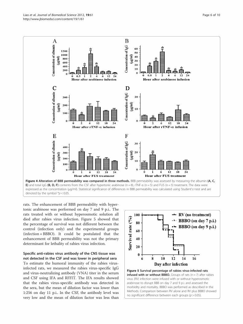
Figure 4 Alteration of BBB permeability was compared in three methods. BBB permeability was assessed by measuring the albumin (A, C,E) and total IgG (B, D, F) contents from the CSF after hypertonic arabinose (n = 8), rTNF-α (n = 5) and FUS (n = 5) treatment. The data wereexpressed as the concentration (μg/ml). Statistical significance of differences in BBB permeability was calculated using Student’s t-test and aredenoted by the symbol *p< 0.05.
Figure 5 Survival percentage of rabies virus-infected ratsinfused with or without BBBO. Groups of rats (n = 7) after rabiesvirus (RV) infection were infused with or without hyperosmoticarabinose to disrupt BBB on day 7 and 9 p.i. and assessed themorbidity and mortality. BBBO was performed as described in theMethods. Comparison between RV alone and RV plus BBBO showedno significant difference between each groups (p> 0.05).
Liao et al. Journal of Biomedical Science 2012, 19:61 Page 6 of 10http://www.jbiomedsci.com/content/19/1/61
rats. The enhancement of BBB permeability with hyper-tonic arabinose was performed on day 7 and 9 p.i.. Therats treated with or without hyperosmotic solution alldied after rabies virus infection. Figure 5 showed thatthe percentage of survival was not different between thecontrol (infection only) and the experimental groups(infection + BBBO). It could be postulated that theenhancement of BBB permeability was not the primarydeterminant for lethality of rabies virus infection.
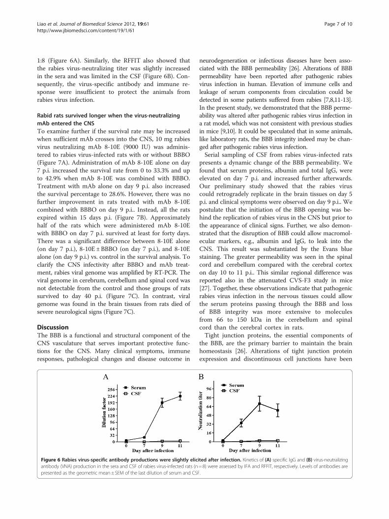
Specific anti-rabies virus antibody of the CNS tissue wasnot detected in the CSF and was lower in peripheral seraTo estimate the humoral immunity of the rabies virus-infected rats, we measured the rabies virus-specific IgGand virus-neutralizing antibody (VNA) titer in the serumand CSF using IFA and RFFIT. The IFA results showedthat the rabies virus-specific antibody was detected inthe sera, but the mean of dilution factor was lower than1:256 on day 11 p.i.. In the CSF, the antibody level wasvery low and the mean of dilution factor was less than
Liao et al. Journal of Biomedical Science 2012, 19:61 Page 7 of 10http://www.jbiomedsci.com/content/19/1/61
1:8 (Figure 6A). Similarly, the RFFIT also showed thatthe rabies virus-neutralizing titer was slightly increasedin the sera and was limited in the CSF (Figure 6B). Con-sequently, the virus-specific antibody and immune re-sponse were insufficient to protect the animals fromrabies virus infection.
Rabid rats survived longer when the virus-neutralizingmAb entered the CNSTo examine further if the survival rate may be increasedwhen sufficient mAb crosses into the CNS, 10 mg rabiesvirus neutralizing mAb 8-10E (9000 IU) was adminis-tered to rabies virus-infected rats with or without BBBO(Figure 7A). Administration of mAb 8-10E alone on day7 p.i. increased the survival rate from 0 to 33.3% and upto 42.9% when mAb 8-10E was combined with BBBO.Treatment with mAb alone on day 9 p.i. also increasedthe survival percentage to 28.6%. However, there was nofurther improvement in rats treated with mAb 8-10Ecombined with BBBO on day 9 p.i.. Instead, all the ratsexpired within 15 days p.i. (Figure 7B). Approximatelyhalf of the rats which were administered mAb 8-10Ewith BBBO on day 7 p.i. survived at least for forty days.There was a significant difference between 8-10E alone(on day 7 p.i.), 8-10E ±BBBO (on day 7 p.i.), and 8-10Ealone (on day 9 p.i.) vs. control in the survival analysis. Toclarify the CNS infectivity after BBBO and mAb treat-ment, rabies viral genome was amplified by RT-PCR. Theviral genome in cerebrum, cerebellum and spinal cord wasnot detectable from the control and those groups of ratssurvived to day 40 p.i. (Figure 7C). In contrast, viralgenome was found in the brain tissues from rats died ofsevere neurological signs (Figure 7C).
DiscussionThe BBB is a functional and structural component of theCNS vasculature that serves important protective func-tions for the CNS. Many clinical symptoms, immuneresponses, pathological changes and disease outcome in
Figure 6 Rabies virus-specific antibody productions were slightly elicantibody (VNA) production in the sera and CSF of rabies virus-infected rats (npresented as the geometric mean±SEM of the last dilution of serum and CSF
neurodegeneration or infectious diseases have been asso-ciated with the BBB permeability [26]. Alterations of BBBpermeability have been reported after pathogenic rabiesvirus infection in human. Elevation of immune cells andleakage of serum components from circulation could bedetected in some patients suffered from rabies [7,8,11-13].In the present study, we demonstrated that the BBB perme-ability was altered after pathogenic rabies virus infection ina rat model, which was not consistent with previous studiesin mice [9,10]. It could be speculated that in some animals,like laboratory rats, the BBB integrity indeed may be chan-ged after pathogenic rabies virus infection.Serial sampling of CSF from rabies virus-infected rats
presents a dynamic change of the BBB permeability. Wefound that serum proteins, albumin and total IgG, wereelevated on day 7 p.i. and increased further afterwards.Our preliminary study showed that the rabies viruscould retrogradely replicate in the brain tissues on day 5p.i. and clinical symptoms were observed on day 9 p.i.. Wepostulate that the initiation of the BBB opening was be-hind the replication of rabies virus in the CNS but prior tothe appearance of clinical signs. Further, we also demon-strated that the disruption of BBB could allow macromol-ecular markers, e.g., albumin and IgG, to leak into theCNS. This result was substantiated by the Evans bluestaining. The greater permeability was seen in the spinalcord and cerebellum compared with the cerebral cortexon day 10 to 11 p.i.. This similar regional difference wasreported also in the attenuated CVS-F3 study in mice[27]. Together, these observations indicate that pathogenicrabies virus infection in the nervous tissues could allowthe serum proteins passing through the BBB and lossof BBB integrity was more extensive to moleculesfrom 66 to 150 kDa in the cerebellum and spinalcord than the cerebral cortex in rats.Tight junction proteins, the essential components of
the BBB, are the primary barrier to maintain the brainhomeostasis [26]. Alterations of tight junction proteinexpression and discontinuous cell junctions have been
ited after infection. Kinetics of (A) specific IgG and (B) virus-neutralizing=8) were assessed by IFA and RFFIT, respectively. Levels of antibodies are.
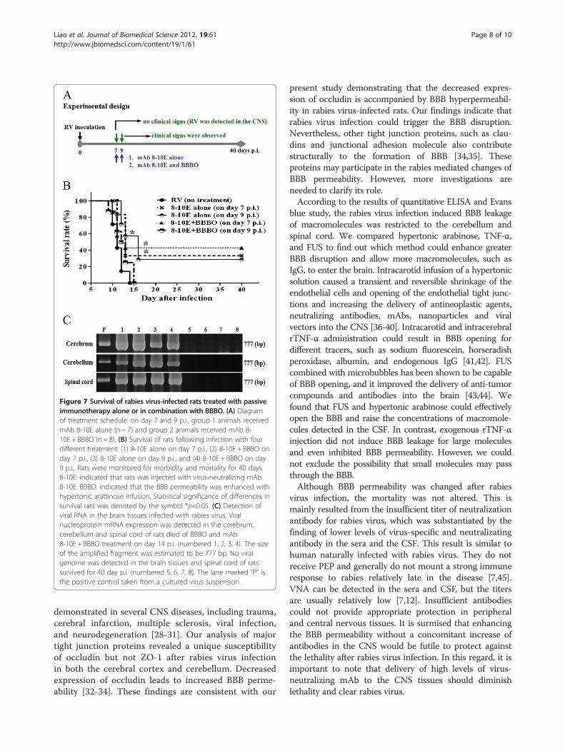
Figure 7 Survival of rabies virus-infected rats treated with passiveimmunotherapy alone or in combination with BBBO. (A) Diagramof treatment schedule: on day 7 and 9 p.i., group 1 animals receivedmAb 8-10E alone (n = 7) and group 2 animals received mAb 8-10E + BBBO (n= 8), (B) Survival of rats following infection with fourdifferent treatment: (1) 8-10E alone on day 7 p.i., (2) 8-10E + BBBO onday 7 p.i., (3) 8-10E alone on day 9 p.i., and (4) 8-10E + BBBO on day9 p.i.. Rats were monitored for morbidity and mortality for 40 days.8-10E: indicated that rats was injected with virus-neutralizing mAb8-10E. BBBO: indicated that the BBB permeability was enhanced withhypertonic arabinose infusion. Statistical significance of differences insurvival rats was denoted by the symbol *p<0.05. (C) Detection ofviral RNA in the brain tissues infected with rabies virus. Viralnucleoprotein mRNA expression was detected in the cerebrum,cerebellum and spinal cord of rats died of BBBO and mAb8-10E + BBBO treatment on day 14 p.i. (numbered 1, 2, 3, 4). The sizeof the amplified fragment was estimated to be 777 bp. No viralgenome was detected in the brain tissues and spinal cord of ratssurvived for 40 day p.i. (numbered 5, 6, 7, 8). The lane marked “P” isthe positive control taken from a cultured virus suspension.
Liao et al. Journal of Biomedical Science 2012, 19:61 Page 8 of 10http://www.jbiomedsci.com/content/19/1/61
demonstrated in several CNS diseases, including trauma,cerebral infarction, multiple sclerosis, viral infection,and neurodegeneration [28-31]. Our analysis of majortight junction proteins revealed a unique susceptibilityof occludin but not ZO-1 after rabies virus infectionin both the cerebral cortex and cerebellum. Decreasedexpression of occludin leads to increased BBB perme-ability [32-34]. These findings are consistent with our
present study demonstrating that the decreased expres-sion of occludin is accompanied by BBB hyperpermeabil-ity in rabies virus-infected rats. Our findings indicate thatrabies virus infection could trigger the BBB disruption.Nevertheless, other tight junction proteins, such as clau-dins and junctional adhesion molecule also contributestructurally to the formation of BBB [34,35]. Theseproteins may participate in the rabies mediated changes ofBBB permeability. However, more investigations areneeded to clarify its role.According to the results of quantitative ELISA and Evans
blue study, the rabies virus infection induced BBB leakageof macromolecules was restricted to the cerebellum andspinal cord. We compared hypertonic arabinose, TNF-α,and FUS to find out which method could enhance greaterBBB disruption and allow more macromolecules, such asIgG, to enter the brain. Intracarotid infusion of a hypertonicsolution caused a transient and reversible shrinkage of theendothelial cells and opening of the endothelial tight junc-tions and increasing the delivery of antineoplastic agents,neutralizing antibodies, mAbs, nanoparticles and viralvectors into the CNS [36-40]. Intracarotid and intracerebralrTNF-α administration could result in BBB opening fordifferent tracers, such as sodium fluorescein, horseradishperoxidase, albumin, and endogenous IgG [41,42]. FUScombined with microbubbles has been shown to be capableof BBB opening, and it improved the delivery of anti-tumorcompounds and antibodies into the brain [43,44]. Wefound that FUS and hypertonic arabinose could effectivelyopen the BBB and raise the concentrations of macromole-cules detected in the CSF. In contrast, exogenous rTNF-αinjection did not induce BBB leakage for large moleculesand even inhibited BBB permeability. However, we couldnot exclude the possibility that small molecules may passthrough the BBB.Although BBB permeability was changed after rabies
virus infection, the mortality was not altered. This ismainly resulted from the insufficient titer of neutralizationantibody for rabies virus, which was substantiated by thefinding of lower levels of virus-specific and neutralizatingantibody in the sera and the CSF. This result is similar tohuman naturally infected with rabies virus. They do notreceive PEP and generally do not mount a strong immuneresponse to rabies relatively late in the disease [7,45].VNA can be detected in the sera and CSF, but the titersare usually relatively low [7,12]. Insufficient antibodiescould not provide appropriate protection in peripheraland central nervous tissues. It is surmised that enhancingthe BBB permeability without a concomitant increase ofantibodies in the CNS would be futile to protect againstthe lethality after rabies virus infection. In this regard, it isimportant to note that delivery of high levels of virus-neutralizing mAb to the CNS tissues should diminishlethality and clear rabies virus.
Liao et al. Journal of Biomedical Science 2012, 19:61 Page 9 of 10http://www.jbiomedsci.com/content/19/1/61
It has also been reported that exogenous neutralizingantibodies accompanied with the enhancement of BBBpermeability can raise the survival more than the adminis-tration of monoclonal antibodies alone [46,47]. In ourstudy, PEP treatment using virus-neutralizing mAb 8-10Ecombined with BBB disruption elevated the survival inLEW rats inoculated with rabies virus on day 7 p.i., how-ever; the effect was diminished on day 9 p.i.. One possibleexplanation may be that the adverse effects of osmoticinfusion, such as seizures, reinforce damage to the brainalready exhibiting neuronal dysfunction [48]. It is surpris-ing to note that neutralizing mAb 8-10E treatment aloneincreased the survival rate by 33.3% and 28.6%. respect-ively, on day 7 and 9 p.i. We hypothesize that administra-tion of sufficient neutralizing mAbs into the CNS couldrescue the rats from rabies-induced lethality and earlytreatment with mAb also increase survival rate.Until now, besides the success of the Wisconsin’s
patient treated with the Milwaukee Protocol, severalothers have employed similar treatment regimens withminimal beneficial outcomes [6,49-51]. Although a frac-tion of rabies virus-infected rats could be saved withpassive neutralizing mAb treatment, whether greaterimprovement could be achieved with immune therapyneeds more studies. In future development, a passivelyadministrated mAb or such a humanized mAb com-bined with effective and global BBB breakdown may pro-vide an alternative therapy for rescuing patients whohave been diagnosed with rabies.
ConclusionsThe present study revealed that BBB permeability wasaltered after pathogenic rabies virus infection in rats.Sufficient virus-neutralizing antibody plays a major rolein determining the survival from rabies virus infection.Our study indicates that enhancing BBB opening com-bined with delivering sufficient virus-neutralizing mAbto the brain may provide an effective treatment whenthe CNS infection has been established.
Competing interestsThe authors declare that they have no competing interests.
Authors’ contributionsPHL performed the major experiments and wrote the manuscript. HHY andPTC were responsible for virus isolation and mAb production. PCC and HLLwere for technical support of FUS study. MHW participated in datainterpretation and manuscript improvement. LKC conceived the study,designed the experiments and analyzed the data. All authors read andapproved the final manuscript.
AcknowledgmentThis work was supported by Buddhist Tzu Chi General Hospital grantsTCRD97-12, TCRD98-03, TCSP-9803, and TCSP-9903.
Author details1Institute of Medical Sciences, Tzu Chi University, Hualien, Taiwan.2Department of Emerging Infectious Pathogen Research Laboratory, BuddhistTzu Chi General Hospital, Hualien, Taiwan. 3Department of Life Science, Tzu
Chi University, Hualien, Taiwan. 4Department of Electrical Engineering,Chang-Gung University, Taoyuan, Taiwan. 5Department of LaboratoryDiagnostics, Medical School, Tzu Chi University, Hualien, Taiwan.6Department of Clinical Pathology, Buddhist Tzu Chi General Hospital,Hualien, Taiwan. 7Department of Clinical Pathology, Buddhist Tzu Chi GeneralHospital, Medical School, Tzu Chi University, No 707, Section 3, Chung-YangRoad, Hualien 970, Taiwan.
Received: 9 April 2012 Accepted: 13 June 2012Published: 26 June 2012
References1. WHO: Expert Consultation on rabies. World Health Organ Tech Rep Ser
2005, 931:1–88. back cover.2. Jackson AC: Therapy of rabies encephalitis. Biomedica 2009, 29:169–176.3. Nigg AJ, Walker PL: Overview, prevention, and treatment of rabies.
Pharmacotherapy 2009, 29:1182–1195.4. McDermid RC, Saxinger L, Lee B, Johnstone J, Gibney RT, Johnson M,
Bagshaw SM: Human rabies encephalitis following bat exposure: failureof therapeutic coma. CMAJ 2008, 178:557–561.
5. Schmiedel S, Panning M, Lohse A, Kreymann KG, Gerloff C, Burchard G,Drosten C, Schmiedel S, Panning M, Lohse A, Kreymann KG, Gerloff C,Burchard G, Drosten C: Case report on fatal human rabies infection inHamburg, Germany, March 2007. Euro Surveill 2007, 12:E070531–E070535.
6. Hemachudha T, Sunsaneewitayakul B, Desudchit T, Suankratay C, Sittipunt C,Wacharapluesadee S, Khawplod P, Wilde H, Jackson AC: Failure oftherapeutic coma and ketamine for therapy of human rabies. J Neurovirol2006, 12:407–409.
7. Human rabies--Indiana and California. MMWR Morb Mortal Wkly Rep 2007,56:361–365.
8. Willoughby RE Jr, Tieves KS, Hoffman GM, Ghanayem NS, Amlie-Lefond CM,Schwabe MJ, Chusid MJ, Rupprecht CE: Survival after treatment of rabieswith induction of coma. N Engl J Med 2005, 352:2508–2514.
9. Roy A, Hooper DC: Lethal silver-haired bat rabies virus infection can beprevented by opening the blood–brain barrier. J Virol 2007, 81:7993–7998.
10. Roy A, Phares TW, Koprowski H, Hooper DC: Failure to open the blood–brain barrier and deliver immune effectors to central nervous systemtissues leads to the lethal outcome of silver-haired bat rabies virusinfection. J Virol 2007, 81:1110–1118.
11. Human rabies–Tennessee. MMWR Morb Mortal Wkly Rep 2002, 51:828–829.12. Human rabies–Minnesota. MMWR Morb Mortal Wkly Rep 2008, 57:460–462.13. Despond O, Tucci M, Decaluwe H, Gregoire MC, S Teitelbaum J, Turgeon N:
Rabies in a nine-year-old child: The myth of the bite. Can J Infect Dis2002, 13:121–125.
14. Laothamatas J, Hemachudha T, Mitrabhakdi E, Wannakrairot P,Tulayadaechanont S: MR imaging in human rabies. Am J Neuroradiol 2003,24:1102–1109.
15. Awasthi M, Parmar H, Patankar T, Castillo M: Imaging findings in rabiesencephalitis. Am J Neuroradiol 2001, 22:677–680.
16. Huang YL, Saljo A, Suneson A, Hansson HA: A new approach for multiplesampling of cisternal cerebrospinal fluid in rodents with minimal traumaand inflammation. J Neurosci Methods 1995, 63:13–22.
17. van den Berg MP, Romeijn SG, Verhoef JC, Merkus FW: Serial cerebrospinalfluid sampling in a rat model to study drug uptake from the nasalcavity. J Neurosci Methods 2002, 116:99–107.
18. Esen F, Erdem T, Aktan D, Orhan M, Kaya M, Eraksoy H, Cakar N, Telci L:Effect of magnesium sulfate administration on blood–brain barrier in arat model of intraperitoneal sepsis: a randomized controlledexperimental study. Crit Care 2005, 9:R18–R23.
19. Rapoport SI, Fredericks WR, Ohno K, Pettigrew KD: Quantitative aspects ofreversible osmotic opening of the blood–brain barrier. Am J Physiol 1980,238:R421–R431.
20. Robinson PJ, Rapoport SI: Size selectivity of blood–brain barrierpermeability at various times after osmotic opening. Am J Physiol 1987,253:R459–R466.
21. Rapoport SI: Osmotic opening of the blood–brain barrier: principles,mechanism, and therapeutic applications. Cell Mol Neurobiol 2000,20:217–230.
22. Liu HL, Wai YY, Hsu PH, Lyu LA, Wu JS, Shen CR, Chen JC, Yen TC, Wang JJ:In vivo assessment of macrophage CNS infiltration during disruption of
Liao et al. Journal of Biomedical Science 2012, 19:61 Page 10 of 10http://www.jbiomedsci.com/content/19/1/61
the blood–brain barrier with focused ultrasound: a magnetic resonanceimaging study. J Cerebral Blood Flow Metab 2010, 30:674.
23. Liu HL, Hua MY, Yang HW, Huang CY, Chu PC, Wu JS, Tseng IC, Wang JJ,Yen TC, Chen PY, Wei KC: Magnetic resonance monitoring of focusedultrasound/magnetic nanoparticle targeting delivery of therapeuticagents to the brain. Proc Natl Acad Sci U S A 2010, 107:15205–15210.
24. Zalan E, Wilson C, Pukitis D: A microtest for the quantitation of rabiesvirus neutralizing antibodies. J Biol Stand 1979, 7:213–220.
25. Smith JS, Yager PA, Baer GM: A rapid reproducible test for determiningrabies neutralizing antibody. Bull World Health Organ 1973, 48:535–541.
26. Hawkins BT, Davis TP: The blood–brain barrier/neurovascular unit inhealth and disease. Pharmacol Rev 2005, 57:173–185.
27. Phares TW, Kean RB, Mikheeva T, Hooper DC: Regional differences inblood–brain barrier permeability changes and inflammation in theapathogenic clearance of virus from the central nervous system.J Immunol 2006, 176:7666–7675.
28. Dallasta LM, Pisarov LA, Esplen JE, Werley JV, Moses AV, Nelson JA, AchimCL: Blood–brain barrier tight junction disruption in humanimmunodeficiency virus-1 encephalitis. Am J Pathoy 1999, 155:1915–1927.
29. Ng I, Yap E, Tan WL, Kong NY: Blood–brain barrier disruption followingtraumatic brain injury: roles of tight junction proteins. Ann Acad MedSingapore 2003, 32(Suppl 5):S63–S66.
30. Kirk J, Plumb J, Mirakhur M, McQuaid S: Tight junctional abnormality inmultiple sclerosis white matter affects all calibres of vessel and isassociated with blood–brain barrier leakage and active demyelination.J Pathog 2003, 201:319–327.
31. Mark KS, Davis TP: Cerebral microvascular changes in permeability andtight junctions induced by hypoxia-reoxygenation. Am J Physiol Heart CircPhysiol 2002, 282:H1485–H1494.
32. Brooks TA, Hawkins BT, Huber JD, Egleton RD, Davis TP: Chronicinflammatory pain leads to increased blood–brain barrier permeabilityand tight junction protein alterations. Am J Physiol Heart Circ Physiol 2005,289:H738–H743.
33. Nishioku T, Yamauchi A, Takata F, Watanabe T, Furusho K, Shuto H, DohguS, Kataoka Y: Disruption of the blood–brain barrier in collagen-inducedarthritic mice. Neurosci Lett 2010, 482:208–211.
34. Nusrat A, Turner JR, Madara JL: Molecular physiology andpathophysiology of tight junctions. IV. Regulation of tight junctions byextracellular stimuli: nutrients, cytokines, and immune cells. Am J PhysiolGastrointest Liver Physiol 2000, 279:G851–G857.
35. Huber JD, Egleton RD, Davis TP: Molecular physiology andpathophysiology of tight junctions in the blood–brain barrier. TrendsNeurosci 2001, 24:719–725.
36. Blanchette M, Fortin D: Blood–brain barrier disruption in the treatment ofbrain tumors. Methods Mol Biol 2011, 686:447–463.
37. Rapoport SI: Advances in osmotic opening of the blood–brain barrier toenhance CNS chemotherapy. Expert opin investigat drugs 2001, 10:1809–1818.
38. Rousseau V, Denizot B, Pouliquen D, Jallet P, Le Jeune JJ: Investigation ofblood–brain barrier permeability to magnetite-dextran nanoparticles(MD3) after osmotic disruption in rats. MAGMA 1997, 5:213–222.
39. Nilaver G, Muldoon LL, Kroll RA, Pagel MA, Breakefield XO, Davidson BL,Neuwelt EA: Delivery of herpesvirus and adenovirus to nude ratintracerebral tumors after osmotic blood–brain barrier disruption. ProcNatl Acad Sci U S A 1995, 92:9829–9833.
40. Hicks JT, Albrecht P, Rapoport SI: Entry of neutralizing antibody to measlesinto brain and cerebrospinal fluid of immunized monkeys after osmoticopening of the blood–brain barrier. Exp Neurol 1976, 53:768–779.
41. Abraham CS, Deli MA, Joo F, Megyeri P, Torpier G: Intracarotid tumornecrosis factor-alpha administration increases the blood–brain barrierpermeability in cerebral cortex of the newborn pig: quantitative aspectsof double-labelling studies and confocal laser scanning analysis. NeurosciLett 1996, 208:85–88.
42. Wright JL, Merchant RE: Blood–brain barrier changes followingintracerebral injection of human recombinant tumor necrosis factor-alpha in the rat. J Neuro-oncol 1994, 20:17–25.
43. Kinoshita M, McDannold N, Jolesz FA, Hynynen K: Targeted delivery ofantibodies through the blood–brain barrier by MRI-guided focusedultrasound. Bioche Biophys Res Comm 2006, 340:1085–1090.
44. Kinoshita M, McDannold N, Jolesz FA, Hynynen K: Noninvasive localizeddelivery of Herceptin to the mouse brain by MRI-guided focused
ultrasound-induced blood–brain barrier disruption. Proc Natl Acad Sci U SA 2006, 103:11719–11723.
45. Human rabies–Mississippi, 2005. MMWR Morb Mortal Wkly Rep 2006,55:207–208.
46. Neuwelt EA, Barnett PA, Hellstrom I, Hellstrom KE, Beaumier P, McCormickCI, Weigel RM: Delivery of melanoma-associated immunoglobulinmonoclonal antibody and Fab fragments to normal brain utilizingosmotic blood–brain barrier disruption. Cancer Res 1988, 48:4725–4729.
47. Boockvar JA, Tsiouris AJ, Hofstetter CP, Kovanlikaya I, Fralin S, KesavabhotlaK, Seedial SM, Pannullo SC, Schwartz TH, Stieg P, Zimmerman RD, KnopmanJ, Scheff RJ, Christos P, Vallabhajosula S, Riina HA: Safety and maximumtolerated dose of superselective intraarterial cerebral infusion ofbevacizumab after osmotic blood–brain barrier disruption for recurrentmalignant glioma. Clinical article. J Neurosurg 2011, 114:624–632.
48. Bellavance MA, Blanchette M, Fortin D: Recent advances in blood–brainbarrier disruption as a CNS delivery strategy. AAPS J 2008, 10:166–177.
49. Jackson AC: Rabies in the critical care unit: diagnostic and therapeuticapproaches. Can J Neurol Sci 2011, 38:689–695.
50. Hunter M, Johnson N, Hedderwick S, McCaughey C, Lowry K, McConville J,Herron B, McQuaid S, Marston D, Goddard T, Harkess G, Goharriz H, Voller K,Solomon T, Willoughby RE, Fooks AR: Immunovirological correlates inhuman rabies treated with therapeutic coma. J Med Virol 2010, 82:1255–1265.
51. Badillo R, Mantilla JC, Pradilla G: Human rabies encephalitis by a vampirebat bite in an urban area of Colombia. Biomedica 2009, 29:191–203.
doi:10.1186/1423-0127-19-61Cite this article as: Liao et al.: Sufficient virus-neutralizing antibody inthe central nerve system improves the survival of rabid rats. Journal ofBiomedical Science 2012 19:61.
Submit your next manuscript to BioMed Centraland take full advantage of:
• Convenient online submission
• Thorough peer review
• No space constraints or color figure charges
• Immediate publication on acceptance
• Inclusion in PubMed, CAS, Scopus and Google Scholar
• Research which is freely available for redistribution
Submit your manuscript at www.biomedcentral.com/submit