Embed Size (px)
Citation preview


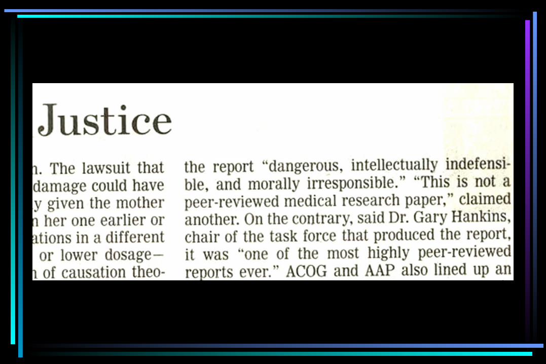
Facts
• CP rate 1.5/1000• No change in last 40 years• No geographic/economic
boundaries

0
5
10
15
20
25
1970 1975 1980 1985 1990 1995 2000
Cesarean Section Rate
Cerebral Palsy Rate
Clark S, Hankins GDV. Am J Obstet Gynecol 2003;188(3):628-633.

• 27 yo G2P1 28 wks/EGA
• PMH – IUFD at 31 wks– Severe preeclampsia
• Maternal transport – severe preeclampsia
Case Study

• Rx:
– MgSO4
– Oxytocin
– Hydralazine
PRN
Case Study
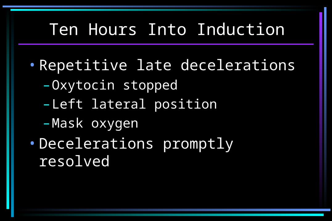
Ten Hours Into Induction
• Repetitive late decelerations– Oxytocin stopped– Left lateral position– Mask oxygen
• Decelerations promptly resolved


C-Section – Low Vertical
General Anesthesia
Case Study

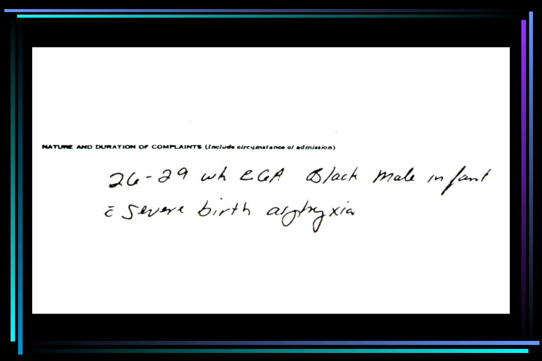
970 gram infant
Apgar 1/0/0/0/0
Pronounced at 20 minutes
Case Study

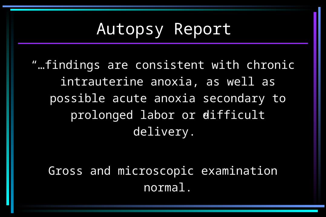
Autopsy Report
“…findings are consistent with chronic intrauterine anoxia, as well as possible acute anoxia secondary to prolonged
labor or difficult delivery.”
Gross and microscopic examination normal.
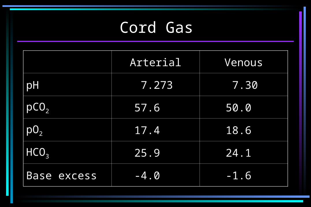
Cord Gas
Arterial Venous
pH 7.273 7.30
pCO2 57.6 50.0
pO2 17.4 18.6
HCO3 25.9 24.1
Base excess -4.0 -1.6

Charge by Dr. Frank Miller
“To create a multidisciplinary task force to review and consider the current state of scientific knowledge about the mechanisms and timing of possible etiologic events which may result in neonatal encephalopathy and cerebral palsy.”

Task Force on Neonatal Encephalopathy and Cerebral Palsy
Committee Members
Mary D’Alton, MD, FACOGMark Ira Evans, MD, FACOGLarry Gilstrap, MD, FACOGRichard Depp, III, MD, FACOGRoger K. Freeman, MD,
FACOGRichard P. Green, MD, FACOG
Gary Hankins, MD, FACOG, Chair

James A. McGregor, MD, FACOG
Karin B. Nelson, MDSusan Ramin, MD, FACOGRobert Resnik, MD, FACOGMichael Speer, MDLouis Weinstein, MD, FACOGStanley Zinberg, MD, MS
Task Force on Neonatal Encephalopathy and Cerebral Palsy
Committee Members

Neonatal Encephalopathy andCerebral Palsy Task Force Consultants
James Barkovich, MD – Professor of Radiology, Neurology, Pediatrics, and Neurosurgery
Kurt Benirschke, MD – Professor Emeritus of Pathology and Reproductive Medicine
Robert R. Clancy, MD – Professor of Neurology and Pediatrics, Pediatric Regional Epilepsy Program
Gabrielle A. DeVeber, MD – Director, Canadian Pediatric Ischemic Stroke Registry
Ronald Gibbs, MD – Professor and Chairman, OB/Gyn
Gregory Locksmith, MD – Assistant Professor, OB/Gyn

Jeffrey Perlman, MD, PhD – Professor of Pediatrics and OB/Gyn
Julian N. Robinson, MD – Assistant Professor, OB/Gyn
Dwight J. Rouse, MD – Associate Professor, OB/Gyn George R. Saade, MD – Professor, OB/Gyn
Diana E. Schendel, PhD – Centers for Disease Control and Prevention
Rodney E. Willoughby, Jr., MD – Associate Professor, Pediatrics
Marjorie R. Grafe, MD – Professor, Placental Pathology
Neonatal Encephalopathy andCerebral Palsy Task Force Consultants

Methods
• Five meetings over 3 years• Extensive consultation with
clinicians and scientists• Consensus development• Draft and redraft
Methodology
• Throughout the process, primary source documents were cited to the fullest extent possible.
• Comments solicited from a number of professional organizations:– American Academy of Pediatrics– The Canadian Paediatric Society– The Child Neurology Society– The Society for Maternal-Fetal Medicine– The March of Dimes Birth Defects
Foundation

• The Centers for Disease Control and Prevention, Department of Health and Human Services
• The National Institute of Child Health and Human Development
• The Royal Australian and New Zealand College of Obstetricians and Gynaecologists
• The Society of Obstetricians and Gynaecologists of Canada.
Methodology

• Throughout the process, primary source documents were cited to the fullest extent possible.
• Comments solicited from a number of professional organizations:– American Academy of Pediatrics– The Canadian Paediatric Society– The Child Neurology Society– The Society for Maternal-Fetal Medicine– The March of Dimes Birth Defects
Foundation
Methodology

• The Centers for Disease Control and Prevention, Department of Health and Human Services
• The National Institute of Child Health and Human Development
• The Royal Australian and New Zealand College of Obstetricians and Gynaecologists
• The Society of Obstetricians and Gynaecologists of Canada.
Methodology

Most Peer-Reviewed Document
on the Subject
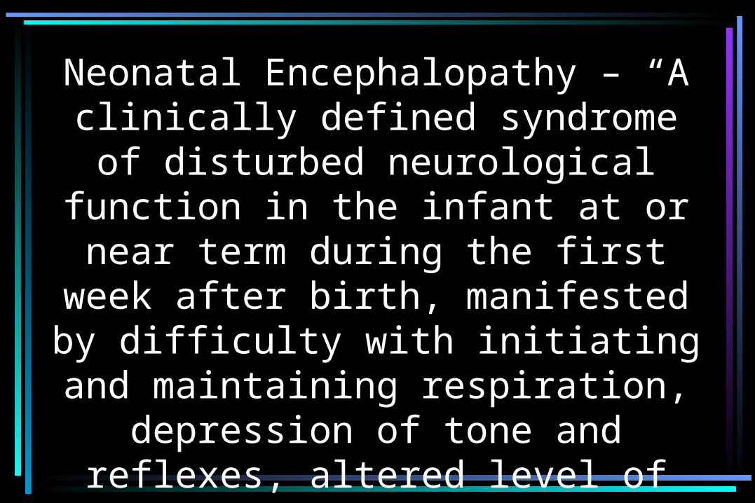
Neonatal Encephalopathy – “A clinically defined syndrome of
disturbed neurological function in the infant at or near term during
the first week after birth, manifested by difficulty with
initiating and maintaining respiration, depression of tone and reflexes, altered level of
consciousness, and often seizures.”

Differential Dx: Neonatal Encephalopathy
• Developmental abnormalities
• Metabolic abnormalities
• Autoimmune disorders
• Coagulation disorders
• Infections
• Trauma
• Hypoxia
• IUGR
• Multiple gestations
• Antepartum hemorrhage
• Chromosomal abnormalities
• Persistent breech/transverse lie

On the Diagnosis of Birth Asphyxia
• Relies on:– Clinical markers of fetal distress (MSAF/Abn
FHR)– Laboratory markers (Cord pH or base excess)– Newborn status (Apgars, time to respirations)
• No data to support:– Equivalence of criteria– Specificity to intrapartum asphyxia
• Potential for misclassification enormousBlair E. Dev Med Child Neurol 1993;35:449-52.

Relationship of Intrapartum Asphyxia to Neonatal Encephalopathy and Cerebral
Palsy
IntrapartumAsphyxia
NeonatalEncephalopathy
CerebralPalsy
x
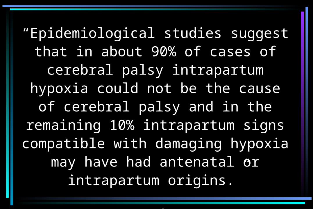
“Epidemiological studies suggest that in about 90% of cases of cerebral
palsy intrapartum hypoxia could not be the cause of cerebral palsy and in the remaining 10% intrapartum signs compatible with damaging hypoxia
may have had antenatal or intrapartum origins.”
Template

Key Publications - ACOG• Committee Opinion #49, Nov 1986/1989
– Use and Misuse of the Apgar Score– Committee on Obstetrics: MFM (ACOG)– Committee on Fetus and Newborn (AAP)
• ACOG Technical Bulletin #163, January 1992– Fetal and Neonatal Neurologic Injury
• Committee on Obstetric Practice #137, April 1994– Fetal Distress and Birth Asphyxia
• Committee on Obstetric Practice #197, Feb 1998– Inappropriate Use of the Terms Fetal Distress and Birth
Asphyxia

Antepartum Risk Factors for Newborn Encephalopathy: The Western Australian Case-
Control Study• Metropolitan Western Australia June 93-Sept 95• All 164 term infants with moderate/severe
encephalopathy• Controls – 400 randomly selected• Stats
– Birth prevalence of moderate/severe newborn encephalopathy 3.8/1000 term live births
– Neonatal Fatality 9.1%• Conclusions
– Causes of newborn encephalopathy are heterogeneous and many of the causal pathways start before birth
Badawi N, et al. BMJ 1998;317:1549-53.

3.57
2.97
2.07
3.82
2.23
2.17
2.73
2.55
0 0.5 1 1.5 2 2.5 3 3.5 4 4.5
Intrapartum Fever
Mod/Severe Antepartum Bleeding
Viral Illness
Family Hx Neurological Disorder
Family Hx Seizure
Instrumental Delivery
Emergency C/S
Abnormal Placental Appearance
Adjusted Odds Ratio
Preconceptional
Intrapartum
Antepartum
Risk Factors for Newborn Encephalopathy
Badawi N, et al. BMJ 1998;317:1549-53.
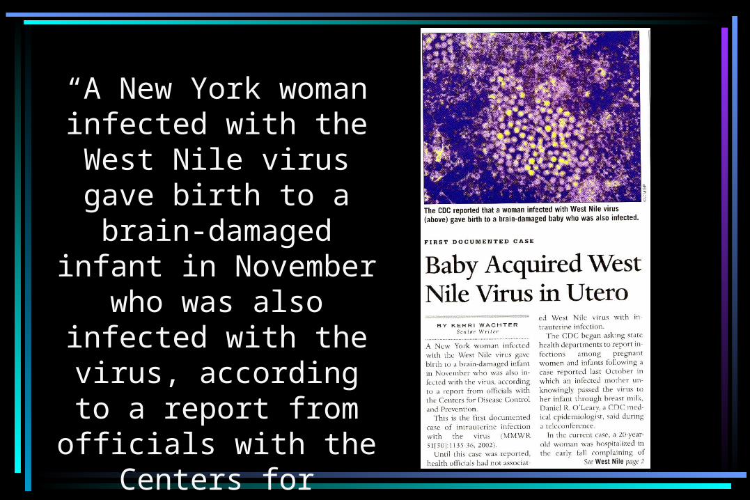
“A New York woman infected with the
West Nile virus gave birth to a brain-
damaged infant in November who was
also infected with the virus, according to a report from officials with the Centers for Disease Control and
Prevention.”

Risk Factors for Newborn Encephalopathy
9.7
6.3
4.37
4.44
4.29
4.43
0 2 4 6 8 10 12
Maternal ThyroidDisease
Severe Preeclampsia
Acute IntrapartumEvent
Infertility Treatment
IUGR 3-9%tile
OP Presentation
Adjusted Odds Ratio
Preconceptional
Intrapartum
Antepartum
Badawi N, et al. BMJ 1998;317:1549-53.

38.23
2.07
0 5 10 15 20 25 30 35 40 45
IUGR <3%tile
Abnormal PlacentalAppearance
Antepartum
Risk Factors for Newborn Encephalopathy

0.17
0.17
00.51
Elective C/S
No Labor
Risk Factors for Newborn Encephalopathy

Distribution of Risk Factors for Newborn Encephalopathy
Antepartum risk factors only (69%)
Antepartum risk factors and intrapartum
hypoxia (25%)
Intrapartum hypoxia only (4%)
Unknown (2%)
Badawi N, et al. BMJ 1998;317:1554-8.

Intrapartum Asphyxia: A Rare Cause of Cerebral Palsy
“It was estimated that in only 8%
(15/183) of all the children with
spastic cerebral palsy was
intrapartum asphyxia the possible
cause of their brain damage.”
Blair E, et al. J Pediatr 1988;112:515-9.

Theoretical Scenarios for Timing of Neurological Insult in Newborn
Encephalopathy
Antepartum period Intrapartum period Neonatal period Newborn outcome
1 Insult Encephalopathy
2 Insult Further Insult Encephalopathy
3 Insult Encephalopathy
4 Insult Encephalopathy
Badawi N, et al. BMJ 1998;317:1554-8.


Uncertain Value of Electronic Fetal Monitoring in Predicting Cerebral
Palsy• Population– Four California counties 1983-1985– Singleton – Birthweight > 2500 gm– Survival to age 3 and EFM (N=78)– Moderate/severe cerebral palsy
• Abnormal FHR– Multiple late decelerations– Decreased beat to beat
• Controls– Matched appropriately (N=300)
Nelson KB, et al. N Engl J Med 1996;334:613-8.

Uncertain Value - Continued
PatternCP
N=78ControlN=300
OR
Tachycardia N (%) N (%)
> 160 22 (28.2) 85 (28.3) 1.0 (0.6-1.7)
> 180 5 (6.4) 16 (5.3) 1.3 (0.4-3.4)
Bradycardia
< 100 27 (34.6) 75 (25.0) 1.5 (0.9-2.5)
< 80 13 (16.7) 35 (11.7) 1.5 (0.8-3.0)
Multiple Lates 11 (14.1) 12 (4.0) 3.9 (1.7-9.3)
Mult. Lates or BTB 21 (26.9) 28 (9.3) 3.6 (1.9-6.7)
Nelson KB, et al. N Engl J Med 1996;334:613-8.

Nelson
“The 21 children with cerebral palsy who had multiple late decelerations or decreased variability in heart rate monitoring represented only 0.19
percent of singleton infants with birth weights of 2500 g or more who had these fetal-monitoring findings, for a
false positive rate of 99.8%.Nelson KB, et al. N Engl J Med 1996;334:613-8.
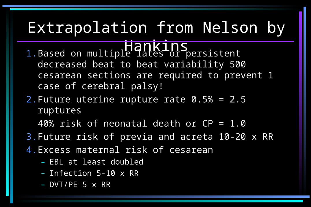
Extrapolation from Nelson by Hankins
1. Based on multiple lates or persistent decreased beat to beat variability 500 cesarean sections are required to prevent 1 case of cerebral palsy!
2. Future uterine rupture rate 0.5% = 2.5 ruptures40% risk of neonatal death or CP = 1.0
3. Future risk of previa and acreta 10-20 x RR4. Excess maternal risk of cesarean
– EBL at least doubled– Infection 5-10 x RR– DVT/PE 5 x RR

“It is not possible at the current time to prospectively recognize during labor the point in time when a reduction in cerebral
perfusion results in irreversible brain injury.”
Jeffrey M. Perlman, M.D.

“Any claims to the contrary are
not supported by any scientific
merit.”
ACOG Task Force on Neonatal
Encephalopathy and Cerebral Palsy

Criteria Required to Define an Acute Intrapartum Hypoxic Event As Sufficient
to Cause Cerebral Palsy
Essential criteria (must meet all four)
1. Evidence of a metabolic acidosis in fetal, umbilical cord arterial blood obtained at delivery (pH 7.00 and base deficit >12 mmol/L).
2. Early onset of severe or moderate neonatal encephalopathy in infants of 34 or more weeks of gestation. ACOG/AAP

3. Cerebral palsy of the spastic quadriplegic or dyskinetic type*.
4. Exclusion of other identifiable etiologies such as trauma, coagulation disorders, infectious conditions, or genetic disorders.
Criteria Required to Define an Acute Intrapartum Hypoxic Event As Sufficient
to Cause Cerebral Palsy
ACOG/AAP
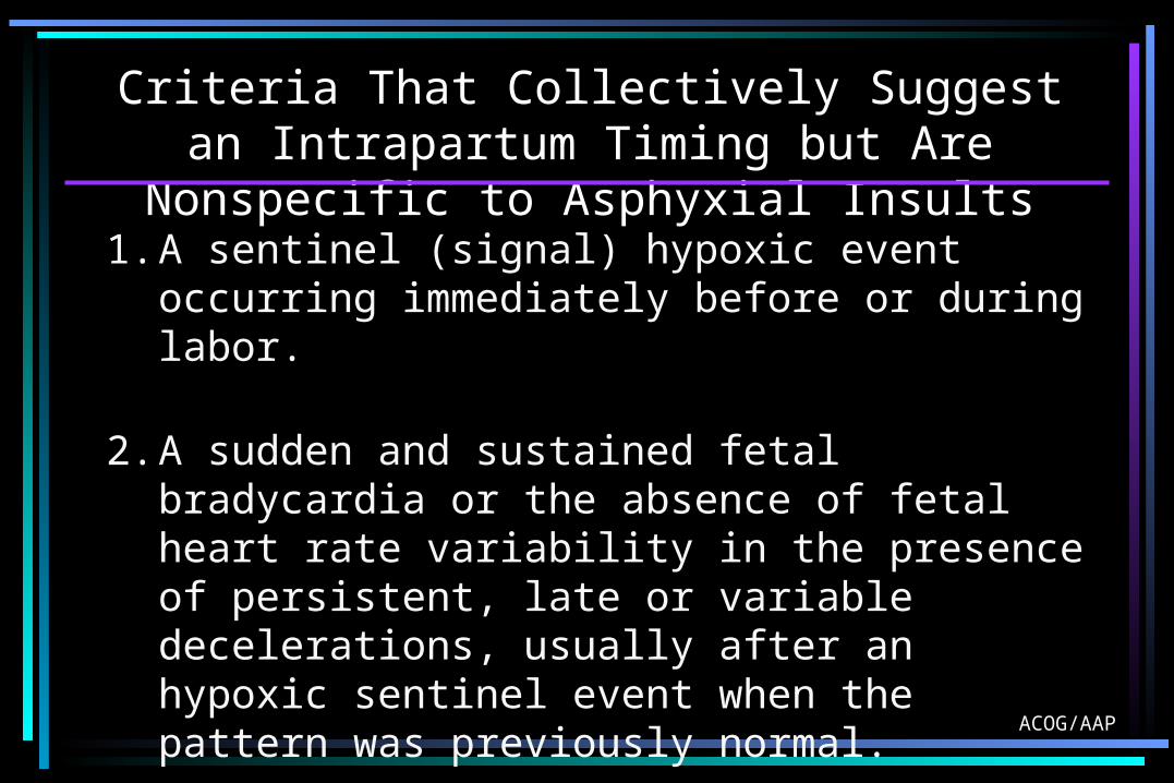
Criteria That Collectively Suggest an Intrapartum Timing but Are Nonspecific to
Asphyxial Insults1. A sentinel (signal) hypoxic event occurring
immediately before or during labor.
2. A sudden and sustained fetal bradycardia or the absence of fetal heart rate variability in the presence of persistent, late or variable decelerations, usually after an hypoxic sentinel event when the pattern was previously normal.
ACOG/AAP

3. Apgar scores of 0-3 beyond 5 minutes.
4. Evidence of multi-system involvement up to 72 hours.
5. Early imaging study showing evidence of acute nonfocal cerebral abnormality.
Criteria That Collectively Suggest an Intrapartum Timing but Are Nonspecific to
Asphyxial Insults
ACOG/AAP

Relationship of Intrapartum Asphyxia to Neonatal Encephalopathy and Cerebral
Palsy
IntrapartumAsphyxia
NeonatalEncephalopathy
CerebralPalsy
x

IMPORTANT
All other things being equal,
neonatal resuscitation can
potentially prevent or further the
injury.


![Kris Allen [ No Boundaries ]](https://img.pdfslide.us/doc/110x75/546fff83af795987048b45b5/kris-allen-no-boundaries--5584abc25fc18.jpg)
![Kris Allen [ No boundaries ]](https://img.pdfslide.us/doc/110x75/546fff85af7959f4758b471a/kris-allen-no-boundaries-.jpg)