Embed Size (px)
Citation preview
256 J. Neurosurg. / Volume 95 / August, 2001
HRONIC subdural hematomas have been reported tohave recurrence rates of 3 to 20% after burr-holesurgery;1–3,7,21however, the factors influencing their
recurrence have not been sufficiently investigated.In our prior report on the relationship between the po-
sition of a drainage catheter and postoperative recurrenceof these lesions,10 we found that frontal drainage is associ-ated with the lowest recurrence rate and the least subdu-ral air collection after closed system drainage for CSDH.In the present study, potential indicators of postoperativerecurrence of CSDHs in 106 patients were investigated byexamining the relationships between postoperative recur-rence and both the natural progression of CSDHs and theextent of their feeding vessels,19 which are considered toprovide a significant influence on the risk of recurrence ofthese lesions.
Clinical Material and Methods
Patient Population
Among the 144 patients with CSDHs who were treated
at Tokyo Kosei Nenkin Hospital between January 1989and April 1998, 106 consecutive patients (82 men and 24women) were studied. Bilateral hematomas were presentin 20 patients and thus there were a total of 126 lesions.
We defined CSDH as an SDH surrounded by a thin cap-sule (hematoma membrane) and consisting of dark red-dish liquefied blood at operation. If the date of head trau-ma was clear, a CSDH was defined as a hematoma thathad persisted more than 3 weeks after the patient sufferedhead trauma.
Nine patients with one or more risk factors that alreadyhad been documented to influence recurrence1 were ex-cluded from the study: four with thrombocytopenia; fourreceiving anticoagulant or thrombolytic drugs; one re-ceiving hemodialysis; and one with a ventriculoperitonealshunt. Another 29 patients were also excluded: nine whodid not undergo surgery and 20 who could not be observedfor 3 months postoperatively.
In all cases, we examined the patient’s age and sex, thetime from head injury to initial CT scanning, the type ofhematoma categorized according to our new classificationof internal architecture, and the intracranial extension ofthe lesion on preoperative and postoperative CT scans.
At our hospital, CT scanning is generally performedfive times in every patient with CSDH: preoperatively, onDays 1 to 3, on Day 7, and on approximately Day 30 andDay 90 postoperatively.
J Neurosurg 95:256–262, 2001
Factors in the natural history of chronic subduralhematomas that influence their postoperative recurrence
HIROSHI NAKAGUCHI , M.D., PH.D., TAKEO TANISHIMA , M.D., PH.D.,AND NORIO YOSHIMASU, M.D., PH.D.
Department of Neurosurgery, Teraoka Memorial Hospital, Hiroshima; and Department ofNeurosurgery, Tokyo Kosei Nenkin Hospital, Tokyo, Japan
Object. Factors affecting the postoperative recurrence of chronic subdural hematomas (CSDHs) have not beensufficiently investigated. The authors have attempted to determine features of CSDHs that are associated with a highor low recurrence rate on the basis of the natural history of these lesions and their intracranial extension.
Methods. One hundred six patients (82 men and 24 women) harboring 126 CSDHs who were treated at TokyoKosei Nenkin Hospital between January 1989 and April 1998 were studied. Types of CSDHs were classified ac-cording to hematoma density and internal architecture, and the intracranial extension of the hematomas were inves-tigated. The postoperative recurrence rate was calculated for each factor.
Based on the internal architecture and density of each hematoma, the CSDHs were classified into four types, includ-ing homogeneous, laminar, separated, and trabecular types. The recurrence rate associated with the separated type washigh, whereas that associated with the trabecular type was low.
Chronic subdural hematomas are believed to develop initially as the homogeneous type, after which they sometimesprogress to the laminar type. A mature CSDH is represented by the separated stage and the hematoma eventually pass-es through the trabecular stage during absorption.
Based on the intracranial extension of each hematoma, CSDHs were classified into three types, including convexi-ty, cranial base, and interhemispheric types. The recurrence rate of cranial base CSDHs was high and that of convex-ity CSDHs was low.
Conclusions. Classification of CSDHs according to the internal architecture and intracranial extension may beuseful for predicting the risk of postoperative recurrence.
KEY WORDS • chronic subdural hematoma • hematoma recurrence • natural history •middle meningeal artery • computerized tomography
C
Abbreviations used in this paper: CSDH = chronic subdural he-matoma; CT = computerized tomography; MMA = middle menin-geal artery; PR = postoperative recurrence; RO = repeated opera-tion; SDH = subdural hematoma.
All patients, except one who displayed neurologicaldeficits caused by CSDH, underwent surgical interventionincluding creation of one burr hole, irrigation of the he-matoma with sterile saline, and postoperative closed sys-tem drainage with the aid of a silicone tube (Type B ven-tricular drainage catheter; Hanako Medical Co., Urawa,Japan).
Craniotomy was performed in one patient with a recur-rent hematoma of the interhemispheric type.
Regardless of the presence of residual hematoma andsubdural air on CT scans 1 day postsurgery, all drainagecatheters were removed within 48 hours after surgery andthe daily activities of the patients were not restricted after-ward. Until the catheter was removed, patients were re-quired always to keep the head as high as the heart.
Recurrence of CSDH was defined as an increase in thevolume of the SDH on the operated side and compressionof the brain surface observed on CT scans obtained with-in 3 months postoperatively, when compared with find-ings 1 day after surgery. Repeated operations were per-formed when neurological symptoms reappeared or thecerebral sulci were diffusely effaced by recurrent hemato-ma. Patients without neurological deficits or with a smallamount of residual hematoma were observed. The PR rateand the RO rate were calculated and compared among thevarious factors that we investigated.
Natural History of CSDHs
There were 18 patients with CSDHs who initially dis-played no marked neurological deficits and received nosurgical intervention for several weeks or more, while be-ing monitored by CT scanning, which was performed atleast twice. We examined changes in the density and inter-nal architecture of the CSDHs on these CT scans to inves-tigate the natural history of CSDHs.
Interval From Head Injury to Initial CT Scan
Although there were many patients in whom a historyof head injury was unknown or unclear, the date of headtrauma was able to be determined in 63 patients (59%),and the interval from head injury to the initial CT scanwas calculated for these patients. We analyzed the correla-tion between this interval and both the recurrence rate andthe hematoma type.
Internal Architecture of the Hematoma
All hematomas were classified into four types accord-ing to their internal architecture, which corresponded topossible stages in the natural history of CSDH: homoge-neous, laminar, separated, and trabecular types.
The homogeneous type was defined as a hematoma thatexhibited a homogeneous density (low–high). The lami-nar type was defined as a subtype of the homogeneoustype that had a thin high-density layer along the innermembrane. The separated type was defined as a hemato-ma containing two components of different densities witha clear boundary lying between them; that is, a lower den-sity component located above a higher density compo-nent. If the boundary was indistinct, with the low densityand high density being mingled at the border, this wascalled the “gradation” type and was considered to be asubtype of the separated type. The trabecular type was de-fined as a hematoma with inhomogeneous contents and ahigh-density septum running between the inner and outermembrane on a low-density to isodense background.
Three series of CT scans (that is, scans obtained pre-operatively, scans obtained 1 day after surgery, and scansobtained 7 days after surgery) were assessed according tothis classification.
Intracranial Extension of CSDHs
The CSDHs were classified into three types accordingto their intracranial extension: hematomas localized at theconvexity without involvement of the cranial base werenamed the convexity type; hematomas that extended tothe cranial base were named cranial base CSDHs; andhematomas that extended to the interhemispheric fissurewere named interhemispheric CSDHs.
The cranial base type was divided into two subtypes:hematomas limited to frontal base were called frontalbase CSDHs; whereas those extending from the temporalbase to the frontal base were named frontotemporal baseCSDHs.
The convexity type was also divided into two subtypes:hematomas limited to the frontal region were called fron-tal convexity CSDHs and those spanning the frontaland parietal regions were called frontoparietal convexityCSDHs.
There were no patients in whom the CSDH was local-ized to the parietal convexity, occipital convexity, tempo-ral base, or posterior fossa.
Statistical Analysis
For statistical analysis, the Fisher exact test or the chi-
J. Neurosurg. / Volume 95 / August, 2001 257
Factors influencing chronic subdural hematoma recurrence
TABLE 1 Relationship between sex of patient and PR rate of CSDHs
No. of PRPatient’s Sex Cases (%) Rate (%) p Value*
male 82 (77) 20 0.5541female 24 (23) 13total 106 (100) 17
* The Fisher exact test was used for analysis of differences.
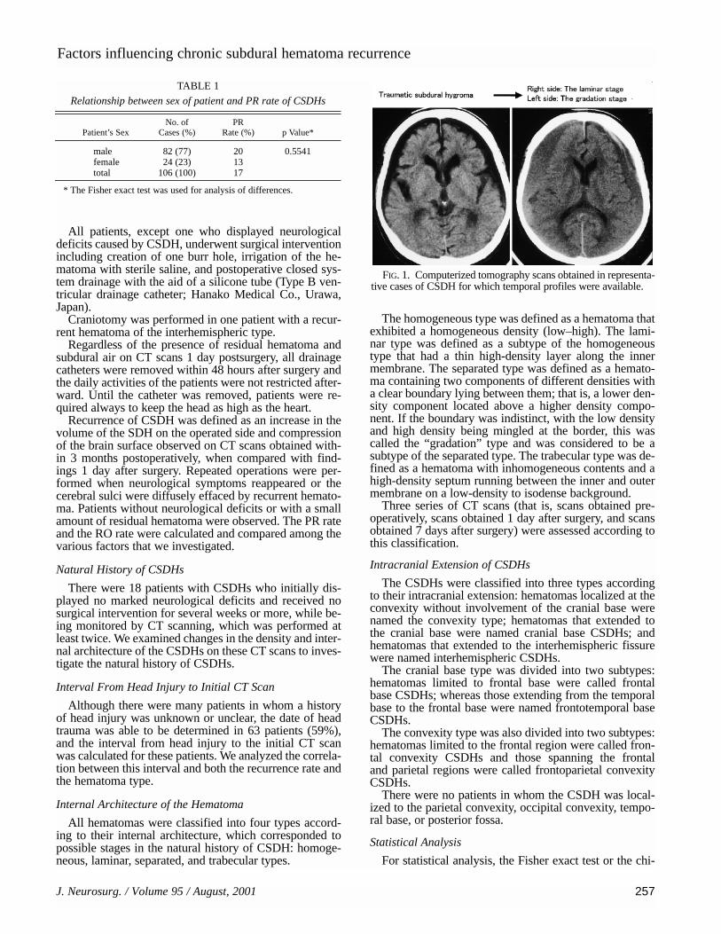
FIG. 1. Computerized tomography scans obtained in representa-tive cases of CSDH for which temporal profiles were available.
square test for independence was performed using com-mercially available statistical computer software (StatView R J Version 4.11 software; SAS Institute, Cary, NC).
Results
For the 126 CSDHs in 106 patients, 142 surgical proce-dures were performed. There were 82 men (77%) and 24women (23%) in the study, ranging in age from 20 to 96years with an average age of 67 years.
The PR rate was 17% (21 lesions) and the RO rate was13% (16 lesions). The PR and RO rates were 20% (16 le-sions) and 16% (13 lesions), respectively, in male patients,whereas the rates were 13% (three lesions) and 4% (onelesion), respectively, in female patients (Table 1). Al-though men had a higher RO rate, no significant differ-ence was seen between the sexes.
The PR rate of patients aged less than 60 years was 16%and that of patients aged 70 years or older was 15%. Therewas no significant difference between younger and olderpatient groups.
Natural History of CSDHs
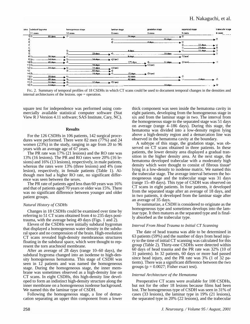
Changes in 18 CSDHs could be examined over time byreferring to 51 CT scans obtained from 4 to 235 days post-trauma, with the average being 49 days (Figs. 1 and 2).
Eleven of the CSDHs were initially subdural hygromasthat displayed a homogeneous water density in the subdu-ral space and no compression of the brain. High-resolutionCT scans revealed high-density membranous structuresfloating in the subdural space, which were thought to rep-resent the torn arachnoid membrane.
After an average of 28 days (range 10–60 days), thesubdural hygroma changed into an isodense to high-den-sity homogeneous hematoma. This stage of CSDH wasseen in 12 patients and was named the homogeneousstage. During the homogeneous stage, the inner mem-brane was sometimes observed as a high-density line onCT scans. In eight CSDHs, this high-density line devel-oped to form an indistinct high-density structure along theinner membrane on a homogeneous isodense background.We named this the laminar type of CSDH.
Following the homogeneous stage, a line of demar-cation separating an upper thin component from a lower
thick component was seen inside the hematoma cavity ineight patients, developing from the homogeneous stage insix and from the laminar stage in two. The interval fromthe homogeneous stage to the separated stage was 51 dayson average (range 4–186 days). During this stage, thehematoma was divided into a low-density region lyingabove a high-density region and a demarcation line wasobserved in the hematoma cavity at the boundary.
A subtype of this stage, the gradation stage, was ob-served on CT scans obtained in three patients. In thesepatients, the lower density area displayed a gradual tran-sition in the higher density area. At the next stage, thehematoma developed trabeculae with a moderately highdensity which were thought to consist of fibrous tissue6
lying in a low-density to isodense matrix. We named thisthe trabecular stage. The average interval between the ho-mogeneous stage and the trabecular stage was 31 days(range 15–49 days). This type of CSDH was observed onCT scans in eight patients. In four patients, it developedfrom the separated stage after an average of 18 days, andin two patients, it developed from the laminar stage afteran average of 35 days.
To summarize, a CSDH is considered to originate as thehomogeneous type and sometimes develops into the lam-inar type. It then matures as the separated type and is final-ly absorbed as the trabecular type.
Interval From Head Trauma to Initial CT Scanning
The date of head trauma was able to be determined in63 patients (59%) and the number of days from head inju-ry to the time of initial CT scanning was calculated for thisgroup (Table 2). Thirty-one CSDHs were detected within60 days of head trauma and the PR rate was 32% (10 of31 patients). In 32 patients, 60 days or more had passedsince head injury, and the PR rate was 3% (1 of 32 pa-tients). There was a significant difference between the twogroups (p = 0.0027; Fisher exact test).
Internal Architecture of the Hematoma
Preoperative CT scans were available for 108 CSDHs,but not for the other 18 lesions because films had beenlost. The homogeneous type of CSDH was seen in 31% ofcases (33 lesions), the laminar type in 19% (21 lesions),the separated type in 20% (22 lesions), and the trabecular
258 J. Neurosurg. / Volume 95 / August, 2001
H. Nakaguchi, et al.
FIG. 2. Summary of temporal profiles of 18 CSDHs in which CT scans could be used to document temporal changes in the densities andinternal architectures of the lesions. ope = operation.
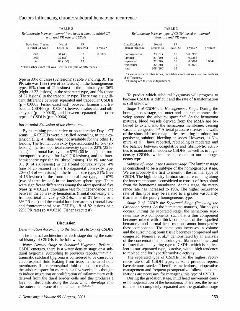
type in 30% of cases (32 lesions) (Table 3 and Fig. 3). ThePR rate was 15% (five of 33 lesions) in the homogeneoustype, 19% (four of 21 lesions) in the laminar type, 36%(eight of 22 lesions) in the separated type, and 0% (noneof 32 lesions) in the trabecular type. There was a signifi-cant difference between separated and trabecular CSDHs(p = 0.0003, Fisher exact test), between laminar and tra-becular CSDHs (p = 0.0204), between trabecular and oth-er types (p = 0.0026), and between separated and othertypes of CSDHs (p = 0.0064).
Intracranial Extension of the Hematoma
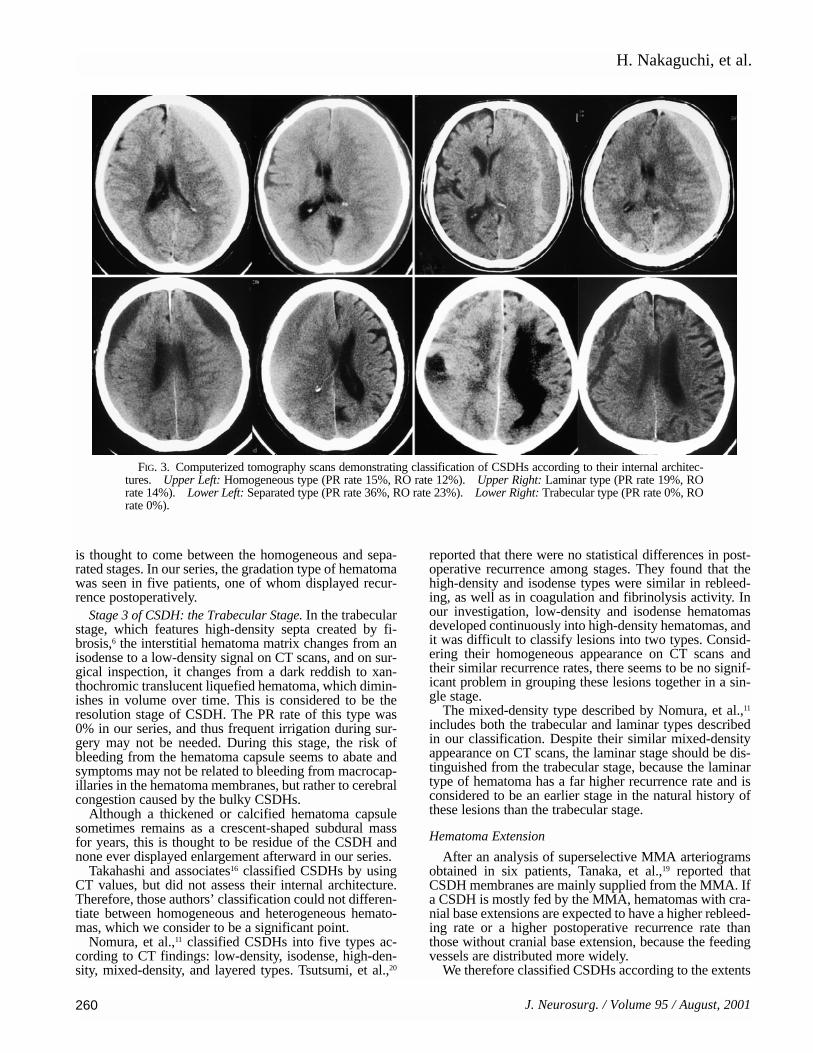
By examining preoperative or postoperative Day 1 CTscans, 116 CSDHs were classified according to their ex-tension (Fig. 4); data were not available for the other 10lesions. The frontal convexity type accounted for 5% (sixlesions), the frontoparietal convexity type for 22% (25 le-sions), the frontal base type for 57% (66 lesions), the fron-totemporal base type for 14% (16 lesions), and the inter-hemispheric type for 3% (three lesions). The PR rate was0% (0 of six lesions) in the frontal convexity type, 4%(one of 25 lesions) in the frontoparietal convexity type,20% (13 of 66 lesions) in the frontal base type, 31% (fiveof 16 lesions) in the frontotemporal base type, and 67%(two of three lesions) in the interhemisphere type. Therewere significant differences among the aforespecified fivetypes (p = 0.0221; chi-square test for independence) andbetween the convexity hematomas (frontal convexity andfrontoparietal convexity CSDHs, one of 31 lesions or a3% PR rate) and the cranial base hematomas (frontal baseand frontotemporal base CSDHs, 18 of 82 lesions or a22% PR rate) (p = 0.0218, Fisher exact test).
Discussion
Determination According to the Natural History of CSDHs
The internal architecture at each stage during the natu-ral history of CSDHs is the following.
Water Density Stage or Subdural Hygroma. Before aCSDH emerges, there is a water density stage or a sub-dural hygroma. According to previous reports,4,8,9,12,13,18,22
traumatic subdural hygroma is considered to be caused bycerebrospinal fluid leaking from tears in the arachnoidmembrane. If a cerebrospinal fluid collection remains inthe subdural space for more than a few weeks, it is thoughtto induce migration or proliferation of inflammatory cellsderived from the dural border cells and this produces alayer of fibroblasts along the dura, which develops intothe outer membrane of the hematoma.4,6,13,15,17
To predict which subdural hygromas will progress tobecome CSDHs is difficult and the rate of transformationis still unknown.
Stage 1 of CSDH: the Homogeneous Stage. During thehomogeneous stage, the outer and inner membranes de-velop around the subdural space.6,14,17 As the hematomamatures, blood vessels derived from the MMA are be-lieved to extend into the hematoma membrane, causingvascular congestion.6,19 Arterial pressure stresses the wallsof the sinusoidal microcapillaries, resulting in minor, butpersistent, subdural bleeding episodes. However, as No-mura, et al.,11 have reported, rebleeding is moderate andthe balance between coagulative and fibrinolytic activi-ties is maintained in isodense CSDHs, as well as in high-density CSDHs, which are equivalent to our homoge-neous type.
Subtype of Stage 1: the Laminar Stage. The laminar stageis considered to be a subtype of the homogeneous stage.We are probably the first to mention the laminar type ofCSDH. The high-density laminar structure running alongthe inner membrane is considered to consist of fresh bloodfrom the hematoma membrane. At this stage, the recur-rence rate has increased to 19%. The higher recurrencerate of this type may be related to a greater vascularitythan that of the purely homogeneous type.
Stage 2 of CSDH: the Separated Stage (Including theGradation Stage). As the hematoma matures, fibrinolysisoccurs. During the separated stage, the hematoma sepa-rates into two components, such that a thin componentbecomes mixed with a thick component of the liquefiedhematoma and normal head motion cannot homogenizethese components. The hematoma increases in volumeand the surrounding brain tissue becomes compressed andcongested. Nomura, et al.,11 demonstrated by an analysisof the concentrations of fibrinogen, fibrin monomer, andd-dimer that the layering type of CSDH, which is equiva-lent to our separated type, is active, with a high tendencyto rebleed and for hyperfibrinolytic activity.
The separated type of CSDHs had the highest recur-rence rate of all CSDH types, as some previous reportshave demonstrated.5,11Therefore, meticulous perioperativemanagement and frequent postoperative follow-up exam-inations are necessary for managing this type of CSDH.
During the gradation stage, mild head movement caus-es homogenization of the hematoma. Therefore, the hema-toma is not completely separated and the gradation stage
J. Neurosurg. / Volume 95 / August, 2001 259
Factors influencing chronic subdural hematoma recurrence
TABLE 2 Relationship between interval from head trauma to initial CT
scan and PR rate of CSDHs
Days From Trauma No. of PRto Initial CT Scan Cases (%) Rate (%) p Value*
�60 31 (49) 32 0.0027�60 32 (51) 3total 63 (100) 17
* The Fisher exact test was used for analysis of differences.
TABLE 3Relationship between type of CSDH based on internal
structure and PR rates
Classification of No. of PRInternal Structure Lesions (%) Rate (%) p Value* p Value†
homogeneous 33 (31) 15 �0.9999laminar 21 (19) 19 0.7388separated 22 (20) 36 0.0064 0.0042trabecular 32 (30) 0 0.0026total 108 (100) 16
* Compared with other types; the Fisher exact test was used for analysisof differences.
† Chi-square test for independence.
is thought to come between the homogeneous and sepa-rated stages. In our series, the gradation type of hematomawas seen in five patients, one of whom displayed recur-rence postoperatively.
Stage 3 of CSDH: the Trabecular Stage. In the trabecularstage, which features high-density septa created by fi-brosis,6 the interstitial hematoma matrix changes from anisodense to a low-density signal on CT scans, and on sur-gical inspection, it changes from a dark reddish to xan-thochromic translucent liquefied hematoma, which dimin-ishes in volume over time. This is considered to be theresolution stage of CSDH. The PR rate of this type was0% in our series, and thus frequent irrigation during sur-gery may not be needed. During this stage, the risk ofbleeding from the hematoma capsule seems to abate andsymptoms may not be related to bleeding from macrocap-illaries in the hematoma membranes, but rather to cerebralcongestion caused by the bulky CSDHs.
Although a thickened or calcified hematoma capsulesometimes remains as a crescent-shaped subdural massfor years, this is thought to be residue of the CSDH andnone ever displayed enlargement afterward in our series.
Takahashi and associates16 classified CSDHs by usingCT values, but did not assess their internal architecture.Therefore, those authors’ classification could not differen-tiate between homogeneous and heterogeneous hemato-mas, which we consider to be a significant point.
Nomura, et al.,11 classified CSDHs into five types ac-cording to CT findings: low-density, isodense, high-den-sity, mixed-density, and layered types. Tsutsumi, et al.,20
reported that there were no statistical differences in post-operative recurrence among stages. They found that thehigh-density and isodense types were similar in rebleed-ing, as well as in coagulation and fibrinolysis activity. Inour investigation, low-density and isodense hematomasdeveloped continuously into high-density hematomas, andit was difficult to classify lesions into two types. Consid-ering their homogeneous appearance on CT scans andtheir similar recurrence rates, there seems to be no signif-icant problem in grouping these lesions together in a sin-gle stage.
The mixed-density type described by Nomura, et al.,11
includes both the trabecular and laminar types describedin our classification. Despite their similar mixed-densityappearance on CT scans, the laminar stage should be dis-tinguished from the trabecular stage, because the laminartype of hematoma has a far higher recurrence rate and isconsidered to be an earlier stage in the natural history ofthese lesions than the trabecular stage.
Hematoma Extension
After an analysis of superselective MMA arteriogramsobtained in six patients, Tanaka, et al.,19 reported thatCSDH membranes are mainly supplied from the MMA. Ifa CSDH is mostly fed by the MMA, hematomas with cra-nial base extensions are expected to have a higher rebleed-ing rate or a higher postoperative recurrence rate thanthose without cranial base extension, because the feedingvessels are distributed more widely.
We therefore classified CSDHs according to the extents
260 J. Neurosurg. / Volume 95 / August, 2001
H. Nakaguchi, et al.
FIG. 3. Computerized tomography scans demonstrating classification of CSDHs according to their internal architec-tures. Upper Left:Homogeneous type (PR rate 15%, RO rate 12%).Upper Right:Laminar type (PR rate 19%, ROrate 14%). Lower Left:Separated type (PR rate 36%, RO rate 23%).Lower Right:Trabecular type (PR rate 0%, ROrate 0%).
to which these lesions involved the MMA: the frontopari-etal convexity type was related to the peripheral parietaland frontal branches of the MMA; the frontal convexitytype was related only to the peripheral frontal branch ofthe MMA; the frontal base type was related to the frontaltrunk of the MMA; and the frontotemporal base type wasrelated to the two main trunks of the MMA. The inter-hemispheric type of CSDH was also differentiated fromthe others because the MMA does not play an importantrole in feeding this type of hematoma. Using this classifi-cation, the cranial base type was found to have the highestrecurrence rate, as we had predicted.
In addition to the extent of feeding vessels from theMMA, a more enlarged subdural space and greater dif-ficulty in hematoma removal encountered with the cra-nial base CSDHs, compared with convexity CSDHs, areconsidered to be factors influencing the higher recur-rence rate.
The extremely low recurrence rate of the convexity typeof CSDH indicates that the bridging veins at the con-vexity do not have an important role in the recurrence ofCSDHs, although arachnoid tears in the vicinity of theconvexity bridging veins are considered to induce the ear-lier stage of CSDH.
Timing of Surgery and CT Scanning After Head Trauma
When surgical intervention was performed less than 60days after head trauma occurred, there was an extremelyhigh postoperative recurrence rate and this was especially
noted for the homogeneous type of CSDH. When the in-terval from head trauma to initial CT scanning was lessthan 60 days, the recurrence rate of the homogeneous typewas far higher than when the interval was 60 days or more(48% compared with 5%). We consider this, however, on-ly to reflect the fact that fibrosis of the capsule or trabec-ulae tends to be immature and organization of the hemato-ma is limited in younger CSDHs, because there was nopredominant difference in internal architecture of CSDHsbetween lesions less than 60 days old and lesions 60 daysor more after head trauma.
To sum up, it is important to classify CSDH into lesions
J. Neurosurg. / Volume 95 / August, 2001 261
Factors influencing chronic subdural hematoma recurrence
FIG. 4. Computerized tomography scans demonstrating classification of CSDHs according to their intracranial exten-sions. Upper Left:Convexity Type 1 or frontal convexity type (PR rate 0%, RO rate 0%).Upper Right:ConvexityType 2 or frontoparietal convexity type (PR rate 4%, RO rate 0%).Lower Left:Cranial base Type 1 or frontal base type(PR rate 20%, RO rate 18%).Lower Center:Cranial base Type 2 or frontotemporal base type (PR rate 31%, RO rate25%). Lower Right:Interhemispheric type (arrows)(PR rate 67%, RO rate 33%).
TABLE 4 Summary of results of classifications of internal structure and
intracranial extension of CSDHs with respect to PR rate
Type of CSDH PR Rate (%)
conditions for a lower PR rate*trabecular type 0convexity type 3
conditions for a higher PR rate†separated type 36
separated type on cranial base 38cranial base type 22
* All factors were proved statistically to have a low recurrence rate (p�0.05).
† All factors were proved statistically to have a high recurrence rate (p�0.05).
262 J. Neurosurg. / Volume 95 / August, 2001
with a high recurrence rate and lesions with a low recur-rence rate according to our system (Table 4), and to selectappropriate surgical procedures and postoperative man-agement to treat this condition efficiently. If a CSDH isclassified as the convexity or trabecular type, it is expect-ed to exhibit quite a low recurrence rate (one [2%] of 50CSDHs) and frequent irrigation during surgery may notbe needed. By contrast, if a CSDH is determined to beneither the convexity nor trabecular type, most of whichare properly classified as the cranial base type, and at thesame time as the homogeneous or separated type, it is ex-pected to have quite a high recurrence rate (14 [29%] of49 CSDHs). If they are found to be in such a higher re-currence group, patients should be informed of the highpostoperative recurrence rate on admission and meticu-lous perioperative management should be undertaken toreduce postoperative recurrence, including placing the tipof the drainage catheter in the frontal convexity and re-moving subdural air sufficiently, as we proved in our priorreport;10 frequent postoperative follow-up examinationsare also necessary.
Conclusions
Three major points should be reiterated. 1) We believethat CSDHs originate in the homogeneous stage andsometimes develop in the laminar stage. The hematomabecomes mature during the separated stage, and is finallyabsorbed during the trabecular stage. 2) According to theinternal architecture and density of hematomas, CSDHswere classified into four types: homogeneous, laminar,separated, and trabecular types. The recurrence rate of theseparated type was high and that of the trabecular typewas low. 3) According to their intracranial extension,CSDHs were classified into three types: convexity, cranialbase, and interhemispheric types. The recurrence rate ofthe cranial base type of CSDHs was high and that of theconvexity type was low.
References
1. Arbit E, Patterson RH Jr, Fraser RAR: An implantable subduraldrain for treatment of chronic subdural hematoma. Surg Neu-rol 15:175–177, 1980
2. Asano Y, Hasuo M, Takahashi I, et al: [Recurrent cases ofchronic subdural hematoma—its clinical review and serial CTfindings.] No to Shinkei 44:827–831, 1992 (Jpn)
3. Ernestus RI, Beldzinski P, Lanfermann H, et al: Chronic subdu-ral hematoma: surgical treatment and outcome in 104 patients.Surg Neurol 48:220–225, 1997
4. Friede RL, Schachenmayr W: The origin of subdural neomem-branes. II. Fine structure of neomembranes. Am J Pathol 92:69–84, 1978
5. Fujioka S, Matsukado Y, Kaku M, et al: [CT analysis of 100cases with chronic subdural hematoma with respect to clinicalmanifestation and the enlarging process of the hematoma.]Neurol Med Chir 21:1153–1160, 1981 (Jpn)
6. Kawano N, Endo M, Saito M, et al: [Origin of the capsule ofchronic subdural hematoma-an electron microscopic study.] NoShinkei Geka 16:747–752, 1988 (Jpn)
7. Kotwica Z, Brzezinski J: Chronic subdural haematoma treatedby burr holes and closed system drainage: personal experiencein 131 patients. Br J Neurosurg 5:461–465, 1991
8. Markwalder TM: Chronic subdural hematomas: a review. JNeurosurg 54:637–645, 1981
9. Nagata K, Asano T, Basugi N, et al: [Studies on the factorsaffecting the reduction of chronic subdural hematoma: effect ofpreoperative factors with special reference to cerebral atrophy.]No Shinkei Geka 16:1347–1353, 1988 (Jpn)
10. Nakaguchi H, Tanishima T, Yoshimasu N: Relationship be-tween drainage catheter location and postoperative recurrenceof chronic subdural hematoma after burr hole irrigation andclosed system drainage. J Neurosurg 93:791–795, 2000
11. Nomura S, Kashiwagi S, Fujisawa H, et al: Characterization oflocal hyperfibrinolysis in chronic subdural hematomas by SDS-PAGE and immunoblot. J Neurosurg 81:910–913, 1994
12. Sato S, Suzuki J: Ultrastructural observations of the capsule ofchronic subdural hematoma in various clinical stages. J Neuro-surg 43:569–578, 1975
13. Schachenmayr W, Friede RL: The origin of subdural neomem-branes. I. Fine structure of the dura-arachnoid interface in man.Am J Pathol 92:53–68, 1978
14. Shimoji T, Sato K, Ishii S: [A pathogenesis of chronic subduralhematoma; its relationship to subdural membrane.] No ShinkeiGeka 20:131–137, 1992 (Jpn)
15. Takahashi Y: [Pathogenetic consideration on chronic subduralhematoma after intracranial surgery for nontraumatic lesions.]Neurol Med Chir 27:1080–1086, 1987 (Jpn)
16. Takahashi Y, Mikami J, Ueda M, et al: [Analysis of chronicsubdural hematoma based on CT (part 3)-clinical stage classifi-cation based on CT findings.] Neurol Med Chir 24:607–614,1984 (Jpn)
17. Takahashi Y, Mikami J, Ueda M, et al: [The origin of chronicsubdural hematoma considered on the basis of hematoma mem-brane findings and contained fluid findings.] Neurol Med Chir25:998–1009, 1985 (Jpn)
18. Tanaka T, Fujimoto S, Saito K, et al: [Histological study of op-erated cases of chronic subdural hematoma in adults: relation-ship between dura mater and outer membrane.] No ShinkeiGeka 25:701–705, 1997 (Jpn)
19. Tanaka T, Fujimoto S, Saitoh K, et al: [Superselective an-giographic findings of ipsilateral middle meningeal artery ofchronic subdural hematoma in adults.] No Shinkei Geka 26:339–347, 1998 (Jpn)
20. Tsutsumi K, Maeda K, Iijima A, et al: The relationship of pre-operative magnetic resonance imaging findings and closed sys-tem drainage in the recurrence of chronic subdural hematoma.J Neurosurg 87:870–875, 1997
21. Wakai S, Hashimoto K, Watanabe N, et al: Efficacy of closed-system drainage in treating chronic subdural hematoma: a pro-spective comparative study. Neurosurgery 26:771–773, 1990
22. Yamashima T, Friede RL: [Light and electron microscopicstudies on the subdural space, the subarachnoid space and thearachnoid membrane.] Neurol Med Chir 24:737–746, 1984(Jpn)
Manuscript received July 31, 2000.Accepted in final form April 24, 2001.Address reprint requests to:Hiroshi Nakaguchi, Department of
Neurosurgery, Teraoka Memorial Hospital, 37, Ooaza Shinichi,Shinichi town, Ashina gun, Hiroshima prefecture, 729–3103, Japan.email: [email protected].
H. Nakaguchi, et al.


![The Myth of the New Phoenicians Are Lebanese People ...masakimizobuchi0316.s2.weblife.me/_src/sc1193/1428715303.pdf| MEDITERRANEAN REVIEW | Vol. 6, No. 1 [June 2013]: 83~112 The Myth](https://img.pdfslide.us/doc/110x75/5ac2de2e7f8b9ad73f8e9eec/the-myth-of-the-new-phoenicians-are-lebanese-people-masakimizobuchi0316s2-mediterranean.jpg)