Embed Size (px)
Citation preview
Facial Trauma, Maxillary and Le Fort Fractures Author: Kris S Moe, MD, FACS, Chief, Division of Facial Plastic and Reconstructive Surgery, Department of Otolaryngology-Head and Neck
Surgery, University of Washington School of Medicine; Clinical Associate Professor of Surgery, Division of Head and Neck Surgery, University of California, San Diego Coauthor(s): Patrick Byrne, MD, Associate Professor, Department of Head and Neck Surgery, Division of Facial Plastic and Reconstructive
Surgery, Johns Hopkins University School of Medicine; David W Kim, MD, Assistant Professor, Department of Otolaryngology-Head and Neck Surgery, Director, Division of Facial Plastic and Reconstructive Surgery, University of California at San Francisco; Adel R Tawfilis, DDS, Assistant Clinical Professor, Department of Surgery, Division of Plastic Surgery, University of California at San Diego Medical Center
Contributor Information and Disclosures
Updated: Dec 15, 2009
Introduction
The maxilla represents the bridge between the cranial base superiorly and the dental occlusal plane inferiorly. Its intimate association with the oral cavity, nasal cavity, and orbits and the multitude of structures contained within and adjacent to it make the maxilla a functionally and cosmetically important structure. Fracture of these bones is potentially life-threatening as well as disfiguring.1 Timely and systematic repair of these fractures provides the best chance to correct deformity and prevent unfavorable sequelae.
For excellent patient education resources, visit eMedicine's Back, Ribs, Neck, and Head Center, Breaks, Fractures, and Dislocations Center, and Teeth and Mouth Center. Also, see eMedicine's patient education articles Facial Fracture, Broken Nose, and Broken or Knocked-out Teeth.
Frequency
Maxillary fractures account for approximately 6-25% of all facial fractures.
Etiology
Maxillary fractures often result from high-energy blunt force injury to the facial skeleton. Typical mechanisms of trauma include motor vehicle accidents, altercations, and falls.2 With increased legislation requiring seat belt use, injuries from driver impact with the steering wheel have shifted from chest trauma to facial trauma.
Pathophysiology
Much of the understanding of patterns of fracture propagation in midface trauma originates from the work of René Le Fort. In 1901, he reported his work on cadaver skulls that were subjected to blunt forces of various magnitudes and directions. He concluded that predictable patterns of fractures follow certain types of injuries. Three predominant types were described.
Le Fort I fractures (horizontal) may result from a force of injury directed low on the maxillary alveolar rim in a downward direction. The fracture extends from the nasal septum to the lateral pyriform rims, travels horizontally above the teeth apices, crosses below the zygomaticomaxillary junction, and traverses the pterygomaxillary junction to interrupt the pterygoid plates.
Typical Le Fort I fracture pattern.
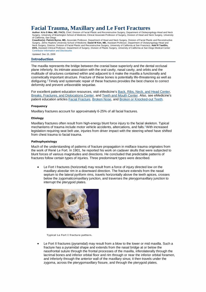
Le Fort II fractures (pyramidal) may result from a blow to the lower or mid maxilla. Such a fracture has a pyramidal shape and extends from the nasal bridge at or below the nasofrontal suture through the frontal processes of the maxilla, inferolaterally through the lacrimal bones and inferior orbital floor and rim through or near the inferior orbital foramen, and inferiorly through the anterior wall of the maxillary sinus; it then travels under the zygoma, across the pterygomaxillary fissure, and through the pterygoid plates.
Typical Le Fort II fracture pattern.
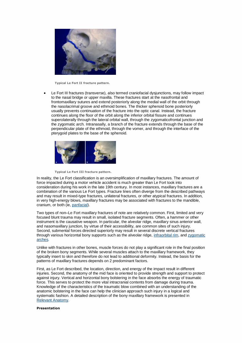
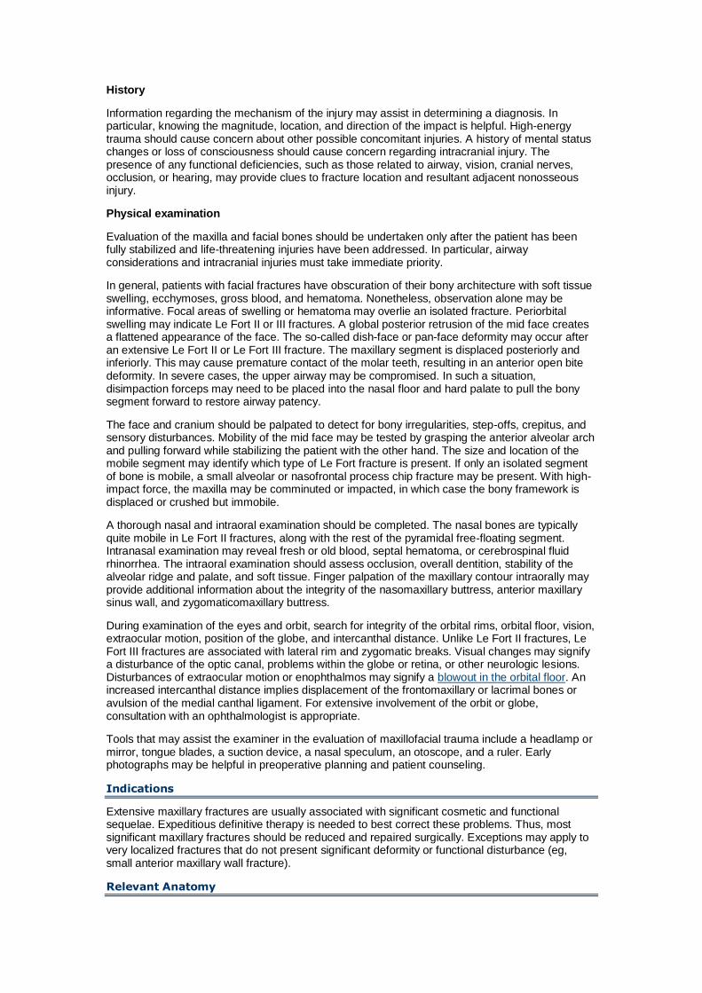
Le Fort III fractures (transverse), also termed craniofacial dysjunctions, may follow impact to the nasal bridge or upper maxilla. These fractures start at the nasofrontal and frontomaxillary sutures and extend posteriorly along the medial wall of the orbit through the nasolacrimal groove and ethmoid bones. The thicker sphenoid bone posteriorly usually prevents continuation of the fracture into the optic canal. Instead, the fracture continues along the floor of the orbit along the inferior orbital fissure and continues superolaterally through the lateral orbital wall, through the zygomaticofrontal junction and the zygomatic arch. Intranasally, a branch of the fracture extends through the base of the perpendicular plate of the ethmoid, through the vomer, and through the interface of the pterygoid plates to the base of the sphenoid.
Typical Le Fort III fracture pattern.
In reality, the Le Fort classification is an oversimplification of maxillary fractures. The amount of force impacted during a motor vehicle accident is much greater than Le Fort took into consideration during his work in the late 19th century. In most instances, maxillary fractures are a combination of the various Le Fort types. Fracture lines often diverge from the described pathways and may result in mixed-type fractures, unilateral fractures, or other atypical fractures. In addition, in very high-energy blows, maxillary fractures may be associated with fractures to the mandible, cranium, or both (ie, panfacial).
Two types of non–Le Fort maxillary fractures of note are relatively common. First, limited and very focused blunt trauma may result in small, isolated fracture segments. Often, a hammer or other instrument is the causative weapon. In particular, the alveolar ridge, maxillary sinus anterior wall, and nasomaxillary junction, by virtue of their accessibility, are common sites of such injury. Second, submental forces directed superiorly may result in several discrete vertical fractures through various horizontal bony supports such as the alveolar ridge, infraorbital rim, and zygomatic arches.
Unlike with fractures in other bones, muscle forces do not play a significant role in the final position of the broken bony segments. While several muscles attach to the maxillary framework, they typically insert to skin and therefore do not lead to additional deformity. Instead, the basis for the patterns of maxillary fractures depends on 2 predominant factors.
First, as Le Fort described, the location, direction, and energy of the impact result in different injuries. Second, the anatomy of the mid face is oriented to provide strength and support to protect against injury. Vertical and horizontal bony bolstering in the face absorbs the energy of traumatic force. This serves to protect the more vital intracranial contents from damage during trauma. Knowledge of the characteristics of the traumatic blow combined with an understanding of the anatomic bolstering in the face can help the clinician approach such injury in a logical and systematic fashion. A detailed description of the bony maxillary framework is presented in Relevant Anatomy.
Presentation
History
Information regarding the mechanism of the injury may assist in determining a diagnosis. In particular, knowing the magnitude, location, and direction of the impact is helpful. High-energy trauma should cause concern about other possible concomitant injuries. A history of mental status changes or loss of consciousness should cause concern regarding intracranial injury. The presence of any functional deficiencies, such as those related to airway, vision, cranial nerves, occlusion, or hearing, may provide clues to fracture location and resultant adjacent nonosseous injury.
Physical examination
Evaluation of the maxilla and facial bones should be undertaken only after the patient has been fully stabilized and life-threatening injuries have been addressed. In particular, airway considerations and intracranial injuries must take immediate priority.
In general, patients with facial fractures have obscuration of their bony architecture with soft tissue swelling, ecchymoses, gross blood, and hematoma. Nonetheless, observation alone may be informative. Focal areas of swelling or hematoma may overlie an isolated fracture. Periorbital swelling may indicate Le Fort II or III fractures. A global posterior retrusion of the mid face creates a flattened appearance of the face. The so-called dish-face or pan-face deformity may occur after an extensive Le Fort II or Le Fort III fracture. The maxillary segment is displaced posteriorly and inferiorly. This may cause premature contact of the molar teeth, resulting in an anterior open bite deformity. In severe cases, the upper airway may be compromised. In such a situation, disimpaction forceps may need to be placed into the nasal floor and hard palate to pull the bony segment forward to restore airway patency.
The face and cranium should be palpated to detect for bony irregularities, step-offs, crepitus, and sensory disturbances. Mobility of the mid face may be tested by grasping the anterior alveolar arch and pulling forward while stabilizing the patient with the other hand. The size and location of the mobile segment may identify which type of Le Fort fracture is present. If only an isolated segment of bone is mobile, a small alveolar or nasofrontal process chip fracture may be present. With high-impact force, the maxilla may be comminuted or impacted, in which case the bony framework is displaced or crushed but immobile.
A thorough nasal and intraoral examination should be completed. The nasal bones are typically quite mobile in Le Fort II fractures, along with the rest of the pyramidal free-floating segment. Intranasal examination may reveal fresh or old blood, septal hematoma, or cerebrospinal fluid rhinorrhea. The intraoral examination should assess occlusion, overall dentition, stability of the alveolar ridge and palate, and soft tissue. Finger palpation of the maxillary contour intraorally may provide additional information about the integrity of the nasomaxillary buttress, anterior maxillary sinus wall, and zygomaticomaxillary buttress.
During examination of the eyes and orbit, search for integrity of the orbital rims, orbital floor, vision, extraocular motion, position of the globe, and intercanthal distance. Unlike Le Fort II fractures, Le Fort III fractures are associated with lateral rim and zygomatic breaks. Visual changes may signify a disturbance of the optic canal, problems within the globe or retina, or other neurologic lesions. Disturbances of extraocular motion or enophthalmos may signify a blowout in the orbital floor. An increased intercanthal distance implies displacement of the frontomaxillary or lacrimal bones or avulsion of the medial canthal ligament. For extensive involvement of the orbit or globe, consultation with an ophthalmologist is appropriate.
Tools that may assist the examiner in the evaluation of maxillofacial trauma include a headlamp or mirror, tongue blades, a suction device, a nasal speculum, an otoscope, and a ruler. Early photographs may be helpful in preoperative planning and patient counseling.
Indications
Extensive maxillary fractures are usually associated with significant cosmetic and functional sequelae. Expeditious definitive therapy is needed to best correct these problems. Thus, most significant maxillary fractures should be reduced and repaired surgically. Exceptions may apply to very localized fractures that do not present significant deformity or functional disturbance (eg, small anterior maxillary wall fracture).
Relevant Anatomy
The maxillary bones are paired pyramidal bones that in many ways serve as the cornerstones of the facial skeleton. In a superoinferior direction, the maxilla bridges the cranial-frontoethmoid complex above to the mandible and to the occlusal plate below. In a transverse plane, it bridges the 2 zygomaticoorbital complexes.
Each individual maxilla can be conceptualized as a 5-sided structure, the base of which makes up the lateral nasal wall. The remaining 4 sides of the pyramid are composed of the orbital floor superiorly, the alveolar ridge inferiorly, the front wall of the maxillary sinus anteriorly, and the anterior face of the pterygopalatine fossa posterolaterally.
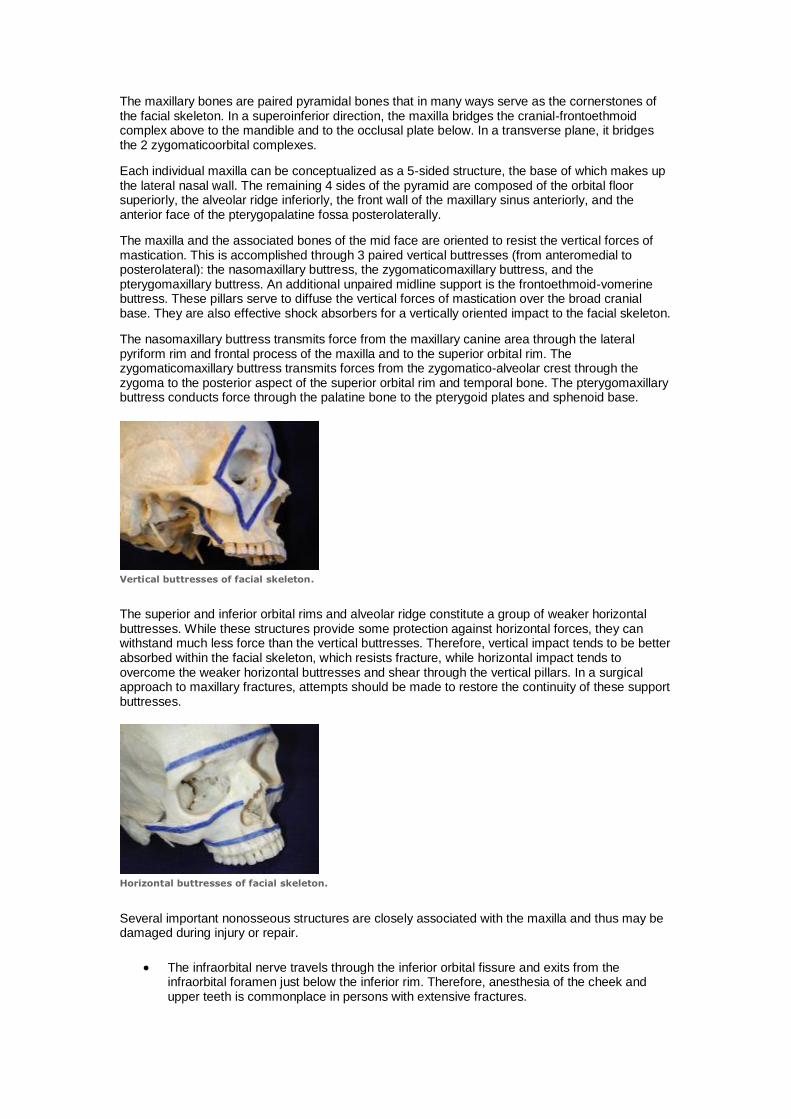
The maxilla and the associated bones of the mid face are oriented to resist the vertical forces of mastication. This is accomplished through 3 paired vertical buttresses (from anteromedial to posterolateral): the nasomaxillary buttress, the zygomaticomaxillary buttress, and the pterygomaxillary buttress. An additional unpaired midline support is the frontoethmoid-vomerine buttress. These pillars serve to diffuse the vertical forces of mastication over the broad cranial base. They are also effective shock absorbers for a vertically oriented impact to the facial skeleton.
The nasomaxillary buttress transmits force from the maxillary canine area through the lateral pyriform rim and frontal process of the maxilla and to the superior orbital rim. The zygomaticomaxillary buttress transmits forces from the zygomatico-alveolar crest through the zygoma to the posterior aspect of the superior orbital rim and temporal bone. The pterygomaxillary buttress conducts force through the palatine bone to the pterygoid plates and sphenoid base.
Vertical buttresses of facial skeleton.
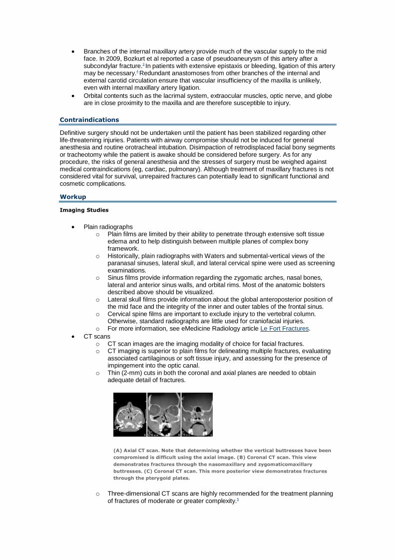
The superior and inferior orbital rims and alveolar ridge constitute a group of weaker horizontal buttresses. While these structures provide some protection against horizontal forces, they can withstand much less force than the vertical buttresses. Therefore, vertical impact tends to be better absorbed within the facial skeleton, which resists fracture, while horizontal impact tends to overcome the weaker horizontal buttresses and shear through the vertical pillars. In a surgical approach to maxillary fractures, attempts should be made to restore the continuity of these support buttresses.
Horizontal buttresses of facial skeleton.
Several important nonosseous structures are closely associated with the maxilla and thus may be damaged during injury or repair.
The infraorbital nerve travels through the inferior orbital fissure and exits from the infraorbital foramen just below the inferior rim. Therefore, anesthesia of the cheek and upper teeth is commonplace in persons with extensive fractures.
Branches of the internal maxillary artery provide much of the vascular supply to the mid face. In 2009, Bozkurt et al reported a case of pseudoaneurysm of this artery after a subcondylar fracture.3 In patients with extensive epistaxis or bleeding, ligation of this artery may be necessary.4 Redundant anastomoses from other branches of the internal and external carotid circulation ensure that vascular insufficiency of the maxilla is unlikely, even with internal maxillary artery ligation.
Orbital contents such as the lacrimal system, extraocular muscles, optic nerve, and globe are in close proximity to the maxilla and are therefore susceptible to injury.
Contraindications
Definitive surgery should not be undertaken until the patient has been stabilized regarding other life-threatening injuries. Patients with airway compromise should not be induced for general anesthesia and routine orotracheal intubation. Disimpaction of retrodisplaced facial bony segments or tracheotomy while the patient is awake should be considered before surgery. As for any procedure, the risks of general anesthesia and the stresses of surgery must be weighed against medical contraindications (eg, cardiac, pulmonary). Although treatment of maxillary fractures is not considered vital for survival, unrepaired fractures can potentially lead to significant functional and cosmetic complications.
Workup
Imaging Studies
Plain radiographs o Plain films are limited by their ability to penetrate through extensive soft tissue
edema and to help distinguish between multiple planes of complex bony framework.
o Historically, plain radiographs with Waters and submental-vertical views of the paranasal sinuses, lateral skull, and lateral cervical spine were used as screening examinations.
o Sinus films provide information regarding the zygomatic arches, nasal bones, lateral and anterior sinus walls, and orbital rims. Most of the anatomic bolsters described above should be visualized.
o Lateral skull films provide information about the global anteroposterior position of the mid face and the integrity of the inner and outer tables of the frontal sinus.
o Cervical spine films are important to exclude injury to the vertebral column. Otherwise, standard radiographs are little used for craniofacial injuries.
o For more information, see eMedicine Radiology article Le Fort Fractures.
CT scans o CT scan images are the imaging modality of choice for facial fractures. o CT imaging is superior to plain films for delineating multiple fractures, evaluating
associated cartilaginous or soft tissue injury, and assessing for the presence of impingement into the optic canal.
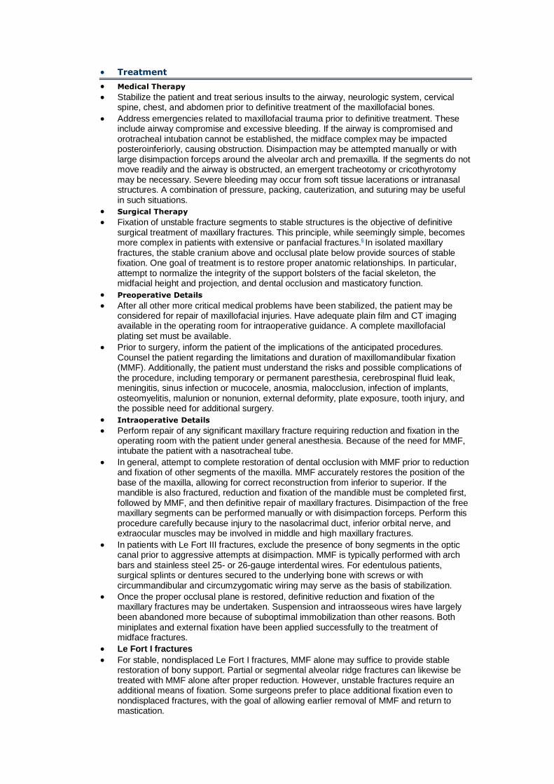
o Thin (2-mm) cuts in both the coronal and axial planes are needed to obtain adequate detail of fractures.
(A) Axial CT scan. Note that determining whether the vertical buttresses have been
compromised is difficult using the axial image. (B) Coronal CT scan. This view
demonstrates fractures through the nasomaxillary and zygomaticomaxillary
buttresses. (C) Coronal CT scan. This more posterior view demonstrates fractures
through the pterygoid plates.
o Three-dimensional CT scans are highly recommended for the treatment planning of fractures of moderate or greater complexity.5
Treatment
Medical Therapy
Stabilize the patient and treat serious insults to the airway, neurologic system, cervical spine, chest, and abdomen prior to definitive treatment of the maxillofacial bones.
Address emergencies related to maxillofacial trauma prior to definitive treatment. These include airway compromise and excessive bleeding. If the airway is compromised and orotracheal intubation cannot be established, the midface complex may be impacted posteroinferiorly, causing obstruction. Disimpaction may be attempted manually or with large disimpaction forceps around the alveolar arch and premaxilla. If the segments do not move readily and the airway is obstructed, an emergent tracheotomy or cricothyrotomy may be necessary. Severe bleeding may occur from soft tissue lacerations or intranasal structures. A combination of pressure, packing, cauterization, and suturing may be useful in such situations.
Surgical Therapy
Fixation of unstable fracture segments to stable structures is the objective of definitive surgical treatment of maxillary fractures. This principle, while seemingly simple, becomes more complex in patients with extensive or panfacial fractures.6 In isolated maxillary fractures, the stable cranium above and occlusal plate below provide sources of stable fixation. One goal of treatment is to restore proper anatomic relationships. In particular, attempt to normalize the integrity of the support bolsters of the facial skeleton, the midfacial height and projection, and dental occlusion and masticatory function.
Preoperative Details
After all other more critical medical problems have been stabilized, the patient may be considered for repair of maxillofacial injuries. Have adequate plain film and CT imaging available in the operating room for intraoperative guidance. A complete maxillofacial plating set must be available.
Prior to surgery, inform the patient of the implications of the anticipated procedures. Counsel the patient regarding the limitations and duration of maxillomandibular fixation (MMF). Additionally, the patient must understand the risks and possible complications of the procedure, including temporary or permanent paresthesia, cerebrospinal fluid leak, meningitis, sinus infection or mucocele, anosmia, malocclusion, infection of implants, osteomyelitis, malunion or nonunion, external deformity, plate exposure, tooth injury, and the possible need for additional surgery.
Intraoperative Details
Perform repair of any significant maxillary fracture requiring reduction and fixation in the operating room with the patient under general anesthesia. Because of the need for MMF, intubate the patient with a nasotracheal tube.
In general, attempt to complete restoration of dental occlusion with MMF prior to reduction and fixation of other segments of the maxilla. MMF accurately restores the position of the base of the maxilla, allowing for correct reconstruction from inferior to superior. If the mandible is also fractured, reduction and fixation of the mandible must be completed first, followed by MMF, and then definitive repair of maxillary fractures. Disimpaction of the free maxillary segments can be performed manually or with disimpaction forceps. Perform this procedure carefully because injury to the nasolacrimal duct, inferior orbital nerve, and extraocular muscles may be involved in middle and high maxillary fractures.
In patients with Le Fort III fractures, exclude the presence of bony segments in the optic canal prior to aggressive attempts at disimpaction. MMF is typically performed with arch bars and stainless steel 25- or 26-gauge interdental wires. For edentulous patients, surgical splints or dentures secured to the underlying bone with screws or with circummandibular and circumzygomatic wiring may serve as the basis of stabilization.
Once the proper occlusal plane is restored, definitive reduction and fixation of the maxillary fractures may be undertaken. Suspension and intraosseous wires have largely been abandoned more because of suboptimal immobilization than other reasons. Both miniplates and external fixation have been applied successfully to the treatment of midface fractures.
Le Fort I fractures
For stable, nondisplaced Le Fort I fractures, MMF alone may suffice to provide stable restoration of bony support. Partial or segmental alveolar ridge fractures can likewise be treated with MMF alone after proper reduction. However, unstable fractures require an additional means of fixation. Some surgeons prefer to place additional fixation even to nondisplaced fractures, with the goal of allowing earlier removal of MMF and return to mastication.
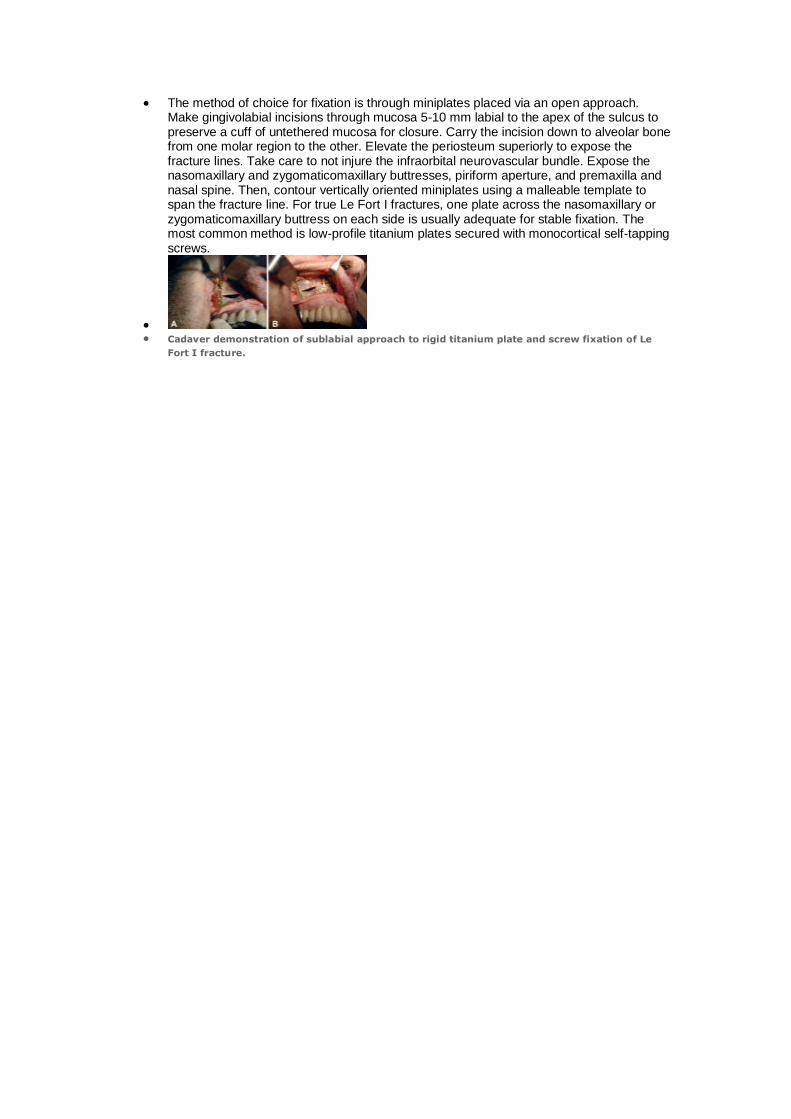
The method of choice for fixation is through miniplates placed via an open approach. Make gingivolabial incisions through mucosa 5-10 mm labial to the apex of the sulcus to preserve a cuff of untethered mucosa for closure. Carry the incision down to alveolar bone from one molar region to the other. Elevate the periosteum superiorly to expose the fracture lines. Take care to not injure the infraorbital neurovascular bundle. Expose the nasomaxillary and zygomaticomaxillary buttresses, piriform aperture, and premaxilla and nasal spine. Then, contour vertically oriented miniplates using a malleable template to span the fracture line. For true Le Fort I fractures, one plate across the nasomaxillary or zygomaticomaxillary buttress on each side is usually adequate for stable fixation. The most common method is low-profile titanium plates secured with monocortical self-tapping screws.
Cadaver demonstration of sublabial approach to rigid titanium plate and screw fixation of Le
Fort I fracture.
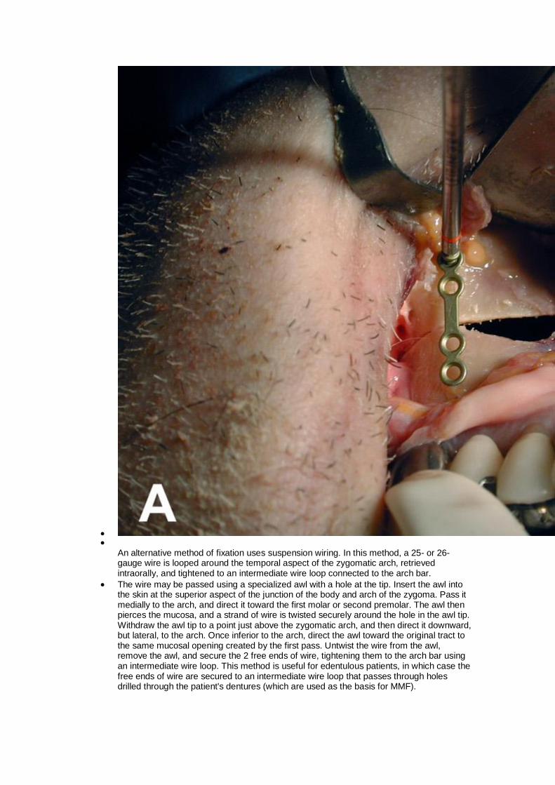
An alternative method of fixation uses suspension wiring. In this method, a 25- or 26-gauge wire is looped around the temporal aspect of the zygomatic arch, retrieved intraorally, and tightened to an intermediate wire loop connected to the arch bar.
The wire may be passed using a specialized awl with a hole at the tip. Insert the awl into the skin at the superior aspect of the junction of the body and arch of the zygoma. Pass it medially to the arch, and direct it toward the first molar or second premolar. The awl then pierces the mucosa, and a strand of wire is twisted securely around the hole in the awl tip. Withdraw the awl tip to a point just above the zygomatic arch, and then direct it downward, but lateral, to the arch. Once inferior to the arch, direct the awl toward the original tract to the same mucosal opening created by the first pass. Untwist the wire from the awl, remove the awl, and secure the 2 free ends of wire, tightening them to the arch bar using an intermediate wire loop. This method is useful for edentulous patients, in which case the free ends of wire are secured to an intermediate wire loop that passes through holes drilled through the patient's dentures (which are used as the basis for MMF).
Take care to not overtighten suspension wires because the zygomatic arch resides somewhat posterior to the true vertical plane of the maxilla. Overaggressive pulling may result in superoposterior displacement of the inferior fracture segment.
Le Fort II fractures
Just as for Le Fort I fractures, disimpaction, MMF, and sublabial incisions and exposure of maxillary bone and fracture lines are performed. Additional exposure is often necessary superiorly for adequate exploration of the orbital rim. This may be achieved through subciliary or transconjunctival incisions. More extensive degloving of the soft tissue envelope through exposure of the piriform aperture and frontomaxillary region may be facilitated by columellar-septal transfixion incisions.
In general, the pyramidal free maxillary segment is stabilized to the intact zygoma. Because rigid fixation is a traumatic procedure, do not perform it until reduction is optimized. Fixation may be completed directly using noncompression miniplates that span the break in the region of the zygomaticomaxillary buttresses. If instability persists, additional plates may be placed in the nasomaxillary buttresses or inferior orbital rim. Any plating must be placed in areas of adequately robust bone (ie, buttresses). Accurate contouring of the plates using malleable templates is important for precise reduction and fixation. Monocortical, self-tapping screws are ideal. Place plates so that at least 2 screws holes are on each side of the fracture. Thus, if needed, additional screws can be placed for more support.
An alternative to miniplates is interosseous wiring. In this method, place small holes into the appropriate bony segments on either side of the fracture line with a minidriver. Then, pass 28-gauge steel wire through the hole on one side of the fracture and retrieve it outward from the gap between the bony segments. Pull the free end of the wire through the opposite drill hole with a loop of 30-gauge wire. Tighten the 2 free ends of wire. In general, place wires from stable to unstable segments. Because this method is less stable than miniplating, perform several areas of fixation (eg, nasomaxillary, zygomaticomaxillary, inferior orbital rim buttresses). If this method is used, implement a longer duration of MMF than with plating.
Circumzygomatic suspension wiring of Le Fort II fractures has been described. While this method may be effective for clean, true Le Fort II fractures, it is discouraged for 3 main reasons. First, these injuries often have multiple segments, in which case comminution and compression of the maxilla may follow efforts to pull the maxilla en bloc. Second, reduction depends on a vector force that is imperfect. In most patients, the vector from the classic fracture line of Le Fort II fractures to the zygomatic arch is at least 15° askew from the ideal axis for fracture reduction. Finally, other methods have the advantage of more precise application of fixation forces immediately at the site of fracture, minimizing micromotion, maximizing bone healing, and allowing for earlier return to mastication.
Le Fort III fractures
In repairing Le Fort III fractures, stabilize the mobile segments of bone to the stable mandible below and cranium above. Initially, the maxilla must be disimpacted and MMF implemented. Soft tissue incisions may be made in the same locations as for Le Fort II fractures. Lateral brow incisions, glabellar fold incisions, or bicoronal scalp flaps can be used for additional exposure to the frontozygomatic buttress.
The bicoronal flaps may be extended to achieve access to the zygomatic arches. The bicoronal flap must be designed cautiously to avoid injury to the frontal branch of the facial nerve. The plane of dissection is between the galea and pericranium. Once the soft tissue flap is rolled over the superior orbital rims, the pericranium may be incised just above the rims to preserve the supraorbital and supratrochlear vascular supply to the flap.
Laterally, perform dissection just superficial to the temporalis fascia. In approaching the zygomatic arch, incise the temporalis fascia well above it. Develop a plane deep to the fascia down to the fractured zygomatic arch. The fracture can then be levered into reduction with a rigid elevator. If impacted or comminuted, direct fixation may be required. Do not use the bicoronal flap in situations in which soft tissue flaps based on the superficial temporal arteries are needed. A receding hairline also may prompt the surgeon to use other incisions.
Prior to fixation of the involved maxillary fractures, reduce and stabilize any mandibular and cranial fractures. Once this is performed and the fractured maxillary segments are exposed, fixation may be undertaken.
Miniplate fixation is currently the most reliable and rigid method. Use malleable templates; accurate contouring of plates; and monocortical, self-tapping screws. Use plates that span the involved major buttresses. For true Le Fort III fractures, bilateral zygomaticofrontal
fixation may suffice. However, more commonly, additional points of fixation are needed (eg, nasomaxillary, nasofrontal, inferior orbital rim, zygomatic arch). Use as few plates as possible to achieve fixation; excessive plating is not necessary.
Interosseous wiring and suspension wiring have been described for Le Fort III fractures but are less reliable than miniplate fixation because vectors of forces to maintain reduction are less accurate and micromotion is increased.
Extraskeletal fixation is not usually necessary for simple Le Fort fractures. In patients with more extensive panfacial fractures, external fixation may be the only means of stabilization. If possible, avoid this method because it can place excessive or misdirected force onto the fracture segments and therefore cause shortening or further deformity of the mid face.
For all maxillary fractures, suspension of the soft tissue of the mid face should be performed prior to closing the intraoral incisions with 3-0 chromic suture and closing the skin incisions with absorbable subcutaneous sutures and permanent skin sutures. Bicoronal flaps may be closed with skin staples.
Postoperative Details
To minimize postoperative edema, a light pressure dressing consisting of gauze and a head wrap may be placed over the operated areas. If the dressing remains dry, it may be removed after 2-5 days.
Surgeons' opinions are divided regarding the need for postoperative antibiotics. If the original fracture sites were open to the external environment or in communication with intraoral or intranasal spaces, implement prophylactic antibiotics covering gram-positive and anaerobic organisms for 5-10 days.
After surgery, observe patients overnight for bleeding, airway problems, and vomiting. If wire fixation was used for MMF, place wire cutters near the patient at all times in the early postoperative period to allow the patient to expel vomited material. Remove wires or rubber bands if the patient begins to feel nauseated.
Prior to discharge, instruct patients on how to remove the MMF in case of vomiting. Also, counsel patients regarding limiting their diet to pureed or liquid intake.
Follow-up
Perform a follow-up evaluation at 5-7 days (skin sutures may be removed at this time), 2-4 weeks, and then at 3-8 weeks for removal of the MMF. Longer-term follow-up care may be needed to monitor postoperative complications or deformity.
The most important goal during the early postoperative period is maintaining a state of immobilization. Depending on the age and general health of the patient, the extensiveness and displacement of the fractures, and the repair technique used, this period may range from 4-8 weeks. This requires that MMF be maintained during this period. During this period, emphasize to the patient to maintain oral hygiene with diligent teeth and arch bar brushing and oral rinses with saline or antiseptic mouthwash each morning and evening and after each meal.
Throughout the postoperative course, the stability of the facial skeleton may be tested by palpating the patient's maxillary teeth during clenching and relaxing of the muscles of mastication. Minimal conducted motion is acceptable, but excessive mobility may indicate poor healing. Postoperative films (ie, mandible series, Panorex dental views, facial series, CT scan) may be helpful in patients in whom malunion is suggested.
Once the facial skeleton is deemed to be well healed and normal occlusion is present, the MMF may be removed. Minimal vertical mobility of the mid face likely resolves with time. Excessive motion indicates that it is too early for the arch bars to be removed or that a problem exists with union. In general, the MMF is removed earlier for fractures repaired with miniplate fixation and later for those repaired with interosseous or suspension wires.
Complications
Soft tissue complications result from technical pitfalls or problems with wound healing. In general, unfavorable scarring may be avoided by closing facial incisions in a 2-layered fashion, with deeper subcutaneous absorbable sutures placed to remove tension from the skin closure. Skin closure should be performed with nontraumatic handling of wound edges and should result in the wound edges being slightly everted.
Intraoral incisions may dehisce partially or completely because of inadequate closure during surgery, poor oral hygiene, local trauma, or excessive motion. When designing the gingivolabial incisions, a cuff of mucosa should be maintained on the gingiva to allow for adequate soft tissue upon which to suture. This may be accomplished by placing the
incision slightly labial to the deepest part of the gingivolabial sulcus. If dehiscence occurs, maintaining local hygiene alone allows for eventual healing.
Lower lid ectropion may follow a subciliary approach to the maxilla. This complication may be avoided by performing meticulous dissection between the orbital septum and orbicularis oculi muscle and, for patients in whom laxity is present, superolateral suspension of the muscle to the periosteum of the lateral orbital wall. If severe ectropion occurs, breaking up the scar with Z-plasty or skin grafting from the opposite lid skin may be necessary. Lower lid transconjunctival incisions decrease the likelihood of ectropion and should be considered in high-risk patients.
Nerve injury may have occurred prior to surgery from the initial traumatic insult. Therefore, the status of the main sensory and motor nerves of the face and forehead must be documented prior to surgery. Care should be taken to identify and preserve the supraorbital and infraorbital neurovascular pedicles while the soft tissue flaps are raised. More commonly, supraorbital nerve injury results from nerve stretching in retracting the soft tissue and orbital tissues to gain access to the superior and medial orbital rims. The frontal branch of the facial nerve may be injured from excess traction on the forehead flap.
Anatomic disruption of the nerve may occur if the improper plane is used to access the zygomatic arch. The nerve is known to cross the arch superficially to the superficial layer of the deep temporalis fascia. Therefore, dissection should be performed deep into this layer. The appropriate plane is accessed by incising the temporalis fascia well above the arch and dissecting deeply to fascia down to the fractured arch. Nerve injury is often incomplete and temporary.
Injury to tooth roots from misplaced screw holes may result in nonviable teeth. If fracture lines are low and do not allow an area adequate to avoid teeth when placing plates, suspension or interosseous wire fixation may be considered.
Postoperative infections are more apt to occur in the setting of extensive soft tissue injury, contaminated wounds, open fractures, fractures communicating with intranasal or intraoral spaces, or nonevacuated sinus blood. If empiric antibiotic therapy does not clear the infection, debridement and drainage may be required. Cultures should be obtained if purulent material is encountered, and specific antibiotic treatment should be instituted. Long-term unchecked infection may cause osteomyelitis around the sites of the screws or wires. Removal of these implants and debridement of bone may be necessary if antibiotics are unsuccessful. Sinusitis may occur if fracture lines involve the sinus drainage ostia. In such instances, decongestants and antibiotics should be started; intranasal surgical drainage should be performed for nonresolving cases.
Malunion and resultant malocclusion and deformity occur if reduction is not precise or if loosening of fixation occurs during the postoperative period. This can be avoided with meticulous surgical technique and adequate fixation, preferably with carefully placed miniplates. Patient noncompliance with MMF and early mastication may result in micromotion, which leads to poor bone healing. If malunion is discovered early, attempts to optimize reduction may be made by loosening the MMF tension and adjusting the wire closure forces or elastics in order to normalize occlusion. If this fails, rigid fixation (wires or plates) must be removed and replaced for better stabilization.
For delayed presentations in which the bones have healed into malposition, osteotomies must be performed through or near the original fracture sites and the bones must be repositioned with rigid fixation. In rare instances, bone resorbs as a result of malunion and motion, and osseous interposition grafts or overlay grafts may be required. Split calvarial grafts are well suited for midface work, but rib grafts may be used as an alternative.
Total nonunion is less common than malunion. In most cases, maintaining an extended period of fixation and immobility results in eventual healing. For persistent nonunion, fracture sites must be reexplored, freshened, and refixated. Again, areas of gaps may need to be addressed with osseous grafts.
Outcome and Prognosis
A lack of prospective studies on trauma patients makes assessment of outcome measures for patients treated for maxillary fractures difficult. Repair of simple maxillary fractures typically restores bony aesthetic contour and function; however, complex fractures often leave the patient with some long-term cosmetic and functional deficits. Early and meticulous surgery is most likely to produce results that restore the patient to the pretrauma state.
Future and Controversies
The continuing trend in facial fracture repair is toward rigid osteosynthesis with miniplates and screws. The advantage of this technique is that a higher degree of stability is gained, allowing for earlier removal of MMF and return to mastication.
Opponents of this technique who favor suspension techniques cite the disadvantage that anatomic realignment must be perfect at the time of surgery. Whereas suspension techniques allow postoperative adjustment of segments by changing the MMF to compensate for slight deviations from perfect reduction, rigid techniques are much less forgiving. Unrecognized displacement of midface or mandibular segments results in inevitable malunion. Also cited are the higher cost of the materials, the difficulty in contouring plates to the surface of the bone, and increased surgical time. Despite these disadvantages, rigid techniques are gaining in popularity. As long as surgical technique is proficient, rigid osteosynthesis is generally believed to lead to better long-term results and faster recovery.
Absorbable plating systems composed of polylactic acids have recently become available and are gaining popularity for maxillofacial repair. These systems have the advantage of providing rigid osseous fixation without permanent foreign body implantation. This theoretically reduces the risk of infection and plate exposure. The other main advantage of these systems is the ability to contour plates with thermal manipulation (hot saline sponge or specialized heated instruments) even after the plates have been positioned in situ. This facilitates contouring plates to a precise and appropriate shape across fracture lines.
The use of endoscopically-assisted techniques allows for limited incisions for the reduction of facial fractures. These techniques have been pioneered for use in the reduction of condylar and orbital fractures but have recently been applied to more extensive procedures.7,8,9 The use of endoscopic techniques allows for limited incisions, faster recovery periods, and shorter hospital stays.8,9 Despite the advantages afforded with these techniques, the indications for open procedures have not been drastically altered. Facial trauma that involves severely dislocated or comminuted fractures of the facial skeleton and major reconstruction of the facial support structures still requires the use of open techniques and direct visualization.
Reconstruction of the facial skeleton involves the reestablishment of the original contours of the face with the precise alignment of fractures. The advancement of image guidance systems has assisted the surgeon in preoperative evaluation and surgical planning, but its recent introduction into the operative arena allows real-time localization of displaced facial skeletal segments during reduction and internal fixation. The use of this technology can help the surgeon obtain a postoperative result that most closely approximates the pre-trauma skeletal structure. This may be most useful in cases where the adjacent bony anatomic landmarks are also displaced or altered and the continuing incorporation of computer-aided guidance of reduction of facial fractures will help to optimize surgical results.10