Embed Size (px)
Citation preview

International Journal of Anatomy, Radiology and Surgery. 2016 Jul, Vol-5(3): RO24-RO3024
Original Article DOI: 10.7860/IJARS/2016/20367:2160
ABSTRACTIntroduction: Facial fractures consist of most common injuries in cases of road traffic accidents. Road traffic injuries are a major public health problem worldwide. Each year, an estimated 1.2 million people die in road traffic accidents and around 50 million suffer from non-fatal severe injuries. In big cities like Bangalore, motor vehicle accidents are very frequent. In such accidents MDCT is the modality of choice for detection and prognosticating facial injuries.
Aim: The study was conducted to assess MDCT findings in facial fractures and to elaborate incidence and spectrum of these injuries.
Materials and Methods: Cases were collected retrospec-tively over a period of 24 months, from January 2014- December 2015, in Department of Radio-diagnosis, Bangalore Medical College and Research Institute, Bangalore. 6 slice CT GE SOMATOM scanner was used to obtain images in bone and soft tissue window and thin reconstructions were done. From the data obtained which consisted of 650 positive cases, they were divided and assessed based on age, gender predominance, site of fracture and associated complications. A specific consideration was given to clear sinus sign. The statistical
analysis was performed using Microsoft Excel 2010 for Windows (Microsoft Corporation, Redmond, Washington).
Results: Of the 650 cases, males constituted 533 cases and the rest were females. Most common age group was between 21-30 years. Cases with multiple fractures were more common as compared to a single fracture. Fractures of nasal bone topped the list with 20% of total cases being fracture of either single or both nasal bones, with or without other fracturesfollowed by orbital, skull base, and maxillary fractures. The Le Fort fractures are most commonly associated with opacification of the sinus indicating hemosinus.
Conclusion: Road traffic accidents are very common in metropolitan cities like Bangalore and facial injuries constitute a significant number. Young and middle aged males are more prone for injury before of increased habit of risk taking and more exposure to risk. MDCT plays a major role in evaluation of patients with maxillofacial trauma. It not only gives information about site and displacement of the fracture; but also helps in detection of adjacent soft tissue injury and airway. In cases of trauma, imaging is very essential in diagnosis, treatment planning, and in prognosticating.
Rad
iolo
gy
Sec
tionMulti Detector Computed
Tomography Evaluation of Spectrum of Facial Fractures in Motor Vehicle Accidents
Vijay KumaR K R, BhaRath B DaS, VenKateSha mP
INTRODUCTIONFacial trauma is the most common injury in patients with motor vehicle accidents [1]. Road traffic injuries are a major public health problem worldwide. Each year, an estimated 1.2 million people die in road traffic accidents and up to 50 million suffer non-fatal injuries [2]. Maxillofacial injuries can sometimes be life threatening as they may cause obstruction of patient’s airway and hence Prompt recognition and stabilization of such
fractures are important [3]. Furthermore, they are frequently associated with orbital injuries, cervical spine injuries, intra cranial haemorrhages and cardio thoracic injuries [4, 5] and thus making prompt and accurate diagnosis is essential. In big cities like Bangalore, motor vehicle accidents are very common. Advances in technology and rapidity of imaging technique has made accurate diagnosis and associated injuries detection possible and thus contributing significantly
Keywords: Clear sinus sign, Le-Fort fracture, Collision, Unifocal and multifocal fractures, sentinel fracture.
www.ijars.net Vijay Kumar K R et al., Multi Detector Computed Tomography Evaluation of Spectrum of Facial Fractures in Motor Vehicle Accidents
International Journal of Anatomy, Radiology and Surgery. 2016 Jul, Vol-5(3): RO24-RO30 25
Keywords: Clear sinus sign, Le-Fort fracture, Collision, Unifocal and multifocal fractures, sentinel fracture.
in overall patient care. Multi Detector Computed Tomography (MDCT) is the imaging modality of choice owing to its high image resolution and thin-section acquisitions allows the detection of even subtle nondisplaced fractures of the facial skeleton. It also gives better delineation of osseous and soft-tissue features and offers both multiplanar and three-dimensional image reconstruction. Scans are performed more quickly than radiography, with easier patient positioning. The purpose of this study is to assess MDCT findings in facial fractures and to elaborate incidence and spectrum of these injuries.
MeThODS AND MATeRIAlSThis retrospective descriptive study was conducted in the Department of Radio-diagnosis, Bangalore Medical College and Research Institute, Bangalore which is a referral as well as a teaching institute. Ethical clearance is obtained from the ethical committee BMC&RI.
6 slice CT GE SOMATOM scanner was used to evaluate every patient who was referred to the department with motor vehicle accident. Scans were acquired in bone soft tissue algorithm and thinner reconstructions, multiplanar imaging and volume rendering reconstruction were done in workstation. Each case was evaluated for the presence of facial fracture, site and associated fractures or complications. Cases were collected retrospectively over a period of 24 months, from January 2014- December 2015. Using Picture Archiving and Communication System (PACS) all cases of motor vehicle accidents with suspected facial injury were retrieved. Among these, patients who fulfilled all inclusion criteria were included in the study and patients who had even one of exclusion criteria were excluded from the study. The criteria’s are enumerated as follows –
Inclusion Criteria• Historyofmotorvehicleaccidentirrespectiveofagegroup
and includes both rider and pillion rider in two wheelers and driver and passengers in other motor vehicle groups.
• Suspected facial injury on clinical examination andconformation of the same on MDCT imaging.
• Passengercar/largervehicles,Twowheelersareincludedin study.
• Bothcollisionsandrunoff roadaccidentsare involved instudy.
excluison Criteria• Nohistoryofmotorvehicleaccidents
• Facialinjuriesduetoothercauses–exampleassaultetc
• Pedestrianandcyclistsarenotinvolvedinstudybecauseof different mechanism of accident.
• Pregnantpatients
• Cases with inadequate or technically sub-optimalexamination.
Thus, the patients who fulfilled all the above inclusion criterias were included in the study. Total 650 patients were included but among them only 513 patients had positive MDCT findings of maxilla facial fracture and rest 137 patients had no fracture.
The MDCT scans were interpreted using clinical workstations (BARCO systems) by two radiologists. The readouts were performed independently and all disagreements were settled by additional consensus readout. The inter-observer variability was around 6%. Injuries were categorized into 13 groups: nasal bone fractures, naso-orbito-ethmoid fractures (NOE), orbital fractures, frontal bone fractures, zygomaticarch fractures, zygomatic complex (ZMC) fractures, maxillary fractures, LeFort I (LF I), LeFort II (LF II), and LeFort III (LF III) fractures, mandibular fractures, skull base fractures, and other fractures.
The frontal bone fractures were classified into five subgroups according to Manolidis [6]: type 1 (anterior wall fracture, minimal comminution), type 2 (anterior wall fracture, comminution), type 3 (anterior and posterior wall fracture), type 4 (anterior and posterior wall with dural injury and cerebrospinal fluid leak), and type 5 (as type 4 with additional soft tissue or bone loss or severe disruption of the anterior cranial fossa).
Furthermore, the mandibular fractures were divided into condylar, subcondylar, coronoid, ramus, angle, body, parasymphyseal, symphyseal, and isolated alveolar process fractures.
In addition, effusions of the paranasal sinuses (maxillary, frontal, ethmoid, and sphenoid) were recorded. Chronic appearing thickening of the paranasal sinus mucosa was not considered to represent free intra-sinus fluid [7].
STATISTICAl ANAlySISThe statistical analysis was performed using Microsoft Excel 2010 for Windows (Microsoft Corporation, Redmond, Washington). Data were analysed using percentages and proportions.
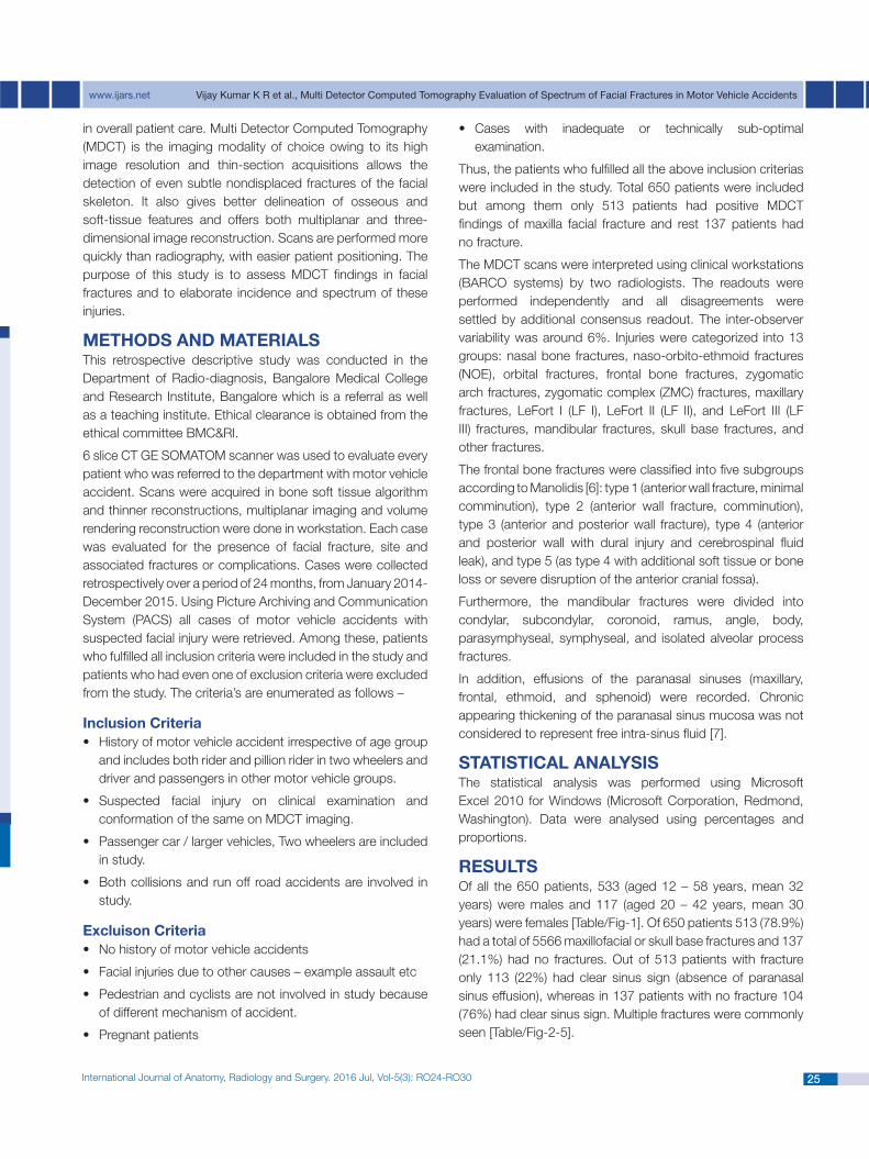
ReSUlTSOf all the 650 patients, 533 (aged 12 – 58 years, mean 32 years) were males and 117 (aged 20 – 42 years, mean 30 years) were females [Table/Fig-1]. Of 650 patients 513 (78.9%) had a total of 5566 maxillofacial or skull base fractures and 137 (21.1%) had no fractures. Out of 513 patients with fracture only 113 (22%) had clear sinus sign (absence of paranasal sinus effusion), whereas in 137 patients with no fracture 104 (76%) had clear sinus sign. Multiple fractures were commonly seen [Table/Fig-2-5].
International Journal of Anatomy, Radiology and Surgery. 2016 Jul, Vol-5(3): RO24-RO3026
Vijay Kumar K R et al., Multi Detector Computed Tomography Evaluation of Spectrum of Facial Fractures in Motor Vehicle Accidents www.ijars.net
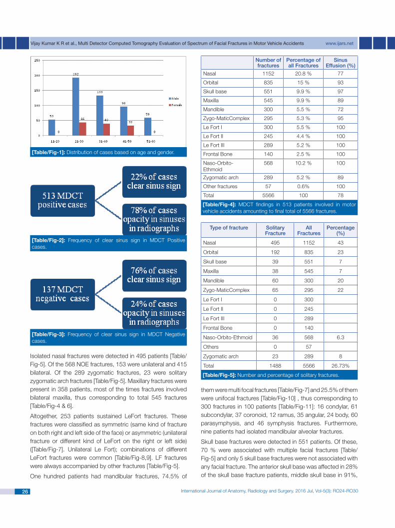
them were multi focal fractures [Table/Fig-7] and 25.5% of them were unifocal fractures [Table/Fig-10] , thus corresponding to 300 fractures in 100 patients [Table/Fig-11]: 16 condylar, 61 subcondylar, 37 coronoid, 12 ramus, 35 angular, 24 body, 60 parasymphysis, and 46 symphysis fractures. Furthermore, nine patients had isolated mandibular alveolar fractures.
Skull base fractures were detected in 551 patients. Of these, 70 % were associated with multiple facial fractures [Table/Fig-5] and only 5 skull base fractures were not associated with any facial fracture. The anterior skull base was affected in 28% of the skull base fracture patients, middle skull base in 91%,
[Table/Fig-1]: Distribution of cases based on age and gender.
number of fractures
Percentage of all Fractures
Sinus effusion (%)
Nasal 1152 20.8 % 77
Orbital 835 15 % 93
Skull base 551 9.9 % 97
Maxilla 545 9.9 % 89
Mandible 300 5.5 % 72
Zygo-MaticComplex 295 5.3 % 95
Le Fort I 300 5.5 % 100
Le Fort II 245 4.4 % 100
Le Fort III 289 5.2 % 100
Frontal Bone 140 2.5 % 100
Naso-Orbito-Ethmoid
568 10.2 % 100
Zygomatic arch 289 5.2 % 89
Other fractures 57 0.6% 100
Total 5566 100 78
[Table/Fig-4]: MDCT findings in 513 patients involved in motor vehicle accidents amounting to final total of 5566 fractures.
type of fracture Solitary Fracture
all Fractures
Percentage (%)
Nasal 495 1152 43
Orbital 192 835 23
Skull base 39 551 7
Maxilla 38 545 7
Mandible 60 300 20
Zygo-MaticComplex 65 295 22
Le Fort I 0 300
Le Fort II 0 245
Le Fort III 0 289
Frontal Bone 0 140
Naso-Orbito-Ethmoid 36 568 6.3
Others 0 57
Zygomatic arch 23 289 8
Total 1488 5566 26.73%
[Table/Fig-5]: Numberandpercentageofsolitaryfractures.
[Table/Fig-2]: Frequency of clear sinus sign in MDCT Positive cases.
[Table/Fig-3]: Frequency of clear sinus sign in MDCT Negativecases.
Isolated nasal fractures were detected in 495 patients [Table/Fig-5].Ofthe568NOEfractures,153wereunilateraland415bilateral. Of the 289 zygomatic fractures, 23 were solitary zygomatic arch fractures [Table/Fig-5]. Maxillary fractures were present in 358 patients, most of the times fractures involved bilateral maxilla, thus corresponding to total 545 fractures [Table/Fig-4 & 6].
Altogether, 253 patients sustained LeFort fractures. These fractures were classified as symmetric (same kind of fracture on both right and left side of the face) or asymmetric (unilateral fracture or different kind of LeFort on the right or left side) ([Table/Fig-7]. Unilateral Le Fort); combinations of different LeFort fractures were common [Table/Fig-8,9]. LF fractures were always accompanied by other fractures [Table/Fig-5].
One hundred patients had mandibular fractures, 74.5% of

www.ijars.net Vijay Kumar K R et al., Multi Detector Computed Tomography Evaluation of Spectrum of Facial Fractures in Motor Vehicle Accidents
International Journal of Anatomy, Radiology and Surgery. 2016 Jul, Vol-5(3): RO24-RO30 27
[Table/Fig-6]: Left LE fort I, II, III fractures, left tripod fracture, fracture of left frontal bone, left hemi mandibleand bilateral maxillary sinus walls fractures.A-H-Axialsectionsbonewindow-Fracture involving fronto-nasal junction (whitearrow),left pterygomaxillary disjunction/Le Fort I fracture (red arrow), Fracture of postero-lateral wall of left maxillary sinus (yellow arrow), zygomatic arrow (peach arrow), lateral wall of left orbit (blue arrow) and associated left frontal bone fracture with pneumocephalus (green and black arrows), bilateral nasal bones and nasal septal fractures ( white arrow) and left squamous temporal bone fracture (curved red arrow). I-L- VRT images- Left Le Fort II fracture (white and yellow arrows), left Le Fort III fracture ( white, peach and blue arrows), left tripod fracture (yellow, dark blue and peach arrow), left squamous temporal bone fracture (curved red arrow), left frontal bone fracture (green arrow) and left hemi-mandibular fracture (blue arrow).
type of Le Fort Fracture number of patients
% tage
LF I (U) 22 9
LF I (B) 14 5.5
LF II (U) 8 3.1
LF II (B) 8 3.1
LF III (U) 38 15.2
LF III (B) 8 3.1
LF I (U) + LF II (U) 4 1.6
LF I (B) + LF II (B) 4 1.6
LF II (U) + LF III (U) 4 1.6
LF II (B) + LF III (B) 8 3.1
LF III (U) + LF I (U) 8 3.1
LF III (U) + LF I (B) 14 5.5
LF III (B) + LF I (B) 4 1.6
LF I (U) + LF II (U) + LF III(U) 14 5.5
LF I (B) + LF II (B) + LF III (B) 68 26.8
LF I (B) + LF II (B) + LF III(U) 8 3.1
LF I (B) + LF II (U) + LF III (U) 11 4.3
LF I (B) + LF II (U) + LF III (B) 8 3.1
[Table/Fig-8]: Distribution of Le Fort I, II, III Fractures in 253 patients.
[Table/Fig-7]: Bilateral LE forts I fracture, right para-symphysis fracture, fracture of right mandibular ramus, left condylar process – A, B, C – Axial and coronal sections in bone window – Fracture involving bilateral both pterygoid plates and pterygomaxillary disjunction (blue arrows) with associated mandibular fractures ( red arrow).D, E, F- VRT images- Bilateral Le Fort I fractures (blue arrow) and associated mandibular fractures (red arrows) and traumatic dislocation of right lateral incisor (arrow head).
and posterior skull base in 16%. Altogether, 140 patients had frontal bone fractures: 10 type 1, 25type 2, 63 type 3, 36 type 4, and 6 type 5 [Table/Fig-9,12].
Zygomatic complex/ Tripod fractures constitute 22% of all fractures- total 295 fractures among which 65 fractures are solitary [Table/Fig-13].
DISCUSSIONRoad traffic injuries are a major public health problem worldwide. Each year, an estimated 1.2 million people die in road traffic accidents and up to 50 million suffer non-fatal injuries [8]. In Finland, with a population of 5.3 million, in 2008, there were a total of 6,881 road traffic accidents involving personal injury, in which 344 people were killed and 8,513 were injured [9]. At our institution, an average of 20 - 25 patients per month is scanned with MDCT due to a suspected facial injury caused by a motor vehicle accident. Of these, 20 – 22 patients per month are diagnosed with a facial or skull base fracture.
Motor vehicle accidents-induced injuries are the result of the remarkable amounts of kinetic energy released when the steady state of a passenger is changed by sudden deceleration or acceleration; both speed and stopping distance have a significant influence [10].
International Journal of Anatomy, Radiology and Surgery. 2016 Jul, Vol-5(3): RO24-RO3028
Vijay Kumar K R et al., Multi Detector Computed Tomography Evaluation of Spectrum of Facial Fractures in Motor Vehicle Accidents www.ijars.net
S. no. Location of Fracture number Percentage (%)
1. Condylar process 16 5.3
2. Sub condylar 61 20.4
3. Angle of mandible 35 11.7
4. Ramus 12 4
5. Body of mandible 24 8
6. Coronoid process 37 12.3
7. Symphysis 46 15.3
8. Parasymphysis 60 20
9. Alveolar ridge 9 3
Total 300
[Table/Fig-11]: Classification of Mandibular fractures based on location with percentage out of 300 fractures in 100 patients.
S. no. type of Frontal Bone Fracture
number Percentage (%)
1. Type 1 10 7
2. Type 2 25 18
3. Type 3 63 45
4. Type 4 36 26
5. Type 5 6 4
Total 140
[Table/Fig-12]: Distribution of types of frontal bone fractures in 140 patients based on Manolidis classification.
[Table/Fig-13]: Right tripod fracture, right coronoid process fracture, complex fracture of right temporal boneA, B- VRT - Fracture of right temporal bone (Yellow arrow), right coronoid process (blue arrow) and right tripod fracture (Red arrow).
[Table/Fig-10]: Types of mandibular fractures.
[Table/Fig-9]: Bilateral Le fort I, II, III fractures, bilateral tripod fracture with associated mandibular fracture and depressed fracture of frontal bone.A – Scannogram showing depressed fracture of face with white arrows depicting probable direction of impact.B-H–Axialsectionbonewindow–Bilateralpterygomaxillarydisjunction/LeFortIfracture(peach arrow), fronto-nasal disjunction and nasal bones fractures (white arrow), bilateral zygomatic arch fracture (dark blue arrow), bilateral lateral wall of orbit fracture (brown arrow), bilateral maxillary wall fractures (yellow arrow), bilateral lamina papyracea fracture (orange arrow).I-N-VRT-BilateralLeFortIfracture(orangearrow),bilateralLeFortIIfracture(whitearrow),bilateral Le Fort III fracture ( white, blue and green) and bilateral Tripod fracture (right- green arrow, left- blue arrow) and associated mandibular fracture with left TM joint dislocation (red and black arrow).O, P- Surface rendering technique – Showing depression of face (Black arrows)
Road traffic accidents morbidity and mortality rates are higher in men than in women [8]. Furthermore, young males are particularly more involved in accidents than middle aged drivers [11]. Road trauma related facial injuries are predominantly an affliction of young men [12]. Similarly, in our study 82% of all
patients were males and among them 46% of them were less than 30 years. This high accident rates in young male drivers can be attributed to deliberate risk taking, carelessness and over estimation of skills [11]. The gender difference can be predominantly attributed to difference in exposure risk [13].
NasalandorbitalfracturesarefrequentlydetectedtogetherinMVA accidents, 50% of orbital fractures were accompanied with associated nasal fracture. Solitary injuries are more commonly seen with nasal, orbital, maxillary and mandibular fractures [Table/Fig-5].
These are considered as low energy fractures due to its occurrence even with low energy trauma. But in some
www.ijars.net Vijay Kumar K R et al., Multi Detector Computed Tomography Evaluation of Spectrum of Facial Fractures in Motor Vehicle Accidents
International Journal of Anatomy, Radiology and Surgery. 2016 Jul, Vol-5(3): RO24-RO30 29
instances they can be “Sentinel injuries” suggesting high energy fractures such as Le Forts fractures. Furthermore low energy fractures, isolated zygomatic arch fractures were never detected alone; in 80% of cases an underlying skull base fracture was detected. Therefore from this study we can come to a consensus that patients who have sustained low energy fractures should undergo MDCT to reveal any high energy injuries.
In circumstances where MDCT is not available, the detection of low energy sentinel injuries in radiography of victims should alert radiologists to search more vigorously for high energy fractures. In this view, Clear Sinus Sign seems to be a valuable aid for detecting MVA injuries. In our study there were no clear sinuseswithfrontal,NOE,LFI,II,III[Table/Fig-4].andisolatedpterygoid plate fractures.
Frontal bone fractures are considered to be seen in high energy trauma [14,15]. In our study, severe comminuted fractures were seen and frontal bone fractures were frequently (66%) accompanied by Le Fort fractures.
In Le Fort fractures – Asymmetric and combined entity were common. Further Le fort fractures were also accompanied by other fractures. Thus, if any Le Fort fracture is detected, high index of suspicion should be raised and more extensive read out should be done to look for other injuries.
With the data from our study we have seen that some fractures never occur as solitary injuries [Table/Fig-5]. Radiologists should therefore be aware that in MVA facial fractures frequently occur as multiple fractures unlike due to other causes e.g. Assaults [16].
The high number of injured two wheeled riders might be due to vulnerability and limited restraint system of the vehicles. These victims had high incidence of orbital and skull base fractures.
Role of MDCT in trauma is about Diagnosis, Treatment planning and assessing outcome [17,18]. The recognition of sentinel fractures leads to active searching and detection of subtle other important fractures. MDCT also is useful in detection of complication and planning for surgical fixation if any. Advances in MDCT in terms of thin section acquisition throws light into even minor pathology details and isotropic nature of high spatial resolution data sets enables display in arbitrary planes so that there is no need to specific positioning prior to acquisition of image [19, 20]. Thus MDCT has revolutionised imaging of suspected facial fractures and improved yield of findings [21].
Other imaging modalities like plain radiograph only shows obvious large fracture as there is significant overlap and lack of imaging of inner bones of face and base of skull. MRI does not show bony abnormality so well.
Studies including such a large number of subjects in a single institute are less, especially in the recent years; hence this study contributes to recent trend of motor vehicle accidents in a metropolitan city. Especially from south India, there is no record of statistic of facial fractures.
This study is on MDCT evaluation of facial injuries which is the best modality available for imaging of bone injuries; hence no significant limitations are noted. The sample size and study design are adequate and satisfactory.
CONClUSIONIn motor vehicle accidents, a number of different fractures and injury patterns occur, and MDCT is a straightforward and well-accepted imaging method. Nasal fractures werethe most common fractures, followed by orbital, skull base, and maxillary fractures. Skull base fractures may extend to the optic canal requiring special attention because of the risk oftraumaticopticneuropathy.Negativeclearsinussignandlow-energy sentinel injuries should be trusted as indications of undetected injuries in MVA victims. The fractures often occur in multitudes and thus, emergency radiologists should be familiar with the complexity of the injuries.
ReFeReNCeSNakhgevany KB, LiBassi M, Esposito B. Facial trauma in[1] motor vehicle accidents: etiological factors. Am J Emerg Med. 1994;12:160–63.WorldHealthOrganization.Globalstatusreportonroadsafety:[2] time for action. Geneva. 2009Perry M, Morris C. Advanced Trauma Life Support (ATLS)and [3] facial trauma: can on size fit all? Part 2: ATLS, maxillofacialinjuries and airway management dilemmas. Int J Oral Maxillofac Surg. 2008;27:309–20.Lindqvist C, Kasvovammat K et al. Traumatologia, 7[4] th edn. KandidaattikustannusOy,Helsinki.2010.HohlriederM,HinterhoelzlJ,UlmerHetal.Maxillofacialfractures[5] masking traumatic intracranial hemorrhages. Int J Oral Maxillofac Surg. 2004;33:389–95.Manolidis S. Frontal sinus injuries: associated injuries and [6] surgical management of 93 patients. J Oral Maxillofac Surg. 2004;62:882– 91.LambertDM,MirvisSE,ShanmuganathanKetal.Computed[7] tomography exclusion of osseous paranasal sinus injury in blunt trauma patients: the “clear sinus” sign. J Oral Maxillofac Surg. 1997;55: 1207–10.WorldHealthOrganization.Globalstatusreportonroadsafety:[8] time for action. Geneva. 2009Statistics Finland, Road traffic accidents 2008. MultiprintOy, [9] Helsinki.2009.BrookesCN.Maxillofacial andocular injuries inmotor vehicle[10] crashes. Ann R Coll Surg Engl. 2004;86:149–55.Elvik R. Why some road safety problems are more difficult to [11] solve than others. Accid Anal Prev. 2010;42(4)1089–96.Batstone M, Monsour F, Pattel P. Transfer of facially injured [12] road trauma victims and its impact on treatment. ANZ J Surg. 2005;75:411–14. Peden M, Scurfield R, Sleet D et al. World report on road traffic [13] injury prevention. World Health Organization, Geneva. 2004

International Journal of Anatomy, Radiology and Surgery. 2016 Jul, Vol-5(3): RO24-RO3030
Vijay Kumar K R et al., Multi Detector Computed Tomography Evaluation of Spectrum of Facial Fractures in Motor Vehicle Accidents www.ijars.net
authOR(S):1. Dr.VijayKumarKR2. Dr. Bharath B Das3. Dr. Venkatesha MP
PaRtiCuLaRS OF COntRiButORS:1. Associate Professor, Department of Radiodiagnosis,
Bangalore Medical College and Research Institute, Bangalore, India.
2. Resident, Department of Radiodiagnosis, Bangalore Medical College and Research Institute, Bangalore, India.
3. Resident, Department of Radiodiagnosis, Bangalore Medical College and Research Institute, Bangalore, India.
name, aDDReSS, e-maiL iD OF the CORReSPOnDinG authOR:Dr. Bharath B DasDepartment of Radiodiagnosis, Centenary building, Victoria HospitalCampus,KRRoad,Bangalore-560002,India.E-mail: [email protected]
FinanCiaL OR OtheR COmPetinG inteReStS: None.
Date of Publishing: jul 01, 2016
DodsonMA. Fracture, frontal sinus. In: BrackerMD (ed) The [14] 5-minute sports medicine consult, 2nd edn. Lippincott Williams & Wilkins, Philadelphia. 2011Lakhani RS, Shibuya TY, Mathog RH et al. Titanium mesh[15] repair of the severely comminuted frontal sinus fracture. Arch Otolaryngol Head Neck Surg. 2001;127:665–69.SalonenEM,KoivikkoMP,KoskinenSK.Violence-relatedfacial[16] trauma: analysis of multidetector computed tomographyfindings of 727 patients. Dento maxillo fac Radiol. 2010;39:107–12.Shintaku WH, Venturin JS, Azevedo B et al. Applications[17] ofcone-beam computed tomography in fractures of the maxillofacialcomplex. Dent Traumatol. 2009;25:358–66.
GeijerM,El-KhouryGY.MDCTintheevaluationofskeletaltrauma:[18] principles, protocols, and clinical applications. Emerg Radiol. 2006;13:7–18.Brem MH, Zamani AA, Riva R et al. Multidetector CT of[19] theparanasal sinus: potential for radiation dose reduction. Radiology. 2007;243:847–52.Turner BG, Rhea JT, Thrall JH et al. Trends in the use of[20] CTand radiography in the evaluation of facial trauma, 1992–2002:implications for current costs. Am J Roentgenol. 2004;183:751–54.Broder J, Warshauer DM. Increasing utilization of [21] computedtomography in the adult emergency department, 2000–2005. EmergRadiol. 2006;13:25–30.


![Corneal Wavefront Aberrations in Patients Wearing ...concentric multifocal center-distance contact lenses [23- 25]. Orthokeratology, unlike unifocal spectacle lenses and contact lenses,](https://img.pdfslide.us/doc/110x75/5f4f5f12a0837a551d15d105/corneal-wavefront-aberrations-in-patients-wearing-concentric-multifocal-center-distance.jpg)