Embed Size (px)
Citation preview
8/3/2019 Fabrizio Gelain, Daniele Bottai, Angleo Vescovi and Shuguang Zhang- Designer Self-Assembling Peptide Nanofiber S…
http://slidepdf.com/reader/full/fabrizio-gelain-daniele-bottai-angleo-vescovi-and-shuguang-zhang-designer 1/11
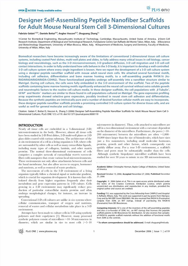
Designer Self-Assembling Peptide Nanofiber Scaffoldsfor Adult Mouse Neural Stem Cell 3-Dimensional CulturesFabrizio Gelain1,2,3, Daniele Bottai2,4, Angleo Vescovi2,3, Shuguang Zhang1*
1 Center for Biomedical Engineering, Massachusetts Institute of Technology, Cambridge, Massachusetts, United States of America, 2 Stem CellResearch Institute, Department of Biological and Technological Research, Fondazione Centro San Raffaele del Monte Tabor, Milan, Italy, 3 Bioscienceand Biotechnology Department, University of Milan-Bicocca, Milan, Italy, 4 Department of Medicine, Surgery and Dentistry, Faculty of Medicine,
University of Milan, Milan, Italy
Biomedical researchers have become increasingly aware of the limitations of conventional 2-dimensional tissue cell culture
systems, including coated Petri dishes, multi-well plates and slides, to fully address many critical issues in cell biology, cancer
biology and neurobiology, such as the 3-D microenvironment, 3-D gradient diffusion, 3-D cell migration and 3-D cell-cell
contact interactions. In order to fully understand how cells behave in the 3-D body, it is important to develop a well-controlled
3-D cell culture system where every single ingredient is known. Here we report the development of a 3-D cell culture system
using a designer peptide nanofiber scaffold with mouse adult neural stem cells. We attached several functional motifs,
including cell adhesion, differentiation and bone marrow homing motifs, to a self-assembling peptide RADA16 (Ac-
RADARADARADARADA-COHN2). These functionalized peptides undergo self-assembly into a nanofiber structure similar to
Matrigel. During cell culture, the cells were fully embedded in the 3-D environment of the scaffold. Two of the peptide
scaffolds containing bone marrow homing motifs significantly enhanced the neural cell survival without extra soluble growth
and neurotrophic factors to the routine cell culture media. In these designer scaffolds, the cell populations with b -Tubulin+,
GFAP+
and Nestin+
markers are similar to those found in cell populations cultured on Matrigel. The gene expression profilingarray experiments showed selective gene expression, possibly involved in neural stem cell adhesion and differentiation.
Because the synthetic peptides are intrinsically pure and a number of desired function cellular motifs are easy to incorporate,
these designer peptide nanofiber scaffolds provide a promising controlled 3-D culture system for diverse tissue cells, and are
useful as well for general molecular and cell biology.
Citation: Gelain F, Bottai D, Vescovi A, Zhang S (2006) Designer Self-Assembling Peptide Nanofiber Scaffolds for Adult Mouse Neural Stem Cell 3-Dimensional Cultures. PLoS ONE 1(1): e119. doi:10.1371/journal.pone.0000119
INTRODUCTIONNearly all tissue cells are embedded in a 3-dimensional (3-D)
microenvironment in the body. However, almost all tissue cells
have been studied in 2-D Petri dishes, 2-D multi-well plates or 2-D
glass slides coated with various substrata. The architecture of thein situ environment of a cell in a living organism is 3-D, where cells
are surrounded by other cells as well as many extracellular ligands,
including many types of collagens, laminin, and other matrix
proteins. The normal three-dimensional environment of cells
comprises a complex network of extracellular matrix nanoscale
fibers with nanopores that create various local microenvironments.
These environments not only allow attachments between cells and
the basal membrane, but also allow access to oxygen, hormones
and nutrients, as well as removal of waste products.
The movements of cells in the 3-D environment of a living
organism typically follow a chemical signal or molecular gradient,
which is crucial for organism development. It is known that cells
isolated directly from higher organisms frequently alter their
metabolism and gene expression patterns in 2-D culture. Cells
growing in a 2-D environment may significantly reduce pro-duction of particular extracellular matrix proteins and often
undergo morphological changes, for instance, an increase in
spreading.
Conventional 2-D cell cultures are unlike in vivo systems where
cellular communication, transport of oxygen and nutrients,
removal of wastes and cellular metabolism take place in a 3-D
environment.
Attempts have been made to culture cells in 3-D using synthetic
polymers and their copolymers [1]. However, many processed
synthetic polymers consist of microfibers ,10–50 micrometers in
diameter, which are similar in size to most cells ( ,5–10
micrometers in diameter). Thus, cells attached to microfibers are
still in a two-dimensional environment with a curvature dependent
on the diameter of the microfibers. Furthermore, the pores ( ,10–
200 micrometers) between the microfibers are often ,1,000–
10,000 times larger than the size of biomolecules, which have sizes
just a few nanometers, including small molecular hormones,proteins, growth and other factors, which consequently can
quickly diffuse away. For a true 3-D environment, a scaffold’s
fibers and pores must be substantially smaller than the cells.
Although synthetic biopolymer microfiber scaffolds have been
studied for over 30 years to mimic in vivo 3D microenvironment,
Academic Editor: Christophe Herman, Baylor College of Medicine, United Statesof America
Received October 13, 2006; Accepted November 27, 2006; Published December27, 2006
Copyright: ß 2006 Gelain et al. This is an open-access article distributed underthe terms of the Creative Commons Attribution License, which permits
unrestricted use, distribution, and reproduction in any medium, provided theoriginal author and source are credited.
Funding: F.G. was supported by the Cross fellowship from CARIPLO and HospitalS. Raffaele, Milano, Italy. This work was supported in part by grants from OlympusCorp and by NIH BRP Grant EB003805. We also received free RADA16 (Puramatrix)samples from 3DM, an MIT startup, instead of purchasing the RADA16(Puramatrix) from BD Biosciences.
Competing Interests: SZ is one of the inventors of the self-assembling peptidesand also a co-founder of 3DM, Inc., an MIT startup that licenses the peptidescaffold patents to BD Biosciences for distribution. SZ also receives free samplesof RADA16, peptide scaffold materials without the addition of functional motifsfrom 3DM as the controls.
* To whom correspondence should be addressed. E-mail: [email protected]
PLoS ONE | www.plosone.org 1 December 2006 | Issue 1 | e119
8/3/2019 Fabrizio Gelain, Daniele Bottai, Angleo Vescovi and Shuguang Zhang- Designer Self-Assembling Peptide Nanofiber S…
http://slidepdf.com/reader/full/fabrizio-gelain-daniele-bottai-angleo-vescovi-and-shuguang-zhang-designer 2/11
concerns about their degradation products and chemicals involved
in their synthesis are still important issues requiring further
improvements.
Animal derived biomaterials such as collagen gels, laminin,
poly-glycosaminoglycans and materials from basement mem-
branes, including MatrigelTM have also widely been used in cell
cultures [2–9]. While they are representative of the correct
nanolength scale, they often contain residual growth factors,
undefined constituents or non-quantified substances [2–6]. Thisnot only makes it difficult to conduct well-controlled studies with
these materials, but also poses problems if such scaffolds are ever
to be used for growing tissues for human therapies.
An ideal 3-D cell culture system should be fabricated from
a synthetic biological material with defined constituents. We
previously reported the discovery of a self-assembling peptide
system, made from natural amino acids, that can undergo
spontaneous assembly into nanofiber scaffolds, ,10 nm in fiber
diameter with pores between 5–200 nm [10–12]. These peptides
have been chemically produced in large quantity using standard
solid phase synthesis method and purified to homogeneity. They
have not only been used for the study of cell attachment, survival
and proliferation but also to incorporate other motifs [11–16], and
inject into animals [17–19]. These self-assembling peptides form
nanofibers that can be controlled at physiological pH by altering salt concentration [10–11]. Because the self-assembled nanofibers
are several thousand times thinner than synthetic polymer
microfibers and cells, thus it is believed that the peptide nanofibers
surround cells in a manner similar to the natural extracellular
matrix. However a systematic study of different motifs and an
examination of how their nanofiber scaffolds interact with cells in
details have not been carried out.
Here we report the use of designer peptide nanofiber scaffolds to
produce 3-D cultures for the study of mouse adult neural stem
cells. We synthesized 18 different peptides that directly incorporate
various functional motifs with the self-assembling peptide
RADA16. These motifs include sequences shown to promote cell
adhesion, differentiation and bone marrow homing activities.
These functionalized peptides self-assemble into nanofiber scaf-folds where cells can be fully embedded by the scaffold in 3-D.
Without addition of soluble growth factors and neurotrophic
factors, two of these scaffolds functionalized with bone marrow
homing motifs [20] not only significantly enhanced survival of the
neural stem cells, but also promoted differentiation towards cells
expressing neuronal and glial markers. This is the first example
suggesting that designer peptide scaffolds alone without additional
extra growth factors could influence neural stem cell differentia-
tion towards neural and glial phenotypes.
RESULTS
Designer peptide synthesisThis class of peptide is short and can be readily molecular-
designed from knowledge gained in the literature, we designed andproduced the functionalized self-assembling peptides. The self-
assembling peptide RADA16 was appended with motifs from
various types of collagen [21–22], laminin [23], fibrin [24],
fibronectin [25–26], osteopontin and osteogenic peptides [26–27],
bone marrow homing peptides (BMHP) [20] and myelo-regulatory
peptides [28–29]. The incorporation of these functional motifs did
not prevent the designer peptides from forming well-ordered
nanofibers using SEM examination.
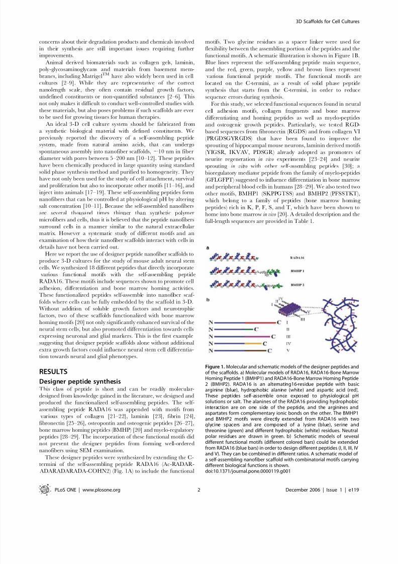
These designer peptides were synthesized by extending the C-
termini of the self-assembling peptide RADA16 (Ac-RADAR-
ADARADARADA-COHN2) (Fig. 1A) to include the functional
motifs. Two glycine residues as a spacer linker were used for
flexibility between the assembling portion of the peptides and the
functional motifs. A schematic illustration is shown in Figure 1B.
Blue lines represent the self-assembling peptide main sequence,
and the red, green, purple, yellow and brown lines represent
various functional peptide motifs. The functional motifs are
located on the C-termini, as a result of solid phase peptide
synthesis that starts from the C-termini, in order to reduce
sequence errors during synthesis.For this study, we selected functional sequences found in neural
cell adhesion motifs, collagen fragments and bone marrow
differentiating and homing peptides as well as myelo-peptides
and osteogenic growth peptides. Particularly, we tested RGD-
based sequences from fibronectin (RGDS) and from collagen VI
(PRGDSGYRGDS) that have been found to improve the
sprouting of hippocampal mouse neurons, laminin derived motifs
(YIGSR, IKVAV, PDSGR) already adopted as promoters of
neurite regeneration in vivo experiments [23–24] and neurite
sprouting in vitro with other self-assembling peptides [30]; a
bioregulatory mediator peptide from the family of myelo-peptides
(GFLGFPT) suggested to influence differentiation in bone marrow
and peripheral blood cells in humans [28–29]. We also tested two
other motifs, BMHP1 (SKPPGTSS) and BMHP2 (PFSSTKT),
which belong to a family of peptides (bone marrow homing
peptides) rich in K, P, F, S, and T, which have been shown to
home into bone marrow in vivo [20]. A detailed description and the
full-length sequences are provided in Table 1.
Figure 1. Molecular and schematic models of the designer peptides andof the scaffolds. a) Molecular models of RADA16, RADA16-Bone MarrowHoming Peptide 1 (BMHP1) and RADA16-Bone Marrow Homing Peptide2 (BMHP2). RADA16 is an alternating16-residue peptide with basicarginine (blue), hydrophobic alanine (white) and aspartic acid (red).
These peptides self-assemble once exposed to physiological pHsolutions or salt. The alanines of the RADA16 providing hydrophobicinteraction are on one side of the peptide, and the arginines andaspartates form complementary ionic bonds on the other. The BMHP1and BMHP2 motifs were directly extended from RADA16 with twoglycine spacers and are composed of a lysine (blue), serine andthreonine (green) and different hydrophobic (white) residues. Neutralpolar residues are drawn in green. b) Schematic models of severaldifferent functional motifs (different colored bars) could be extendedfrom RADA16 (blue bars) in order to design different peptides (I, II, III, IVand V). They can be combined in different ratios. A schematic model of a self-assembling nanofiber scaffold with combinatorial motifs carryingdifferent biological functions is shown.doi:10.1371/journal.pone.0000119.g001
3D Scaffolds for Cell Cultures
PLoS ONE | www.plosone.org 2 December 2006 | Issue 1 | e119
8/3/2019 Fabrizio Gelain, Daniele Bottai, Angleo Vescovi and Shuguang Zhang- Designer Self-Assembling Peptide Nanofiber S…
http://slidepdf.com/reader/full/fabrizio-gelain-daniele-bottai-angleo-vescovi-and-shuguang-zhang-designer 3/11
All of the designer peptides are soluble in aqueous solution and
self-assemble into nanofiber scaffolds upon exposure to cell culture
medium, PBS, and in neutral pH solutions.
The peptides of RADA16, RADA16-BMHP1 and RADA16-
BMHP2 are modeled using the van der Waals representation and
colored using the residue type method (Fig. 1). The original mainsequence of alternating basic (blue), hydrophobic (white) and acid
(red) residues is the RADA16 sequence necessary for the self-
assembling property of the functionalized scaffold. In this manner,
a simplified designer peptide scaffold model is proposed with
different functional motifs that could be easily incorporated (Fig. 1B).
Designer peptide nanofiber scaffolds and cells
embedded in themUpon incorporation of functional motifs to the RADA16 peptide,
there was concern that the appended peptide motifs would inhibit
the self-assembling peptide nanofiber formation. To address the
concern, we used scanning electron microscopy (SEM) to examine
the nanofiber structure of these designer peptides. We show here
the expected nanofibers of the pure RADA16 and of the 100%
functionalized peptides after self-assembly both in PBS and in cellculture medium (Fig. 2).
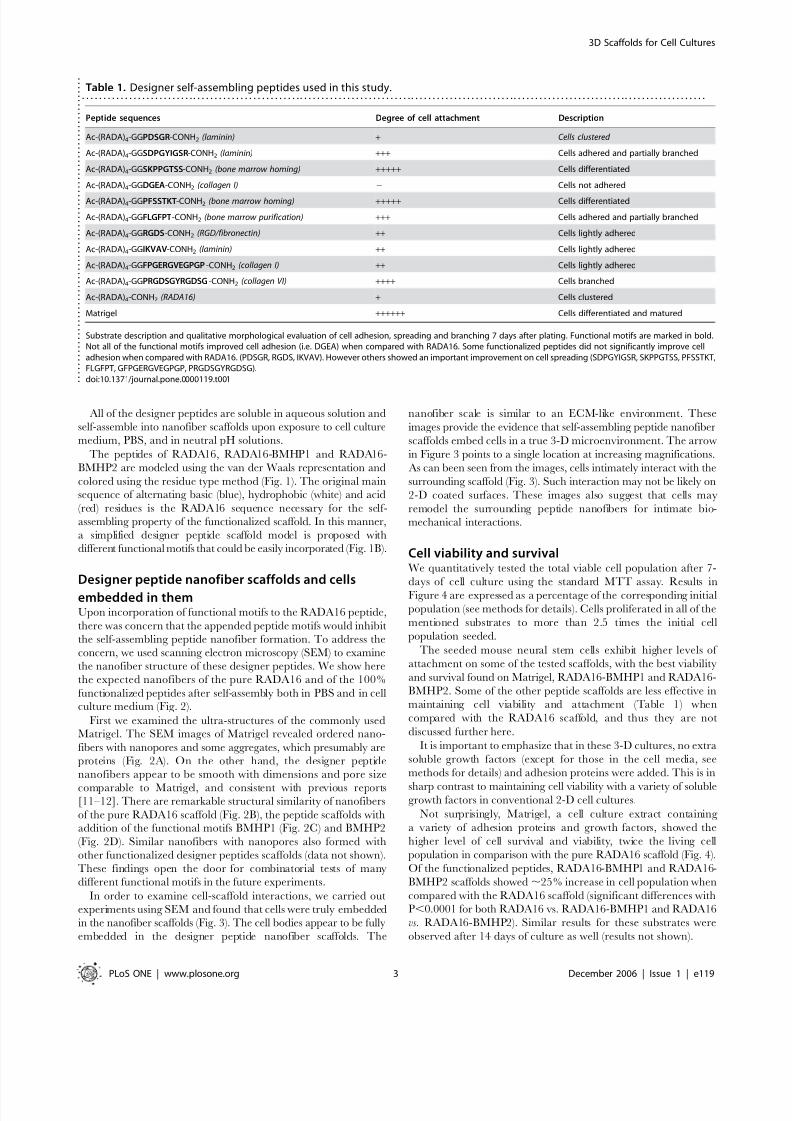
First we examined the ultra-structures of the commonly usedMatrigel. The SEM images of Matrigel revealed ordered nano-
fibers with nanopores and some aggregates, which presumably are
proteins (Fig. 2A). On the other hand, the designer peptide
nanofibers appear to be smooth with dimensions and pore size
comparable to Matrigel, and consistent with previous reports[11–12]. There are remarkable structural similarity of nanofibers
of the pure RADA16 scaffold (Fig. 2B), the peptide scaffolds withaddition of the functional motifs BMHP1 (Fig. 2C) and BMHP2
(Fig. 2D). Similar nanofibers with nanopores also formed with
other functionalized designer peptides scaffolds (data not shown).
These findings open the door for combinatorial tests of many
different functional motifs in the future experiments.
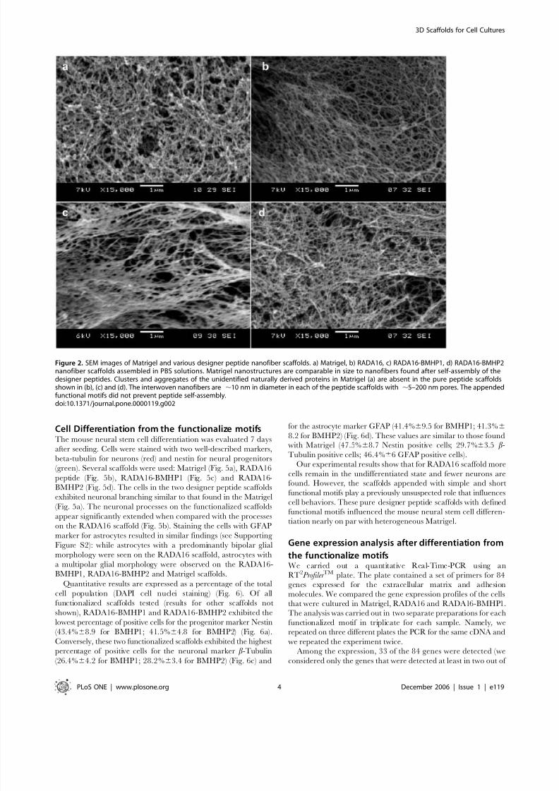
In order to examine cell-scaffold interactions, we carried out
experiments using SEM and found that cells were truly embedded
in the nanofiber scaffolds (Fig. 3). The cell bodies appear to be fully
embedded in the designer peptide nanofiber scaffolds. The
nanofiber scale is similar to an ECM-like environment. These
images provide the evidence that self-assembling peptide nanofiber
scaffolds embed cells in a true 3-D microenvironment. The arrow
in Figure 3 points to a single location at increasing magnifications.
As can been seen from the images, cells intimately interact with the
surrounding scaffold (Fig. 3). Such interaction may not be likely on
2-D coated surfaces. These images also suggest that cells may
remodel the surrounding peptide nanofibers for intimate bio-
mechanical interactions.
Cell viability and survivalWe quantitatively tested the total viable cell population after 7-
days of cell culture using the standard MTT assay. Results in
Figure 4 are expressed as a percentage of the corresponding initial
population (see methods for details). Cells proliferated in all of the
mentioned substrates to more than 2.5 times the initial cell
population seeded.
The seeded mouse neural stem cells exhibit higher levels of
attachment on some of the tested scaffolds, with the best viability
and survival found on Matrigel, RADA16-BMHP1 and RADA16-
BMHP2. Some of the other peptide scaffolds are less effective in
maintaining cell viability and attachment (Table 1) when
compared with the RADA16 scaffold, and thus they are not
discussed further here.
It is important to emphasize that in these 3-D cultures, no extra
soluble growth factors (except for those in the cell media, see
methods for details) and adhesion proteins were added. This is in
sharp contrast to maintaining cell viability with a variety of solublegrowth factors in conventional 2-D cell cultures.
Not surprisingly, Matrigel, a cell culture extract containing
a variety of adhesion proteins and growth factors, showed the
higher level of cell survival and viability, twice the living cell
population in comparison with the pure RADA16 scaffold (Fig. 4).
Of the functionalized peptides, RADA16-BMHP1 and RADA16-
BMHP2 scaffolds showed ,25% increase in cell population when
compared with the RADA16 scaffold (significant differences with
P,0.0001 for both RADA16 vs. RADA16-BMHP1 and RADA16
vs. RADA16-BMHP2). Similar results for these substrates were
observed after 14 days of culture as well (results not shown).
Table 1. Designer self-assembling peptides used in this study.. . . . . . . . . . . . . . . . . . . . . . . . . . . . . . . . . . . . . . . . . . . . . . . . . . . . . . . . . . . . . . . . . . . . . . . . . . . . . . . . . . . . . . . . . . . . . . . . . . . . . . . . . . . . . . . . . . . . . . . . . . . . . . . . . . . . . . . . . . . . . . . . . .
Peptide sequences Degree of cell attachment Description
Ac-(RADA)4-GGPDSGR-CONH2 (laminin) + Cells clustered
Ac-(RADA)4-GGSDPGYIGSR-CONH2 (laminin) +++ Cells adhered and partially branched
Ac-(RADA)4-GGSKPPGTSS-CONH2 (bone marrow homing) +++++ Cells differentiated
Ac-(RADA)4-GGDGEA-CONH2 (collagen I) 2 Cells not adhered
Ac-(RADA)4-GGPFSSTKT-CONH2 (bone marrow homing) +++++ Cells differentiated
Ac-(RADA)4-GGFLGFPT-CONH2 (bone marrow purification) +++ Cells adhered and partially branched
Ac-(RADA)4-GGRGDS-CONH2 (RGD/fibronectin) ++ Cells lightly adhered
Ac-(RADA)4-GGIKVAV-CONH2 (laminin) ++ Cells lightly adhered
Ac-(RADA)4-GGFPGERGVEGPGP -CONH2 (collagen I) ++ Cells lightly adhered
Ac-(RADA)4-GGPRGDSGYRGDSG-CONH2 (collagen VI) ++++ Cells branched
Ac-(RADA)4-CONH2 (RADA16) + Cells clustered
Matrigel ++++++ Cells differentiated and matured
Substrate description and qualitative morphological evaluation of cell adhesion, spreading and branching 7 days after plating. Functional motifs are marked in bold.Not all of the functional motifs improved cell adhesion (i.e. DGEA) when compared with RADA16. Some functionalized peptides did not significantly improve celladhesion when compared with RADA16. (PDSGR, RGDS, IKVAV). However others showed an important improvement on cell spreading (SDPGYIGSR, SKPPGTSS, PFSSTKT,FLGFPT, GFPGERGVEGPGP, PRGDSGYRGDSG).doi:10.1371/journal.pone.0000119.t001 .
.
.
.
.
.
.
.
.
.
.
.
.
.
.
.
.
.
.
.
.
.
.
.
.
.
.
.
.
.
.
.
.
.
.
.
.
.
.
.
. . . .
.
.
.
.
.
.
.
.
.
.
.
.
.
.
.
.
.
.
.
.
.
.
.
.
.
.
.
3D Scaffolds for Cell Cultures
PLoS ONE | www.plosone.org 3 December 2006 | Issue 1 | e119
8/3/2019 Fabrizio Gelain, Daniele Bottai, Angleo Vescovi and Shuguang Zhang- Designer Self-Assembling Peptide Nanofiber S…
http://slidepdf.com/reader/full/fabrizio-gelain-daniele-bottai-angleo-vescovi-and-shuguang-zhang-designer 4/11
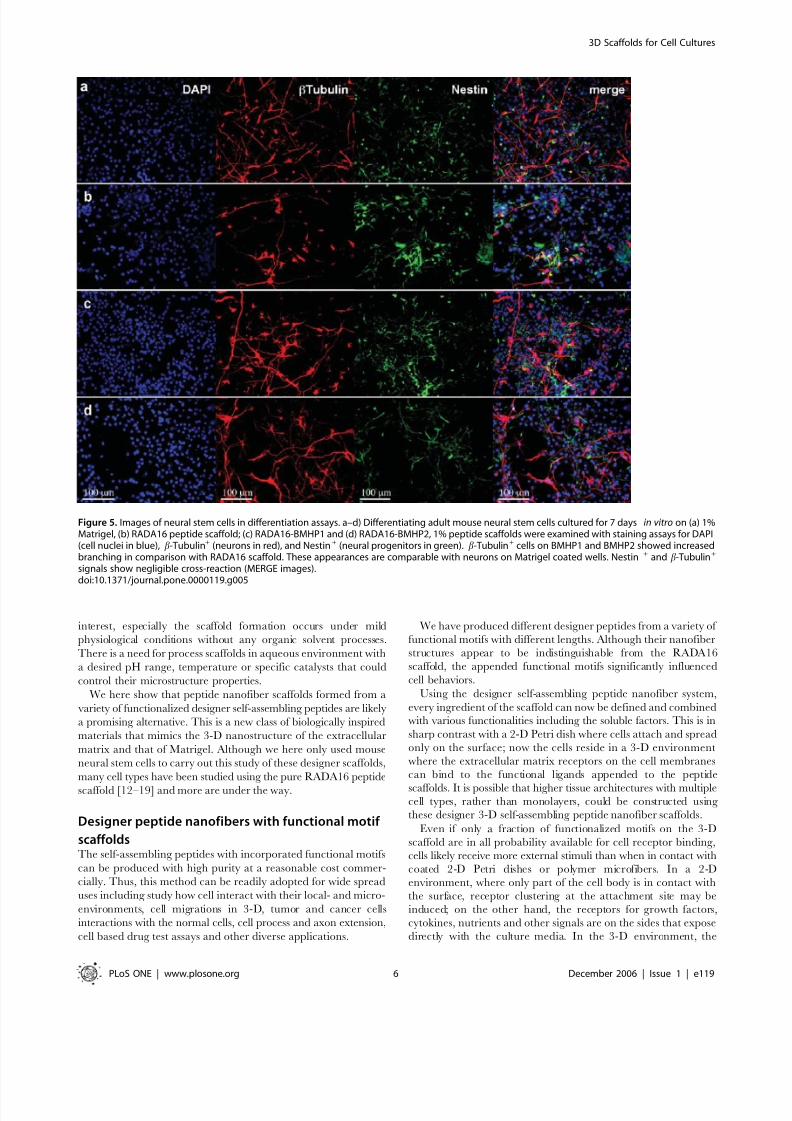
Cell Differentiation from the functionalize motifsThe mouse neural stem cell differentiation was evaluated 7 days
after seeding. Cells were stained with two well-described markers,
beta-tubulin for neurons (red) and nestin for neural progenitors
(green). Several scaffolds were used: Matrigel (Fig. 5a), RADA16
peptide (Fig. 5b), RADA16-BMHP1 (Fig. 5c) and RADA16-
BMHP2 (Fig. 5d). The cells in the two designer peptide scaffolds
exhibited neuronal branching similar to that found in the Matrigel
(Fig. 5a). The neuronal processes on the functionalized scaffolds
appear significantly extended when compared with the processes
on the RADA16 scaffold (Fig. 5b). Staining the cells with GFAP
marker for astrocytes resulted in similar findings (see Supporting
Figure S2): while astrocytes with a predominantly bipolar glial
morphology were seen on the RADA16 scaffold, astrocytes witha multipolar glial morphology were observed on the RADA16-
BMHP1, RADA16-BMHP2 and Matrigel scaffolds.
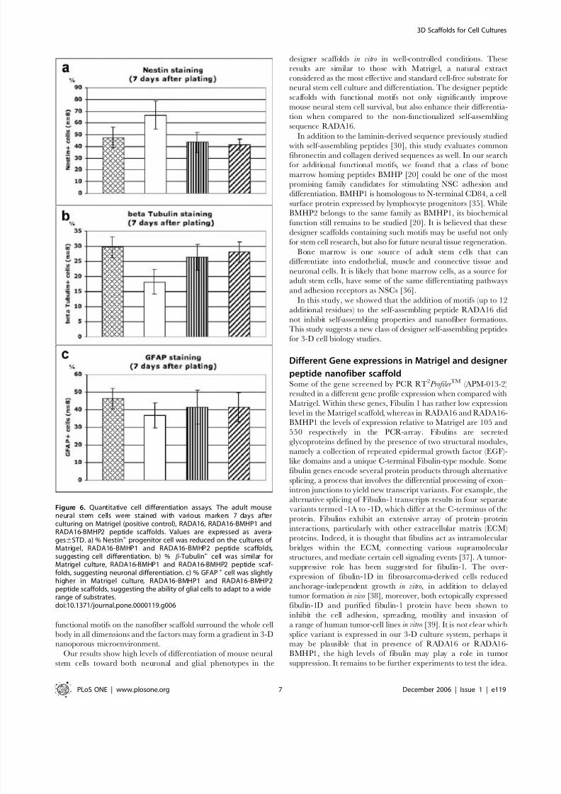
Quantitative results are expressed as a percentage of the total
cell population (DAPI cell nuclei staining) (Fig. 6). Of all
functionalized scaffolds tested (results for other scaffolds not
shown), RADA16-BMHP1 and RADA16-BMHP2 exhibited the
lowest percentage of positive cells for the progenitor marker Nestin
(43.4%68.9 for BMHP1; 41.5%64.8 for BMHP2) (Fig. 6a).
Conversely, these two functionalized scaffolds exhibited the highest
percentage of positive cells for the neuronal marker b-Tubulin
(26.4%64.2 for BMHP1; 28.2%63.4 for BMHP2) (Fig. 6c) and
for the astrocyte marker GFAP (41.4%69.5 for BMHP1; 41.3%6
8.2 for BMHP2) (Fig. 6d). These values are similar to those foundwith Matrigel (47.5%68.7 Nestin positive cells; 29.7%63.5 b-
Tubulin positive cells; 46.4%66 GFAP positive cells).
Our experimental results show that for RADA16 scaffold more
cells remain in the undifferentiated state and fewer neurons arefound. However, the scaffolds appended with simple and short
functional motifs play a previously unsuspected role that influences
cell behaviors. These pure designer peptide scaffolds with defined
functional motifs influenced the mouse neural stem cell differen-
tiation nearly on par with heterogeneous Matrigel.
Gene expression analysis after differentiation from
the functionalize motifsWe carried out a quantitative Real-Time-PCR using an
RT2Profiler TM plate. The plate contained a set of primers for 84
genes expressed for the extracellular matrix and adhesion
molecules. We compared the gene expression profiles of the cells
that were cultured in Matrigel, RADA16 and RADA16-BMHP1.
The analysis was carried out in two separate preparations for each
functionalized motif in triplicate for each sample. Namely, we
repeated on three different plates the PCR for the same cDNA and
we repeated the experiment twice.
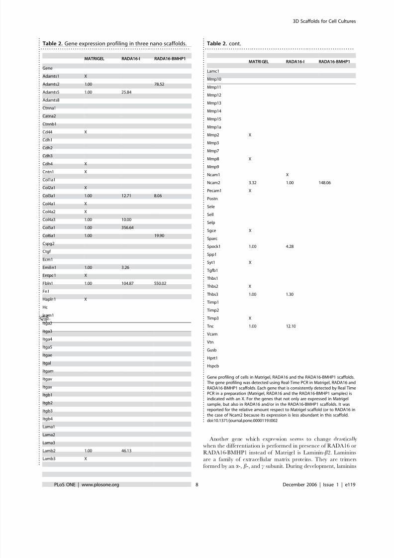
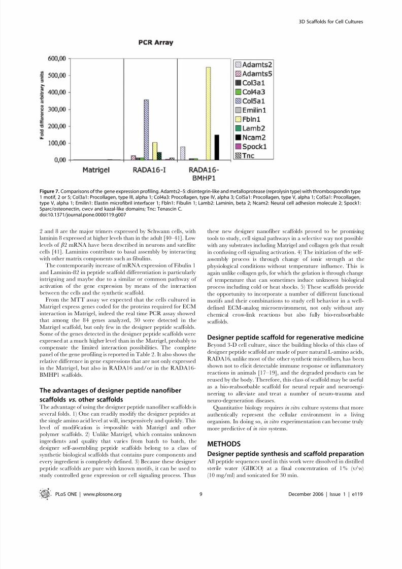
Among the expression, 33 of the 84 genes were detected (we
considered only the genes that were detected at least in two out of
Figure 2. SEM images of Matrigel and various designer peptide nanofiber scaffolds. a) Matrigel, b) RADA16, c) RADA16-BMHP1, d) RADA16-BMHP2nanofiber scaffolds assembled in PBS solutions. Matrigel nanostructures are comparable in size to nanofibers found after self-assembly of thedesigner peptides. Clusters and aggregates of the unidentified naturally derived proteins in Matrigel (a) are absent in the pure peptide scaffoldsshown in (b), (c) and (d). The interwoven nanofibers are ,10 nm in diameter in each of the peptide scaffolds with ,5–200 nm pores. The appendedfunctional motifs did not prevent peptide self-assembly.doi:10.1371/journal.pone.0000119.g002
3D Scaffolds for Cell Cultures
PLoS ONE | www.plosone.org 4 December 2006 | Issue 1 | e119
8/3/2019 Fabrizio Gelain, Daniele Bottai, Angleo Vescovi and Shuguang Zhang- Designer Self-Assembling Peptide Nanofiber S…
http://slidepdf.com/reader/full/fabrizio-gelain-daniele-bottai-angleo-vescovi-and-shuguang-zhang-designer 5/11
three PCR experiments); 30 in the Matrigel sample, 12 in
RADA16 sample, 5 in the RADA16-BMHP1 (Table 2).
Three of the 84 genes were detectable in all three samples: Fbln1 (Fibulin 1), procollagen type III a1 and neural cell adhesion
molecule 2 and fibulin. Seven genes were detected in Matrigel and
RADA16 including a disintegrin-like and metalloprotease (repro-
lysin type) with thrombospondin type 1 motif 5, elastin microfibril
interfacer 1, protocollagen type IV a3, protocollagen type V a3,
thrombospondin 3, tenascin C, Laminin b2. Two genes were
present at the same time in the samples Matrigel and RADA16-
BMHP1: A disintegrin-like and metalloprotease (reprolysin type)
with thrombospondin type 1 motif 2, procollagen type VI a1. 15
genes were measurable only in the Matrigel sample: disintegrin-
like and metalloprotease (reprolysin type) with thrombospondin
type 1 motif 1, cadherin 4, contactin 1, procollagen type II a1,
procollagen type IV a1, procollagen type IV a2, ectonucleoside
triphosphate diphosphohydrolase 1, hyaluronan and proteoglycan
link protein 1, laminin b3, matrix metalloproteinase 2, platelet/
endothelial cell adhesion molecule, thrombospondin 2, tissue
inhibitor of metalloproteinase 3. Cadherin 1 and proteoglycan 1
were only detectable in the RADA16-BMHP2 samples; whereas
neural cell adhesion molecule 1 was detectable only in mRNA
isolated from cells cultured in RADA16.
For the genes that are detected at least in two different samples
we performed a comparison calculating the fold differences
assigning to the sample that had the lower level the value 1.
Fibulin 1 has low expression level in the Matrigel sample whereas
in RADA16, RADA16-BMHP1 the levels of expression relative to
Matrigel are 105, 550 higher respectively. Neural cell adhesion
molecule 2 is highly expressed in RADA16-BMHP1. The gene
expression profiling is showed in Fig. 7 and in Table 2
DISCUSSION
Synthetic polymer microfibers and self-assembling
peptide nanofibersMost of the synthetic polymer biomaterials currently used in tissue
cell cultures and regenerative medicine usually have microfiber
structures [1,31–34]. Since the size of most cells (5–10 m ) are
similar or smaller to these microfibers ( ,10–100 m ), upon
attachment to the microfibers the cells still exhibit a 2-D topologywith a curvature depending on the diameter of the microfibers.
Polymeric materials are often functionalized to promote desired
biological activities through chemical reactions or coating. Because
of their micro-scale sizes, their mechanical strength usually
prevents further material structural adaptations from the forces
exerted by cells during their adhesion, migration and maturation
processes. Thus, although these microfibers provide an artificial
extracellular environment, they are still far from the natural
nanoscale ECM.
For the encapsulation of labile bioactive substances and living
cells, physically cross-linked nanofiber scaffolds are of great
Figure 3. SEM images of adult mouse neural stem cells (NSC)embedded in designer peptide nanofiber scaffold RADA16-BMHP1(1% v/w) after 14 day in vitro cultures. I) Cluster of three visible NSCs(white circle) embedded in 3-D self-assembling RADA16-BMHP1. II) Asingle cell at different magnification with extended processesembedded in the scaffold is shown (a–c). White arrows point to theimage areas enlarged in the consecutive pictures. d) High-magnificationpicture focusing on the interface between the nanofiber scaffold andthe round shaped cell body. The black arrow in (b) points to a cellularprocess. Cells and processes are thus embedded in the self-assemblingpeptide nanofiber scaffold in a true 3-D environment, which may likelypromote cell adhesions in 3-D similar to the natural cellularenvironment. Adult mouse neural stem cells have been cultured andcould be differentiated in vitro for several weeks. The scale bars areshown on each image.doi:10.1371/journal.pone.0000119.g003
Figure 4. MTT cell proliferation assays of adult mouse neural stem cellsafter 7-day culture. Cells were seeded on Matrigel or peptide scaffolds.The results are expressed as cell % increases from the seedingpopulation on first day. BMHP1 and BMHP2 peptide scaffolds allowfor higher cell proliferation in comparison to RADA16 peptide scaffold(t =27.28 and t=25.28 for respectively RADA16 vs. RADA16-BMHP1and RADA16 vs. RADA16-BMHP2 with p,0.0001% in both cases). Notsurprisingly, Matrigel containing various unknown quantity of growthfactors showed considerable cell population increase. Similar increasesof total cell populations were confirmed for 14-day cultures (notshown). The cell proliferation using the designer peptide scaffolds could
be further improved from addition of soluble neurotrophic factors.doi:10.1371/journal.pone.0000119.g004
3D Scaffolds for Cell Cultures
PLoS ONE | www.plosone.org 5 December 2006 | Issue 1 | e119
8/3/2019 Fabrizio Gelain, Daniele Bottai, Angleo Vescovi and Shuguang Zhang- Designer Self-Assembling Peptide Nanofiber S…
http://slidepdf.com/reader/full/fabrizio-gelain-daniele-bottai-angleo-vescovi-and-shuguang-zhang-designer 6/11
interest, especially the scaffold formation occurs under mild
physiological conditions without any organic solvent processes.
There is a need for process scaffolds in aqueous environment with
a desired pH range, temperature or specific catalysts that could
control their microstructure properties.
We here show that peptide nanofiber scaffolds formed from a
variety of functionalized designer self-assembling peptides are likely
a promising alternative. This is a new class of biologically inspired
materials that mimics the 3-D nanostructure of the extracellular
matrix and that of Matrigel. Although we here only used mouse
neural stem cells to carry out this study of these designer scaffolds,
many cell types have been studied using the pure RADA16 peptide
scaffold [12–19] and more are under the way.
Designer peptide nanofibers with functional motif
scaffoldsThe self-assembling peptides with incorporated functional motifs
can be produced with high purity at a reasonable cost commer-
cially. Thus, this method can be readily adopted for wide spread
uses including study how cell interact with their local- and micro-
environments, cell migrations in 3-D, tumor and cancer cells
interactions with the normal cells, cell process and axon extension,
cell based drug test assays and other diverse applications.
We have produced different designer peptides from a variety of
functional motifs with different lengths. Although their nanofiber
structures appear to be indistinguishable from the RADA16
scaffold, the appended functional motifs significantly influenced
cell behaviors.
Using the designer self-assembling peptide nanofiber system,
every ingredient of the scaffold can now be defined and combinedwith various functionalities including the soluble factors. This is in
sharp contrast with a 2-D Petri dish where cells attach and spread
only on the surface; now the cells reside in a 3-D environment
where the extracellular matrix receptors on the cell membranes
can bind to the functional ligands appended to the peptide
scaffolds. It is possible that higher tissue architectures with multiplecell types, rather than monolayers, could be constructed using
these designer 3-D self-assembling peptide nanofiber scaffolds.
Even if only a fraction of functionalized motifs on the 3-D
scaffold are in all probability available for cell receptor binding,
cells likely receive more external stimuli than when in contact with
coated 2-D Petri dishes or polymer microfibers. In a 2-D
environment, where only part of the cell body is in contact with
the surface, receptor clustering at the attachment site may be
induced; on the other hand, the receptors for growth factors,
cytokines, nutrients and other signals are on the sides that expose
directly with the culture media. In the 3-D environment, the
Figure 5. Images of neural stem cells in differentiation assays. a–d) Differentiating adult mouse neural stem cells cultured for 7 days in vitro on (a) 1%Matrigel, (b) RADA16 peptide scaffold; (c) RADA16-BMHP1 and (d) RADA16-BMHP2, 1% peptide scaffolds were examined with staining assays for DAPI(cell nuclei in blue), b-Tubulin+ (neurons in red), and Nestin+ (neural progenitors in green). b-Tubulin+ cells on BMHP1 and BMHP2 showed increasedbranching in comparison with RADA16 scaffold. These appearances are comparable with neurons on Matrigel coated wells. Nestin + and b-Tubulin+
signals show negligible cross-reaction (MERGE images).doi:10.1371/journal.pone.0000119.g005
3D Scaffolds for Cell Cultures
PLoS ONE | www.plosone.org 6 December 2006 | Issue 1 | e119
8/3/2019 Fabrizio Gelain, Daniele Bottai, Angleo Vescovi and Shuguang Zhang- Designer Self-Assembling Peptide Nanofiber S…
http://slidepdf.com/reader/full/fabrizio-gelain-daniele-bottai-angleo-vescovi-and-shuguang-zhang-designer 7/11
functional motifs on the nanofiber scaffold surround the whole cell
body in all dimensions and the factors may form a gradient in 3-D
nanoporous microenvironment.
Our results show high levels of differentiation of mouse neural
stem cells toward both neuronal and glial phenotypes in the
designer scaffolds in vitro in well-controlled conditions. These
results are similar to those with Matrigel, a natural extract
considered as the most effective and standard cell-free substrate for
neural stem cell culture and differentiation. The designer peptide
scaffolds with functional motifs not only significantly improve
mouse neural stem cell survival, but also enhance their differentia-
tion when compared to the non-functionalized self-assembling
sequence RADA16.
In addition to the laminin-derived sequence previously studiedwith self-assembling peptides [30], this study evaluates common
fibronectin and collagen derived sequences as well. In our search
for additional functional motifs, we found that a class of bone
marrow homing peptides BMHP [20] could be one of the most
promising family candidates for stimulating NSC adhesion and
differentiation. BMHP1 is homologous to N-terminal CD84, a cell
surface protein expressed by lymphocyte progenitors [35]. While
BMHP2 belongs to the same family as BMHP1, its biochemical
function still remains to be studied [20]. It is believed that these
designer scaffolds containing such motifs may be useful not only
for stem cell research, but also for future neural tissue regeneration.
Bone marrow is one source of adult stem cells that can
differentiate into endothelial, muscle and connective tissue and
neuronal cells. It is likely that bone marrow cells, as a source for
adult stem cells, have some of the same differentiating pathwaysand adhesion receptors as NSCs [36].
In this study, we showed that the addition of motifs (up to 12
additional residues) to the self-assembling peptide RADA16 did
not inhibit self-assembling properties and nanofiber formations.
This study suggests a new class of designer self-assembling peptides
for 3-D cell biology studies.
Different Gene expressions in Matrigel and designer
peptide nanofiber scaffoldSome of the gene screened by PCR RT2Profiler TM (APM-013-2)
resulted in a different gene profile expression when compared with
Matrigel. Within these genes, Fibulin 1 has rather low expression
level in the Matrigel scaffold, whereas in RADA16 and RADA16-
BMHP1 the levels of expression relative to Matrigel are 105 and
550 respectively in the PCR-array. Fibulins are secreted
glycoproteins defined by the presence of two structural modules,
namely a collection of repeated epidermal growth factor (EGF)-
like domains and a unique C-terminal Fibulin-type module. Some
fibulin genes encode several protein products through alternative
splicing, a process that involves the differential processing of exon–
intron junctions to yield new transcript variants. For example, the
alternative splicing of Fibulin-1 transcripts results in four separate
variants termed -1A to -1D, which differ at the C-terminus of the
protein. Fibulins exhibit an extensive array of protein–protein
interactions, particularly with other extracellular matrix (ECM)
proteins. Indeed, it is thought that fibulins act as intramolecular
bridges within the ECM, connecting various supramolecular
structures, and mediate certain cell signaling events [37]. A tumor-suppressive role has been suggested for fibulin-1. The over-
expression of fibulin-1D in fibrosarcoma-derived cells reduced
anchorage-independent growth in vitro, in addition to delayed
tumor formation in vivo [38], moreover, both ectopically expressed
fibulin-1D and purified fibulin-1 protein have been shown to
inhibit the cell adhesion, spreading, motility and invasion of
a range of human tumor-cell lines in vitro [39]. It is not clear which
splice variant is expressed in our 3-D culture system, perhaps it
may be plausible that in presence of RADA16 or RADA16-
BMHP1, the high levels of fibulin may play a role in tumor
suppression. It remains to be further experiments to test the idea.
Figure 6. Quantitative cell differentiation assays. The adult mouseneural stem cells were stained with various markers 7 days afterculturing on Matrigel (positive control), RADA16, RADA16-BMHP1 andRADA16-BMHP2 peptide scaffolds. Values are expressed as avera-ges6STD. a) % Nestin+ progenitor cell was reduced on the cultures of Matrigel, RADA16-BMHP1 and RADA16-BMHP2 peptide scaffolds,
suggesting cell differentiation. b) % b-Tubulin+
cell was similar forMatrigel culture, RADA16-BMHP1 and RADA16-BMHP2 peptide scaf-folds, suggesting neuronal differentiation. c) % GFAP+ cell was slightlyhigher in Matrigel culture, RADA16-BMHP1 and RADA16-BMHP2peptide scaffolds, suggesting the ability of glial cells to adapt to a widerange of substrates.doi:10.1371/journal.pone.0000119.g006
3D Scaffolds for Cell Cultures
PLoS ONE | www.plosone.org 7 December 2006 | Issue 1 | e119
8/3/2019 Fabrizio Gelain, Daniele Bottai, Angleo Vescovi and Shuguang Zhang- Designer Self-Assembling Peptide Nanofiber S…
http://slidepdf.com/reader/full/fabrizio-gelain-daniele-bottai-angleo-vescovi-and-shuguang-zhang-designer 8/11
Another gene which expression seems to change drastically
when the differentiation is performed in presence of RADA16 or
RADA16-BMHP1 instead of Matrigel is Laminin-b2. Laminins
are a family of extracellular matrix proteins. They are trimers
formed by an a-, b-, and c subunit. During development, laminins
Table 2. Gene expression profiling in three nano scaffolds.. . . . . . . . . . . . . . . . . . . . . . . . . . . . . . . . . . . . . . . . . . . . . . . . . . . . . . . . . . . . . . . . . . . . . .
MATRIGEL RADA16-I RADA16-BMHP1
Gene
Adamts1 X
Adamts2 1.00 78.52
Adamts5 1.00 25.84
Adamts8
Ctnna1
Catna2
Ctnnb1
Cd44 X
Cdh1
Cdh2
Cdh3
Cdh4 X
Cntn1 X
Col1a1
Col2a1 X
Col3a1 1.00 12.71 8.06
Col4a1 X
Col4a2 X
Col4a3 1.00 10.00
Col5a1 1.00 356.64
Col6a1 1.00 19.90
Cspg2
Ctgf
Ecm1
Emilin1 1.00 3.26
Entpd1 X
Fbln1 1.00 104.87 550.02
Fn1Hapln1 X
Hc
Icam1
Itga2
Itga3
Itga4
Itga5
Itgae
Itgal
Itgam
Itgav
Itgax
Itgb1
Itgb2
Itgb3
Itgb4
Lama1
Lama2
Lama3
Lamb2 1.00 46.13
Lamb3 X .
.
.
.
.
.
.
.
.
.
.
.
.
.
.
.
.
.
.
.
.
.
.
.
.
.
.
.
.
.
.
.
. . .
.
.
.
.
.
.
.
.
.
.
.
.
.
.
.
.
.
.
.
.
.
.
.
.
.
.
.
.
.
.
.
.
.
.
.
.
.
.
.
.
.
.
. . .
.
.
.
.
.
.
.
.
.
.
.
.
.
.
.
.
.
.
.
.
.
.
.
.
.
.
.
.
.
.
.
.
.
.
.
.
.
.
.
.
.
.
.
. . .
.
.
.
.
.
.
.
.
.
.
.
.
.
.
.
.
.
.
.
.
.
.
.
.
.
.
.
.
.
.
.
.
.
.
.
.
.
.
.
.
.
.
. . .
.
.
.
.
.
.
.
.
.
.
.
.
.
.
.
.
.
.
.
.
.
.
.
.
.
.
.
.
MATRI GEL RADA16-I RADA16-BMHP1
Lamc1
Mmp10
Mmp11
Mmp12
Mmp13
Mmp14
Mmp15
Mmp1a
Mmp2 X
Mmp3
Mmp7
Mmp8 X
Mmp9
Ncam1 X
Ncam2 3.32 1.00 148.06
Pecam1 X
Postn
Sele
Sell
Selp
Sgce X
Sparc
Spock1 1.00 4.28
Spp1
Syt1 X
Tgfb1
Thbs1
Thbs2 X
Thbs3 1.00 1.30
Timp1
Timp2
Timp3 X
Tnc 1.00 12.10
Vcam
Vtn
Gusb
Hprt1
Hspcb
Gene profiling of cells in Matrigel, RADA16 and the RADA16-BMHP1 scaffolds.The gene profiling was detected using Real-Time PCR in Matrigel, RADA16 andRADA16-BMHP1 scaffolds. Each gene that is consistently detected by Real TimePCR in a preparation (Matrigel, RADA16 and the RADA16-BMHP1 samples) isindicated with an X. For the genes that not only are expressed in Matrigelsample, but also in RADA16 and/or in the RADA16-BMHP1 scaffolds. It wasreported for the relative amount respect to Matrigel scaffold (or to RADA16 inthe case of Ncam2 because its expression is less abundant in this scaffold.doi:10.1371/journal.pone.0000119.t002 .
.
.
.
.
.
.
.
.
.
.
.
. . .
.
.
.
.
.
.
.
.
.
.
.
.
.
.
.
.
.
.
.
.
.
.
.
.
.
.
.
.
.
.
.
.
.
.
.
.
.
.
.
.
.
.
. . .
.
.
.
.
.
.
.
.
.
.
.
.
.
.
.
.
.
.
.
.
.
.
.
.
.
.
.
.
.
.
.
.
.
.
.
.
.
.
.
.
.
.
.
. . .
.
.
.
.
.
.
.
.
.
.
.
.
.
.
.
.
.
.
.
.
.
.
.
.
.
.
.
.
.
.
.
.
.
.
.
.
.
.
.
.
.
.
. . .
.
.
.
.
.
.
.
.
.
.
.
.
.
.
.
.
.
.
.
.
.
.
.
.
.
.
.
.
Table 2. cont.. . . . . . . . . . . . . . . . . . . . . . . . . . . . . . . . . . . . . . . . . . . . . . . . . . . . . . . . . . . . . . . . . . . . . .
3D Scaffolds for Cell Cultures
PLoS ONE | www.plosone.org 8 December 2006 | Issue 1 | e119
8/3/2019 Fabrizio Gelain, Daniele Bottai, Angleo Vescovi and Shuguang Zhang- Designer Self-Assembling Peptide Nanofiber S…
http://slidepdf.com/reader/full/fabrizio-gelain-daniele-bottai-angleo-vescovi-and-shuguang-zhang-designer 9/11
2 and 8 are the major trimers expressed by Schwann cells, withlaminin 8 expressed at higher levels than in the adult [40–41]. Low
levels of b2 mRNA have been described in neurons and satellite
cells [41]. Laminins contribute to basal assembly by interacting
with other matrix components such as fibulins.
The contemporarily increase of mRNA expression of Fibulin 1
and Laminin-ß2 in peptide scaffold differentiation is particularlyintriguing and maybe due to a similar or common pathway of
activation of the gene expression by means of the interaction
between the cells and the synthetic scaffold.
From the MTT assay we expected that the cells cultured inMatrigel express genes coded for the proteins required for ECM
interaction in Matrigel, indeed the real time PCR assay showedthat among the 84 genes analyzed, 30 were detected in the
Matrigel scaffold, but only few in the designer peptide scaffolds.Some of the genes detected in the designer peptide scaffolds were
expressed at a much higher level than in the Matrigel, probably to
compensate the limited interaction possibilities. The complete
panel of the gene profiling is reported in Table 2. It also shows the
relative difference in gene expressions that are not only expressed
in the Matrigel, but also in RADA16 and/or in the RADA16-
BMHP1 scaffolds.
The advantages of designer peptide nanofiber
scaffolds vs. other scaffoldsThe advantage of using the designer peptide nanofiber scaffolds is
several folds. 1) One can readily modify the designer peptides at
the single amino acid level at will, inexpensively and quickly. This
level of modification is impossible with Matrigel and other
polymer scaffolds. 2) Unlike Matrigel, which contains unknown
ingredients and quality that varies from batch to batch, the
designer self-assembling peptide scaffolds belong to a class of
synthetic biological scaffolds that contains pure components and
every ingredient is completely defined. 3) Because these designer
peptide scaffolds are pure with known motifs, it can be used to
study controlled gene expression or cell signaling process. Thus
these new designer nanofiber scaffolds proved to be promising
tools to study, cell signal pathways in a selective way not possible
with any substrates including Matrigel and collagen gels that result
in confusing cell signaling activation. 4) The initiation of the self-
assembly process is through change of ionic strength at the
physiological conditions without temperature influence. This is
again unlike collagen gels, for which the gelation is through change
of temperature that can sometimes induce unknown biological
process including cold or heat shocks. 5) These scaffolds provide
the opportunity to incorporate a number of different functional
motifs and their combinations to study cell behavior in a well-defined ECM-analog microenvironment, not only without any
chemical cross-link reactions but also fully bio-reabsorbable
scaffolds.
Designer peptide scaffold for regenerative medicineBeyond 3-D cell culture, since the building blocks of this class of
designer peptide scaffold are made of pure natural L-amino acids,
RADA16, unlike most of the other synthetic microfibers, has been
shown not to elicit detectable immune response or inflammatory
reactions in animals [17–19], and the degraded products can be
reused by the body. Therefore, this class of scaffold may be useful
as a bio-reabsorbable scaffold for neural repair and neuroengi-
neering to alleviate and treat a number of neuro-trauma and
neuro-degeneration diseases.Quantitative biology requires in vitro culture systems that more
authentically represent the cellular environment in a living
organism. In doing so, in vitro experimentation can become truly
more predictive of in vivo systems.
METHODS
Designer peptide synthesis and scaffold preparation All peptide sequences used in this work were dissolved in distilled
sterile water (GIBCO) at a final concentration of 1% (v/w)
(10 mg/ml) and sonicated for 30 min.
Figure 7. Comparisons of the gene expression profiling. Adamts2–5: disintegrin-like and metalloprotease (reprolysin type) with thrombospondin type
1 motif, 2 or 5; Col3a1: Procollagen, type III, alpha 1; Col4a3: Procollagen, type IV, alpha 3; Col5a1: Procollagen, type V, alpha 1; Col5a1: Procollagen,type V, alpha 1; Emilin1: Elastin microfibril interfacer 1; Fbln1: Fibulin 1; Lamb2: Laminin, beta 2; Ncam2: Neural cell adhesion molecule 2; Spock1:Sparc/osteonectin, cwcv and kazal-like domains; Tnc: Tenascin C.doi:10.1371/journal.pone.0000119.g007
3D Scaffolds for Cell Cultures
PLoS ONE | www.plosone.org 9 December 2006 | Issue 1 | e119
8/3/2019 Fabrizio Gelain, Daniele Bottai, Angleo Vescovi and Shuguang Zhang- Designer Self-Assembling Peptide Nanofiber S…
http://slidepdf.com/reader/full/fabrizio-gelain-daniele-bottai-angleo-vescovi-and-shuguang-zhang-designer 10/11
Cell viability and differentiation assay tests were conducted by
pouring aqueous solution of functionalized sapeptide (30 ml per
well) so as to evenly cover the bottom surface of each well
(approximately 30 mm gel layer thickness) of 96 multi-well plates
(BD Biosciences). In the case of SEM imaging both for Matrigel
and the sapeptides, the total amount of biomaterial was reduced to
10 ml. The experimental protocol included control tests conducted
with non-functionalized RADA16 (negative control) and Matrigel
coating (positive control). RADA16 and other functionalizedsapeptides: poured 30 ml/well of a 1% (w/v) distilled sterile water
solution, followed by slow addition of 200 ml/well of basal
medium. Allowed to self-assemble at +37uC for 30 minutes and
rinsed once with control medium to wash away any residual acid
residues remaining from peptide synthesis. Matrigel GF-reduced
(from EHS sarcoma, BD Biosciences): diluted 1:100 in basal
medium, poured at 100 ml/well, 309 incubation at 37uC, then
rinsed with basal medium. In the case of SEM imaging, no dilution
was adopted in order to guarantee the necessary stiffness for a
3-dimensional scaffold.
Cell cultures and seedingNeural precursor cultures were established and expanded as
described in the supplementary methods.
In the case of adhesion and differentiation tests, cell seeding
(at a concentration of 2–86104 cells/cm2 ) was performed two days
after the last mechanical dissociation in order to seed the
maximum percentage of stem cells. Cells were seeded on the
top-surface of each assembled scaffold, where they were able to
settle into the nanofiber matrices. Over time cells penetrate the
self-assembled layer (see Supporting Fig. S1).
In the case of SEM imaging, cells were acutely mixed with 8 ml
of aqueous gel solution at a final concentration of 5–86103 cells/
ml in a total final volume of 10 ml per each sample. Self-assembling
was then initiated by adding basal medium slowly and placing
seeded scaffolds mounted on copper grids (Ted Pella Inc.) at
+37uC for 30 minutes. Cells were thus already embedded in the
matrices.
For both adhesion and differentiation tests and SEM imaging,cells were cultured with basal medium supplemented with bFGF
(10 ng/ml), added to enhance neuronal progeny differentiation.
After 3 days, the medium was shifted to a medium containing
Leukemia Inhibitory Factor (LIF, Chemicon) (20 ng/ml) and
Brain Derived Neurotrophic Factor (BDNF, Peprotech) (20 ng/
ml) to pursue the neuronal and glial population maturation in
NSC progeny [42]. Cells were fed every three days with the same
fresh culture medium.
Cell proliferation assayTo assess the viability of NSCs seeded on scaffolds made of various
peptides, a quantitative method, MTT assay (Sigma), was used (see
supplementary methods). Four independent experiments compris-
ing three replicates each were performed. For this viability test thedirect proportional linearity between the optical density and the
viability/metabolic activity of the cell populations was assessed
from verifying the linearity of 5 different standard curves at 6
increasing cell concentrations, ranging from 56103 to
56105 cells/well. Results are expressed as percent increase in cell
population from the population seeded on day one.
ImmunocytochemistryNeuronal and glial differentiation was assessed by double and
single immunostaining with lineage-specific antibodies: anti-Nestin
(1:150, Chemicon) for progenitor cells, rabbit anti-b-Tubulin
(1:500, Covance) for neurons, mouse anti-Glial Fibrillary Acidic
Protein (1:200, Chemicon) for astrocytes. Primary antibodies were
then stained with secondary ALEXA 488 goat anti-mouse (1:1000
Molecular Probes) and CY3 AffiniPure F(ab’)2 Anti-Rabbit IgG
antibodies (1:100 Jackson Immuno Research). Cell nuclei were
counterstained with DAPI (Molecular Probes). The samples were
then examined by inverted fluorescence microscope. Quantitative
analyses were performed by counting 100–300 cells for each of 10
non-overlapping (and randomly chosen) fields. Four independentexperiments comprising two replicates each were performed.
SEM sample preparation and imaging After seeding NSCs within the self-assembled scaffolds as
previously described (cell seeding section), cells were cultured for
7 and 14 days in NSC basal medium. The peptide matrices were
prepared for SEM as described in the supplementary methods and
examined using a JOEL JSM 6060 SEM at 2,000–100,0006
magnification, 6KV acceleration voltage, 29–32 spot size, and
12 mm electronic working distance.
RNA isolation and DNAse1 treatmentTwo independent preparations were used per each functional
motif scaffolds. Total RNA was isolated in accordance to themanufacturer’s instructions. To remove the chromosomal DNA,
the samples were incubated with DNase I (2 U/1 mg of RNA)
(Ambion, Austin, TX) at 37uC for 60 min in a total volume of
50 ml. DNase-treated RNA was extracted again with phenol-
chloroform-isoamyl-alcohol and precipitated in ethanol.
cDNA synthesisSynthesis of single-strand cDNA was carried out using 300 ng of
random hexamers (Invitrogen, San Diego, CA), and 5 mg of RNA
in a total volume of 11 ml at 65uC for 10 min and chilled on ice.
Then, 8 ml of reaction mix (1 first strand buffer, 5 mM
dithiothreitol (DTT), 2.5 mM of dNTPs, 20 U RNAoutTM
(Invitrogen, San Diego, CA) and 200 U M-MLV Reverse
Transcriptase III (Invitrogen, San Diego, CA), was added. Thereaction was incubated at 25uC for 5 min, at 42uC for 1 hour and
at 70uC for 10 min.
SYBRH Green I real-time PCRReal-time PCR was performed in a MJ Chromo 4 using BrilliantH
SYBRH Green QPCR Master Mix (Stratagene, La Jolla, CA).
Amplification was performed in a total volume of 25 ml containing
BrilliantH SYBRH Green QPCR Master Mix 16 and template
cDNA. After a cycle of 95uC for 10 min, the reactions were cycled
40 times under the following parameters: 95uC for 30 sec, then
40 s, 56uC for 30 sec, 72uC for 1 min. At the end of the PCR, the
temperature was increased from 60uC to 95uC at a rate of 2uC/
min, and the fluorescence was measured every 15 sec to construct
the melting curve. A non-template control (NTC) was run withevery assay, and all determinations were performed in triplicate in
two separated experiments. The analysis was performed using an
RT2Profiler TM (APM-013-2) PCR Array from SuperArray Bio-
science Corporation.
SUPPORTING INFORMATION
Figure S1 Confocal image (LEICA) of neural stem cells 3 weeks
after plating initially seeded on the top surface of BMHP1 scaffold.
Living cells were labeled in green with Live/Dead assay kit from
Molecular Probes and cell nuclei were labeled in blue with
3D Scaffolds for Cell Cultures
PLoS ONE | www.plosone.org 10 December 2006 | Issue 1 | e119
8/3/2019 Fabrizio Gelain, Daniele Bottai, Angleo Vescovi and Shuguang Zhang- Designer Self-Assembling Peptide Nanofiber S…
http://slidepdf.com/reader/full/fabrizio-gelain-daniele-bottai-angleo-vescovi-and-shuguang-zhang-designer 11/11
Hoechst 33342. Cells penetrated the self-assembled matrix
underneath them and formed a 3D cellular network.Found at: doi:10.1371/journal.pone.0000119.s001 (4.62 MB TIF)
Figure S2 Images of astrocytes cell assays. (a) Staining for DAPI
(cell nuclei in blue) and GFAP+ cells (astrocytes in green) of adult
mouse neural stem cells seeded after 7 days in vitro on 1%
Matrigel coated wells, (b) RADA16; (c) RADA16-BMH1 and (d)
RADA16-BMH2 peptide scaffolds. Astrocytes cells were detected
in the all peptide scaffolds tested.Found at: doi:10.1371/journal.pone.0000119.s002 (9.65 MB TIF)
ACKNOWLEDGMENTSWe thank Angus Hucknall for carefully reading it and make critical
comments.
Author Contributions
Conceived and designed the experiments: SZ FG. Performed the
experiments: FG DB. Analyzed the data: SZ FG DB AV. Contributed
reagents/materials/analysis tools: FG DB. Wrote the paper: SZ FG DB
AV.
REFERENCES
1. Lanza R, Langer R, Vacanti J (2000) Principles of Tissue Engineering, 2nd
Academic Press: San Diego, USA.
2. Kleinman HK, McGarvey ML, Hassell JR, Star VL, Cannon FB, et al. (1986)Basement membrane complexes with biological activity. Biochemistry 25:312–318.
3. Weaver VM, Howlett AR, Langton-Webster B, Petersen OW, Bissell MJ (1995)The development of a functionally relevant cell culture model of progressivehuman breast cancer. Seminar in Cancer Biol. 6: 175–184.
4. Spancake KM, Anderson CB, Weaver VM, Matsunami N, Bissell MJ, et al.(1999) E7-transduced human breast epithelial cells show partial differentiation inthree-dimensional culture. Cancer Res. 59: 6042–6045.
5. Zhau HE, Goodwin TJ, Chang SM, Baker TL, Chung LW (1997) Establishment
of a three-dimensional human prostate organoid coculture under microgravity-simulated conditions: evaluation of androgen-induced growth and PSAexpression. Cell Dev. Biol. Anim. 33: 375–380.
6. Cukierman E, Pankov R, Stevens DR, Yamada KM (2001) Taking cell-matrixadhesions to the third dimension. Science 294: 1708–1712.
7. Bissell MJ, Rizki A, Mian IS (2003) Tissue architecture: the ultimate regulator of breast epithelial function. Curr. Opin. Cell Biol. 15: 753–762.
8. Schmeichel KL, Bissell MJ (2003) Modeling tissue-specific signaling and organfunction in three dimensions. J. Cell Sci. 116: 2377–2388.
9. Lelievre SA, Weaver VM, Nickerson JA, Larabell CA, Bhaumik A, et al. (1998)Tissue phenotype depends on reciprocal interactions between the extracellularmatrix and the structural organization of the nucleus. Proc. Natl. Acad. Sci.USA 95: 14711–14716.
10. Zhang S, Holmes T, Lockshin C, Rich A (1993) Spontaneous assembly of a self-complementary oligopeptide to form a stable macroscopic membrane. Proc.Natl. Acad. Sci. USA 90: 3334–3338.
11. Zhang S, Holmes T, DiPersio M, Hynes RO, Su X, et al. (1995) Self-complementary oligopeptide matrices support mammalian cell attachment.Biomaterials 16: 1385–1393.
12. Holmes TC, De Lacalle S, Su X, Liu G, Rich A, et al. (2000) Extensive neuriteoutgrowth and active synapse formation on self-assembling peptide scaffolds.Proc. Natl. Acad. Sci. USA 97: 6728–6733.
13. Kisiday J, Jin M, Kurz B, Hung H, Semino C, et al. (2002) Self-assembling peptide hydrogel fosters chondrocyte extracellular matrix production and celldivision: implications for cartilage tissue repair. Proc. Natl. Acad. Sci. USA 99:9996–10001.
14. Narmoneva DA, Oni O, Sieminski AL, Zhang S, Gertler JP, et al. (2005) Self- Assembling short oligopeptides and the promotion of angiogenesis. Biomaterials26: 4837–4846.
15. Bokhari MA, Akay G, Zhang S, Birch MA (2005) The enhancement of osteoblast growth and differentiation in vitro on a peptide hydrogel–polyHIPEpolymer hybrid material. Biomaterials 26: 5198–5208.
16. Zhang S, Zhao X, Spirio L (2005) PuraMatrix: Self-assembling peptidenanofiber scaffolds In Scaffolding in Tissue Engineering. (Ed. Ma and Elisseeff)CRC Press: Boca Raton, FL. pp. 217–238.
17. Davis ME, Motion JPM, Narmoneva DA, Takahashi T, Hakuno D, et al. (2005)Injectable Self-Assembling Peptide Nanofibers Create Intramyocardial Micro-environments for Endothelial Cells. Circulation 111: 442–450.
18. Davis ME, Hsieh PCH, Takahashi T, Song Q, Zhang S, et al. (2006) Localmyocardial IGF-1 delivery with biotinylated peptide nanofibers improves celltherapy for myocardial infarction. Proc. Natl. Acad. Sci. USA 103: 8155–8160.
19. Ellis-Behnke R, Liang YX, You SW, Tay D, Zhang S, et al. (2006) Nano neuroknitting: peptide nanofiber scaffold for brain repair and axon regeneration withfunctional return of vision. Proc. Natl. Acad. Sci. 103: 5054–5059.
20. Nowakowski GS, Dooner MS, Valinski HM, Mihaliak AM, Quesenberry PJ, etal. (2004) A specific heptapeptide from a phage display peptide library homes tobone marrow and binds to primitive hematopoietic stem cells. Stem Cells 22:1030–1038.
21. Helfrich MH, Nesbitt SA, Lakkakorpi PT, Barnes MJ, Bodary SC, et al. (1996)Beta 1 integrins and osteoclast function: involvement in collagen recognition andbone resorption. Bone 19: 317–328.
22. Knight CG, Morton LF, Peachey AR, Tuckwell DS, Farndale RW, et al. (2000)The collagen-binding A-domains of integrins alpha(1)beta(1) and alpha(2)beta(1)recognize the same specific amino acid sequence, GFOGER, in native (triple-helical) collagens. J. Biol. Chem. 275: 35–40.
23. Ranieri JP, Bellamkonda R, Bekos EJ, Vargo TG, Gardella JA Jr, et al. (1995)Neuronal cell attachment to fluorinated ethylenepropylene films with covalentlyimmobilized laminin oligopeptides YIGSR and IKVAV. J. Biomed. Mater. Res.29: 779–785.
24. Schense JC, Bloch J, Aebischer P, Hubbell JA (2000) Enzymatic incorporation of bioactive peptides into fibrin matrices enhances neurite extension. Nat. Biotech.18: 415–419.
25. Ruoslahti E, Pierschbacher MD (1987) New perspectives in cell adhesion: RGD
and integrins. Science 238: 491–497.26. Kreis T, Vale R (1999) Guide book to the extracellular matrix, anchor and
adhesion proteins. 2nd Ed. Oxford University Press: Oxford, UK.27. Greenberg Z, Gavish H, Muhlrad A, Chorev M, Shteyer A, et al. (1997)
Isolation of osteogenic growth peptide from osteoblastic MC3T3 E1 cell culturesand demonstration of osteogenic growth peptide binding proteins. J. CellBiochem. 65: 359–367.
28. Petrov RV, Mikhailova AA, Fonina LA (1997) Bone marrow immunoregulatorypeptides (myelopeptides): isolation, structure, and functional activity. Biopoly-mers 43: 139–146.
29. Mikhailova A, Fonina L, Kirilina E, Gur’yanov S, Efremov M, et al. (2003)Peculiarities of immunocorrective effects of the bone marrow regulatory peptides(myelopeptides). Regul. Pept. 114: 183–187.
30. Silva GA, Czeisler C, Niece KL, Beniash E, Harrington DA, et al. (2004)Selective differentiation of neural progenitor cells by high-epitope densitynanofibers. Science 303: 1352–1355.
31. Palsson B, Hubell J, Plonsey R, Bronzino JD (2003) Tissue engineering:Principles and applications in engineering. CRC Press: Boca Raton, FL.
32. Durselen L, Dauner M, Hierlemann H, Planck H, Claes LE, et al. (2001)
Resorbable polymer fibers for ligament augmentation. J Biomed Mater Res. 58:666–672.
33. Sarkar S, Lee GY, Wong JY, Desai TA (2006) Development and characteriza-tion of a porous micro-patterned scaffold for vascular tissue engineering applications. Biomaterials 27: 4775–4782.
34. Tuzlakoglu K, Alves CM, Mano JF, Reis RL (2004) Production andcharacterization of chitosan fibers and 3-D fiber mesh scaffolds for tissueengineering applications. Macromol. Biosci. 4: 811–819.
35. Palou E, Pirotto F, Sole J, Freed JH, Peral B, et al. (2000) Genomiccharacterization of CD84 reveals the existence of five isoforms differing in theircytoplasmic domains. Tissue Antigens 55: 118–127.
36. Bjornson CR, Rietze RL, Reynolds BA, Magli MC, Vescovi AL (1999) Turning brain into blood: a hematopoietic fate adopted by adult neural stem cells in vivo.Science 283: 534–537.
37. Twal WO, Czirok A, Hegedus B, Knaak C, Chintalapudi MR, et al. (2001)(Fibulin-1 suppression of fibronectin-regulated cell adhesion and motility. J. CellSci. 114: 4587–4598.
38. Qing J, Maher VM, Tran H, Argraves WS, Dunstan RW, et al. (1997)Suppression of anchorage-independent growth and matrigel invasion and
delayed tumor formation by elevated expression of fibulin-1D in humanfibrosarcoma-derived cell lines. Oncogene 15: 2159–2168.39. Hayashido Y, Lucas A, Rougeot C, Godyna S, Argraves WS, et al. (1998)
Estradiol and fibulin-1 inhibit motility of human ovarian- and breast-cancer cellsinduced by fibronectin. Int. J. Cancer 75: 654–658.
40. Wallquist W, Patarroyo M, Thams S, Carlstedt T, Stark B, et al. (2002) Lamininchains in rat and human peripheral nerve: distribution and regulation during development and after axonal injury. J Comp Neurol 454: 284–293.
41. LeBeau JM, Liuzzi FJ, Depto AS, Vinik AI (1994) Differential laminin geneexpression in dorsal root ganglion neurons and nonneuronal cells. Exp Neurol127: 1–8.
42. Galli R, Pagano SF, Gritti A, Vescovi AL (2000) Regulation of neuronaldifferentiation in human CNS stem cell progeny by leukemia inhibitory factor.Dev. Neurosci. 22: 86–95.
3D Scaffolds for Cell Cultures
PLoS ONE | www.plosone.org 11 December 2006 | Issue 1 | e119