Embed Size (px)
Citation preview
Vol. 1January 2018
Vol. 1January 2018
February 2021
1
February 2021
IAP ExEcutIvE cOMMIttEE - 2021
PRESIDENT DR PIYUSH GUPTA PRESIDENT-ELECT DR REMESH KUMAR R IMM. PAST PRESIDENT DR BAKUL JAYANT PAREKH VICE-PRESIDENT (CENTRAL ZONE) DR N RAVI KUMAR VICE-PRESIDENT (EAST ZONE) DR ATANU BHADRA VICE-PRESIDENT (NORTH ZONE) DR SANGEETA YADAV VICE-PRESIDENT (SOUTH ZONE) DR MAJ K NAGARAJU VICE-PRESIDENT (WEST ZONE) DR JAYANTKUMAR V UPADHYE SECRETARY GENERAL DR G V BASAVARAJA TREASURER DR PURNA A KURKURE EDITOR-IN-CHIEF, IP DR DEVENDRA MISHRA EDITOR-IN-CHIEF, IJPP DR S THANGAVELU JT. SECRETARY - LIAISON DR HARISH KUMAR PEMDE JT. SECRETARY - ADMIN DR SAMIR HASAN DALWAI
Executive MembersDR BATCHU RAJSEKHAR (AP)DR P ANIL KUMAR (AP)DR DEVAJIT KUMAR SARMA (Assam)DR BIRENDRA KUMAR SINGH (Bihar)DR SUJIT KUMAR SINHA (Bihar)DR ARUN BANSAL (Chandigarh)DR ARVIND PRAKASH SAVANT (Chhattisgarh)DR ANURAG AGARWAL (Delhi)DR PEEYUSH KHANNA Delhi)DR AJAY KUMAR GUPTA Delhi)DR SHIVANAND Y GAUNS (Goa)DR CHETAN G TRIVEDI (Gujarat)DR YOGESH N PARIKH (Gujarat)DR SANDIP K TRIVEDI (Gujarat)DR SATISH KUMAR SHARMA (Haryana)DR NARENDER S YADAV (Haryana)DR RAVINDER K GUPTA (J & K)DR JOY BHADURI (Jharkhand)DR MALLESH GOWDA GS (Karnataka)
DR PREETI M GALAGALI (Karnataka)DR SUMITHA NAYAK (Karnataka)DR RAJAKUMAR N MAROL (Karnataka)DR RIAZ I (Kerala)DR JEESON C UNNI (Kerala)DR MK NANDAKUMAR (Kerala)DR JAYAKUMAR C PANICKER (Kerala)DR KEWAL KISHORE ARORA (MP)DR MAHESH MAHESWARI (MP)DR ANAND S DESHPANDE (Maharashtra)DR ANIL SHRIRAM RAUT (Maharashtra,)DR NARENDRA R NANIVADEKAR (Maharashtra)DR PRASHANT B JADHAV (Maharashtra)DR PARAMANAD G ANDANKAR (Mumbai)DR BISHWAJIT MISHRA (Odisha)DR NARAYAN P MODI (Odisha)DR HARPREET SINGH (Punjab)DR MANMEET KAUR SODHI (Punjab)DR DEVENDRA SAREEN (Rajasthan)
DR RAKESH KUMAR JORA (Rajasthan)DR SUDIP DUTTA (Sikkim)DR JANANI SHANKAR (Tamil Nadu)DR R PALANI RAMAN (Tamil Nadu)DR R SOMASEKAR (Tamil Nadu)DR A JALEEL AHAMED (Tamil Nadu)DR A YASHOWANTH RAO (Telangana)DR K NARAHARI (Telangana)DR UPPIN NARAYAN REDDY (Telangana)DR PARTHASARTHI CHAKRABARTI (Tripura)DR RAJEEV KRASHAK (UP)DR SANJAY NIRANJAN (UP)DR OM SHANKAR CHAURASIYA (UP)DR UTKARSH SHARMA (Uttarakhand)DR ASOK KUMAR DATTA (West Bengal)DR SUBROTO CHAKRABARTTY(West Bengal)DR SWAPAN KUMAR RAY (West Bengal)DR SHEILA S MATHAI (Services)DR RUCHIRA GUPTA (Chief Org. Secretary)
Vol. 1January 2018
Vol. 1January 2018
February 2021
2
Editor’s Note
Dear Colleagues,
The Feb issue of Child India focuses on children with congenital malformations of the heart as we celebrate the Congenital Heart Defects (CHD) Awareness Week from Feb. 7 – 14.
CHD is the most frequently occurring congenital disorder, responsible for 28% of all congenital birth defects. The birth prevalence of CHD is reported to be 8-12/1000 live births. Considering a birth
prevalence of congenital heart disease as 9/1000, the estimated number of children born with CHD in India is more than 2 lakh per year, including an estimated 40 thousand with complex and severe defects which necessitate surgical intervention early in infancy. To make this possible we need
Government initiatives (there are many schemes in place) to reach out to even the marginalised inhabitants in the difficult to access areas of our country. At present advanced cardiac care is available mainly in private hospitals in urban settings. There is an urgent need to overcome the issues of cost with adequate publicity regarding these Govt schemes, public awareness of the advances in this field and means to access appropriate care.
The National Pediatric and Fetal Cardiology Forum (Cardiology Chapter IAP) has submitted excellent articles for this issue of Child India and we thank them profusely for the same.
Dr Piyush Gupta, IAP President 2021, is rolling out his academic brilliance through all his Action Plans. The research methodology and thesis and journal publishing workshops have been planned meticulously. Early Childhood Development (ECD), his theme project for the year is getting its finishing touches and Parenting Guidelines are being released, one per week.
We request all IAP members to please send in photos with captions of IAP activities so that each IAP grassroot worker gets recognised for their contribution to child welfare in our country.
With warmest regards,
Jai Hind
Jai IAP
Dr Jeeson C Unni Editor-in-Chief
Vol. 1January 2018
Vol. 1January 2018
February 2021
3
Dear colleagues,
Greeting to all IAPians.
I hope all of you are enjoying surfing through the articles in Child India. We had 3 very good articles from experts of the Women’s Wing of IAP on The Girl Child in India for our Jan issue.
This month we celebrated the Congenital Heart Defects (CHD) Awareness Week from Feb. 7 – 14 and therefore we have a few good articles from our pediatric cardiologists for this issue, Thanks are due to the National Pediatric and Fetal Cardiology Forum (Cardiology Chapter IAP).
All IAP members must involve ourselves in creating an awareness of the issues related to CHD in our country. Raising awareness is important because not only are most people unaware of CHD, the more awareness we raise, the more likely it is that schemes for care of these children are availed of so as to improve the lives of our children and adults living with CHD.
Awareness could save a life.
To make meaningful reductions in mortality and morbidity from CHD, it is imperative to focus on comprehensive newborn and infant cardiac care. However, improvements in maternal and child health services must occur simultaneously. So far, little emphasis has been placed on preventive measures for CHD. Mass immunization against Rubella has made a start in the UIP and that needs to be propagated. RBSK workers need to be able to pick up at least some of these children early. Screening all newborns with pulse oximetry to diagnose critical CHD should become a part of newborn care.
IAP is committed to evidence based care of all children and children with CHD need special attention. Our Action Plans for ECD and Parental Guidelines would consider their care as priority
Warm regards,
Piyush Gupta National President, IAP 2021
President’s Address

Vol. 1January 2018
Vol. 1January 2018
February 2021
4
Contribute Your Bit To Nation Building Devote just 20 Minutes
My Fellow Pediatricians,
Greetings from the Indian Academy of Pediatrics!
As you know, Ingraining the concept of “Nurturing Care for Early childhood development” in the mainstream practice of parents through you all is the flagship program of IAP in 2021-22. We have committed to train almost 10,000 pediatricians, so as to reach at least 10 million parents in next 2 years. Before we start training, it is absolutely essential to know the baseline practices of the same in pediatricians. For this I need your help.
We have designed a brief questionnaire that will not take more than 20 minutes of your valuable time. With folded hands, I appeal you to please join the Academy in its efforts to improve the developmental index of the Nation by taking this survey (the link is given below). I assure you that your identity will be kept confidential. For your knowledge, the study has been approved by the Ethics Committee.
This first step is MOST crucial to the health of children of the country for whom you work, we work!
Yours ONLY,
Piyush Gupta President, IAP 2021
Link for questionnaire . https://forms.gle/LGsovToMTx1ZZtS76

Vol. 1January 2018
Vol. 1January 2018
February 2021
5
Dear All
“Children are likely to live up to what you believe of them.” — Lady Bird Johnson, Former First Lady of the United States
Greetings! It gives me immense pleasure to start by congratulating all the IAP members for the success of IAP Pedicon 2021 hosted by CIAP in Mumbai. The 4-day event, i.e from 4th-7th Feb, saw the largest gathering ever having about 25000 people attending virtually.
Despite the pandemic, Pedicon 2021 will most certainly remain in the minds of people for the exceptional organisation. “Nurturing Care for Early Childhood Development” - the theme in itself was an emotional one and delegates participating from India & Abroad added to the limelight.
It’s a moment of proudness to me to say that we have conducted over 1000 virtual academic events including a dozen interactive conferences with a cumulative viewership in access of 2 million. This robust digital infrastructure coupled with unparalleled academic content and expertise has given birth to the first hybrid education (online plus offline) platform in 2021.
Secondly, the Office Bearers meeting, Executive Board meeting, Annual General body meetings were conducted in the month of February and the meetings have been fruitful. Committee meetings via video conferencing with WHO – IAP COVID Registry meeting, IAP Pulmonology Fellowship Monitoring Committee meeting, IAP Website Committee Meeting, IAP CRC Committee Meeting, ECD Stakeholder Meeting, IAP Finance Committee Meeting have been extremely productive. Administrative zonal meetings via video conferencing were also very result-oriented.
My heartfelt thanks to everyone for participating in these meetings and for making the month of February a grand success.
National TOTs like Mission School Uday, Mission Co-Win Uday, NTEP Workshop held at Rajkot during Gujarat PEDICON, TASK Module National, South Zone Cowin ToT via virtual platforms and video conferencing saw a huge growth in participation and I wish to see many such ToTs to be conducted in near future too.
I would like to mention some great branch activities of IAP Tamilnadu State Chapter’s Undergraduate Clinic in Pediatrics Award Entry, IAP Delhi Branch’s WAR Workshop, IAP AP State’s Regional CME and much more. My appreciation to the team for doing a great job.
Last but not the least, IAP NRP FGM National Consultative Meet, National Consultative Meet of National Stakeholders, WHO Plenary Session was conducted and has been successful.
Letters are not enough to cover the complete activities and to show my gratitude towards my beloved members. Just can say, Together let us build IAP where every member is reached and heard.
Jai IAP!! Jai Hind!!
Sincere Regards,
Dr G V Basavaraja Hon. Secretary General 2020-21
Secretary’s Message
Vol. 1January 2018
Vol. 1January 2018
February 2021
6
IntroductionCongenital Heart disease (CHD) is the most common serious birth defect that affects children and is responsible for more deaths than any other birth defect. Between 8–10 children for 1000 live births have CHD. Between 5-6/1000 require specialized interventions and approximately 50 percent of these are patients during the first few months of life.1 A classification of various heart diseases based on severity is presented in the table below.
A declining trend in infant mortality rates is now being witnessed in almost every part of the
Ensuring Timely Treatment of Every Child with Congenital Heart Disease:
The Remarkable Success Story of Kerala’s Hridyam Program
R Krishna Kumar MD DM FAHAClinical Professor and Head
Pediatric Cardiology, Amrita Institute of Medical Sciences and Research Centre, cochin, 682041, Kerala, India | [email protected]
world. This decline is almost entirely because of reductions in mortality from common childhood infections such as diarrhea and pneumonia. This decline has allowed Congenital heart defects to surface as a significant health problem among infants and newborns in several parts of India and other low and middle income countries.2
The clinical spectrum of CHD varies from critical CHD that requires `immediate attention to minor CHD that needs occasional follow up. The broad categories of CHD along with the implications for survival and the appropriate treatment approaches are presented as Table 1 below.
Table 1: Broad Categories of Congenital Heart Disease Classified on Basis of Severity
Broad category Implications for survival and treat-ment Examples*
Critical CHD Incompatible with survival without specific intervention in first few months of life
Transposition of the great arteries (TGA) , total anoma-lous pulmonary venous return (TAPVC)
Major CHD Intervention is required, often in infancy, for optimal long-term outcome
Tetralogy of Fallot (TOF), large ventricular septal defect (VSD) and Patent ductus arteriosus (PDA), complete atrioventricular canal (AV canal), Anomalous left coro-nary artery from pulmonary artery (ALCAPA)
CHD that typi-cally manifests at an older age
Diagnosis seldom made in early childhood; intervention required to prevent long-term problems in adulthood
Atrial septal defect (ASD), some forms of coarctation, less severe forms of aortic and pulmonary valve stenosis
Minor CHD Long-term, symptom-free survival can be expected without any specif-ic intervention in most cases
Small left-to-right shunts (ASD, VSD, PDA), bicuspid aor-tic valve
*These examples are not a comprehensive list of conditions; many conditions are not listed. Numerous combinations are possible.
Vol. 1January 2018
Vol. 1January 2018
February 2021
7
Estimates of Burden of Congenital Heart Disease in India:
Two recent surveys at birth have provided the basis of estimating the burden of CHD in India. 3,4 They suggest that the occurrence and pattern of CHD in India is similar to elsewhere in the world. A community-based survey from Himachal Pradesh estimated a prevalence of 6.3/10005 and a school based survey form Andhra Pradesh estimated a prevalence of 9.2/1000.6 These numbers translate into a massive national burden. Using the generally accepted statistic for all forms of CHD of 8/1000, it can be estimated that there are approximately 250,000 children born with CHD in India every year. This number would include CHD that do not require intervention of any kind because they may eventually close (such as a restrictive VSD) and defects such as atrial septal defect that need to be addressed later in life. Using estimates of major CHD incidence at birth2 it can be estimated that approximately 100,000 babies are born each year with CHD requiring intervention in the first year of life. Of these, ~37000 have critical CHD requiring intervention in the first month of life. It is very difficult to estimate the burden of uncorrected CHD in older children and in adults.
Caring for CHD in the very young- Essential requirements
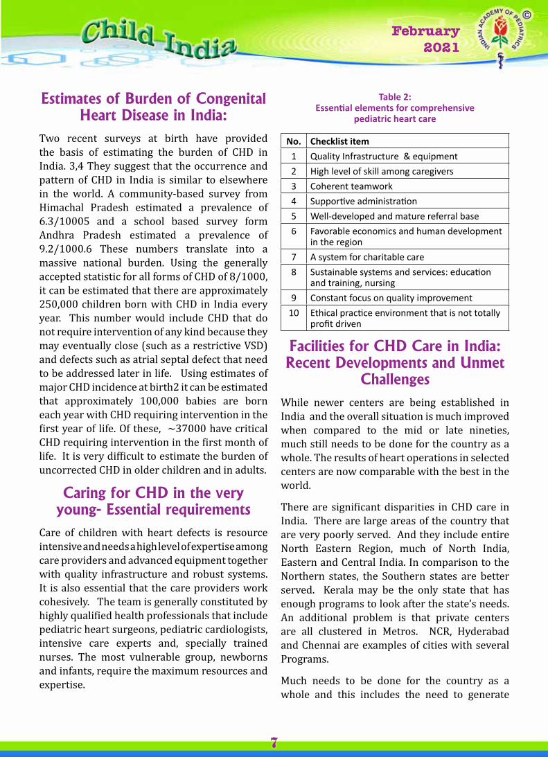
Care of children with heart defects is resource intensive and needs a high level of expertise among care providers and advanced equipment together with quality infrastructure and robust systems. It is also essential that the care providers work cohesively. The team is generally constituted by highly qualified health professionals that include pediatric heart surgeons, pediatric cardiologists, intensive care experts and, specially trained nurses. The most vulnerable group, newborns and infants, require the maximum resources and expertise.
Table 2: Essential elements for comprehensive
pediatric heart care
No. Checklist item1 Quality Infrastructure & equipment2 High level of skill among caregivers3 Coherent teamwork4 Supportive administration5 Well-developed and mature referral base6 Favorable economics and human development
in the region7 A system for charitable care8 Sustainable systems and services: education
and training, nursing9 Constant focus on quality improvement
10 Ethical practice environment that is not totally profit driven
Facilities for CHD Care in India: Recent Developments and Unmet
ChallengesWhile newer centers are being established in India and the overall situation is much improved when compared to the mid or late nineties, much still needs to be done for the country as a whole. The results of heart operations in selected centers are now comparable with the best in the world.
There are significant disparities in CHD care in India. There are large areas of the country that are very poorly served. And they include entire North Eastern Region, much of North India, Eastern and Central India. In comparison to the Northern states, the Southern states are better served. Kerala may be the only state that has enough programs to look after the state’s needs. An additional problem is that private centers are all clustered in Metros. NCR, Hyderabad and Chennai are examples of cities with several Programs.
Much needs to be done for the country as a whole and this includes the need to generate
Vol. 1January 2018
Vol. 1January 2018
February 2021
8
extensive awareness among pediatricians and other medical personnel about the magnitude of the problem. Additionally, there is a need for sustained advocacy on the need to develop regional centers with expertise in pediatric cardiac care.
Economic barriers come in the way of most Indian children receiving timely care for their heart conditions. The hardships faced by young parents that have just begun a family are enormous given their limited financial reserves. Furthermore, most medical insurance providers have specifically listed congenital heart defects as an exception to the list of conditions covered. Heart operations and catheter procedures in children are expensive because of the resources utilized (costs of consumables, intensive care etc.). As a result the economic impact on families who pay from out of their pockets is often devastating. 7 (http://journals.plos.org/plosone/article?id=10.1371/journal.pone. 0131348 )
The massive burden of congenital heart disease in India can only be tackled through a systematic plan that is developed and implemented by the government.
The Way forward: A Population based approach to CHD care
In 2012, the Government of India started the Rashtriya Bal Swasthya Karyakram (RBSK), a national child health initiative for screening and treatment of childhood diseases and disabilities, including CHD. This program administered by the National Health Mission (within the Ministry of Health and Family Welfare) provides funding and technical assistance to individual States. With the addition of funds and commitment by the Government of Kerala, financial resources were made available for a dedicated program to address the CHD burden in the state.
This program is named “Hridyam—for little hearts,”. (www.hridyam.in)
The program seeks to look at the entire life course of CHD from screening to identification to referral, surgery and follow up rather than providing for the surgery alone. This is in keeping with the vision of the RBSK program of the government of India.
A comprehensive web-based application, has been developed to enable referral and track the track the progress through each stage. Any physician within Kerala can add a name; there are no layers of medical hierarchy or bureaucracy to impede or delay this process.
This program also functions as a registry for children 0-18 years with suspected CHD of all types.
Once registered, each child’s progress is coordinated at the local level by the District Early Intervention Centre (DEIC, under the National Health Mission), but monitored centrally by the National Health Mission under Department of Health. The incentive for universal registration is that Hridyam serves as the sole entry point for accessing government-funded treatment via the above-mentioned Government schemes. Once listed, a pediatric cardiologist is obliged to review the online record within 24 hours and classify the case according to urgency for treatment. (In situations of insufficient information, that same cardiologist may direct the DEIC to acquire further tests.) A referral is made, based on a protocol that takes into consideration, the geographic location, acuity, parental preferences and institutional capacity. Outcomes are tracked on the same website, and the Hridyam protocol also directs the nature and timing of follow-up care. Everything concerning each registrant’s case occurs in real-time, is tracked and directed as needed by the government agency, and can be accessed by any interested and qualified in-state party.
Comprehensive pediatric cardiac care that includes surgery and catheter procedures are delivered in two government and five private institution across the state through a unique
Vol. 1January 2018
Vol. 1January 2018
February 2021
9
public-private partnership model. The highest standards of care are ensured and there is careful monitoring of results of treatment.
The results have been most gratifying. The Hridyam registry website was activated in August 2017. A steady growth in case registrations and surgical treatments has documented, as well as a trend toward newborn and infant procedures.
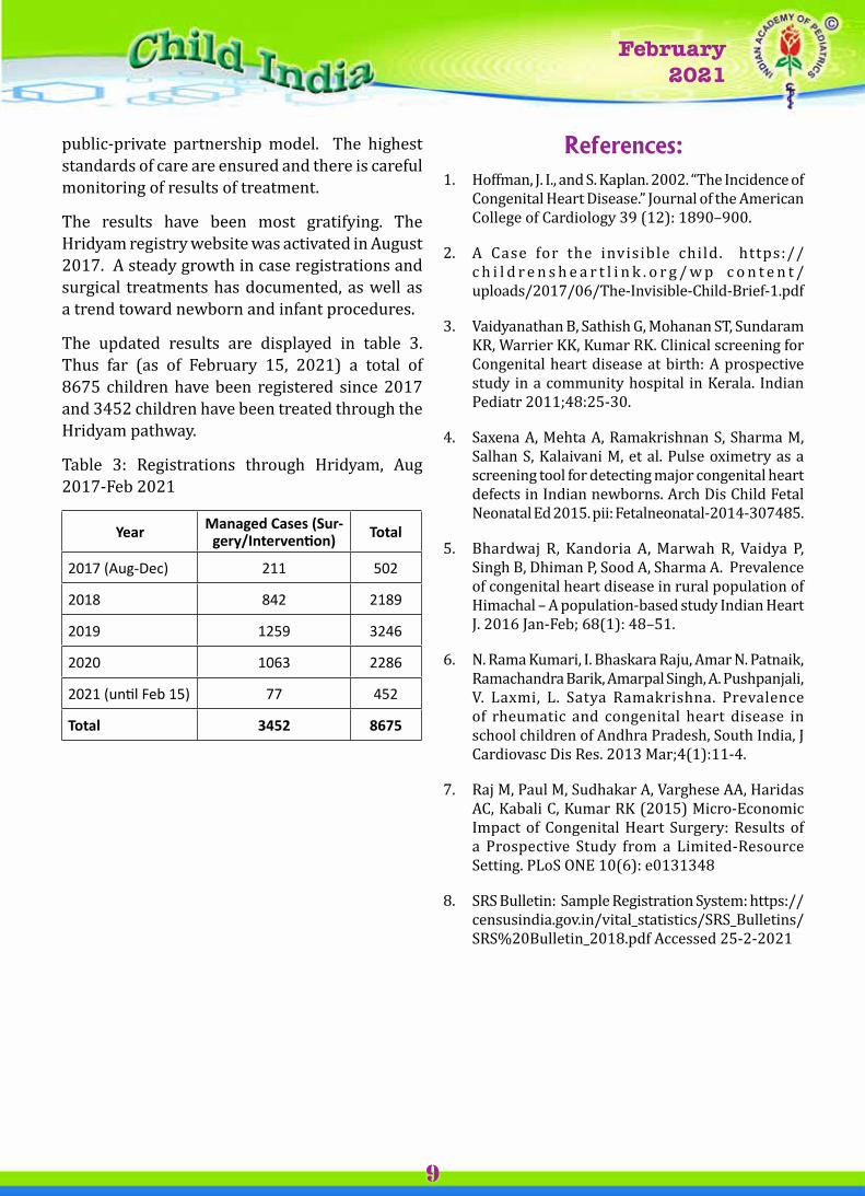
The updated results are displayed in table 3. Thus far (as of February 15, 2021) a total of 8675 children have been registered since 2017 and 3452 children have been treated through the Hridyam pathway.
Table 3: Registrations through Hridyam, Aug 2017-Feb 2021
Year Managed Cases (Sur-gery/Intervention) Total
2017 (Aug-Dec) 211 502
2018 842 2189
2019 1259 3246
2020 1063 2286
2021 (until Feb 15) 77 452
Total 3452 8675
References:1. Hoffman, J. I., and S. Kaplan. 2002. “The Incidence of
Congenital Heart Disease.” Journal of the American College of Cardiology 39 (12): 1890–900.
2. A Case for the invisible child. https://c h i l d r e n s h e a r t l i n k . o r g / w p c o n t e n t /uploads/2017/06/The-Invisible-Child-Brief-1.pdf
3. Vaidyanathan B, Sathish G, Mohanan ST, Sundaram KR, Warrier KK, Kumar RK. Clinical screening for Congenital heart disease at birth: A prospective study in a community hospital in Kerala. Indian Pediatr 2011;48:25-30.
4. Saxena A, Mehta A, Ramakrishnan S, Sharma M, Salhan S, Kalaivani M, et al. Pulse oximetry as a screening tool for detecting major congenital heart defects in Indian newborns. Arch Dis Child Fetal Neonatal Ed 2015. pii: Fetalneonatal-2014-307485.
5. Bhardwaj R, Kandoria A, Marwah R, Vaidya P, Singh B, Dhiman P, Sood A, Sharma A. Prevalence of congenital heart disease in rural population of Himachal – A population-based study Indian Heart J. 2016 Jan-Feb; 68(1): 48–51.
6. N. Rama Kumari, I. Bhaskara Raju, Amar N. Patnaik, Ramachandra Barik, Amarpal Singh, A. Pushpanjali, V. Laxmi, L. Satya Ramakrishna. Prevalence of rheumatic and congenital heart disease in school children of Andhra Pradesh, South India, J Cardiovasc Dis Res. 2013 Mar;4(1):11-4.
7. Raj M, Paul M, Sudhakar A, Varghese AA, Haridas AC, Kabali C, Kumar RK (2015) Micro-Economic Impact of Congenital Heart Surgery: Results of a Prospective Study from a Limited-Resource Setting. PLoS ONE 10(6): e0131348
8. SRS Bulletin: Sample Registration System: https://censusindia.gov.in/vital_statistics/SRS_Bulletins/SRS%20Bulletin_2018.pdf Accessed 25-2-2021
Vol. 1January 2018
Vol. 1January 2018
February 2021
10
The ECG is an important tool for the diagnosis of childhood heart disease, especially congenital heart disease. It is very often specific for anatomical defects when combined with other clinical findings and diagnostic tests. It also gives us a road map in many CHD and the more sophisticated investigations are often helped by the ECG data.
In children we should take a 14 lead ECG including V3R and V4R; the other leads being standard limb leads (I, II, III), augmented limb leads (aVR, aVL, aVF) and precordial leads (V1-V6). In suspected dextrocardia, one should order additional leads to the right. They are V5R and V6R.
ECG can be recorded in single channel or multi-channel machine. In the standard ECG, paper speed is 25 mm/sec i.e. I second is divided into 25 small divisions of 1 mm each. Time is measured horizontally, the distance between two thick lines being 0.2 seconds (200 msecs) and distance between two thin lines representing 40 msecs. (0.04 sec). Voltage is represented vertically, 1 mm being 0.1 mv. Paper speed is made 50 mm/sec for a more accurate assessment of various intervals, detection of AF etc.
Overall, pediatricians all over the world tend to be a bit overawed by ECG either adult or pediatric. This phenomenon is quite uncalled for and we can overcome it by a systematic analysis of a given ECG. ECG interpretation in the young is so different from that of an adult because almost all ECG parameters – normal and abnormal are age dependent and is also related to changes in the circulation especially so in the newborn. The transitional circulation in the very young makes interpretation more difficult. Pediatric training generally is soft on ECG interpretation, particularly arrhythmias. These are compounded by the fact that many structural lesions, both congenital and acquired need not produce definite and well
HOW TO READ ECG IN THE YOUNGAnd
How To Know Where Red Flags AreM. Zulfikar Ahamed
Thiruvananthapuram [email protected]
defined abnormalities in ECG. We will be dealing with normal and abnormal ECG’s in children and their interpretation and go on to special issues, problems and solutions later.
NORMAL ECG1. Calculating Heart RateWhen reading an ECG we usually take note of
the standardization. Then we calculate the heart rate. The best method is dividing 1500 by the number of small divisions between 2 R’s (RR interval). RR intervals may vary slightly, but, by not more than 3 small divisions, if variation is more than 120 msec (3 divisions) it is called sinus arrhythmia. It can be respiratory or non respiratory. Respiratory sinus arrhythmia is more common.
Heart rate is age dependent and bradycardia is said to exist in an older child when HR is less than 60/mt. The cut off for bradycardia is 90/mt in newborn. Tachycardia is considered when in a neonate, heart rate is >150/mt, in the child >120/mt and older child >100/mt.
2. Rhythm and P WaveNext we look at the P wave to know whether
rhythm is sinus or not. P wave is due to atrial depolarization. First half of the P Wave is inscribed by RA depolarization and second half by LA depolarization. In situs solitus (normal situation) P wave is always +ve in I, II, V4 & V6 and -ve in aVR and variable in other leads. It could be either positive or negative in aVL, V1-V3 and III. In aVF it is almost always upright. In situs inversus, P wave is positive in aVR and negative in I, II, V5-V6.
The Axis of P wave is normally between 30o-60o. If it is >75o it is most likely abnormal. Axis of >90o indicates situs inversus.
Vol. 1January 2018
Vol. 1January 2018
February 2021
11
Widest P wave can be up to 100 msec. Abnormal width of P wave is > 120 msec (3m).Maximum amplitude of P wave is 2.5 small divisions. So amplitude of >3mV (3mm) is abnormal. Morphology of P wave can vary. It can be monophasic, biphasic, notched, inverted etc.
Wandering pacemaker is a benign finding seen in ECG where there are continual changes in P wave morphology, amplitude and PR interval.
3. PR IntervalPhysiologically it represents the interval between
onset of atrial depolarization and ventricular depolarization. The distance between beginning of P wave to beginning of QRS complex is measured. Normal PR interval in a child is 110-160 msec and in a neonate it is < 140 msec. Adult value is 120-200 msec. The longest PR interval in any lead is taken for measuring.
Prolonged PR interval is commonly found in Rheumatic carditis, Rheumatic valvar disease and in CHD like Ebstein and L- TGA. Short PR interval (<100 msec) is seen in very small infants, WPW syndrome, LGL syndrome and Pompe’s disease.
PR segment is the interval between end of P wave and beginning of QRS complex.
3. QRS ComplexQRS indicates ventricular depolarization. The
direction is from endocardium to epicardium. QRS duration is traditionally measured in limb leads or V1-V2. The average duration is between 50-100 msec. If it is >100 msec, it is abnormal – may indicate intraventricular conduction defect or bundle branch block.
I. AmplitudeLow voltage QRS complexes are defined as
having < 5mm height in limb leads and <10 mm height in precordial leads. High voltage QRS complexes are > 20mm in limbs leads and > 30mm in precordial leads. High voltage complexes need not be abnormal. Tallest R are found in V4, V5.
2. Q Wave & R/SSmall q is normally is seen in 1, aVL, V4 -V6 and
aVF. Depth of such q is < 3 mm and width <1mm. A q wave of > 4 mm depth is considered abnormal. Usually depth of Q is less than 25% of corresponding R height. In adults and in older children R/S in V1 is always less than I and in V6 more than I.
3. TransitionThis is the point at which dominant polarity
changes in the precordial leads. It usually occurs in
V3 or V4. If it occurs in V2 it is called early transition and if it occurs in V5 it is late transition.
4. QRS AxisAxis determination is crucial in evaluation
of CHD, especially in congenital cyanotic heart disease where axis may point to a definite diagnostic possibility, like left axis deviation in Tricuspid Atresia.
According to NYHANormal axis 0o to + 90oRight Axis Deviation +90o to + 180oLeft Axis Deviation 0o to -90oRight Upper Quadrant Axis 90o to +80o
From birth to 1 year, normal axis is from +90o to + 150o. Between 1 – 8 yrs it varies between +45o and +105o. Above 8 years it is <90o.
To determine the axis, initially concentrate on QRS complexes in I and aVF.
A. If I and aVF.Are both +ve = Normal AxisI is –ve; aVF is +ve = RADI is +ve; aVF is –ve = LADBoth are –ve = RUQ AxisOnce this broad categorization is done, go on to
calculate axis more specificallyIf axis is normal, look at III and aVL. If III is
equiphasic axis is +30o; If aVL is equiphasic axis is +60o.
It is RAD look at II and aVR. If aVR is equiphasic axis is +120o; If II is equiphasic axis is +150o.
If it is LAD look at II and aVR again. If II is equiphasic axis is -30o. If aVR is equiphasic, axis is -60o.
If the axis is RUQ, it could be extreme RAD or extreme LAD. To determine which, we look at the loop of QRS. There are two types of loops – clockwise (q in II. III. aVF) and counter clockwise (q in I. aVL). If in RUQ axis QRS is clockwise it is extreme RAD and if it is counter clockwise it is extreme LAD.
5. ST SegmentPoint of end of S wave and beginning of ST
segment is called J point. From J point to beginning of T wave is called ST segment. The most important information is regarding its deviation from base line (T-P segment). Normally, it should be always <1mm. In precordial leads some ST elevation is seen in the young in V1-V3. Any ST depression in precordial leads is abnormal.
Vol. 1January 2018
Vol. 1January 2018
February 2021
12
6. T WaveIt represents ventricular repolarisation. The
polarity is that of QRS complex. Up to 3 days of life T is upright in V3R, V1, and V2. After the 3rd day it is inverted in V3R, V1 and can be so even up to V3. In older children inverted T is rare in V3. Maximum height of T is 6 mm in limb leads and 10 mm in precordial leads.
7. QTc (Corrected QT Interval)It represents the duration of ventricular electrical
systole. It is measured from beginning of QRS to end of T wave. QT varies with heart rate, so corrected QT is calculated. For this divide QT in milli seconds obtained by square root of R-R interval in seconds. Normal QTC is <440 msec.
U WaveIt probably represents repolarisation of His
Purkinje system. It has one fourth amplitude of T wave and is positive.
ABNORMAL EGG
Having described a Normal ECG we shall go to abnormal pattern. First we take up chamber enlargement.1. Left Atrial Enlargement Best seen in lead II. V1. a. Morris criteria – Negative deflection of P wave in
V1 is more than 1 mm in depth and width.b. P wave duration > 120 msec.c. In a bifid P wave, inter peak distance > 40 msec.d. Macruz index – P duration / PR segment is > 1.6.e. P axis - < 30o.2. Right Atrial Enlargement
Best seen in II. V1. a. Tall, peaked P wave, > 3mm, in lead II.b. P axis > 60o.c. P initial force in V1 > 1.5 mm.Previously LAE was called as P mitrale and RAE
was called as P pulmonale and congenitale. These terms are no longer in current use.
3. Left Ventricular Hypertrophy(Modified Lyon – Sokolow criteria in children)
a. R in V5/V6 + S in V1 > 45 mmb. R in V5/V6 > 35 mmc. R in aVL, aVF > 20 mmd. Additional criteria – prolonged QRS, LAD, LAE.
Estes scoring is yet another criteria to diagnose LVH incorporating QRS amplitude, strain pattern,
LAE, LAD, QRS duration LVH may bea. Volume overload1. Prominent q in 1, aVL. V5-V62. Tall R in V5-V63. Tall symmetrical upright T in V5-V6b. Pressure overload1. No significant q in aVL. V5-V62. Slurred upstroke of R in Left precordial leads.3. Asymmetrical T inversion with sagging ST
segment in V5-V64. Right Ventricular Hypertrophya. Upright T in V1-V3 R after 3 days of life.b. R/S in V1 > 1 after 6 years.> 1.5 between 3-6 years> 3 between 6 months – 3 years> 5 in infants (<6 months)c. qR in V3 R or V1. d. R in V1 > 7 mm in child above 6 years.e. R in V1 + S in V6 > 11 mm.f. R in AVR > 5 mmg. RAD > + 110o There can be three morphological types of RVH.1. Type A RVH – monophasic R in V1 or qR
(Pressure overload)2. Type B RVB – rSR in V1 or V3R (Volume
overload)3. Type C RVH – rS in V1-V6.5. Biventricular Hypertrophy1. Independently RVH + LVH2. LVH + RAD3. LVH + Clockwise loop4. RVH + LAD5. Katz – Wachtel Phenomenon – tall R and
deep S in mid precordial leads measuring > 60 mm.6. Heart BlocksCan be divided into SA blocks AV Blocks Bundle Branch Blocks1. SA BlockSA Blocks are uncommon in children except
in postoperative patients. It is divided into I, II, III. degree heart blocks. 1o is not detected on surface ECG and in 11o SA block there is presence of long cycle between P waves. In IIIo SA Block, only an escape rhythm is evident on surface ECG.
Vol. 1January 2018
Vol. 1January 2018
February 2021
13
2. AV BlockCan also be of three types – 1o, 11o, 111o. (CHB).Io AV Block: All atrial impulses are conducted to
ventricles with AV delay. So PR interval is prolonged (> 200 msec.).
IIo AV Block: Not all atrial impulses are conducted to ventricles. There are two types of blocks.
a. Mobitz Type I (Wenckebach) – There is gradual prolongation of PR interval in successive beats followed by dropping of QRS complex.
b. Mobitz Type II – There is no preceding PR prolongation. QRS is abruptly dropped. The ratio of P to QRS may be 2:1 or 3:1 or so.
IIIo AV Block (complete heart block): No impulse from atrium reaches the ventricle so that both atrium and ventricle have their own rates – AV dissociation.
3. Bundle Branch BlockCan be RBBB or LBBBRBBB: 1. QRS duration > 120 m sec. 2. R > r in rSR 3. VAT is > 80 msec 4. ST depression and T inversion.LBBB: 1. QRS duration > 120 msec 2. VAT > 80 msec. 3. Characteristic M pattern or
monophasic tall R in V5V6 4. Absence of Q in V5-V6 5. ST depression and T inversionNote: rSR pattern is found ina. Normalb. Pectus, Straight Back Syndromec. RV volume overload d. Cor pulmonale
e. WPW syndrome7. ARRHYTHMIAS1. Atrial Ectopics1. Premature P wave2. Premature P wave is abnormal and different
from sinus P wave3. Variable PR interval4. Narrow QRS5. Less than full compensatory pause.2. Ventricular Ectopics
1. Premature QRS complex2. No proceeding P wave3. QRS is abnormal and wide4. Secondary ST-T changes.5. Full compensatory pause6. Constant coupling interval.3. WPW SyndromeIt is not an uncommon situation in Pediatric
ECG. Characteristic features are1. PR interval < 120 msec.2. Delta wave3. Abnormal QRS with width > 110 msec.4. Additional ST-T changes.5. Associated SVT complicating.4. Supraventricular TachycardiaIt is an important rhythm abnormality in
children. Rate is around 180-220/mt. with Narrow QRS complexes. P wave is either submerged in QRS or retrograde or abnormal. It can be commonly due to AV re-entrant or AV nodal re-entrant tachycardia.
5. Ventricular TachycardiaUncommon rhythm in children. Can occur in
long QT syndromes.
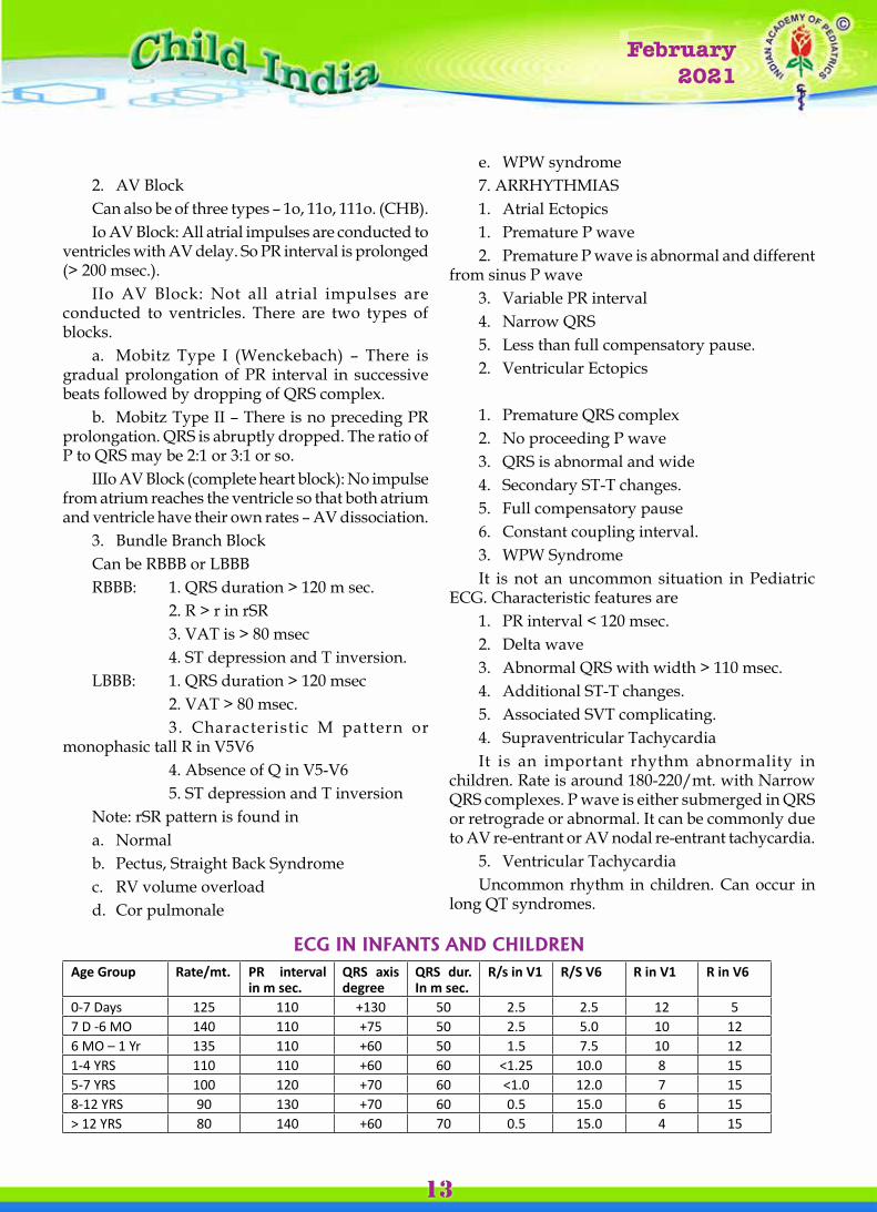
ECG IN INFANTS AND CHILDRENAge Group Rate/mt. PR interval
in m sec.QRS axis degree
QRS dur. In m sec.
R/s in V1 R/S V6 R in V1 R in V6
0-7 Days 125 110 +130 50 2.5 2.5 12 57 D -6 MO 140 110 +75 50 2.5 5.0 10 126 MO – 1 Yr 135 110 +60 50 1.5 7.5 10 121-4 YRS 110 110 +60 60 <1.25 10.0 8 155-7 YRS 100 120 +70 60 <1.0 12.0 7 158-12 YRS 90 130 +70 60 0.5 15.0 6 15> 12 YRS 80 140 +60 70 0.5 15.0 4 15
Vol. 1January 2018
Vol. 1January 2018
February 2021
14
Picking up Red Flags 1. Rate interpretation: SVT: The highest sinus rate achievable in the neonate
and infant rarely crosses 200/mt. Hence a rate above 200 usually indicates supraventricular tachycardia (SVT) if QRS complexes are narrow. Occasionally it may become very difficult to distinguish between sinus tachycardia and SVT.
The following points are useful in such situations. 1. Sinus tachycardia rarely exceeds rate of 200/
mt; Most SVT’s have a rate above 200; more often over 250/mt.
2. Sinus tachycardia usually shows some respiratory variation; SVT has no respiratory variation. It has a monotonously regular fast rate.
3. P waves are usually seen and well defined in sinus tachycardia; P wave in SVT are either not seen or seen immediately succeeding QRS complex- retrograde P wave. In Ectopic Atrial Tachycardia, P waves are well defined and precede QRS complexes, but will usually be inverted in II, III and aVF.
BRADYCARDIA:A very slow heart rate could be due to sinus
bradycardia, sino atrial block or AV block. In sinus bradycardia each QRS will be preceded by a normal looking P wave and there could be mild sinus arrhythmia – RR interval variation > 120 msec (3 small divisions). In second degree SA block, rate will be very slow and if it is 2:1 SA block it will closely mimic sinus bradycardia. Both advanced IIo and Complete AV blocks will have a slower heart rate and P – QRS relationship (AV dissociation) is crucial in determining the cause of ‘bradycardia’ as to CHB.
2. P waves: Are looked at not only for atrial enlargement but
also for situs in congenital heart disease (CHD). In analyzing ECG of a neonate or infant, possibility of a situs problem should be kept in mind. Inverted P in lead I and positive P wave in Avr. Occasionally lead reversal can cause positive P in aVR and negative P in I. But the other leads will show normal pattern in such a situation.
3. PR interval Rarely poses problems either in measuring
or interpretation. One must think of rheumatic etiology in any child with a murmur and prolonged PR interval. Short PR interval should make one to look for WPW syndrome. The other causes of short
PR interval are so rare like Pompe disease and LGL syndrome
4. QRS voltages Are to be designated as normal, low or high
according to the criteria set, not by eyeballing alone. It is likely that low voltages indicate cardiac problem than high voltages. Myocarditis, though said to be an important cause of low voltage complexes usually has near normal voltages in children.
5. Q waves in ECG Can be present normally in young infants and
children. There are criteria for abnormal Q which have been discussed elsewhere. Many normal infants do have reasonably deep Q waves in II, III and aVF. But deep q waves spread over a coronary artery territory – inferolateral, anterior and antero lateral become suspicious. Associated ST-T changes impart more clinical significance to such q waves. Such a situation can occur in ALCAPA, Kawasaki disease and Myocarditis.
Even a small Q in V1 or V3R are abnormal. qR pattern in V1 or V3R are always abnormal and can be due to
i. Ventricular inversion (in L-TGA).ii. Severe Right ventricular hypertension
exceeding left ventricular pressure eg: severe PS. IPAH.
iii. Right ventricular EMF – possibly due to a huge right atrium.
In a cyanotic infant no q in V5-V6 may indicate single ventricle as q in V5, V6 are caused by septal depolarization.
6. Transition of QRS complex If V3R is not taken, an early transition which
occur from V3R to V1 may be missed and V1 will shall a lack of predominant RV activity – small R and deep S and one may wrongly suspect Tricuspid Atresia in a cyanotic heart disease. RVH can also be missed in occasional situations if V3R is not taken. If precordial leads show progressive decline of R wave from V1 to V6 one should suspect dextrocardia and should ask for additional leads V4R, V5R, V6R.
7. QRS AxisMost normal infants will have right axis
deviation (RAD). One must realize that most infants with significant CHD also will have RAD – eg; VSD, ASD, TOF, TGA etc. So QRS axis is not very helpful in diagnosis unless it is unusual – left axis deviation or right upper quadrant axis.
Left axis deviation (LAD) can be found in both

Vol. 1January 2018
Vol. 1January 2018
February 2021
15
cyanotic and acyanotic heart diseases. Acyanotic heart diseases which have LAD are
i. Primum ASDii. Inlet VSD or Multiple muscular VSD.iii. Rarely PDA or Coarctation.Many cyanotic CHD can have LAD. Leading
causes are Tricuspid Atresia, AV canal Defect, single ventricle and L-TGA.Right upper Quadrant axis (RUQ) is peculiar and is found in dysplastic PS in Noonan, CHD in Rubella Syndrome or in extreme right or left axis deviation.
8. ST Segment and T wave Changes are not common among children.
It can occur in digitoxicity, myocarditis, dilated cardiomyopathy and hypertrophic cardiomyopathy. Global T inversion in precordial leads usually indicates a primary myocardial lesion. Asymmetrical T wave inversion can be found in strain pattern associated with LVH or RVH.
9. Calculation of QTC Is very crucial in certain clinical situations and
has to be properly done. Prolonged QTc can be found LQTS (Long QT syndrome) and can also be due to drugs and metabolic abnormalities.
CLINICAL SITUATIONS AND ECG – HOW DOES IT HELP?
1. If an eight month old baby has CHF, No cyanosis and good precordial murmur with normal pulses, the usual diagnosis is VSD. We will expect RAD and BVH. An interesting phenomenon of very tall R waves and deep S waves in mid precordial leads can be present which, more or less settles the clinical diagnosis. This phenomenon is called Katz- Wachtel phenomenon.
If such an infant has a LAD one should think of inlet VSD, multiple muscular VSD or AV septal defect. If associated 1o AV block is also present AVSD is most likely. However if ECG shows RAD, BVH and 1o AV block one must think of possible double outlet right ventricle (DORV) with VSD, PAH. If the infant has clinically asymptomatic VSD with no CHF and has RVH in ECG, a pink TOF has to be thought of.
2. If an asymptomatic child with an insignificant murmur at pulmonary area and mildly hyperkinetic precordium; has RAD and rSR in V1 or V3 R the diagnosis is virtually a secundum ASD. 85-95% of all significant ASD will have rSR pattern.
3. Some infants with PDA can present with
only systolic murmur and high volume pulse. In such a situation ECG will help by demonstrating predominantly LV volume overload than BVH as expected in VSD. Clinical PDA in a neonate with RVH could indicate a compensatory ductus as in severe TOF or pulmonary atresia.
4. If an asymptomatic child has a prominent systolic murmur over 2nd space and RVH in ECG, the diagnosis is most likely PS. In PS there is a definite correlation between severity and R voltages in V1 or V3R. Virtually all severe valvar PS will have an abnormal ECG. If instead of RAD, RUQ axis is found, it could due to dysplastic pulmonary valve as in Noonan syndrome.
5. 6 month old baby with ejection click and murmur at apex and Rt upper sternal border invokes the diagnosis of AS. ECG will not probably show LVH. The infant pattern of RAD and RVH will be present in such a baby. Even in older child ECG is not particularly useful in diagnosis AS. Severe AS can have normal ECG.
6. An infant with severe Coarctation (CoA) will have usually RAD and RVH contrary to expectation. This pattern will be present up to one year. In fact LV forces in infants with suspected CoA will indicate additional problems like PDA or EFE.
7. A 9 month old baby with cyanosis and murmur without CHF or cardiomegaly shows RAD and RVH, the diagnosis is usually TOF. An early transition ie change in polarity of QRS (R/S) from V1 to V2 is almost typical in TOF. RVH with strain is not found in TOF and hence other diagnosis must be thought of.
TOF physiology clinically and ECGi. With LAD – Think of AVCD with PS, L-TGA
with PS, Tricuspid Atresia.ii. With 1o AV block - Think of AVSD with PS,
DORV VSD PS, LTGA VSD PS.iii. With No q in V5 and V6 – Think of single
ventricle. iv. With q in V1 and no q in V6 – L - TGA: VSD
PS. v. Extreme RAD – Think of DORV. VSD. PS or
associated ASD.8. A neonate with congenital cyanotic heart
disease like TGA, TOF, and pulmonary Atresia .VSD will have RAD and RVH. RAD and RG dominance is the rule in normal infants also. Hence true RVH is difficult to identify; it is suggested by.
a. An axis of more than +120o b. Upright T in V1 or V3R
Vol. 1January 2018
Vol. 1January 2018
February 2021
16
c. Monophasic tall R in V1 or V3Rd. RV strain pattern.9. A neonate with cardiomegaly, cyanosis and
CHF with WPW can be having an Ebstein anomaly. Other CHB which can have WPW syndrome are L-TGA and AVSD.
10. If a neonate has RDS, cyanosis with RVH and strain pattern the possibilities are TAPVC – both obstructed and unobstructed, PPHN and severe CoA with LV / RV dysfunction. The usual common significant CCHD like TGA, TOF, etc will not exhibit RV strain in ECG.
11. A recent onset CHF in an infant or child with insignificant murmur could be due to myocarditis or DCM. The diagnostic triad are low voltage QRS, ST segment and T wave changes and tachycardia. However such a triad is found only in 50% of myocarditis.
12. While DCM (Dilated cardiomyopathy) may have nonspecific ECG changes, HCM will invariably have an abnormal ECG. HCM with a normal ECG is virtually unknown.
13. A myocardial infarction pattern in ECG in infants and children will be due to ALCAPA, Kawasaki disease, myocarditis and pompe disease and due to infarction in SLE and familial lipoproteinemias.
Summary
ECG interpretation in the young is a challenging but fruitful exercise in diagnostic evaluation of CHD and acquired heart disease. Systematic analysis of a surface ECG will demystify the enigma of ECG is an infant. One must be also aware of age dependent factors influencing ECG’s and presence of certain correlation between specific ECG patterns and CHD, which will be of great help in a diagnostic conclusion.
Suggested Reading
1. Electrocardiography. A. Garson Jr in Science and Practice of Pediatric Cardiology Ed: A Garson Jr, JT Bricker, DJ Fisher, SR Neish Williams and Wilkins © 1998.
2. Demystifying the Pediatric ECG MZ Ahamed in Pediatric Cardiopulmonary Update 1999 Ed. MZ Ahmed, S Subramony, Iype Joseph. A Pediatric Cardiology Division Publication © 1999.
3. Chou’s Electrocardiography in Clinical Practice. Fifth Edition. Ed. B. Sarawicz and TK Knilans W.B. Saunders 2001.
4. Lippman – Massie Clinical Electro cardio-graphy Ed. MI Dunn, BS Lipman 8th edition Year Book Medical Publishers 1989.
Vol. 1January 2018
Vol. 1January 2018
February 2021
17
IntroductionA newborn with cardiac disease is always a challenging case to diagnose and manage as presentation can vary from completely asymptomatic to life threatening episodes .Most of the presentations can mimic the common ,non cardiac neonatal disorders such as septicemia, shock, respiratory disorders, persistent pulmonary hypertension of newborn (PPHN),inborn errors of metabolism and so on. But with a meticulous approach, presumptive clinical cardiac diagnosis can be made and appropriate management started. (1) The focus of this chapter is approach and management of common neonatal cardiac disorders which a neonatologist comes across in his day to day practice.
HistoryDetailed medical history about antenatal and perinatal events is very important to determine the possibility of cardiac disease. In today’s world; where antenatal diagnosis is getting more and more precise and fetal echo is providing great insights into the disease, neonatologist must look into antenatal reports. (2) Similarly antenatal history about drugs and diseases may point to a specific diagnosis and should be sought. Perinatal events such as fetal distress leading to respiratory distress and cyanosis at birth may point to the diagnosis of non cardiac cause of cyanosis. On the other hand, uneventful delivery with sudden onset of cyanosis or shock after 24-48 hours of birth should give a suspicion of duct dependent circulation. A history of preterm baby with hyaline membrane disease getting better after surfactant therapy and then again worsening with need for ventilator is a
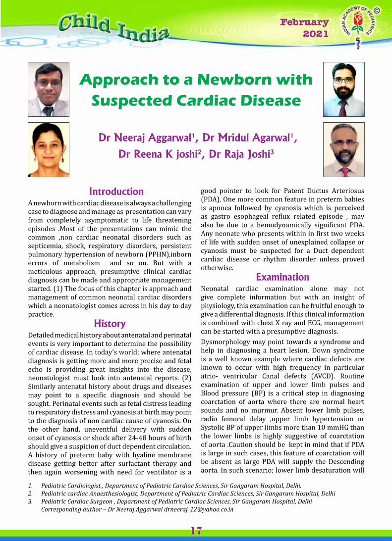
Approach to a Newborn with Suspected Cardiac Disease
Dr Neeraj Aggarwal1, Dr Mridul Agarwal1,
Dr Reena K joshi2, Dr Raja Joshi3
1. Pediatric Cardiologist , Department of Pediatric Cardiac Sciences, Sir Gangaram Hospital, Delhi.2. Pediatric cardiac Anaesthesiologist, Department of Pediatric Cardiac Sciences, Sir Gangaram Hospital, Delhi3. Pediatric Cardiac Surgeon , Department of Pediatric Cardiac Sciences, Sir Gangaram Hospital, Delhi Corresponding author – Dr Neeraj Aggarwal [email protected]
good pointer to look for Patent Ductus Arteriosus (PDA). One more common feature in preterm babies is apnoea followed by cyanosis which is perceived as gastro esophageal reflux related episode , may also be due to a hemodynamically significant PDA. Any neonate who presents within in first two weeks of life with sudden onset of unexplained collapse or cyanosis must be suspected for a Duct dependent cardiac disease or rhythm disorder unless proved otherwise.
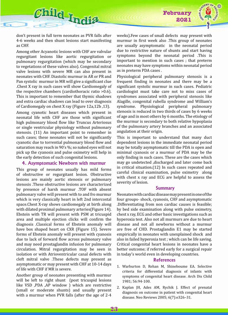
ExaminationNeonatal cardiac examination alone may not give complete information but with an insight of physiology, this examination can be fruitful enough to give a differential diagnosis. If this clinical information is combined with chest X ray and ECG, management can be started with a presumptive diagnosis.Dysmorphology may point towards a syndrome and help in diagnosing a heart lesion. Down syndrome is a well known example where cardiac defects are known to occur with high frequency in particular atrio- ventricular Canal defects (AVCD). Routine examination of upper and lower limb pulses and Blood pressure (BP) is a critical step in diagnosing coarctation of aorta where there are normal heart sounds and no murmur. Absent lower limb pulses, radio femoral delay ,upper limb hypertension or Systolic BP of upper limbs more than 10 mmHG than the lower limbs is highly suggestive of coarctation of aorta .Caution should be kept in mind that if PDA is large in such cases, this feature of coarctation will be absent as large PDA will supply the Descending aorta. In such scenario; lower limb desaturation will
Vol. 1January 2018
Vol. 1January 2018
February 2021
18
help to diagnose this lesion. Low volume pulses in all extremities will suggest left sided obstructive lesion like aortic stenosis. Similarly bounding pulses will be felt in PDA, aortic regurgitation, Truncus Arteriosus and systemic arteriovenous fistula.Pulse oximetry can be a valuable tool and important assistant to clinical examination .Naked eyes cannot diagnose cyanosis in patients where saturation is less than 85 % and so will miss cyanotic heart disease in many cases unless pulse oximetry is used. This may be a valuable exercise to diagnose cyanotic heart diseases in newborn and may be a routine in our neonatal units soon.(3) The best time would be after 24 hours of life as many normal neonates will have lower saturation in first 24 hours of life. The cardiac examination should be performed in a systematic manner. When the child is irritable, it will be difficult to appreciate heart sounds and every attempt should be made to examine a neonate while sleeping. Situs solitus with dextrocardia should point towards a complex cardiac lesion (Figure 1a, 1b, 1c). Left sided precordial bulge will suggest cardiac enlargement and a left parasternal heave indicates right ventricular hypertension. Also look for a palpable precordial thrill. Abnormalities of the second heart sound are often seen in congenital heart disease, making it perhaps the most important element of auscultation in the pediatric patient. Third and fourth heart sounds can be normal. The Grade IV, V, and VI murmurs are associated with a palpable precordial thrill, and are always pathological. The innocent murmurs are soft (Grade I or II) and ejection in quality. Although diastolic murmurs are much less common in the child, the presence of a diastolic murmur indicates that structural heart disease is present.Last, a simple, noninvasive indicator of cardiac output is the capillary refill time. This is obtained by blanching the nail bed or digit, and observing the time to reperfusion, normally less than 3 seconds.
Hemodynamic Considerations in Neonate (4)
Neonates are born with pulmonary arterial hypertension and this makes the assessment of left to right shunts difficult by clinical examination or echocardiography. Appearance of murmur in left to right shunts is delayed for 2-3 weeks till the pulmonary vascular resistance (PVR) drops .This change in hemodynamics is also important for many critical cardiac evaluations .Some examples are important to consider here. A small or moderate
Ventricular Septal Defect (VSD) will not have a PSM at birth (characteristic of a restrictive VSD shunt) due to high PVR and ECHO may not show volume overload of Left atrium and left ventricle .Pulmonary pressures will always be high enough at birth to cause confusion in the estimation of true hemodynamics of VSD shunt till 2-3 weeks of age. In such cases; it is useful to repeat Echo after 2-3 weeks to assess such patients. Similarly right sided obstructive lesions may be under diagnosed in neonatal period with presence of PAH (like pulmonary stenosis and TOF physiology) where gradient across pulmonary valve will be underestimated in presence of pulmonary hypertension and such cases will need follow up evaluation after 1-2 weeks to reassess the severity of true obstruction by Doppler.
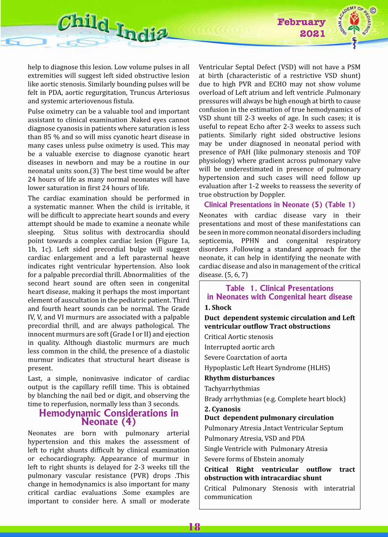
Clinical Presentations in Neonate (5) (Table 1)Neonates with cardiac disease vary in their presentations and most of these manifestations can be seen in more common neonatal disorders including septicemia, PPHN and congenital respiratory disorders .Following a standard approach for the neonate, it can help in identifying the neonate with cardiac disease and also in management of the critical disease. (5, 6, 7)
Table 1. Clinical Presentations in Neonates with Congenital heart disease
1. ShockDuct dependent systemic circulation and Left ventricular outflow tract obstructionsCritical Aortic stenosisInterrupted aortic arch Severe Coarctation of aortaHypoplastic Left Heart Syndrome (HLHS)Rhythm disturbances TachyarrhythmiasBrady arrhythmias (e.g. Complete heart block)2. cyanosis Duct dependent pulmonary circulationPulmonary Atresia ,Intact Ventricular Septum Pulmonary Atresia, VSD and PDA Single Ventricle with Pulmonary AtresiaSevere forms of Ebstein anomalycritical Right ventricular outflow tract obstruction with intracardiac shuntCritical Pulmonary Stenosis with interatrial communication
Vol. 1January 2018
Vol. 1January 2018
February 2021
19
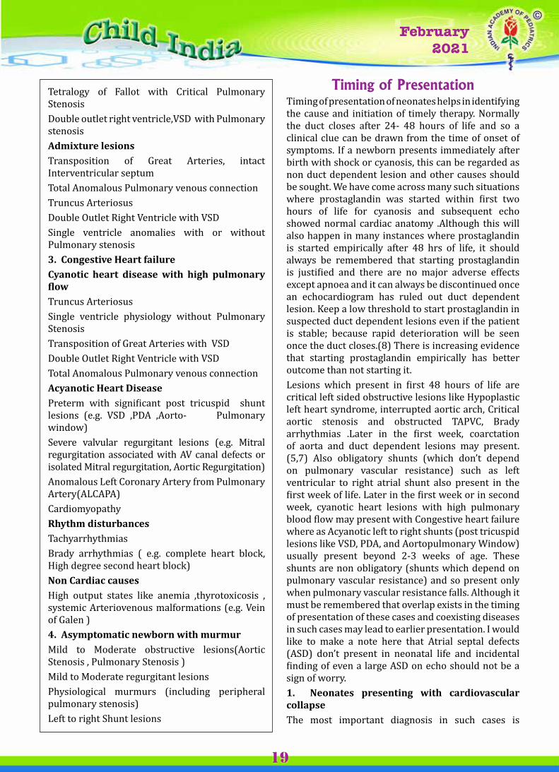
Tetralogy of Fallot with Critical Pulmonary StenosisDouble outlet right ventricle,VSD with Pulmonary stenosisAdmixture lesions Transposition of Great Arteries, intact Interventricular septumTotal Anomalous Pulmonary venous connectionTruncus Arteriosus Double Outlet Right Ventricle with VSDSingle ventricle anomalies with or without Pulmonary stenosis3. Congestive Heart failure Cyanotic heart disease with high pulmonary flow Truncus Arteriosus Single ventricle physiology without Pulmonary StenosisTransposition of Great Arteries with VSDDouble Outlet Right Ventricle with VSDTotal Anomalous Pulmonary venous connection Acyanotic Heart DiseasePreterm with significant post tricuspid shunt lesions (e.g. VSD ,PDA ,Aorto- Pulmonary window)Severe valvular regurgitant lesions (e.g. Mitral regurgitation associated with AV canal defects or isolated Mitral regurgitation, Aortic Regurgitation)Anomalous Left Coronary Artery from Pulmonary Artery(ALCAPA)CardiomyopathyRhythm disturbancesTachyarrhythmias Brady arrhythmias ( e.g. complete heart block, High degree second heart block)Non Cardiac causes High output states like anemia ,thyrotoxicosis , systemic Arteriovenous malformations (e.g. Vein of Galen )4. Asymptomatic newborn with murmur Mild to Moderate obstructive lesions(Aortic Stenosis , Pulmonary Stenosis )Mild to Moderate regurgitant lesions Physiological murmurs (including peripheral pulmonary stenosis)Left to right Shunt lesions
Timing of PresentationTiming of presentation of neonates helps in identifying the cause and initiation of timely therapy. Normally the duct closes after 24- 48 hours of life and so a clinical clue can be drawn from the time of onset of symptoms. If a newborn presents immediately after birth with shock or cyanosis, this can be regarded as non duct dependent lesion and other causes should be sought. We have come across many such situations where prostaglandin was started within first two hours of life for cyanosis and subsequent echo showed normal cardiac anatomy .Although this will also happen in many instances where prostaglandin is started empirically after 48 hrs of life, it should always be remembered that starting prostaglandin is justified and there are no major adverse effects except apnoea and it can always be discontinued once an echocardiogram has ruled out duct dependent lesion. Keep a low threshold to start prostaglandin in suspected duct dependent lesions even if the patient is stable; because rapid deterioration will be seen once the duct closes.(8) There is increasing evidence that starting prostaglandin empirically has better outcome than not starting it. Lesions which present in first 48 hours of life are critical left sided obstructive lesions like Hypoplastic left heart syndrome, interrupted aortic arch, Critical aortic stenosis and obstructed TAPVC, Brady arrhythmias .Later in the first week, coarctation of aorta and duct dependent lesions may present. (5,7) Also obligatory shunts (which don’t depend on pulmonary vascular resistance) such as left ventricular to right atrial shunt also present in the first week of life. Later in the first week or in second week, cyanotic heart lesions with high pulmonary blood flow may present with Congestive heart failure where as Acyanotic left to right shunts (post tricuspid lesions like VSD, PDA, and Aortopulmonary Window) usually present beyond 2-3 weeks of age. These shunts are non obligatory (shunts which depend on pulmonary vascular resistance) and so present only when pulmonary vascular resistance falls. Although it must be remembered that overlap exists in the timing of presentation of these cases and coexisting diseases in such cases may lead to earlier presentation. I would like to make a note here that Atrial septal defects (ASD) don’t present in neonatal life and incidental finding of even a large ASD on echo should not be a sign of worry. 1. Neonates presenting with cardiovascular collapse The most important diagnosis in such cases is
Vol. 1January 2018
Vol. 1January 2018
February 2021
20
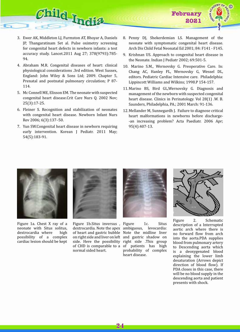
obstructive lesions like critical Aortic stenosis(AS), critical Coarctation with left ventricular dysfunction and also critical pulmonary stenosis (PS) with RV dysfunction .The list also includes cases of Hypoplastic left heart syndrome(HLHS) and obstructive TAPVC (total anomalous pulmonary venous connection) who present in shock. Neonates presenting with sudden unexplained onset of shock after 48 hrs of life should be evaluated for the above mentioned diseases (critical AS, critical PS, coarctation, HLHS, obstructive TAPVC) and also for the other duct dependent lesions mentioned in table 1.This neonate is usually a healthy newborn who presents after 48-72 hrs of life with sudden onset of pallor, grey appearance and breathing difficulty. Typical history includes baby not passing urine and not taking feeds over last 4-6 hrs. There is usually an evidence of metabolic acidosis. These newborns should be started immediately with prostaglandin E1 suspecting duct dependent systemic circulation among other measures to stabilize; including ventilation and inotropes (unless echo rules out cardiac lesion)If PDA is still open ; these newborns may be picked up early during routine evaluation with a harsh systolic murmur of obstructive lesion (aortic stenosis or pulmonary stenosis).Coarctation of aorta will be picked up by careful palpation of all 4 limb pulses and blood pressure .Any evidence of radio femoral delay should prompt the diagnosis of coarctation of aorta. Differential cyanosis with lower limb showing desaturation compared to upper limb should prompt for the cause of PDA shunting right to left (e.g.
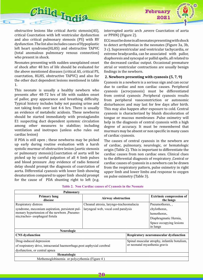
interrupted aortic arch ,severe Coarctation of aorta or PPHN) (Figure 2).ECG must be done in all neonates presenting with shock to detect arrhythmias in the neonates (Figure 3a, 3b, 3 c). Supraventricular and ventricular tachycardia, or extreme bradycardia, can be associated with pallor, diaphoresis and syncopal or pallid spells, all related to the decreased cardiac output. Occasional premature atrial or ventricular contractions are usually benign findings in the newborn. 2. Newborn presenting with cyanosis (5, 7, 9)Cyanosis in a newborn is a serious sign and can occur due to cardiac and non cardiac causes. Peripheral cyanosis (acrocyanosis) must be differentiated from central cyanosis .Peripheral cyanosis results from peripheral vasoconstriction or autonomic disturbances and may last for few days after birth. This may also happen after exposure to cold. Central cyanosis is characterized by bluish discoloration of tongue or mucous membranes .Pulse oximetry will help in the diagnosis of central cyanosis with a high degree of accuracy. It must be remembered that murmurs may be absent or non specific in many cases of cardiac cyanosis.The causes of central cyanosis in the newborn are of cardiac, pulmonary, neurologic, or hematologic origin (Table 2). This is important to differentiate the cardiac causes from non cardiac ones. Clinical clues to the differential diagnosis of respiratory ,Central or cardiac causes of cyanosis in a newborn can be drawn from the respiratory pattern, pulse oximetry in right upper limb and lower limbs and response to oxygen on pulse oximetry (Table 3).
Table 2. Non Cardiac causes of Cyanosis in the Neonate
PulmonaryPrimary lung
disease Airway obstruction Extrinsic compression of the lungs
Respiratory distress syndrome, meconium aspiration, persistent pul-monary hypertension of the newborn ,Pneumo-nia,tracheo- esophageal fistula
Choanal atresia, laryngo-tracheomalacialaryngeal web, vocal cord paralysis
Pneumothorax, , chylothorax,hemothorax,Diaphragmatic Hernia,Space occupying lesions in lungs
NeurologicCNS dysfunction Respiratory neuromuscular dysfunction
Drug-induced depressionof respiratory drive, intracranial haemorrhage,post asphyxial cerebraldysfunction, or central apnea
Spinal muscular atrophy, infantile botulism, or neonatal myasthenia gravis
Hematologic Methemoglobinemia or polycythemia (Figure 4 )
Vol. 1January 2018
Vol. 1January 2018
February 2021
21
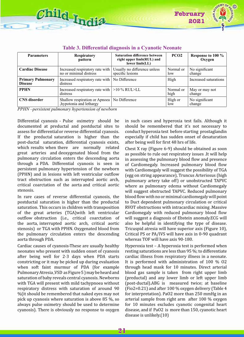
Table 3. Differential diagnosis in a Cyanotic Neonate Parameters Respiratory
patternSaturation difference between right upper limb(RUL) and
lower limb(LL)
PCO2 Response to 100 % Oxygen
Cardiac Disease Increased respiratory rate with no or minimal distress
Usually no difference unless specific lesions
Normal or low
No significant change
Primary Pulmonary Disease
Increased respiratory rate with distress
No Difference High Increased saturations
PPHN Increased respiratory rate with distress
>10 % RUL>LL Normal or high
May or may not change
CNS disorder Shallow respiration or Apnoea ,hypotonia and lethargy
No Difference High or low
No significant change
PPHN –persistent pulmonary hypertension of newborn
in such cases and hyperoxia test fails. Although it should be remembered that it’s not necessary to conduct hyperoxia test before starting prostaglandin especially if child has sudden onset of desaturation after being well for first 48 hrs of life. Chest X ray (Figure 6-9) should be obtained as soon as possible to rule out respiratory issues .It will help in assessing the pulmonary blood flow and presence of Cardiomegaly. Increased pulmonary blood flow with Cardiomegaly will suggest the possibility of TGA (egg on string appearance), Truncus Arteriosus (high pulmonary artery take off) or unobstructed TAPVC where as pulmonary edema without Cardiomegaly will suggest obstructed TAPVC. Reduced pulmonary blood flow with no or minimal cardiomegaly will point to Duct dependent pulmonary circulation or critical RVOT obstructions with intracardiac mixing .Massive Cardiomegaly with reduced pulmonary blood flow will suggest a diagnosis of Ebstein anomaly.ECG will also be helpful in identifying the type of disease. Tricuspid atresia will have superior axis (Figure 10), Critical PS or PA/IVS will have axis in 0-90 quadrant whereas TOF will have axis 90-180.Hyperoxia test – A hyperoxia test is performed when resting saturations are less than 95 %; to differentiate cardiac illness from respiratory illness in a neonate. It is performed with administration of 100 % O2 through head mask for 10 minutes. Direct arterial blood gas sample is taken from right upper limb (preductal) and any lower limb or left upper limb (post-ductal).ABG is measured twice; at baseline (Fio2=0.21) and after 100 % oxygen delivery (Table 4 for interpretation). PaO2 more than 250 mmHg in an arterial sample from right arm after 100 % oxygen for 10 minutes excludes cyanotic congenital heart disease, and if PaO2 is more than 150, cyanotic heart disease is unlikely.(10)
Differential cyanosis - Pulse oximetry should be documented at preductal and postductal sites to assess for differential or reverse differential cyanosis. If the preductal saturation is higher than the post-ductal saturation, differential cyanosis exists, which results when there are normally related great arteries and deoxygenated blood from the pulmonary circulation enters the descending aorta through a PDA. Differential cyanosis is seen in persistent pulmonary hypertension of the newborn (PPHN) and in lesions with left ventricular outflow tract obstruction such as interrupted aortic arch, critical coarctation of the aorta and critical aortic stenosis.In rare cases of reverse differential cyanosis, the postductal saturation is higher than the preductal saturation. This occurs in children with transposition of the great arteries (TGA)with left ventricular outflow obstruction (i.e., critical coarctation of the aorta, interrupted aortic arch, critical aortic stenosis) or TGA with PPHN. Oxygenated blood from the pulmonary circulation enters the descending aorta through PDA.Cardiac causes of cyanosis-These are usually healthy neonates who present with sudden onset of cyanosis after being well for 2-3 days when PDA starts constricting or it may be picked up during evaluation when soft faint murmur of PDA (for example Pulmonary Atresia ,VSD as Figure 5 ) may be heard and saturation of baby reveals central cyanosis. Newborns with TGA will present with mild tachypnoea without respiratory distress with saturation of around 90 %(it should be remembered that naked eyes may not pick up cyanosis where saturation is above 85 %, so always pulse oximetry should be used to determine cyanosis). There is obviously no response to oxygen
Vol. 1January 2018
Vol. 1January 2018
February 2021
22
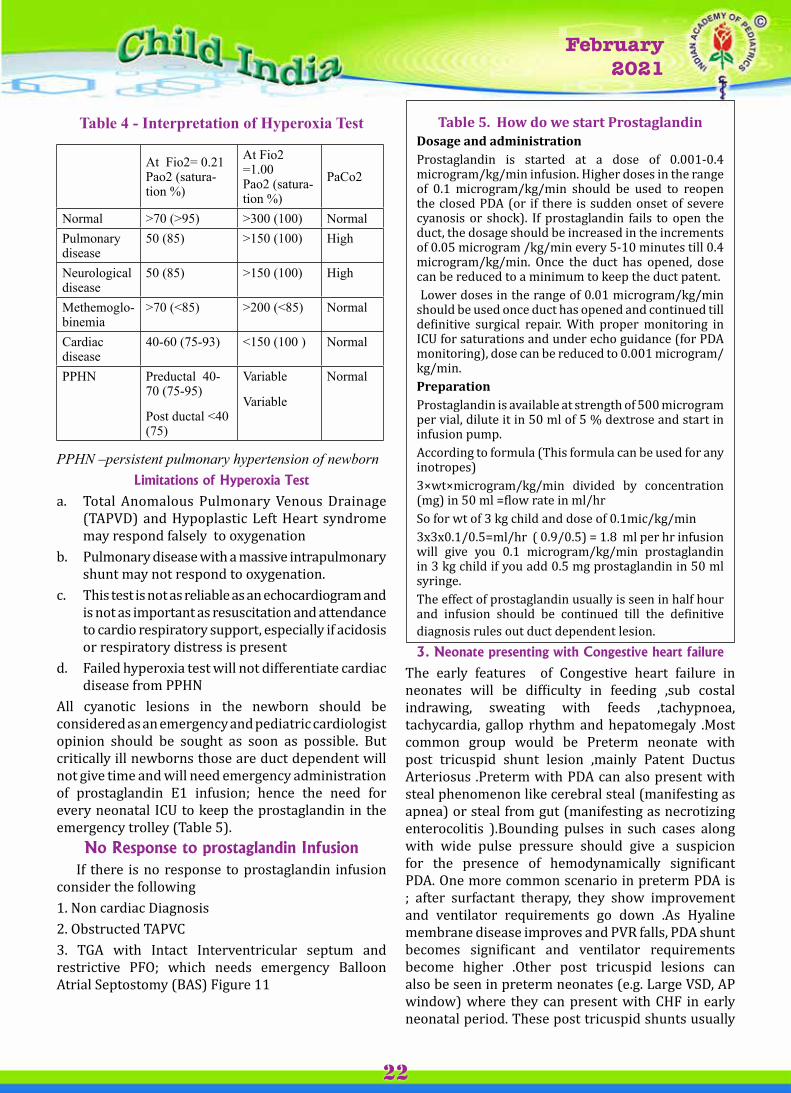
Table 4 - Interpretation of Hyperoxia Test
At Fio2= 0.21 Pao2 (satura-tion %)
At Fio2 =1.00 Pao2 (satura-tion %)
PaCo2
Normal >70 (>95) >300 (100) NormalPulmonary disease
50 (85) >150 (100) High
Neurological disease
50 (85) >150 (100) High
Methemoglo-binemia
>70 (<85) >200 (<85) Normal
Cardiac disease
40-60 (75-93) <150 (100 ) Normal
PPHN Preductal 40-70 (75-95)
Post ductal <40 (75)
Variable
Variable
Normal
PPHN –persistent pulmonary hypertension of newborn Limitations of Hyperoxia Test
a. Total Anomalous Pulmonary Venous Drainage (TAPVD) and Hypoplastic Left Heart syndrome may respond falsely to oxygenation
b. Pulmonary disease with a massive intrapulmonary shunt may not respond to oxygenation.
c. This test is not as reliable as an echocardiogram and is not as important as resuscitation and attendance to cardio respiratory support, especially if acidosis or respiratory distress is present
d. Failed hyperoxia test will not differentiate cardiac disease from PPHN
All cyanotic lesions in the newborn should be considered as an emergency and pediatric cardiologist opinion should be sought as soon as possible. But critically ill newborns those are duct dependent will not give time and will need emergency administration of prostaglandin E1 infusion; hence the need for every neonatal ICU to keep the prostaglandin in the emergency trolley (Table 5).
No Response to prostaglandin Infusion If there is no response to prostaglandin infusion consider the following1. Non cardiac Diagnosis2. Obstructed TAPVC3. TGA with Intact Interventricular septum and restrictive PFO; which needs emergency Balloon Atrial Septostomy (BAS) Figure 11
table 5. How do we start ProstaglandinDosage and administrationProstaglandin is started at a dose of 0.001-0.4 microgram/kg/min infusion. Higher doses in the range of 0.1 microgram/kg/min should be used to reopen the closed PDA (or if there is sudden onset of severe cyanosis or shock). If prostaglandin fails to open the duct, the dosage should be increased in the increments of 0.05 microgram /kg/min every 5-10 minutes till 0.4 microgram/kg/min. Once the duct has opened, dose can be reduced to a minimum to keep the duct patent. Lower doses in the range of 0.01 microgram/kg/min should be used once duct has opened and continued till definitive surgical repair. With proper monitoring in ICU for saturations and under echo guidance (for PDA monitoring), dose can be reduced to 0.001 microgram/kg/min.Preparation Prostaglandin is available at strength of 500 microgram per vial, dilute it in 50 ml of 5 % dextrose and start in infusion pump.According to formula (This formula can be used for any inotropes)3×wt×microgram/kg/min divided by conce ntration (mg) in 50 ml =flow rate in ml/hr So for wt of 3 kg child and dose of 0.1mic/kg/min3x3x0.1/0.5=ml/hr ( 0.9/0.5) = 1.8 ml per hr infusion will give you 0.1 microgram/kg/min prostaglandin in 3 kg child if you add 0.5 mg prostaglandin in 50 ml syringe.The effect of prostaglandin usually is seen in half hour and infusion should be continued till the definitive diagnosis rules out duct dependent lesion.3. Neonate presenting with Congestive heart failure
The early features of Congestive heart failure in neonates will be difficulty in feeding ,sub costal indrawing, sweating with feeds ,tachypnoea, tachycardia, gallop rhythm and hepatomegaly .Most common group would be Preterm neonate with post tricuspid shunt lesion ,mainly Patent Ductus Arteriosus .Preterm with PDA can also present with steal phenomenon like cerebral steal (manifesting as apnea) or steal from gut (manifesting as necrotizing enterocolitis ).Bounding pulses in such cases along with wide pulse pressure should give a suspicion for the presence of hemodynamically significant PDA. One more common scenario in preterm PDA is ; after surfactant therapy, they show improvement and ventilator requirements go down .As Hyaline membrane disease improves and PVR falls, PDA shunt becomes significant and ventilator requirements become higher .Other post tricuspid lesions can also be seen in preterm neonates (e.g. Large VSD, AP window) where they can present with CHF in early neonatal period. These post tricuspid shunts usually
Vol. 1January 2018
Vol. 1January 2018
February 2021
23
don’t present in full term neonates as PVR falls after 4-6 weeks and then shunt lesions start manifesting as CHF.Among other Acyanotic lesions with CHF are valvular regurgitant lesions like aortic regurgitation or pulmonary regurgitation (which may be secondary to vegetations of these valves also). Congenital mitral valve lesions with severe MR can also present in neonates with CHF. Diastolic murmur in AR or PR and Pan systolic murmur in MR will give a significant clue .Chest X ray in such cases will show Cardiomegaly of the respective chambers (cardiothoracic ratio >0.6).This is important to remember that thymic shadows and extra cardiac shadows can lead to over diagnosis of Cardiomegaly on chest X ray (Figure 12a,12b ,13).Among cyanotic heart diseases which present in neonatal life with CHF are those with significant high pulmonary blood flow like Truncus Arteriosus or single ventricular physiology without pulmonary stenosis. (11) An important point to remember in such cases; these neonates will not be significantly cyanotic due to torrential pulmonary blood flow and saturation may reach in 90’s %; so naked eyes will not pick up the cyanosis and pulse oximetry will help in the early detection of such congenital lesions.
4. Asymptomatic Newborn with murmur This group of neonates usually has mild forms of obstructive or regurgitant lesion. Obstructive lesions are mainly aortic stenosis or pulmonary stenosis .These obstructive lesions are characterized by presence of harsh murmur .TOF with absent pulmonary valve will present with to and fro murmur which is very classically heart in left 2nd intercostal space.Chest X-ray shows cardiomegaly at birth along with dilated proximal pulmonary arteries(Figure 14). Ebstein with TR will present with PSM at tricuspid area and multiple ejection clicks will confirm the diagnosis .Classical forms of Ebstein anomaly will have box shaped heart on CXR (Figure 15). Severe forms of Ebstein anomaly will present with cyanosis due to lack of forward flow across pulmonary valve and may need prostaglandin infusion for pulmonary circulation. Mitral regurgitation may be seen in isolation or with Atrioventricular canal defects with cleft mitral valve .These defects may present as asymptomatic or may present with CHF at 10-14 days of life with CHF if MR is severe.Another group of neonates presenting with murmur will be left to right shunt (post tricuspid lesions like VSD ,PDA ,AP window ) which are restrictive (small or moderate shunts) and usually present with a murmur when PVR falls (after the age of 2-4
weeks).Few cases of small defects may present with murmur in first week also .This group of neonates are usually asymptomatic in the neonatal period due to restrictive nature of shunts and start having symptoms beyond the neonatal period .This is important to mention in such cases ; that preterm neonates may have symptoms within neonatal period as in preterm PDA cases .Physiological peripheral pulmonary stenosis is a frequent finding in neonates and there may be a significant systolic murmur in such cases. Pediatric cardiologist must take care not to miss cases of syndromes associated with peripheral stenosis like Alagille, congenital rubella syndrome and William’s syndrome. Physiological peripheral pulmonary stenosis is reduced in two thirds of cases by 6 weeks of age and in most others by 6 months. The etiology of the murmur is secondary to both relative hypoplasia of the pulmonary artery branches and an associated angulation at their origin. This is important to understand that many duct dependent lesions in the immediate neonatal period may be totally asymptomatic till the PDA is open and minimal cyanosis or a murmur of PDA may be the only finding in such cases. These are the cases which may go undetected ,discharged and later come back in critical situation.(12) In such cases repeated and careful clinical examination, pulse oximetry along with chest x ray and ECG are helpful to assess the severity of lesion.
Summary Neonates with cardiac disease may present in one of the four groups- shock, cyanosis, CHF and asymptomatic .Differentiating from non cardiac causes is feasible; by bed side examination along with pulse oximetry, chest x ray, ECG and other basic investigations such as hyperoxia test. Also not all murmurs are due to heart disease and not all newborns without a murmur are free of CHD. Prostaglandin E1 may be started empirically in neonates with unexplained shock and also in failed hyperoxia test ; which can be life saving. Critical congenital heart lesions in neonates have a better outcome; if referred early for a surgical repair in today’s world even in developing countries.
References1. Warburton D, Rehan M, Shineboume EA. Selective
criteria for differential diagnosis of infants with symptoms of congenital heart disease. Arch Dis Child 1981; 56:94-100.
2. Kaplan JH, Ades AM, Rychik J. Effect of prenatal diagnosis on outcome in patient with congenital heart disease. Neo Reviews 2005; 6(7):e326–31.
Vol. 1January 2018
Vol. 1January 2018
February 2021
24
3. Ewer AK, Middleton LJ, Furmston AT, Bhoyar A, Daniels JP, Thangaratinam Set al. Pulse oximetry screening for congenital heart defects in newborn infants: a test accuracy study. Lancet.2011 Aug 27; 378(9793):785-94.
4. Abraham M.R. Congenital diseases of heart: clinical physiological considerations .3rd edition. West Sussex, England: John Wiley & Sons Ltd; 2009. Chapter 5, Prenatal and postnatal pulmonary circulation; P 87-114.
5. Mc Connell ME, Elixson EM. The neonate with suspected congenital heart disease.Crit Care Nurs Q. 2002 Nov; 25(3):17-25.
6. Fleiner S. Recognition and stabilization of neonates with congenital heart disease. Newborn Infant Nurs Rev 2006; 6(3):137–50.
7. Yun SW.Congenital heart disease in newborn requiring early intervention. Korean J Pediatr. 2011 May; 54(5):183-91.
8. Penny DJ, Shekerdemian LS. Management of the neonate with symptomatic congenital heart disease. Arch Dis Child Fetal Neonatal Ed 2001; 84: F141 - F145.
9. Krishnan US. Approach to congenital heart disease in the Neonate. Indian J Pediatr 2002; 69:501-5.
10. Marino S.M., Wernovsky G. Preoperative Care. In: Chang AC, Hanley FL, Wernovsky G, Wessel DL, editors. Pediatric Cardiac Intensive care. Philadelphia: Lippincott Williams and Wilkins; 1998.P 154-157.
11. Marino BS, Bird GL,Wernovsky G. Diagnosis and management of the newborn with suspected congenital heart disease. Clinics in Perinatology. Vol 28(1) .W. B. Saunders, Philadelphia, PA.; 2001 March: 91-136.
12. Mellander M, Sunnegardh J. Failure to diagnose critical heart malformations in newborns before discharge--an increasing problem? Acta Paediatr. 2006 Apr; 95(4):407-13.
Figure 1a. Chest X ray of a neonate with Situs solitus, dextrocardia where high possibility of a complex cardiac lesion should be kept
Figure 1b.Situs inversus , dextrocardia. Note the apex of heart and gastric bubble on right side and liver on left side. Here the possibility of CHD is comparable to a normal sided heart.
Figure 1c. Situs ambiguous, levocardia: Note the midline liver and gastric shadow on right side .This group of patients has high probability of complex heart disease.
Figure 2. Schematic description of a Interrupted aortic arch where there is no forward flow from arch into the aorta.PDA supplies blood from pulmonary artery to Descending aorta which is a deoxygenated blood explaining the lower limb desaturation (Arrows depict direction of blood flow). If PDA closes in this case, there will be no blood supply in the descending aorta and patient presents with shock.
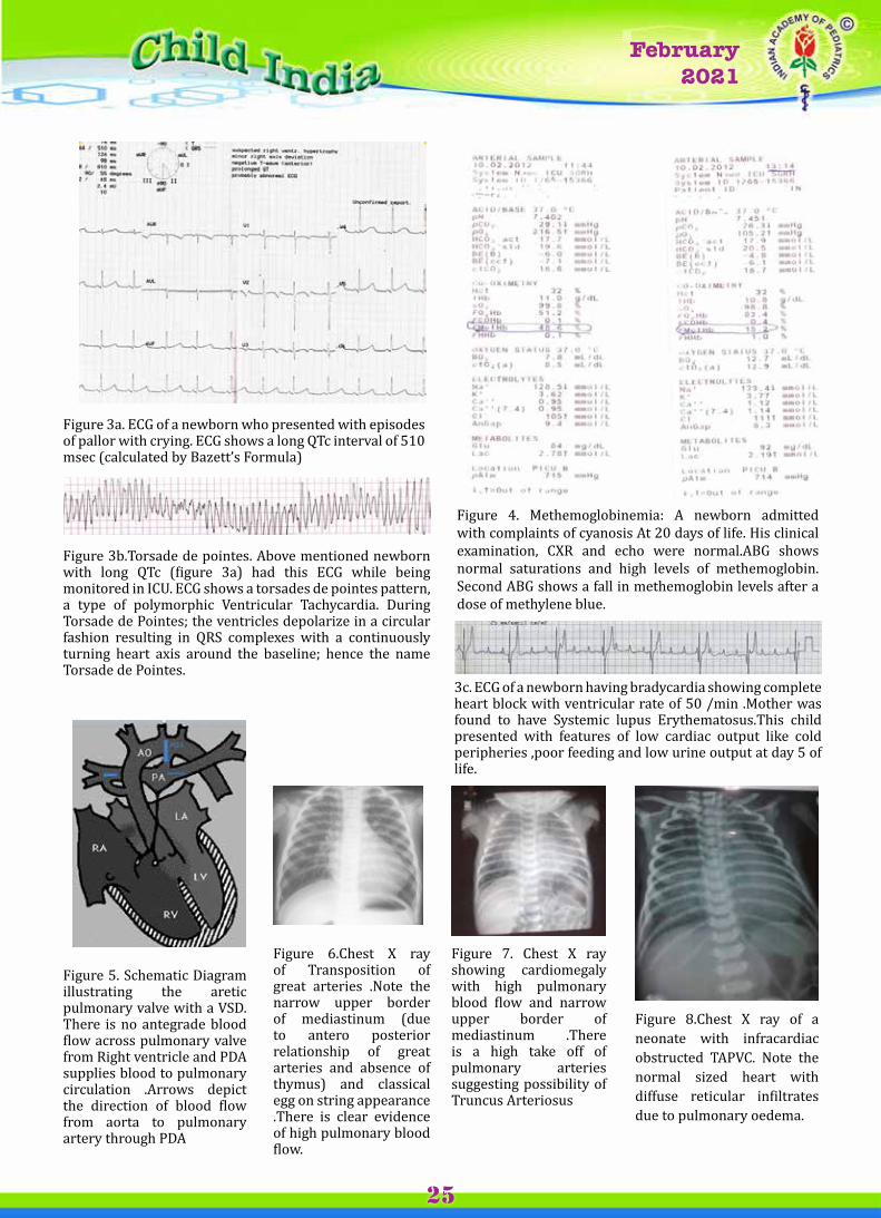
Vol. 1January 2018
Vol. 1January 2018
February 2021
25
Figure 3a. ECG of a newborn who presented with episodes of pallor with crying. ECG shows a long QTc interval of 510 msec (calculated by Bazett’s Formula)
Figure 3b.Torsade de pointes. Above mentioned newborn with long QTc (figure 3a) had this ECG while being monitored in ICU. ECG shows a torsades de pointes pattern, a type of polymorphic Ventricular Tachycardia. During Torsade de Pointes; the ventricles depolarize in a circular fashion resulting in QRS complexes with a continuously turning heart axis around the baseline; hence the name Torsade de Pointes.
3c. ECG of a newborn having bradycardia showing complete heart block with ventricular rate of 50 /min .Mother was found to have Systemic lupus Erythematosus.This child presented with features of low cardiac output like cold peripheries ,poor feeding and low urine output at day 5 of life.
Figure 4. Methemoglobinemia: A newborn admitted with complaints of cyanosis At 20 days of life. His clinical examination, CXR and echo were normal.ABG shows normal saturations and high levels of methemoglobin. Second ABG shows a fall in methemoglobin levels after a dose of methylene blue.
Figure 5. Schematic Diagram illustrating the aretic pulmonary valve with a VSD. There is no antegrade blood flow across pulmonary valve from Right ventricle and PDA supplies blood to pulmonary circulation .Arrows depict the direction of blood flow from aorta to pulmonary artery through PDA
Figure 7. Chest X ray showing cardiomegaly with high pulmonary blood flow and narrow upper border of mediastinum .There is a high take off of pulmonary arteries suggesting possibility of Truncus Arteriosus
Figure 8.Chest X ray of a neonate with infracardiac obstructed TAPVC. Note the normal sized heart with diffuse reticular infiltrates due to pulmonary oedema.
Figure 6.Chest X ray of Transposition of great arteries .Note the narrow upper border of mediastinum (due to antero posterior relationship of great arteries and absence of thymus) and classical egg on string appearance .There is clear evidence of high pulmonary blood flow.
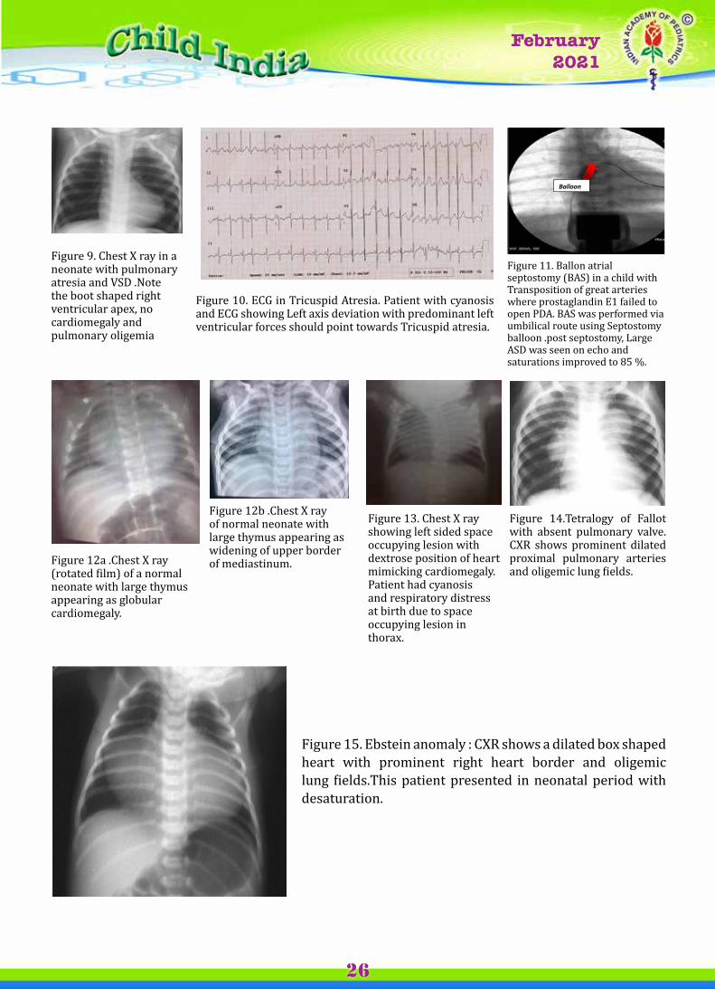
Vol. 1January 2018
Vol. 1January 2018
February 2021
26
Figure 9. Chest X ray in a neonate with pulmonary atresia and VSD .Note the boot shaped right ventricular apex, no cardiomegaly and pulmonary oligemia
Figure 11. Ballon atrial septostomy (BAS) in a child with Transposition of great arteries where prostaglandin E1 failed to open PDA. BAS was performed via umbilical route using Septostomy balloon .post septostomy, Large ASD was seen on echo and saturations improved to 85 %.
Figure 10. ECG in Tricuspid Atresia. Patient with cyanosis and ECG showing Left axis deviation with predominant left ventricular forces should point towards Tricuspid atresia.
Balloon n
Figure 12a .Chest X ray (rotated film) of a normal neonate with large thymus appearing as globular cardiomegaly.
Figure 13. Chest X ray showing left sided space occupying lesion with dextrose position of heart mimicking cardiomegaly. Patient had cyanosis and respiratory distress at birth due to space occupying lesion in thorax.
Figure 14.Tetralogy of Fallot with absent pulmonary valve.CXR shows prominent dilated proximal pulmonary arteries and oligemic lung fields.
Figure 12b .Chest X ray of normal neonate with large thymus appearing as widening of upper border of mediastinum.
Figure 15. Ebstein anomaly : CXR shows a dilated box shaped heart with prominent right heart border and oligemic lung fields.This patient presented in neonatal period with desaturation.
Vol. 1January 2018
Vol. 1January 2018
February 2021
27
The IAP Drug Formulary (IAP DF), first published in 2004-2005, has completed 16 yrs of service to the pediatric specialists in our country and abroad, being the only exclusively pediatric formulary in the world, other than the British National Formulary for Children (BNFc). The IAP DF contains recommendations for drug therapy for & gt; 510 pediatric ailments and detailed, well researched information for 669 Drugs in 231 Drug Classes, that are licensed for use in neonates, children and adolescents.
The IAP DF hard copy textbooks are printed once in 3 - 4 years as after 12-16 web updates, the text would have changed drastically and the IAP Sub Chapters submit latest updates of their respective chapters. The 5th Edition - The IAP Drug Formulary 2019 - part of IAP President’s Action Plan for 2018 - was released by IAP President Dr Santosh Soans, at the IAP CMIC National Conference and IAP WB State Annual at Kolkata on Dec 8/9th 2018. It has sold nearly 8750 copies since Jan 2019.
The December Web Update 2020(3) will be the Edition 58, the 56th web update of the IAP Drug Formulary. The Web Updates are a quarterly exercise where new drugs, changes in use of drugs and new recommendations for drug therapy of pediatric illnesses are updated as and when they are available. This has made the formulary a dynamic and up-to-date reference for pediatric therapeutics.
The IAP DF, in its 17th year required a complete overhaul as it was becoming difficult for our admin to update charts, figures, flow charts and even formatting of text on the old platform. We therefore spent 6.5 lakh last year to create The
IAP Drug FormularyDr Jeeson C Unni
IAP Drug Formulary 2.0. The updates include the following –
Web Portal1) New Design Layout
2) New user Profile Login
a) Using registered Email ID and OTP Authen-tication. Therefore not requiring the user to remember thier password
b) New User Profile Page displaying the list of IAP DF subscriptions along with the respective App Keys and Profile Info.
3) New Admin Pages
a) To moderate all the content of the website. Hence making all the content of the website dynamic.
b) Pre Release preview of all update content before release.
c) Single Point release for all IAP DF Platforms.
d) Advanced User management to Create /Update Users and Manage Subscriptions. Email Feature, which will directly send an Invitation email to subscribers with the relevant App key information. No need for pre-generated App keys anymore.
4) Drug Formulary
a) Refined Search Module along with display of tables and Images
b) Hyper Link to Brands on selection of Generics.
Vol. 1January 2018
Vol. 1January 2018
February 2021
28
5) No Need for cD’s: the users can directly download the Windows Desktop App (If subscribed to) through their Profile Page.
Android and IOS App1) New Apps created on a new platform with
the following Features
a) Login using App Key/Registered Email ID and through email OTP.
b) OFFLINE APP: Unlike the earlier version, the new Apps will work off line. A new Sync Module will download the content on the device for viewing off line.
c) New Feature of viewing Editors, IAP President, Hon. Gen. Secretary, IAP CMIC Chairperson and Secretary’s message along with photographs can be viewed.
d) Contributors List will also be viewable.
e) The Home page has the editors email id and mobile no which can be directly dialled by clicking on the number
f) Advanced Notification feature which will alert the users of any new notifications or updates. The App will auto prompt the users to download whenever there are new updates released.
g) Advanced Drug Formulary
-- Search and view of content, be it in text, tables or in image form.
-- Pinch Zoom in feature to expand any content.
-- Hyperlink to Brands page for the respective Generics.
Windows Desktop App
1) Built on a completely new platform compatible with Windows 7 and above.
2) Easy Installation process wherein the users just need to download a Single Installable EXE file and Login using their Registered Credentials and OTP.
3) The New platform enables search and view of content, be it in text, tables or in image form.
4) Auto Update (No Update Patches required)” Quarterly Updates will be a simple Update Link within the software, wherein the software identifies any updates and automatically updates the new version.
All users need to therefore uninstall their mob apps and re-install the new desktop and mobile versions. The highlight of the update is that the user can, after synchronising the app, scan the contents of the IAP DF offline and the web updates will be automatically updated.
We hope that this year onwards we will be able to offer 1 free mob app key to all IAP members (till now our MOU with IAP CO offered this facility to all members who joined the Academy on or before Dec 31st 2015).
Vol. 1January 2018
Vol. 1January 2018
February 2021
29
As I look back as the last few days, the lasting image is of a family that came together after a near calamity. It was like a dream come true...It was a wonderful experience to welcome all of you to Mumbai.. the city that never sleeps.. city of dreams...for the CIAP PEDICON from the 3rd to the 7th of February 2021! This was a unique Pedicon that made history for several reasons, for being the first hybrid pedicon, for being the most attended, for being organised with consideration of many Government restrictions, and being organised in the shortest time frame of two months! This was the PEDICON of Dr Piyush Gupta where we saw the most loveable side of his immense personality, wit and wisdom in a lovely tete-a-tete during the Presidential Dinner. This was the PEDICON of Dr Bakul Parekh who temporarily deleted the word ‘impossible’ from the dictionary and always remained a step ahead of challenges. This was the PEDICON of the powerhouse of energy, and confidence, Dr Basavaraj who never said ‘No’ to any request. This was the Pedicon of Dr Remesh who supported the team with his ever ready cooperation and spirit. This was the Pedicon of a vibrant team of Mumbai Navi, Mumbai and Thane led
Kabhi Alvida Na Kehna! Roads change, the journey goes on.
Dr Samir H Dalwai
Developmental Pediatrician, Jt Secretary (Admin) & chair - Local Organising committee
cIAP PEDIcON 2021
by Dr Bela Verma, Dr Bipin Shah, Dr Rajesh Chokhani, Dr Jeetendra and Dr Parmanand who braved all odds for the love of IAP. This was the PEDICON of illuminating science that bridged the gap between two eras in medicine- the pre and post Covid periods- a scientific program helmed by Dr Nitin Shah, Dr Vijay Yewale and the indefatigable Dr Dheeraj Shah. This was the PEDICON of the CIAP office and dIAP team who suspended the difference between day and night and toiled relentlessly for a program that ran like clockwork. Above all, this was the PEDICON of the 600 Bravehearts, the common but extraordinary IAPians, who bested all fears, all restrictions all challenges and took that train or boarded that flight to make PEDICON what IAP intended it to be - the annual feast of camaraderie and fellowship of dedicated members of the Academy. We are grateful to all the above, in addition to all our Honourable Past Presidents, especially Dr Y K Amdekar for his inspiring speech as the Chief Guest, Dr CP Bansal for his unstinted support from inception to the CIAP PEDICON, Dr Deepak Ugra for the interview with Gulzar Sahab, Dr S S Kamath for the NRP meeting, Dr M R Lokeshwar, Dr Rohit Agarwal, Dr Pramod
Vol. 1January 2018
Vol. 1January 2018
February 2021
30
Jog, Dr Santosh Soans, Dr Digant Shastri for gracing the occasion with their presence! We are grateful to the national Vice Presidents, Dr Sangita Yadav, Dr Jayant Upadhye, Dr Ravikumar, Dr Atanu Bhadra and Dr Major Nagaraju for their unflinching support and to our amiable Treasurer Dr Purna Kurkure and our unassuming Joint Secretary (Liaison) Dr Hemant Pemde for their academic support, the FIAP awardees who graced the occasion in their ceremonial robes and the various State/City branches and Chapter Award winners who displayed unbridled bonhomie.
We are grateful to the members of the Executive Board of 2021 for their trust and confidence in us and to the Office Bearers and Executive Board of 2020 for having supported this event with all their might.
We are grateful to the Government of India, especially the Honourable Minister of Heath Dr Harasvardhan ji for a tremendously impactful statement, as well as WHO and UNICEF. We are happy we could bring a little cheer and you could enjoy the evenings with the Presidential Dinner, an outstanding internationally reputed DJ- DJ Zulfi and a Bollywood night of swinging melodies from the Golden Era with Shri Ajay Madan and Shri Suraj Sathe. I personally enjoyed sharing these lovely evenings with all of you which brought some cheer after the solitude of the past many months.
Which is why we wanted to make this PEDICON special for each one of you. Our team took special care to select the most safe and secure and yet an awesome venue. We knew you would miss home food being at home for so long, hence our team took special care to ensure excellent meals. Our partners were encouraged to go the extra mile to ensure excellent hospitality commencing from the airport pickup and drop for almost everyone. Ultra smooth check ins and comfortable stays. No ruckus, pure science and excellent entertainment without any chaos.
All this comes at a cost. IAP may have suffered huge losses to have such a grand PEDICON in these times- some may fear. We are very happy to share that against all odds and expectations, the conference has in fact saved more than Rs 1 crore and since this is a CIAP PEDICON, this will all go to CIAP kitty All thanks to the tremendous name and goodwill enjoyed by IAP, and also the microscopic attention to details and budgets given by the committee to save each and every rupee.
As the sands of time flow through the watch glass of life, we look forward to a great year of academics, fellowship and camaraderie in person as well as online.
Thank you once again for making this PEDICON such a happy occasion for us! We wish you and your family a year of happiness , success and good heal th .
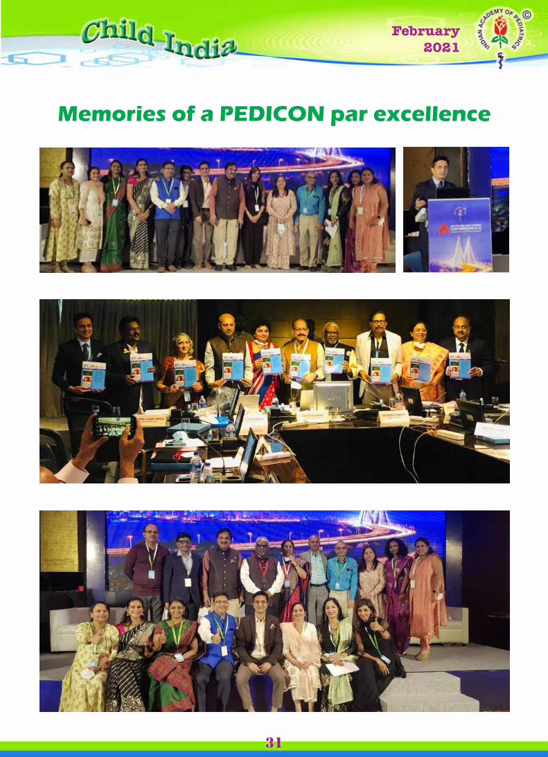
Vol. 1January 2018
Vol. 1January 2018
February 2021
31
Memories of a PEDICON par excellence