Embed Size (px)
Citation preview

UNIFORMED SERVICES UNIVERSITY OF THE HEALTH SCIENCESF. EDWARD HEBERT SCHOOL OF MEDICINE
4301 JONES BRIDGE ROADBETHESD~MARYLAND 20814-4799
(iRAOr.;.-\TE EOUC'ATlO:-.J1,011 2'1)-J'jll
fAX 000:-1'-<.772
APPROVAL SHEET
Title of Dissertation: "Mutations in the Histone-like Nucleoid Structuring Regulatory Gene(hns) Decrease the Adherence of Shiga Toxin-producing Escherichia coli 091 :H21 Strain 82FIto Human Colonic Epithelial Cells and Increase the Production of Hemolysin"
Name of Candidate: Maria ScottDoctor ofPhilosophy Degree19 October 1999
Dissertation and Abstract Approved:
Date
fDate )
Paul Rick~ Ph.D.Department of Microbiology and ImmunologyCqIlJ,fIliaee Chairperso~ /')}t: r ' -\ . . •
~ '",)=ti.d ( ~iVJl,,-"-
Alison O'Brien~ Ph.D.Department of Microbiology and Immunology
Committe,,:~~/!f" 1/ e: I JL~VV;.7 ':~"U.
AmI Jerse. Ph::p.Departmentg[ Microbiology and ImmunologyCommittee Member
·,J1/UYi 7:/nf1.~Teresa Dunn. Ph.D.Department of BiochemistryCommittee Member
!c;-/r·91Date
Date

The author hereby certifies that the use ofany copyrighted material in the thesismanuscript entitled:
'-Mutations in the Histone-like Nucleoid Structuring Regulatory Gene (hns)Decrease the Adherence of Shiga Toxin-producing Escherichia coli 091 :H21
Strain B2F I to Human Colonic Epithelial Cellsand Increase the Production of Hemolysin"
beyond brief excerpts is with the permission of the copyright owner, and will save andhold harmless the Uniformed Services University of the Health Sciences from anydamage which may arise from such copyright violations.
Maria ScottDepartment of Microbiology and ImmunologyUniformed Services University of the Health Sciences
ii

Abstract
Title of Dissertation:
Mutations in the Histone-like Nucleoid Structuring Regulatory Gene (hns) Decrease the
Adherence of Shiga Toxin-producing Escherichia coli 091 :H21 Strain B2F1 to Human
Colonic Epithelial Cells and Increase the Production of Hemolysin
Maria E. Scott
Candidate, Doctor of Philosophy, 1999
Dissertation directed by:
Alison D. O'Brien. Ph.D.Professor and Chairman of Department of Microbiology and Immunology
Non-O157 serotype Shiga toxin-producing E. coli (STEC) cause a significant amount
of food-borne hemorrhagic colitis (HC) and life-threatening hemolytic uremic syndrome
(HUS) worldwide and yet these strains do not encode intimin, a major adherence factor
encoded by the most common serotype 0157:H7 isolated from documented cases ofHC and
HUS. In this investigation, we attempted to isolate specific adhesins critical for attachment
ofintimin-negative STEC 091 :H21 to human colonic epithelial cells. Transposon mutagenesis
of B2F 1 was accomplished with the mini-Tn5phoACm f mobile element and a mutant bank
of B2F 1 colonies that carried putative in-frame PhoA-positive transposon insenions was
isolated. The bank was screened for mutants unable to adhere to the human colonic epithelial
III

T84 cell line in both its nonpolarized and polarized states. An adherence mutant designated
as 34.7 was identified that did not bind to these cells. Subsequently, mutant 34.7 was found
to contain an out-of-frame insertion of the transposon into hns. The hns gene encodes the
H-NS protein that has been reported to modulate the expression ofvarious housekeeping as
well as virulence-associated genes encoded by E. coli and other gram-negative organisms.
Disruption ofhns in 34.7 not only caused reduced adherence to T84 cells but resulted in over
production of STEC-hemolysin encoded on the large -90 kb plasmid carried by this strain.
When H-NS was supplied to 34.7 in trans on a moderate copy number plasmid. both the
reduced adherence phenotype and the hyper-hemolytic phenotype reverted to wild-type.
Because hemolysin can destroy the T84 monolayer over the course of the adherence assay, it
was difficult to assess the direct effect ofthe hns mutation on adherence ofB2FI to T84 cells.
Hence. we sought to separate the reduced adherence phenotype from the hyper-hemolytic
phenotype exhibited by mutant 34.7. To accomplish this, an attempt was made to inactivate
hns in the plasmid cured derivative of B2F I. strain S II. The plasmid- cured strain attached
to T84 cells with a wild-type adherence phenotype. The hns gene encoded by S11 was
partially inactivated by disruption ofhns by insertion ofa suicide plasmid. The resultant hns
mutant. S 11366. I5, was unable to bind to T84 cells and the reduced adherence phenotype was
complemented in trans with wild-type H-NS expressed from a low copy number plasmid.
Hence. mutations in hns that ablated or reduced the activity of H-NS directly affected the
capacity of B2F I or its plasmid-cured derivative, S11, to adhere to T84 cells. These data
suggests that, in B2Ft, H-NS may coordinately regulate the expression ofadherence factors
and the production of hemolysin.
iv

Mutations in the Histone-like Nucleoid Structuring Regulatory
Gene (hns) Decrease the Adherence of Shiga Toxin-producing
Escherichia coli 091:821 Strain 82Fl to Human
Colonic Epithelial Cells and Increase
the Production of Hemolysin
by
Maria Elizabetb Scott
Dissertation submitted to tbe Faculty of tbeDepartment of Microbiology and Immunology Graduate Program
of tbe Uniformed Services University of tbe HealtbSciences in partial fulfillment of tbe
requirements for tbe degree ofDoctor of Pbilosopby 1999
v

DedicatedTo Dr. Henry Wu
Your intelligence was wrapped in wannthand the umbrella ofyour kindness will be felt forever.
I will miss you and never forget you.
vi

Acknowledgements
To my family-You have always believed I would succeed even when I didn't believe it. You
helped me to see my strengths and accept my weaknesses. I thank you Joe for understanding
why I needed to pursue this dream and for helping me make it come true. AJex you have been
a constant source ofjoy and helped me to laugh when I didn't think I could. You both have
been a profound source of happiness throughout this endeavor.
To Alison-You taught me that bacteria are more fun than viruses. Your patience has taught
me patience. Your critical and in-depth approach to each new scientific problem has unveiled
a power in you unsurpassed by few. Your passion for microbial pathogenesis has become my
new found passion in life. I thank you from the bottom of my heart.
To the members of my committee-Drs. Paul Rick, Ann Jerse, Anthony Maurelli, and Teresa
Dunn. the many long hours you have spent with me on my behalfdid not fallon deafears. I
will use each lesson you gave me to attempt to represent you well in my chosen profession.
To my Jab mates-You are all my second family. I especially thank Clare and Angela for
lending me a light when I was scientifically in the dark, Karen for making me laugh so much..
Melody for all those hugs, and to the rest of you for making my experience at USUHS
interesting, fulfilling, and unforgettable.
vii

Table of Contents
Introduction
l. Preface I
II. Purpose and overview 1
III. Overview ofenteric pathogenic Escherichia coli implicated in
diarrheaJ disease . . . . . . . . . . . . . . . . . . . . . . . . . . . . . . . . . . . . . . . . . . . . . 2
A. Enterotoxigenic £ coli (ETEC) 3
B. Enteroinvasive E. coli (EffiC) 3
C. Enteroadherent E. coli: enteroaggregative (EaggEC) 4
D. Enteroadherent E. coli: diffusely adherent (DAEC) 4
E. Enteropathogenic E. coli 4
1. Characteristics of EPEC 4
2. Relationship between AlE lesion fonnation and the LEE
pathogenicity island 5
F. Enterohemorrhagic E. coli (EHEC), a subset ofSTEC 10
1. Discovery of STEC 10
2. Geographic distribution ofSTEC 11
3. ClonaJly divergent subgroups ofSTEC 12
4. Vehicles of transmission ofSTEC 13
5. Illness associated with STEC Infections 14
viii

6. Treatment of HUS 15
7. Isolation and detection of STEC organisms 16
8. Major virulence properties associated with STEC . . . . . . . . . .. 18
a. Shiga toxins.. major virulence factors
encoded by STEC . . . . . . . . . . . . . . . . . . . . . . . . . 18
b. LEE pathogenicity island encoded by STEC 20
c. Adherence ofSTEC groups I and 2 that do
not encode the LEE: subject of
this project 22
9. The -90 kb plasmid encoded by E. coli 0157:H7 and
other STEC and its putative role in pathogenesis 23
a. Description of the -90 kb plasmid 24
b. Hemolysin expressed by STEC 24
c. E. coli 0157:H7 tyPe II secretion system 29
d. Bifunctional catalase-peroxidase (KatP) 29
e. Serine protease EspP 29
f. Distribution of the plasmid-encoded putative
virulence traits among STEC 30
g. Description of probe CVD419 31
IV. The histone-like-nucleoid-structuring protein (H-NS) 31
A. Discovery of H-NS and the types ofsystems regulated 31
ix

B. Proposed mechanisms by which H-NS
modulates gene expression 38
C. Structure-function analyses of H-NS 39
D. Comparison of the structure and function of H-NS
and H-NS-like proteins encoded by other
gram-negative bacteria 41
V. Specific Aims 42
Materials and Methods 43
Cell lines~ bacterial strains., and plasmids 43
Media enzyme.. and biochemicals 43
Qualitative adherence assay 51
Fluorescence actin staining assay (FAS) 52
Q .. dh -.,uantllatlve a erence assay )_
Isolation of plasmids 53
Isolation of total DNA 53
Recombinant DNA methods 54
Southern blot analysis 54
Colony blot analysis 55
DNA sequencing 56
Generalized transduction of B2Ft with PI ::Tn9 bacteriophage 56
Description of the mini-Tn5phoACmr transposon and the delivery
x

system utilized for mutagenesis 57
Procedure for transfer ofmini-Tn5phoACmT by conjugal mating 60
Assay for extracellular hemolytic activity 61
Detection of hemolysis on blood agar plates 62
Preparation of bacterial lysates for hemolysin detection by western
blot analysis ., 63
Vero cell cytotoxicity assay 64
Evaluation of motility of mutants in semi-solid agar 64
Methods used to compare membrane barrier function ofwild-type B2F I and
mutants of B2F1 65
Results 66
I. Absence of the LEE in STEC prototype strain B2F I 66
II. In vitro model ofadherence ofSTEC to human colonic
epithelial cells 69
Human colonic epithelial cell line used to optimize the in vitro
adherence assay 69
EHEC and STEC strains used to optimize the adherence assay 74
Qualitative analysis of STEC adherence to T84 cells 77
Phenotypic comparison of the capacity ofB2FI to adhere to
polarized and non-polarized T84 cells 85
Quantitative analysis of B2Ft adherence to T84 cells 88
xi

III. The role of the large -90 kb plasmid in the adherence to
human colonic epithelial T84 cells by STEC B2F1 96
Transformation ofa non-adherent laboratory E. coli strain DHI OB
with the large plasmid isolated from an isogenic mutant ofB2FI ..... 96
Capacity ofa plasmid-cured derivative of B2F I to bind
to human colonic epithelial T84 cells 99
IV. Attempt to construct by PI transduction an
alkaline phosphatase null mutation in B2F1 104
V. Construction and screening of the mini-Tn5phoACmr mutant
bank prepared from the wild-type strain B2F1 109
Isolation of mini-Tn5phoACmr mutants 109
Isolation of three adherence-deficient mutants
ofSTEC091:H2LstrainB2Fl 110
Characterization ofadherence mutants 120
VI. Restriction mapping of the mini-Tn5phoACmr insertion present
in the adherence mutants 34.3. 34.7 and 30. lOa 127
Southern hybridization analysis of the large plasmid encoded by
the adherence mutants for the presence ofTnphoA insertion 132
VII. Strategy used to clone the mini-Tn5phoACmr disrupted
gene from strain 34.7 135
VIII. Identification of the gene that contained the transPOson insertion 138
xii

IX. The site of insertion of the transposon within the hns gene 145
X. Sequence analysis ofhns encoded by 34.7.. an hns knockout
adherence-deficient mutant of B2F I 145
XI. Isolation of the wild-type hns gene from B2F1 146
XII. Analysis of Shiga-toxin and STEC hemolysin production in an
hns negative adherence mutant strain 34.7 153
Shiga toxin production by hns mutant 34.7 . . . . . . . . . . . . . . . . . . . . . . . . . . .. 153
Titer of STEC hemolysin secreted by the hns mutant and
wild-lyJ>e B2F I 158
Comparison of the level ofSTEC hemolysin produced by 34.7 and
wild-type, B2FI by SDS-PAGE and Western blot analysis 159
XIII. H-NS regulates attachment ofSTEC 091 :H21 to T84 cells
and hemolysin production 164
XIV. Construction ofan hns mutation in the hemolysin-negative
strain.,SII 167
Partial inactivation ofhns following insertion ofa suicide plasmid 180
XV. The effects of partial inactivation of H-NS in strain S11 194
Presence ofH-NS restores the wild-type adherence phenotype
and the regulation ofalkaline phosphatase to
mutant S11366. I 5 203
xiii

Discussion . . . . . . . . . . . . . . . . . . . . . . . . . . . . . . . . . . . . . . . . . . . . . . . . . .. 216
1. Adherence of STEC to human colonic epithelial cells in culture 215
II. Comparison of important characteristics exhibited by adherence
mutants ofstrain B2F1 218
III. Implications of the current investigation and further
studies warranted 222
Bibliography . . . . . . . . . . . . . . . . . . . . . . . . . . . . . . . . . . . . .. 228
xiv

Table I.
Table 2.
Table 3.
Table 4.
Table 5.
Table 6.
Table 7.
List of Tables
Examples ofgenes repressed by H-NS 35
E. coli strains used in this study •••••••••••••••••••.••••••••••.• 45
Plasmids used in this study .•••••.•••••••••••.•••••••••••••••• 48
Characteristics ofbacterial strains used to optimize
the in vitro model ofadherence to T84
cells by STEC organisms •••••••••••••••••••••••.••••••• 76
Results ofconjugal matings for transfer of mini-Tn5phoACmr
to create a mutant bank ofSTEC 091 :H21 strain B2Ft •••••• III
Characteristics ofadherence mutants 34.3. 34.7. and
30.IOa compared to the parental strain B2F1 .••••••••••••• 121
Motility of parental strains B2F1 and S 11 compared
to isogenic mutants grown on semisolid agar at 30°C •••••••••••••• 202
xv

Figure 1.
Figure 2.
Figure 3.
Figure 4.
Figure 5.
Figure 6.
Figure 7.
Figure 8.
Figure 9.
List of Figures
Map ofthe locus ofenterocyte effacement (LEE) 7
The STEC hemolysin operon including a representation of
maturation and export by E. coli 26
Multiple sequence alignment of H-NS homologues 32
Schematic of plasmid pAM400 that carries the mini-Tn5phoACmr
mobile element 58
Restriction fragments derived from the EPEC 35 kb Locus
of Enterocyte Effacement (LEE) that constituted the DNA probes
used for colony blot hybridization of wild-type B2FI 67
Colony blot hybridization ofSTEC strain B2F1 with restricted
DNA fragments derived from the locus ofenterocyte effacement
(LEE) used as probes A and B 70
Colony blot hybridization ofSTEC strain B2F1 with restricted
DNA fragments derived from the locus ofenterocyte effacement
(LEE) used as probes C and D 72
Phase contrast and fluorescent micrographs ofHEp-2 cells
after a six hour incubation with B2FI and 86-24 78
Non-confluent human colonic epithelial T84 cells five hours
XVI

Figure 10.
Figure II.
Figure 12.
Figure 13.
Figure 14.
Figure IS.
Figure 16.
Figure ~ 7.
post-inoculation with B2FI, and H30. or DH5a 80
Confluent T84 cells infected for five hours with B2FI
and intirnin-positive STEC 83
Polarized and non-polarized T84 cells inoculated with B2F1,
86-24 or DH5a and incubated for 5 hours 86
Quantitative adherence assay comparing the amounts of B2F1
bacteria and E. coli K-12 strain DH5a attached to non-polarized
T84 cells 89
Comparison of the number of intirnin-encoding STEC
(86-24 and H30) and intirnin-negative B2Ft adherent to
non-polarized T84 cells three and five hours post-infection 92
Quantitative adherence assay that compared the number of
B2F I bacteria adherent to polarized T84 cells versus the poorly
adherent E.coli K-12 strain DH5a 94
Schematic of the in vitro adherence assay used to evaluate
attachment of STEC strains and mini-Tn5phoACmr mutants 97
E. coli DHIOB(p2B3) transfonnants tested by colony blot
hybridization with probe CVD419 . . . . . . . . . . . . . . . . . . . . . . . . . . . . . 100
Human colonic epithelial T84 cells five hours post-infection
with strain 2B3, B2F I, transfonnant DH1OB(p2B3) and the
parental strain DH lOB 102
xvii

Figure 18.
Figure 19.
Figure 20.
Figure 21.
Figure 22.
Figure 23.
Figure 24.
Figure 25.
Colony hybridization of the plasmid..cured derivative of
82F1. designated as S 11 with probe CVD419 105
Comparison of the adherence of wild-type 82F1 and its plasmid-
cured derivative S II to T84 cells five hours after infection 107
Appearance of mini-Tn5phoACmr mutants designated as 34.3~
34.7 and 30.IOa and parental strain 82FI grown on XP agar 112
Growth rates of three adherence mutants,!, 34.3~ 34.7 and
30. lOa. compared with Parental strain 82FI grown in
Kunon tissue culture adherence assay media at 37DC 114
Capacity ofmini-Tn5phoACmr mutants 30. lOa. 34.3.34.7
to adhere to T84 cells after 5 hours of incubation as compared
to wild-type B2F I 116
The number of bacteria adherent to polarized T84 cells after
three hours of incubation determined for three mini-Tn5phoACm f
mutants. 34.3", 34.7 and 30.l0a and compared to wild-type B2FI ..... 118
Comparison of the hemolytic phenotypes of the wild-type
82Ft.. its isogenic mini-Tn5phoACmf adherence defective
mutants.. and the plasmid-cured strain SI 1 123
Comparison of the growth of 82F1 and adherence mutants
34.3.. 34.7 and 30.1Oa on LB agar that contained 3% instant
nonfat dry milk . . . . . . . . . . . . . . . . . . . . . . . . . . . . . . . . . . . . . . . . . . . . 125
xviii

Figure 26.
Figure 27.
Figure 28.
Figure 29.
Figure 30.
Figure 31.
Figure 32.
Figure 33.
Figure 34.
Southern hybridization ofHindi II-restricted total DNA from B2F I
mini-Tn5phoACmt adherence-defective mutants 34.3,!, 34.7'!' 30.1Oa ... 128
Southern hybridization ofSall-digested'!' Pstl-digested or both
Sail and Pstl-digested total DNA from B2F1 mini-Tn5phoACmt
adherence-defective mutants 34.3,!, 34.7 and 30.1 Oa. and the
parental strain B2F1 . . . . . . . . . . . . . . . . . . . . . . . . . 130
Southern hybridization analysis ofunrestricted plasmid DNA
isolated from mini-Tn5phoACmt adherence-defective strains 34.3,!,
34.7. 30. I Oa.. and controls B2Ft., 933cu,!, 933'!' and 2B3 133
Strategy used to clone the gene that contained the mini-Tn5phoACm t
insertion from the adherence deficient mutant 34.7 136
Agarose gel electrophoresis of the 6kb Hindlll DNA
fragment pool derived from mutant 34.7 139
Drawing of plasmid pAOB347 that carries the 6 kb Hindlll
DNA fragment cloned from the mini-Tn5phoACmT adherence
mutant 34.7 141
Location of mini-TnSphoACmr within the
hns gene encoded by adherence deficient mutant 34.7 143
Nucleic acid sequence ofhns encoded by STEC 091:H21 strain B2Fl 147
Location of the primers used to amplify and to sequence the 1882 bp DNA
peR product from the chromosome ofwild-type
strain B2F1 . . . . . . . . . . . . . . . . . . . . . . . . . . . . . . . . . . . . . . . . . . . . . . . 149
xix

Figure 35.
Figure 36.
Figure 37.
Figure 38.
Figure 39.
Figure 40.
Figure 41.
Figure 42.
Figure 43.
Diagram of plasmid pAOBlOO that carries the 1882 bp region
of DNA cloned from the chromosome of wild-type 82F1 151
Shiga toxin levels produced by the hns::mini-Tn5phoACmr
adherence mutant strain. 34.7 compared to the levels of Shiga
toxin produced by the wild-type~ B2F1 at 30°C 154
Shiga toxin levels produced by the hns::mini-Tn5phoACmr
adherence mutant 34.7 compared to the levels of Shiga toxin
produced by the wild-type strain., 82F1 at 37°C 156
Comparison ofextracellular STEC hemolysin activity
detected in the culture supernatants of the adherence mutants
34.7 and 34.3 to strains. 82Fl(wild-type hemolysin).. DH5a
(hemolysin negative) and SII (hemolysin negative) 160
Comparison of the amount of STEC hemolysin protein
produced by adherence mutant 34.7,34.3 and and wild-tyPe.
B2Ft by SDS-PAGE and Western blot analysis 162
Restoration of the adherence phenotype ofmutant 34.7 to that
of the parent isolate B2Fl in the presence of wild-type H-NS 165
Orientation of the mini-Tn5phoACmr insertion
within the hns gene encoded by adherence mutant 34.7 and the
primers used to amplify the 4.3 kb fragment from its chromosome 168
Plasmid pCR1211 170
Suicide plasmid pMS 1211 173
xx

Figure 44.
Figure 45.
Figure 46.
Figure 47.
Figure 48.
Figure 49.
Figure 50.
Figure 5 I.
Figure 52.
Figure 53.
Figure 54.
Figure 55.
Cointegration of pMS1211 into the S11 chromosome following single-
crossover between hos and flanking galU and ldk sequences .175
PCR and Southern hybridization analysis of two putative
allelic exchange hns mutants S II G3C5 and S 11G3A2. . 178
Ethidium bromide-stained agarose gel electrophoresis of PCR
fragments generated from colonies ofhns: :mini-Tn5phoACmr
strains S II G3C5 and S 11G3A2 with ampicillin specific primers
AMP1 and AMP2 181
Primer set used to generate the 358 bp internal hns PCR product 184
Plasmid map of pCR358 186
Suicide plasmid pM8358 188
PCR analysis of putative mutants ofSll that contain an
insertion of the suicide plasmid pMS358 within hns 191
Southern hybridization ofSall-digested and Bg/ll-digested total
DNA isolated from hns mutants S11366.15 and 811366.19 193
Illustration of the hns gene encoded by S11366.15 following
insertion of the plasmid pMS358 195
Growth rates ofhns mutant 811366.15 and 34.7 compared
with parental strains 811 and B2F I 197
The motility ofhns mutants S 11366.15 and 34.7 compared
to wild-type strains 82F 1 and S 11 200
Appearance ofhns mutants 34.7 and S11366.15 and
xxi
Figure 56.
Figure 57.
Figure 58.
Figure 59.
wild-type strain B2Ft grown on XP agar 204
Adherence ofhns mutants 811366.15 and 34.7 and the plasmid-
cured wild-type strain S 11 to T84 cells three hours post-infection .... 206
Non-polarized T84 cells infected with three hns mutants 811366.15"
S11366.19 and 34.7 and wild-type strains S 11 and B2F1 208
Restoration of the adherence phenotype of mutant S 11366.15 to
that of the parent strain 811 in the presence of wild-type H-NS 210
The presence ofwild-type H-N8 restores regulation of
alkaline phosphatase in the hns mutant S 11366.15 . 213
xxii

INTRODUCTION
I. Preface
For the purpose ofthis dissertation. Escherichia coli that produce one or more types
of Shiga toxin (Stx) will be called STEC whether or not they produce the adhesin intimin.
Intimin is the primary intestinal cell adhesin produced by Escherichia coli 0157:H7 strains
that are responsible for most cases ofhemorrhagic colitis and hemolytic uremic syndrome in
developed countries. The subset ofSTEC that carry a pathogenicity island called the locus
of ~nterocyte~ffacement(LEE) and express intimin encoded in LEE will be referred to as
enterohemorrhagic Escherichia coli (EHEC).
II. Purpose and overview
Shiga toxin-producing E. coli (STEC) serotype 091 :H21 strain B2Fl was originally
isolated from the stools of a 3 year old with kidney failure associated with the hemolytic
uremic s}ndrome (Dr. M. Kannali, personal communication). This human pathogen does not
encode intimin. The genes that encode colonization adherence factors for strain B2FI and for
other intirnin-negative STEC have not been described. The purpose of this thesis was to
define factors important for adherence of this strain and other intimin-negative non-0157
strains to human epithelial cells. During this investigation we found that H-NS, a histone-like
protein.. plays a critical role in the adherence ofB2FI to the human colonic epithelial cell line
T84.
1

2
In the next sections~ I will provide background infonnation on several topics that are
integral to my study. First, I will present a summary of the characteristics of the various
classes ofdiarrheagenic E. coli. I will emphasize enteropathogenic E. coli (EPEC) and STEC~
and I will provide a detailed description ofthe shared and unique features ofthese two groups.
Second~ I will describe important attributes of the histone-like nucleoid-~tructuringprotein.
H-NS~ that regulates the expression ofvarious genes encoded by several strains ofE. coli, and
other gram-negative bacteria.
III. Overview of enteric pathogenic Escherichia coli implicated
in diarrheal disease.
E. coli were initially thought to be benign commensals of the human intestinal tract..
but in the 1940s researchers discovered that some isolates of E. coli that infect the bowel are
in fact frank pathogens (Bray~ (945). Currently~ 6 categories of E. coli that cause distinct
diarrheal disease have been identified (Levine~ 1987; Saravino et aL~ (993). These groupings
are based on: 1.) features ofdisease manifested; 2.) specific virulence detenninants associated
with the infecting strain; 3.) nature of the interactions of the organism with the intestinal
muco~ and; 4.) serotype ofthe strain (Levine 1987). The six classes ofdiarrheagenic E. coli
include: 1) enterotoxigenic (ETEC); 2) enteroinvasive (Ellie); 3) enteroadherent
enteroaggregative (EaggEC): 4) enteroadherent-diffusely adherent (DAEC); 5)
enteropathogenic (EPEC); and" 6) enterohemorrhagic (EHEC)" a subset of (STEC).

3
A. Enterotoligenie E. coli (ETEC).
ETEC are one ofthe most common causes ofdiarrhea among travelers in developing
countries (Levine. 1987). ETEC are also responsible for significant morbidity and monality~
especially among infants and elderly persons in developing countries. ETEC produce heat
labile enterotoxin (Ln~ heat- stable enterotoxin (Sn~ or both (Weikel et al.~ 1986). The heat
labile toxin produced by ETEC organisms is homologous to the toxin encoded by Y: cholerae.
These toxins are the primary cause of the watery diarrhea associated with ETEC infections.
ETEC elaborate several colonization factors~ but the adhesins known to be imponant in
human infections include CFA/I~ CFA/II, E8775.. PCFOI59~ and AAF/l, and Perhaps the
longus pilus encoded by /ngA (Giron et ala 1994).
B. Enteroinvasive E. coli (EIEC).
EIEC resemble Shigella in that they invade and multiply within epithelial cells and
cause severe abdominal Pain.. watery diarrhea, and sometimes dysentery. The high number
of leukocytes in the stools of infected individuals is a hallmark of EIEC infection (Dupont
eta al., 1971). EIEC carry a large plasmid that encodes genes necessary for invasion~ and this
plasmid is similar to the virulence-associated plasmid of Shigella (Hale et al., 1985).
Enterotoxic and cytotoxic activities are present in culture filtrates and cell lysates of EIEC
strains (Fasano et aL in 1990). Shiga toxin type l(Stxl) is not responsible for this activity
because antiserum specific for Stx 1 does not neutralize the EIEC toxin activity. The role of
the EIEC toxins in pathogenesis is not known.

4
C. Eoteroadhereat E. coli; eateroallRiative bacteria (EaggEC).
Diarrhea of long duration, greater than or equal to 14 days. in children in developing
countries has been linked to the enteroaggregative group of E. coli (Savarino.. 1993).
Enteroaggregative E. coli adhere to HEp-2 cells in a stacked brick-like lattice pattern. This
pattern ofadherence to cells is referred to as aggregative adherence (AA). The aggregative
adherence fimbriae is encoded byaaf/J (Nataro et aI... 1992; Savarino et aI ... 1994). A heat
stable enterotoxin that is encoded on a plasmid is produced by EaggEC isolates (Savarino et
aL 1991: Savarino et aI.• 1983).
D. Eoteroadhereat E. CO/ii diffusely adhereat bacteria (DAEC).
Epidemiological studies have implicated diffusely adherent E. coli as agents of
diarrhea in underdeveloped countries (Mathewson et aI... 1987). However.. factors involved
in virulence in humans have not been definitively determined. Two adhesins.. F 1845 (Bilge
et a1.. 1989) and AIDA-l (Benz and Schmidt. 1989).. are present on some DAEC strains.. and
the genes from these adhesins have been cloned.
E. Enteropathogenic E. coli (EPEe).
J. Characteristics ofEPEC
EPEC were isolated in 1945 (Bray.. 1945) from an outbreak of infantile
diarrhea. In many parts of the developing world.. EPEC remains a significant pathogen of
children under one year of age. EPEC are also recognized as the cause of protracted and
sometimes lethal diarrhea (Robins-Browne.. 1987). EPEC do not make LT or ST (Gross et
aL 1976). EPEC form microcolonies on the surface of infected epithelial cells by localized

5
adherence (LA). Initial localized adherence is followed by intimate contact between EPEe
and cells that leads to localized degeneration of the epithelial brush border microvilli. This
adherence pattern is called an attaching and effacing (AiE) lesion, and it was first described
in EPEC-infected gut tissue of piglets (Staley et al. 1969). These observations were
subsequently confinned (Moon et al. 1983; Tzipori et al... 1985) in rabbit and gnotobiotic
piglet animal models and in humans infected with EPEC (Ulshen and Rolio, 1980; Rothbaum
et aI.. 1982). HEp-2 cells have been used extensively to detect the localized adherence pattern
associated with EPEC (Cravioto et aI., 1979). The EPEC- induced AlE lesion (or pedestal)
is associated with the assembly of highly organized cytoskeletal structures in the epithelial
cells immediately beneath the site of the intimately adherent bacteria (Knutton et al., 1989).
Fonnation of AlE lesions following intimate bacterial attachment to epithelial cells is
visualized by staining the polymerized filamentous actin with fluorescein isothiocyanate
(FITC)-conjugated to phalloidin, a fungal toxin that binds to polymerized actin (Knutton et
at.. 1989). This fluorescence actin staining (FAS) test is routinely used to detect EPEC (or
EHEC-see below)-mediated AlE activity.
2. Relationship between A/E lesion formation and the LEE
pathogenicity island.
Fonnation oran AlE lesion by EPEC is dependent on the product of eae. the
E. coli attaching and effacing gene that encodes a 94 kDa adhesin called intimin (Jerse et aI.,
1990). In addition, a large plasmid called EAF <EPEC adherence factor) is common to EPEe
strains. Pili responsible for the first step in adherence ofEPEC to epithelial cells are encoded
on the EAF plasmid (Baldini et aI., 1983). These pili appear to wrap around each other and

6
draw bacteria close together; thus., they are called bundle forming pili (bfpA) (Giron et al..,
1991; Door. =nberg et al.., 1992; Giron et al... 1993). The jllasmid ~ncoded regulatory locus.
per. is located on the EAF plasmid and controls expression of both the eae (for E.. coli
attaching and ~ffacing) and the bfp genes. The eae gene. whose product intimin promotes
close contact of EPEC with host cells (Jerse et aL 1990).. resides within a 35 kb region on the
chromosome~designated as the locus of,nterocyte ,ffacement or LEE (Figure 1). LEE., which
is categorized as a pathogenicity island., encodes all the kno\\n detenninants ofthe EPEC AlE
phenotype (McDaniel et aI.• 1995; McDaniel et al., 1997). The LEE locus is not present in
normal flora £ coli. K-12 E. coli, or enterotoxigenic E.coli. but is found in all EPEe clinical
isolates and in the subset ofSTEC called EHEC (discussed below). The G + C content ofthe
EPEC LEE pathogenicity island is 38.3%., (Elliott et al.. 1998), and the rest of the E. coli
genome is 50.8% G + C (Blattner et al.., 1997). This difference suggests that horizontal
transfer ofthe LEE pathogenicity island into E. coli from another species ofbacteria occurred.
The pathogenicity islands of EPEC strain E2348/69 and ofEHEC serotype OI57:H7 strain
EDL933 are inserted into the E. coli chromosome at minute 82 of the E. coli K-12
chromosome and are downstream ofthe selC locus that encodes the tRNA for selenocysteine
(McDaniel and Kaper. 1997). The insertion of the LEE locus in other isolates ofEPEC and
EHEC can occur at different sites (phelJ) [Wieler et al., 1997]. Indeed Whittam et al., (1993)
reported that the insertion site of the LEE locus correlates with the clonal relatedness of
EHEC.
The LEE locus ofboth EPEC and EHEC is divided into three regions that have known
functions. The middle region contains the eae gene and the tir (for translocated intirnin

7
Figure I. Map of the locus of enterocyte effacement (LEE). Thin arrows denote the
predicted operons. The prophage that is present in EHEC 0157:H7 strains but absent in EPEC
E2348/69 is indicated as a solid bar. This diagram is modified from the data of Kaper et aI.•
1998. Attaching-and-effacing intestinal histopathology and the locus of enterocyte
effacement. p.. 163-182. In J. B. Kaper. A. D. O~Brien (ed.). Escherichia coli 0157:H7 and
other Shiga toxin-producing E. coli strains.

\18, 18 I 18 IIr I15....,0 r10 0IfU-
....112
."zr3,11 I
10,.."
,1 12Lt·L~1~Jt~'~~ rL,a L7 . inri
L~1o 5000 10000 15000 20000 25000 30000 35CXKl 4GOOO
I"" 'e, "'" " I" ,e I Ie Ie I,. t, ,.", ", II I,. e,
bp
LEEt
......... ..."".
LEE2 LEE3
......... .........LEE4 prophage
+ese: adhesin
~esp
+ fi'
• esc+ssp
$ees
¢ ORF with no database homolog
¢ ORF with database homolog+putative prophage OAF
00

9
receptor) gene. Evidence suggest that the tir gene encodes a receptor for intimin that is
translocated into the host cell during infection (Rosenshine et aL.1996; Kenny et al., 1997).
Downstream of eae are the esp (for E.. coli ~creted I2roteins) genes that encode secreted
proteins responsible for inducing the epithelial cell signal transduction events leading to
attachment and effacement of the microvilli. Upstream ofeae and fir are a series of genes
[collectively called esc and sep (for secretion of EPEe proteins)] that encode the type III
secretion system important in the extracellular export ofthe proteins encoded by the esp genes
(Elliott et aL. 1999). The components of the type III secretion system are transcribed from
three polycistronic operons designated LEE1., LEE2 and LEE3. The secreted Esp molecules
are part ofa fourth polycistronic operon called LEE4 (Mellies et al.., 1999). The fer. for LEE
encoded regulator. activates the expression ofLEE2, LEE3 and LEE4. and the expression of
fer is directly activated by the plasmid encoded per gene product. ThUS., the expression ofthe
components of the type ill secretion system and secreted molecules are controlled by the per
and fer regulatory cascade in EPEe (Mellies et al.• 1999). The type III secretions system found
in EPEC and EHEC organisms are similar to other specialized protein secretion systems
found in a variety of gram-negative human, animal and plant pathogens (e.g. Shigella,
Yersinia and Erwinia). The E. coli secreted proteins or Esps are made and secreted in vitro,
but bacterial extracts of these proteins have no effect on the eucaryotic target cell (Jarvis et
aL 1995; Jarvis and Kaper, 1996). Only when the Esps are presented to the epithelial cell in
the context of the bacterium attached to epithelial cells do these proteins induce the various
signal transduction changes in the cell that result In the intimate attachment of bacteria and
microvilli effacement (Kenny and Finley, 1995). In addition, LEE genes are not only encoded
10
by EPEC and EHEC 0157:H7 but have been associated with Hafnia alvei isolated from
children with diarrhea (Albert et al.~ 1992), and with Citrobacter rodentium. an organism that
causes murine colonic hyperplasia but not diarrhea (Schauer and Falkow, 1993). Various E.
coli stnlins that are capable of formation ofthe AlE lesion. both Shiga toxin (SOC) positive and
Stx negative. have been isolated from diarrheal fecal samples from rabbits (Cantey and Blake.
1977). calves (Fischer et aI.., 1994), pigs (Zhu et al~ 1994) and dogs (Drolet et al~ 1994).
F. Enterohemorrhaaie E. col; (EHEC). a subset of STEC.
1. Discovery ofSTEC
[n 1977 Konowalchuk and colleagues recovered 7 clinical isolates of E.coli
from patients with severe diarrheal illness. Each of these isolates produced a toxin that was
lethal to Vero cells in vitro (Konowalchuk et al., 1977). These investigators named that toxin
verotoxin. Independently. O'Brien et al. reported that certain strains ofE.coli produced a
cytotoxin that was lethal for HeLa cells (O'Brien et aI., 1977). Purification and
characterization of the toxin by O~Brien and colleagues showed that this E. coli toxin had
biological properties like that ofShiga toxin produced by Shigella dysenteriae type 1, and the
toxin was neutralized by anti-Shiga toxin antibodies (O~Brien et aI.~ 1982). The original
designation for this E. coli toxin described by O'Brien et al. was Shiga-like toxin (SLT); these
toxins are now called Shiga toxins (Socs) and will be so referenced hereafter. In 1983 O'Brien
et aI. reported that verotoxin and Shiga toxin were the same proteins (O'Brien et al. 1983b)
and that this toxin was produced by a novel agent responsible for a food-borne outbreak of
bloody diarrhea (Riley et aI., 1983). E. coli 0157:H7. Later it became clear that the Shiga
toxin produced by STEC organisms was responsible for the kidney damage suffered by

II
several hemolytic uremic syndrome (HUS) patients (Kannali et al" 1983 and Kannali et al"
1985). Furthermore" Kannali noted that E. coli 0157:H7 was not the only serotype ofSTEC
that was associated with disease in HUS patients" and he sunnised that these non..()157 STEC
may playa significant role in the disease.
2. Geographic disiriblllion ofSTEC
Reports confinn that E. coli 0157:H7 is the most frequently isolated cause of
hemorrhagic colitis (HC) and HUS disease in the United States" Canada, the United Kingdom
and Japan (Reilly" 1998). An unprecedented outbreak in 1996 occurred in Japan with a
reported 9.451 cases ofSTEC infections caused by 0157:H7 (Reilly" 1998). There were 1.808
people (many children) hospitalized and 12 deaths. In the United States. there are an
estimated 20.000 infections and 250 deaths attributed to STEC ofserotype 0157:H7 annually
(Reilly. 1998). However" non-O 157 STEC pose a serious threat to the public health as well.
For example. 0111 :NM and 0104:H21 serotypes caused recent outbreaks in the United States.
These outbreaks would not have been discovered if only routine laboratory screening
procedures had been used because strains 0 III and 0 I04 ferment sorbitol and would not have
been distinguished from nonnal flora E. coli on sorbitol-MacConkey's agar used to identify
sorbitoll non-fermenting 0157:H7 isolates (Banatval~ et al." 1996). Non-OI57 STEC playa
significant role in outbreaks in other parts of the world such as Australi~ Argenti~ and
China (Reilly" 1998). Over 100 non-0157:H7 STEe serotypes have been isolated from
patients with HC and several non-0157:H7 serotyPes have also caused HUS in infected
persons (Willshaw. et al., 1992; Griffin, 1995; Banatvala et al., 1996; Bokete, 1997; Schmidt,

12
1999; Bonnet et al... 1998;). Some of those serotypes most frequently isolated from human
infections are 026, 0103, 0111, 0113 and 0128 (Griffin, 1995).
3. Clonally divergent subgroups ofSTEC.
More than 36 different O:H serotypes have been isolated from humans with
disease and more than 60 divergent serotypes have been cultured from cattle and food
(reported by Lior,!, 1994). Clearly,!, the diversity of E. coli isolates that carry sIx genes is
immense. In an effort to group and classify STEC.. Whittam and colleagues (Whittam, 1998)
used the multilocus enzyme electrophoresis method (Selander et aI., 1986) to estimate the
genetic relationships among E. coli strains that carry the sIX gene. [The similarity of the
electrophoretic mobility ofspecific alleles ofhousekeeping enzymes between strains indicates
relatedness (Whittam et aI. 1993)]. When the data were compiled and analyzed, four major
clonally related groups of Stx-producing E. coli were defmed: EHEC I, EHEC 2, STEC 1,
and STEC 2. Please note that all four groups belong to the large category of STEC.
EHEC I is the most common Stx-positive group associated with human
disease. EHEC 1 consists of0157:H7, its sorbitol fennenting derivative., and the non-motile
variant (Whittam et aI., 1988; Whittam et aI., 1993). The only other 0 type found in this
group is 055:H7. All EHEC I make intimin and contain the LEE locus. Members of the
EHEC 2 group are non-o 157 serotypes. Some ofthe serotypes associated with this group are
0111 :H8, 026:HII along with a variety of other nonmotile or nontypeable (with standard
antisera) isolates. Because EHEC 2 group members have the same virulence factors as E. coli
0157:H7 (Le. encode the LEE and make intimin)and are recovered from HC and HUS patient
stool samples, they are classified together with 0157:H7 as EHEC. However, evolutionary
13
genetic analysis indicates that group EHEC 2 is distinct from E. coli 0 157:H7 (Whittam et
al. 1998).
Strains of STEC I typically do not express intimin nor do they carry the LEE
pathogenicity island. STEC I members are represented by diverse serotypes but many of the
strains express H21 flagella antigen (Whittam.. 1998). The most common serotypes ofSTEC
I are 0113:H2L OX3:H21 and 091:H21. Strain 82FI is included in the STEC 1 group.
Many STEC I strains produce a variant of Stx2. Members of this group are commonly
isolated from humans with disease and cattle in North America., Europe, and Asia. The fourth
group, STEC 2.. is composed of three clonally related serotypes 0103:H2, OI03:H6 and
045:H2 that are also LEE locus and intimin-negative. Clonal analysis indicates that STEC
2 are highly divergent from that ofother STEC groups. Many isolates ofSTEC 2 encode H2
flagella antigen. STEC 2 appear to have a broad host range and a wide geographic
distribution. (Whittam, 1998).
4. Vehicles oftransmission ofSTEC
Contaminated food appears to be the major vehicle of STEC transmission.
Undercooked ground beef, roast beef.. ham.. turkey.. cheese. potatoes, unpasteurized milk and
apple juice, fermented sausage, and raw fruits and vegetables have all been implicated in the
transmission of STEC organisms to humans. Water-borne as well as person to person
transmission (daycare centers and nursing homes) in sporadic cases or outbreaks have also
been reported (Samadpour et aI., 1993; Brewster, et aI., 1994; Cransberg et aI.; 1996 Reilly,
1998). STEC of various serotypes have been isolated from and or detected in the feces and
milk ofsheep and cattle (Gyles et aI., 1998; Meng et aI., 1998; Fagan, 1999). This suggests

14
that herd or food source animals may be an important reservoirs for STEC. Direct contact
with farm animals and birds carrying the organism is also a recognized source of infection
(Renwick et al.. 1993). The role ofasymptomatic food handlers in outbreaks is unclear but
it may be important because of the low infectious dose needed for transmission (Griffin..
1995; Tuttle et al.~ 1999).
5. Illness associated witll STEC Infections.
Individuals infected with STEC manifest clinical symptoms and complications
that are both overlapping and distinct from other acute diarrheal illnesses such as those caused
by Campylobacter, Salmonella, and Shigella spp. (Slutsker et al ... 1997). Symptoms occur
within 3-8 days after ingestion of contaminated food or water. Illness starts with crampy
abdominal pain., a short-lived fever followed by non-bloody diarrhea. Bloody diarrhea follows
within 48 hours in most diagnosed E.coli 0157:H7 infections and is usually accompanied by
increased abdominal pain and tenderness. At this time.. patients are not febrile.. and stool
samples contain few or no leukocytes. Hemorrhagic colitis or Bloody diarrhea due to E. coli
OI57:H7 and other STEC, generally last between four to ten days. However, about 5 to 10%
of patients with HC progress to the major complication of infection by STEC, HUS (Griffin..
1995). HUS is thought to be caused by Shiga toxin that is absorbed systemically from the
intestine and targeted to the vascular endothelial cells ofthe kidney. In vitro studies confirm
that Shiga toxin is extremely toxic to vascular endothelial cells of the kidney (Louise and
Obrig.. 1995). HUS is life threatening in young children and in the elderly. The syndrome is
characterized by hemolytic anemia, thrombocytopenia, with progression to renal failure due
to glomerular damage (Slutsker et al., 1997; Remuzi and Ruggenenti, 1998). About 3-5% of

15
patients with HUS die (Remuzi and Ruggenenti.. 1998). In 10-30% of survivors.. long-tenn
or life-long renal dysfunction occurs (Tarr.. 1994; Remuzi and Ruggenenti.. 1998). Once the
toxin has gained access to the systemic circulatio~ microvascular damage may also develop
at other target organs. Such damage may manifest as thromobotic thrombocytopenic purpura
(TTP). a systemic fonn of thrombotic microangiopathy with neurologic manifestations
(Remuzi and Ruggenenti" 1998) that is sometimes fatal.
6. Treatment ofNUS.
Prospective analysis ofclinical cases indicates that treatment ofOI57:H7
infected patients with trimethoprim-sulfamethoxazole or gentamicin is associated with an
increased risk of HUS or TIP (Ostroff et aI., 1989: Remuzzi and Ruggenenti, 1998). In
contrast. other retrospective clinical studies do not support this finding. However" in vitro
studies show that when E. coli 0157 is grown in the presence of the antibiotic trimethoprim
sulfamethoxazole, the level of Shiga toxin production increases although it is not known if
this phenomenon happens in humans (Karch et aI., 1986). Therefore, treatment of STEC
infections with antibiotics is not recommended in the United States. Anti-motility agents
given to patients for greater than 24 hours increases the risk of E. coli 0157 infection
progressing to HUS (Cimolai et al, 1992; Cimolai, 1993). Thus, except for supportive care
and hemodialysis, no treatment has been shown to decrease the severity of illness. However,
specific chimeric-humanized monoclonal antibodies that neutralize the activity ofStxl, Stx2
and variant Stx2d have been developed (Edwards et aI., 1998). These chimeric monoclonals
protect streptomycin-treated mice fed STEC against the lethal effects of the toxin (Edwards

16
et al.~ 1998). In the future" passive therapy with these monoclonals may serve to reduce the
severity of illness in STEC-infected patients.
7. Isolation and detection ofSTEC organisms.
Sorbitol-MacConkey media (SMAC) is used extensively to isolate 0157 from
patient stool samples and differentiate these organisms from other fecal E. coli that ferment
sorbitol within 24 hours (Wells et ai, 1983; March et aI., 1986). When colorless colonies are
isolated on SMAC.. they are selected and tested with specific 0 serotype antibody to determine
if the isolates are OI57:H7. Test reagents for the serological identification ofthe 0157 antigen
are commercially available (Le. latex aggIutinatio~Remel, USA). Derivations ofthe SMAC
media have been developed that contain the substrate 4-methylumbelliferyl-p-D-glucuronide
(MUG) that when cleaved by P-glucuronidase~ fluoresces and releases a product (4
Methylurnbelliferone) that is detectable with long wave ultra-violet light (Chapman et aI.,
1991; Zadik et al., 1993). Because 0157:H7 strains are negative for p-glucuronidase this test
discriminates them from other fecal E. coli isolates. Only one MUG-positive 0157 EHEC has
been detected in the United States. but MUG positive and sorbitol positive strains of0157 are
not uncommon in Central Europe (Gunzer and Karch, 1993; Hays, et al. 1995). E. coli
0157:NM (non-motile) strains have been isolated in Europe (Gunzeret ai, 1992) and represent
the most frequent serotype recovered from stools of HUS patients in Germany. Sorbitol
fennenting 0157:H7 strains have not been detected by North American researchers who have
used methods that did not rely on SMAC media to identity clinical isolates as 0157 organisms
(reports by Reilly, 1998 and Tarr, 1994).

17
Shiga toxin has been detected in fecal samples or enrichment cultures by the
use ofa commercial enyzme immunoassay (ElA) kit (premier EHEC; Meridian Diagnostics~
Inc.. CincinnatL Ohio). This kit is sensitive (detects ~ 100 pg) and specific but it can not
detect a variant ofShiga toxin, Stx2e~ produced by E. coli OIOI:H19 (Allerberger et aI., 1996),
nor does the kit detect isolates that produce low levels of toxin (Yam et al ... 1998).
Standardized~FDA-approved nucleic acid-based assays are not available for
routine clinical diagnostic labs to detect STEC but such assays have been used by several
reference laboratories around the world to characterize clinical isolates ofSTEC. Multiplex
polymerase chain reaction (PCR) has also been used to amplify from stool samples, primary
fecal cultures. and isolated colonies various target genes that are known to be encoded by
Shiga toxin-producing E. coli (Le. sIX.. eae~ hlyDABC [STEC-hemolysin operon)). Recently~
several researchers have used this method to determine what virulence genes were encoded
by strains isolated from specific clinical cases or outbreaks (Meng et al... 1998; Fagan et aI.~
1999; Schmidt et al.. 1999; Bonnet et aI, 1998).
Many non-O 157 serotypes have been isolated from clinical cases of STEC
related illnesses and have caused significant outbreaks in the United States and in most of the
developed nations ofthe world. Therefore.. it has become critical to develop rapid and reliable
tests that can help identify specific organisms responsible for an outbreak. Subtyping STEC
strains below the species level has become an integral part ofepidemiologic investigations
and has been helpful in tracing the source of outbreaks. Pulsed-field gel electrophoresis
(PFGE) has been used for this purpose and is the most widely used fingerprinting method for
subtyping STEC pathogens. PFGE can be used to determine if isolates recovered from

18
clinical cases during an outbreak are the same as those recovered from suspected sources of
contamination. This approach was used successfully during the 1996 0157:H7 outbreak in
Japan to show that two different strains of 0157:H7 were responsible for outbreaks in
Hiroshima and Sakai (Izumiya, H. et at. 1997).
8. Major virulence properties associated with STEC.
The primary virulence detenninants ofSTEC are the Shiga toxins (described
below). In addition. intimin and the LEE genes encoded by EHEC 1 and EHEC 2 members
are critical for AlE lesion fonnation and in vivo colonization (McKee et at. 1995).
Colonization factors expressed by LEE locus-negative STEC are clearly important in the
pathogenesis ofthese organisms. but the genes that encode these adherence factors have not
been cloned. Other determinants encoded on the large -90 kb plasmid that may contribute
to virulence are: STEC-hemolysin. type II secretion system. catalase-peroxidase. and serine
protease. These putative virulence factors will be discussed under a separate heading.
a.) Shiga toxins, major virulence factors encoded by STEC.
Shiga toxins represent a family ofpotent cytotoxins that are produced
by some strains ofE. coli. There are two antigenic types ofShiga toxin (Scotland et at.• 1985;
Strockbine et a1.. 1986); they are designated as Shiga toxin type 1 and Shiga toxin type 2
(0'Brien et at.• 1992). Structurally. the Stxs consist of 1A:5B non-covatently-linked subunits
(Donohue-Rolfe et at... 1984; Donohue-Rolfe et al.. 1989). The B polypeptide of Stx is
approximately 7.7 kDa and fonns a pentamer that uses globotriaosylceramide (Gb3 ) as its
receptor on eucaryotic cells. The A subunit (32kDa) is nicked by trypsin and reduced to an
AI portion of -28kDa and an A2 peptide of 4kDa. After nicking. the Al portion is non-

19
covalently linked to the B pentamer by the A2 peptide. The B pentamer ofStx encircles the
carboxy tenninus of the Stx A subunit (Stein et al.., 1992)., but the position of the C-tenninal
portion of the A2 is not known. The AI portion of the molecule contains N-glycosidase
activity that causes depurination ofa critical residue in the 28S rRNA of60S ribosomes and
leads to inhibition of protein synthesis.
Neutralization studies have shown that Shiga toxins belong to two
antigenic groups. Group one consists of Stx1 whose activity is neutralized by antiserum
specific to Shiga toxin produced by Shigella dysenleriae (O'Brien et al.., 1982). Group two
Stxs are not neutralized by Stx I specific antisera (Strockbine et aI.., 1985). This group
includes Stx2 and its antigenic variants Stx2c., Stx2d and Stx2e (Perera et aI'" 1988; Schmitt
et al. .. 1991). Both groups ofStxs (Stxl and Stx2) are cytotoxic for Vero (monkey kidney
epithelial) and HeLa (human cervical epithelial) cells., enterotoxic for rabbits and lethal for
rabbits.. mice and other animals. Stxl and Stx2 are only 55% similar at the amino acid level
within the A subunit and only 57% within the B subunits. Stx2 is more potent than Stx I in
several models such as mice (Tesh 1993), human renal endothelial cells (Louise et aI., 1995),
and intestinal endothelial cells (Jacewica et al., 1999). This difference in toxicity is reflected
in streptomycin-treated mice fed STEC strains that make both Stx I and Stx2., or Stx1
(Wadolkowski et aI... 1990; Lindgren 1993). Stx2 is responsible for the severe necrotic renal
tubular lesions and death of STEC-fed mice. Moreover.. epidemiological evidence indicates
that EHEC 0157:H7 strains that encode Stx2 are more frequently associated with HUS than
are strains that contain only six,.

20
Variants exist among the soa group: Stx2c~ Stx2d and Stx2e. The
mature B subunits ofStx2c and Stx2d are identical to each other but differ significantly from
the Stx2e variants~ and these differences may be enough to alter their affinity to receptor or
even to change the receptor utilized. There are two amino acid differences within the C
tenninal end of the A2 subunits between the Stx2c and the Stx2d toxin. These changes in the
A;! subunit of Stx2d are thought to be responsible for the enhanced toxicity of activatable
Stx2d on Vero cells and in the streptomycin-treated mouse model. The exact mechanism
responsible for activation of Stx2d is under investigation. However~ STEC strains that
produce Stx2d (e.g. 82Fl) have an oral LD50 of less than 10 colony forming units (CFU). In
contrast STEC strains that produce Stx2 or Stx2c have oral LDso ofapproximately 1010 CFU
(Lindgren et al ... 1993). Mouse virulence of STEC strains is directly related to the type of
toxin produced. It is not known what advantage., if any, the presence of activatable toxin
confers on STEC organisms during infection in humans.
The sIX genes of0157:H7 are carried on lambda-like bacteriophages
and have been transferred into non-toxigenic strains in the laboratory (O'Brien et aI., 1984;
Newland et al.., 1985; Strockbine et al.., 1986). However, in some STEC strains the six genes
are chromosomally encoded, and this appears to be the case for strain B2F1 that encodes the
activatable toxin Stx2d (Lindgren 1993).
b.) LEE pathogenieity island eneoded by STEC.
Isolates of both EHEC 1 and EHEC 2 groups encode the LEE locus
genes. [The LEE locus was described in detail under enteropathogenic E. coli (EPEC)]. The
LEE is present in the majority ofnon-0157:H7 STEC strains implicated in human disease
21
(reviewed by Nataro and Kaper, 1998)., and the complete LEE loci from 0157:H7 strain
EDL933 and from EPEC E2348/69 have been sequenced and compared. There are major
differences between the EPEC and EHEC LEE pathogenicity islands. The fll'St major
difference is that the 0157:H7 LEE contains 43,359 bp in contrast to the 35,624 bp from
E2348/69 (Kaperet al., 1998). In strain EDL933. most ofthe difference in size is due to a 7.5
kb putative prophage with homology to the P4 family ofprophages that is inserted at the distal
end of the LEE closest to the selC locus. The presence and position ofthe prophage suggests
that it may have been involved in mobilization of the LEE into this strain. but the placement
of the al1 sites contradict this notion (Perna et aI., 1999). The other 41 genes that are present
in both LEEs are arranged in exactly the same order and number. The 41 genes, on average,
are 93.90/0 homologous but the amount ofdivergence is heterogeneous among these 41 genes.
For example. the esc genes that encode the components of the type III secretion system are
highly conserved (Elliot 1998), whereas genes that encode proteins known to directly interact
with the target cells of the host are the most divergent. Examples ofdivergent genes include
esps, eae and fir. The eae gene of EHEC is 87.23% identical to that of EPEC; the 3' end is
the most divergent (Schmidt et aI.. 1993; Willshaw et aI., 1993) and this is the end of the gene
that encodes the putative receptor·binding domain of intimin. The tir gene is the most
divergent of the LEE genes. The Tir protein, produced by EPEe strains, is phosphorylated
when it is inserted into the host membrane. Post·translocation phosphorylation does not occur
in EHEC 0157:H7 strains that encode tir but does occur in EHEC 026:H- (member ofEHEC
group 2) isolates (Deibel et aI.I998). The reason for the phenotypic differences between the
prototypic EHEC 0157:H7 and EPEC E2348/69 is not clear.

22
c.) Adherence of STEC groups I and 2 tbat do not encode tbe LEE:
the subject of tbis project.
STEC groups t and 2 do not encode the LEE pathogenicity island but
many of these strains have not only been isolated from animal and food sources (Johnson et
al.~ 1996) but also from patients with HUS and HC (Rilley. 1998). For example. an STEC
group 1 serotype 091 :H21 strain 82Ft was isolated from a 3 year old with HUS (Dr. M.
Kannali~ Hospital for Sick Children.. Toronto.. Ontario). More recently (1996).. an 091 :H21
strain was isolated from a HUS patient in Clermont-Ferrand. France (Bonnet, et al.• 1998).
In addition. an 0104:H21 isolate ofSTEC 1 caused a milk-borne outbreak ofHC in Montana
in 1994 (Strockbine et al.. 1997; Slutsker et al.. 1997). These fmdings taken together lend
credence to the hYJX>thesis that STECs can and do adhere to human intestinal epithelial cells.
Indeed. in the current study the capacity of 091 :H21 82Fl to adhere to human colonic
epithelial T84 cells was demonstrated. The experimental data available on the factors
(independent of intimin) necessary for adherence ofSTEC are summarized below.
Type I pili are produced by many E. coli species and may playa key role in
commensal host-bacterial interactions. Such pili also appear to be a virulence factor in
urinary tract infections (Schaeffer et al.. 1987; Connel et al., 1996). Type 1 fimbriae mediate
attachment to mannose-containing receptors allowing colonization of many host surfaces
(Brinton.. 1959; Keith et aI., 1986; Sokurenko et al.'l 1994). Specific type 1 pili-dependent
adherence ofsome species ofE. coli is inhibited in the presence of D-mannose (Ashkenazi
et aI.. 1991). Investigators have explored the role of type 1 pili-D-mannose-sensitive
adherence ofSTEe (Duma et al., 1989; Winsor et al., 1992) and concluded that type one pili

23
are not necessary for attachment of STEC to epithelial cells. Indeed.. in this study we
demonstrated that 091 :H21 adherence to T84 cells is not blocked by mannose.
9. The -90 kb plasmidencoded by E. coli 0157:H7andother STEC and
its putative 'ole in pathogenesis.
STEC carry a large plasmid that is -90 kb in size [the size may vary slightly
among strains (Bopp et al... 1987)]. The role of the -90 kb plasmid in the adherence of
prototypic 0157:H7 strains was assessed by several investigators. One study showed that
0157:H7 strains that possessed the large plasmid (-90 kb) elaborated funbriae and adhered
to Henle 407 intestinal cells. whereas the plasmid-cured derivatives failed to express fimbriae
or to adhere to intestinal cells (Karch et aI., 1987). Another group reported that a plasmid
cured derivative of0157:H7 showed decreased adherence to intestinal cells and to HEp-2
cells. but full adherence was restored when the plasmid was transformed back into the strain
(Toth et 81., 1990). In 1993 Fratamico and colleagues reported that E. coli 0157:H7 with the
plasmid and its plasmidless derivative adhered equally well to HEp-2 cells and to Henle 407
cells. In addition. sparse fimbriae were noted on the surfaces of0157:H7 with and without
the plasmid (Fratamico et al .. 1993). Although plasmid p0157, carried by STEC 0157:H7
strain EDL933 .. has been the subject ofnumerous investigations, published data in support of
a correlation between the presence of plasmid pOl57 and adherence ofEDL933 to epithelial
cells has been conflicting. All of the published infonnation gathered to date concerning the
large plasmid encoded by STEC organisms was derived from the study of the large plasmid
encoded by strains of E. coli 0157:H7 and none of these experiments dealt with STEC eae
negative strains. In this study, to address the role of the large plasmid encoded by 82Ft in

24
adherence of this strain to T84 cells" a plasmid cured derivative of B2F I" S 11, was assessed
for its capacity to adhere to human colonic T84 cells.
The streptomycin-treated mouse model was used to show that growth in mucus
and colonization of the small and large intestine were not affected in the plasmid-cured
derivative ofEDL933. But., when the plasmid-cured derivative (EDL933-cu) and the parent
EDL933 strain were fed simultaneously, EDL933-eu was unable to maintain a stable level of
colonization in about two-thirds of the mice tested (Wadolkowski et aI. (990). Other
investigators proved that plasmid pOlS7 from strain 933 was not needed to cause the AlE
lesion or for manifestation ofdisease in gnotobiotic piglets (Tzipori et al., (987).
a.) Description of the -90 kb plasmid
A physical map ofplasmid p01S7, isolated from E. coli OlS7:H7 strain
EDL933, was constructed and the plasmid size was detennined to be --90 kb (Schmidt et aI.,
1996). Two years following the 1996 EHEC outbreak in Japan. the -90 kb plasmid from a
clinical isolate 0157:H7 strain, RIMD 0509952., was sequenced (Makino et al., 1998).
Computer analysis of the sequence data identified 84 potential protein coding open-reading
frames (Makino et al.., 1998), and several of the ORFs did not match any known nucleotide
sequences in the database. However., some of the genes have been cloned by independent
investigators and found to encode some putative virulence detenninants., and these will be
discussed below.
b.) Hemolysin expressed by STEe.
In this study, an H-NS knockout mutant exhibited derepression of
hemolysis production. Therefore, I will provide more infonnation on hemolysin than on the
25
other plasmid-encoded determinants. Please note: The hemolysin encoded on the large
plasmid ofSTEC is hereafter generically called the STEC-hemolysin unless reference is made
to results ofa specific study ofE. coli 0157:H7 hemolysin.
The -90 kb plasmid ofE. coli 0157:H7 strains encodes hemolysins that
are members of the E. coli a-hemolysin family (Schmidt et aI.~ 1994). As indicated by
nucleotide sequence analysis~ the STEC-hlyA gene from E. coli 0157:H7 strain EDL933 is
61 % homologous to the hlyA gene of a-hemolysin (Schmidt et al.~ 1995). Each of the four
genes within the operon that encodes the pO157-encoded hemolysin is highly related to the
genes of the E. coli a-hemolysin operon and other Repeats in Ioxin (RTX) cytolysins
(Schmidt et al., 1995). Like E. coli a-hemolysin., hemolysin genes of EDL933 are organized
in an operon structure in the order of h/y-CABD [Figure 2] (Welch, 1988: Schmidt et al.,
1995). The E.coli 0157:H7- hlyA gene encodes the 107 kDa structural protein of hemolysin.
STEe hemolysin requires posttranslational acylation for activity. This modification is done
by the hlyC gene product, and occurs before the 107 kDa protein is translocated across the
bacterial inner membrane. The HlyB and HlyD proteins serve as the translocator unit for the
functional hemolysin. TolC (an outer membrane protein) encoded by a gene separated from
the a-h/y-CABD operon cooperates with the HlyB and HlyD protein to translocate the HlyA
protein (Wandersman, C. et al.~ 1990). One difference between wild-type E. coli strains that
express the STEC-hemolysin and E.coli strains that express a-hemolysin (such as
uropathogenic strains ofJ96) is that only small turbid zones ofhemolysis are evident around
colonies of the EDL931, whereas large clear zones of hemolysis occur around J96.

26
Figure 2. The STEC-hemolysin operon including a representation of maturation and
export by E. coli. (CM. cytoplasmic membrane; OM. outer membrane; AP, total proton
motive-force). The STEC-hlyA gene encodes inactive prohemolysin.. which is activated by
the acyltransferase., HlyC. HlyA is secreted by a type I process and molecules HlyB and HlyD
serve as the transporter of active hemolysin. The genes of the STEC-hly operon are
represented by capital letters; CABD. Jagged lines attached to hemolysin represent the
acylated-state of STEC-hemolysin. Acyl-ACP stands for acyl donor. This figure was
modified from a review by Stanley et at (Stanley et aI.., 1998).

27
B o
acyltransferase prohemolysin
Target cell
secretion ofactivehemolysin
OM
eM
••T~~cp

28
The a-hemolysin is an important virulence factor in E. coli extraintestinal
infections such as those of the upper urinary tract (Welch et al.~ 1981). This toxin is a
member of a family of membrane-targeted toxins assumed or proven to playa role in the
pathogenesis of not only urinary tract infections but also juvenile periodontitis~pneumoni~
whooping cou~ and wound infections. Toxins within the family include the leukotoxin of
Pasteurella haemolytica (Strathdee and Lo, 1987), the bifunctional adenylate cyclase
hemolysin of Bordetella pertussis (Glaser et al., 1988), and the hemolysins of Proteus
vulgaris (WelclL 1987). These toxins share (i) posttranslational maturation, (ii) a C-tenninal
calcium-binding domain ofacidic glycine-nonapeptide repeats that has led to the RTX (repeat
toxin) family nomenclature; and~ (iii) export out of the cell by type I secretion systems
(Stanley et al.~ 1998).
Like a-hemolysin.. the E. coli 01 57:H7-hemolysin protein contains 13
tandem repeats at the C-terminus. Calcium is bound following export of the protein with two
or more molecules ofcalcium per repeat and binding is a requirement for cytotoxic activity
(Bauer et aI., 1996). The a-hemolysin exhibits little target cell specificity but has a wide
spectrum ofcytocidal activity; attacking erythrocytes~ granulocytes, monocytes, endothelial
cells~ and renal epithelial cells ofmice, ruminants, and primates (Mobley et al. , 1990; Keane
et aL 1987; Stanley et aI., 1998). Unlike a-hemolysin, the E. coli 01 57:H7-hemolysin has
virtually no lytic activity against human leukocytes (Bauer and Welch, 1996). It is believed
(based on experiments that sought to show saturable binding) that receptor-mediated binding
is not essential for the hemolysin to intoxicate target cells (Soloaga et al~ 1999).

29
The precise role, if any. that STEC-hemolysin plays in the pathogenesis of
disease remains to be detennined. Antibody responses against STEC-hemolysin have been
detected in the sera ofpatients recovering from HUS (Schmidt et al., 1995), an indication that
hemolysin is expressed during infection. However'!' not all Shiga toxin-producing E. coli
produce this hemolysin (Schmidt et aI., (999).
ee) E. coli 01S7:H7 type II seeretion system.
A thirteen gene cluster designated as E. coli 0 157:H7 type II secretion
pathway (etpC-O) is upstream ofthe STEC-hemolysin operon. Sequence analysis indicates
that the genes encode a superfamily of proteins involved in various secretion pathways of
gram-negative bacteria (Schmidt et aL, (997). The proteins secreted by this putative transport
system have not been isolated or characterized.
d.) Bifunctional catalase-peroxidase (KatP).
A gene (katP) that encodes a bifunctional catalase-peroxidase is present
on the plasmid pO 157 ofstrain 0 t57:H7 (Bronder et al.. 1996). This protein has significant
similarity to the catalase-peroxidase genes encoded by perA ofBacillus slearothermophilus,
and the chromosomally encoded HPI catalase KatG ofE. coli. The protein produced by katP
is present in wild-type E. coli 0157:H7, mainly in the periplasm. This protein functions as a
catalase and a peroxidase.
e.) Serine protease (EspP).
The espP gene produces an extracellular serine protease that cleaves
human coagulation factor V (Brunder et al., 1997). It is possible that cleavage ofthis critical
blood clotting factor may contribute to the mucosal hemorrhage observed in patients with

30
hemorrhagic colitis (Makino et at., 1998). Homologues of EspP exist in EPEC (EspC),
Shigella jlexneri (SepA), and in an avian-pathogenic E. coli (Tsh) [Stein et at., 1996;
Benjelloun-Touimi et at., 1995; Provence et at., 1994]. The protease is synthesized as a large
precursor and processed extensively at the amino-tenninus and carboxy-terminus during
secretion to yield the mature active form of the protein (104 kDa). Secretion of espP is
regulated by both temperature and pH and expression is optimal when bacteria are grown at
pH 7 at 37° C. Since convalescent sera contain antibodies to EspP, the protease is probably
expressed at some time during infection (Bronder et at... 1997).
f.) Distribution of the plasmid..-encoded putative virulence traits
amongSTEe.
In a recent study, the distribution of plasmid encoded determinants
among STEC isolates was analyzed (Karch et at.. 1998). A total of 50 E. coli 0157 and 50
non-O 157 clinical isolates were evaluated by multiplex-peR. Also, 30 strains each of ETEC,
EaggEC.. EPEC and EIEC bacteria were included as controls. The STEC-hlyA gene and the
type II secretion gene (elpO-C) occurred in almost all the EHEC 0157 isolates but the katP
and espP genes were present in only 66%. The most prevalent marker in non-O157 isolates
was STEC-hlyA (95%) and elpO-C. katP and espP were encoded in 52%, 38% and 36% of
the strains, respectively (Karch et al, 1998). All other strains ofenteric E. coli pathogens were
negative for these markers.
g.) Description of probe CVD419.
Levine et aI. developed a probe (CVD419) for the detection ofthe large
plasmid in STEC. The probe consists ofa 3.4 kb Hindlll fragment ofplasmid pOl57 that is

31
carried by E. coli EDL933 (Levine et al.~ 1987). The 3.4 kb fragment contains a major
portion of the hlyA gene and part ofthe h/yB gene (Schmidt et al., 1995). The probe has been
used successfully in the past to detect 0157:H7 strains but now it is used infrequently for
detection ofother STEC.
IV. The histone-like-nucleoid-structuring protein (H-NS).
G. Discoyery ofH-NS and the types of systems regulated
by H-NS.
The hns gene was initially cloned from E. coli by Pon et ale in 1988. The gene
encodes a protein that consists of 137 amino acids (15.4 kDa) [Figure 3]. H-NS was
originally shown to be involved in the organization of the bacterial chromosome by
modulation of the level of DNA condensation,! much like the eukaryotic histones (Spassky et
aL 1984): thus.. the name H-NS, histone-like nucleoid structuring protein. H-NS is a major
component of the Escherichia coli nucleoid (-20.000 copies/cell) and is. in fact, one of the
most abundant DNA binding proteins in Enterobacteriaceae.
H-NS is a global modulatorofa variety ofunlinked and unrelated genes in E. coli and
Salmonella typhimurium (Ussery et al., 1994; Atlung and Ingmer, 1997; Donnan et ale 1999).
Mutations in hns are pleiotropic and affect the synthesis, both positively and negatively, of
more than 50 £ coli proteins (Bertin et al., 1990; Yamada et al., 1991; Laurent-Winter et al.,
1995: Laurent-Winter et al., 1997) including H-NS itself (Free and Dorman~ 1995).Many

32
Figure 3. Multiple sequence alignment of H-NS homologues. The numbers refer to the
residues from the Escherichia coli H-NS amino acid sequence. Residues in boxes represent
non-identity between residues. The consensus sequence is indicated below the aligned
sequences. The sequences aligned are: H-NS E. coli" Sw:P08936; H-NS Shige/lajlexneri.,
Sw:P09120; H-NS Salmonella typhimurium. Sw;PI7428; and StpA E. coli. Sw:P30017. Sw
refers to the Swissprotein databases. The oligomerization domain.. the linker region., the
nucleic-acid-binding domain, and the region responsible for protein-protein interactions are
indicated. This figure was modified from a publication by Donnan and colleagues (Dorman
et al... 1999).

33
10 10 30 40
H-NS E. coli
H-NS S jle.Tnt!n
H-NS S t.\'phmrurium
StpA £. call
MSEALKILNNIRTLRAQARECTLETLEEMLEKLEVVVNERREEE
MSEALKfLNNIRTLRAQARECTLETLEEMLEKLEVVVNERREEE
MSEALKfLNNIRTLRAQARECTLETLEEMLEKLEVVVNERREEE
M~LNNIRTLRAgAR~FSIDgLEEMLE~V~ERREEEM S E LK f L N N I RTLRAQARECTLETLEEM LEKL EVVVNERREE E
Linker_
OfiIomerizatioB domainProteia.proteia inleradioas (67-137)
50 60 70 80
H-NS E. call S A A A A EVE E R T R K L Q Q Y REM L I A 0 G lOP N ELL N 5 L A A V K 5 G T K A
H-NS S jlt!:mm S A A A A EVE E R T R K L Q Q Y REM L I A 0 G lOP N ELL N 5 L A A V K S G T K A
H-NSS typhlmunum SAAAAEVEERTRKLQQYREMLI A OGIOPNELL~ KSG TKA
SlpA E.colt IEQQQRI~E~~'STW~AOGI~@L1~ AAPRAG K
consensus AAAE E ERTRKLQQYREMLlAOGIOPNELL. . A. K. KA
Nucleic acid-biDdillg domaia
ProteiD-proteill iIIteractioDs
90 100 110 120 130H-NS E. coli K R A Q R P A K Y S Y V 0 ENG E T K T W T G Q G R T P A V (K K A MOE Q G K S L DO F L I K Q
H-NS S Jlcrnen K R A Q R P A K Y S Y V 0 ENG E T K T W T G Q G R T P A VI K K A MOE Q G K 5 L 00 F LI K Q
H-NS S.l)PhlmunumKR~RPAK YSYVOENGETK TWTGQGRTPA VI KKA~GK§L~OFLIK~SrpAE. colt K~RPA K ylK Fjo(!NG ET K TWTGQG RT0 I~A~.G KS L DO F LI
con.sc..'TlSUS K R R P A KYO N G E K T W T G . G R T P I A . G K S L. 0 F LI
34
target genes that are negatively affected by H-NS are regulated by environmental factors such
as osmolarity~ temperature~ oxygen tensio~ pH or growth phase. The expression ofgenes that
code for certain pili and virulence factors is thennoregulated (Le. Pap pili and VirF);
expressed at 37°C but not at 26-30°C. Typically~ H-NS represses transcription of genes in
response to environmental cues.. and mutations in H..NS cause derepression of these genes
(Table 1). Examples ofsystems in which H-NS negatively regulates gene expression include:
proU-osmotic regulation (Higgins et aI.~ 1988; Hinton et al.~ 1990; May et aI., 1990),jimB..
type I pili phase variation (Donato et aI., I997)~ bgl-p..glucosidase (Oefez and DeFelice,
1981 ) and pap-thermoregulation of Pap pili (Goransson et at., 1990).
In addition, a number of H-NS-negatively-modulated genes are also regulated by
positive transcription factors, such as VirB that transactivates the ;pa, mx; and spa virulence
operons of enteroinvasive E. coli in response to increased concentrations of its positive
regulator VirF (Dagberg et aI., 1992). For example, in the wild-type EIEC background the
expression of virB at 30°C is reduced, and invasion virulence genes are not expressed. The
VirF protein activates the expression ofvirB at 37°C to turn on invasion genes in the wild
type background. In contrast~ in a hns null mutant of EIEC, invasion genes are expressed
inappropriately at 30°C because virF expression is derepressed due to the absence of H-NS.
H-NS also activates the expression of some genes. For example. evidence suggests
that hns is a positive regulator of jlhCD, the master regulon responsible for flagellar
biogenesis. In fact, data indicate that null mutations in hns abolish motility in Salmonella

35
Footnotes for Table I
a Presence ofbend in DNA within the promoter region: +~ experimentallydetennined;
(+), predicted bend; (-) no predicted bend according to annotations from GeneBank; ND not
detennined.
b Experimentally detennined in vitro by gel retardation and/or footprinting.
C Expression in hns null or hns wild-type background calculated from data determined
with reporter gene fusions or from direct transcript measurements. Expression detennined
in the presence or absence of inducing stimuli and/or inducing factors.
This table was compiled from a review paper published by Atlung el aI. (Atlung et al ..1997).

Table 1. Examples of senes rep_re_ss_e_d_b..y_"_-N_S_. _
Additional regulationc
Gene FunctionBentaDNA
H·NSbindingb inducing stimuli factor(s) Reference
ad; Arginine decarboxylase (-) ND Low pH Supercoiling Shi et. ai, 1993
bolA Morphogene; affects + NO Stationary phase RpoS Barth et aI., 1995cell division and growth
bgl p-glucosidase + NO Not known CRP, BglC Higgins et aI., 1988
cadBA Lysine decarboxylase (-) NO Low pH Cade Shi et al., 1993
chpA DnaJ homologue + NO Low phosphate RpoS Yamashino et aI.,stationary phase 1994; 1995
c.ygA Curti subunit NO ND Low temperature~ RpoS Amqvist et aI.,low osmolarity 1994
claAB CFA/lpilli NO NO 37°C CfaD Jordi et aI., 1992
limB Recombinases for - + 37°C Lrp Kawula et aI..type I pili 1991; Donato et aI.,
]999
36

Table 1. Continued
Additional regulationBent H-NS
Gene Function DNA binding inducing stimuli factor(s) Reference
Ims Global regulator; + + Cold shock Fis, CspA Atlung et al., 1997DNA binding protein
lrp DNA binding protein ND + Growth phase Lrp Oshima et at, 1995
ompC Outer membrane (+) + High osmolarity~ OmpR, Lrp SUluki et aI., 1996protein
osmC Periplasmic protein ND ND High osmolarity Lrp, CRP, RpoS Barth ct at., 1995stationary phase
papBA PpiJi ND + 37°C Lrp White-Ziegler et al., )998
rrnB Ribosomal RNA + + Not known Fis, ppGpp Tippner et al., 1994
slpA RNA chaperone ND + No leucine StpA, Lrp Zhang et aI., )996; Sonden etal.. 1996
v;rBS11I activator of + + 3rC VirF Tobe et al.. 1993invasion genes
37

38
typhimurium (Hinton et aI., 1992) and in E.coli (Bertin et al., 1994).
8. Proposed mechanisms by which U-NS modulates gene expression.
In vitro studies revealed that at physiological concentrations (concentrations of H-NS
nonnally found in the bacterial cell), H-NS forms homodimers and binds to intrinsically curved
DNA sequences located near prokaryotic promoters (Tanaka et aI., 1991). DNA in vitro
footprinting and gel-retardation studies have shown that H-NS nonnally protects large regions
of DNA and that multiple H-NS molecules bind co-operatively (Lucht et aI., 1994; Tippner et
aL 1994). H-NS itselfcan bend DNA, and this activity may explain how H-NS interacts with
DNA that is not naturally curved (Spurio et aI., 1997). H-NS also binds non-specifically to
DNA in a sequence-independent manner (Tupper et aI.• 1995; Lucht et aI., 1994). Nevertheless,
the exact mechanism by which H-NS affects transcription is not clear for most of the genes that
it regulates. One hyPOthesis is that H-NS affects transcription of a gene by changing the
supercoiling of the DNA, which in turn, modulates promoter activity (Hinton et aI., 1988).
Alternately, H-NS may bind to the promoter region of a gene and interfere with RNA
polymerase interacting productively with a promoter, a possibility suggested by the findings on
H-NS-mediated repression of E. coli proU and EIEC virB (Ueguchi and Mizuno, 1993;
Colonna et aI., 1995). Such a scenario is inadequate to explain the H-NS-mediated repression
of E. coli hns and Salmonella typhimurium prou' because the H-NS binding sites are located
outside the promoter sequences of these genes. Recently, H-NS was also shown to bind avidly
to multiple sites within thefimB promoter element (Donato et aI., 1997) and regulate expression
ofthat gene. Another proposed mechanism by which H-NS negatively affects gene transcription

39
is that it ·silences' a DNA region (Goransson et aL. 1990) by constraining negative supercoils
(Tupper et al., 1994).
c. Structure-function analyses of B-NS.
Mutational analyses have demonstrated that H-NS has a tw<Hiomain structure comprised
of a carboxy-terminal DNA-binding domain (Shindo et aI., 1995) and an amino-terminal
oligomerization region (Ueguchi et aL. 1997; Williams et aI., 1996). These two domains are
linked by a short (residues 76-88) protease-sensitive (exposed) domain (Dorman et aI.., 1999).
The DNA-binding domain ofH-NS is unique in that its three-dimensional structure does not
show any similarity (no zinc-finger motif: etc.) to other known DNA-binding proteins (Shindo
et al.. 1995). Specific mutations such as amino acid deletions or exchanges within the carboxy
terminal domain ofH-NS affect its capacity to bind to DNA. Mutations within the carboxy
tenninal domain ofH-NS between amino acid residues 90 and 120 reduce binding ofH-NS to
the fimB promoter (Donato et aI., 1998). Indeed, a change of tryptophanresidue 108 to
isoleucine renders the H-NS mutant completely unable to bind to the fimB promoter (Donato
et al.. 1999). Furthermore, analysis ofdeletion mutants indicates that the core DNA-binding
motif (i.e. TWTG-GR-P) lies between residues 108 and 116 and is highly conserved among
gram-negative bacteria that encode H-NS and H-NS-like proteins (Figure 3).
Recently, multimerization has been shown to be critical for H-NS repressor function at
both the proU and hns promoter regions (Spurio et aI., 1997; Ueguchi et aI., 1997). Analysis
ofmutants defective in oligomerization indicates that the multimerization region lies between
amino acids 15 to 70 of the H-NS protein and that this region is predicted to fonn a coiled-

40
coiled structure «Ueguchi et 31.~ 1996; Dorman et al... 1999). In addition. this sm311 amino-
tennin31 portion ofH-NS is able to form homodimers and to fonn mixed dimers with intact
H-NS in vivo (Williams et 31.~ 1996). However., mixtures of truncated and wild-type H-NS
dimers are not able to regulate the expression ofproU (Williams et 31.• 1996). This dominant-
negative mutant phenotype demonstrates that H-NS interacts with DNA as an oligomer and that
the carboxy-tenninal portion of the molecule is important in regulation ofgene expression.
In 1994 Bertin et 31., showed that hns null mutants ofE coli K- I2 were non-motile and
that H-NS was a positive regulator oftheflhCD master operon that controls the biogenesis of
tlagella in E. coli. Donato and Kawula (1999) reponed that hns mutants that contained a
substitution of the tryptophan residue 108 for isoleucine were hypermotile. Since tryptophan
residue 108 had been previously shown to be critical for H-NS-DNA binding, the results
indicate that H-NS-DNA binding was not required to regulate flagella biogenesis in E. coli K-
12. Bv contrast, truncation ofamino acids at the carboxv-tenninus ofH-NS resulted in reduced. .or completely nonmotile mutants. Donato and Kawula concluded that H-NS may also regulate
gene expression by protein-protein interactions.. and that confonnation ofthe carboxy-terminus
of H-NS may be important in these protein-protein interactions.
H-NS exists as three isoforms that differ in their pI v31ues (Spassky et aL, 1984; Ussery
et aL 1994; Laurent-Winter et 31.~ 1995; Donato et 31... 1999). The biological function ofeach
isofonn is still unknown. However, the region between amino acids 18 to 26 appears to be the
site of posttranslational modification that yields the three isoforms (Donato et aI., 1999).

41
D. Comparison of the structure and function of H-NS and H-NS-like
proteins encoded kY other gram-negatiye bacteria.
H-NS exhibits strong amino acid conservation among the Enterobacteriaceae (Atlung
et aL 1997) as well as in related bacteria such as Erw;n;a chrysanlhemi and Haemophilus
injluenzae (Bertin et al.~ 1999). Moreover, StpA~ a H-NS-like protein with 57% identity with
H-NS (Figure 3) has been isolated in E. coli (Zhang et aI., 1996). Protein cross-linking and
genetic studies indicate that H-NS and StpA can form heteromeric complexes in vivo. Indeed'!'
data from two-dimensional gel analysis of total proteins in E. coli show that certain genes
require both proteins for normal regulation (Donnan et a1.~ 1999). BpH3, from Borde/ella
pertussis exhibits amino acid conservation with H-NS'!' especially at the C tenninus. A gram
negative bacterium that is phylogenetically distant from E. coli~ Rhodobacter capsula/urs.
encodes a transactivator (HvrA) that has a low but significant similarity with H-NS and other
H-NS-like proteins (Bertin et al..,1999). Interestingly'!' mutations in H-NS that affected such
characteristics as motility were able to be complemented partially or completely by StpA~ BpH3
and other H-NS-like proteins (Bertin et al.~ 1999). Sequence conservation and distribution of
hydrophobic clusters suggest that the secondary and three..<fimensional structures of the C
domains ofthese proteins~ H-NS., StpA., BpH3 and HvrA are similar. Structural topology ofthe
C-terminal domain is conserved; the hydrophobic core ofH-NS that holds this stnlcture together
is built around the interactions of residues WI08 and Y97, Y99., Fl34 and 1135.

42
v. Specific Aim
The main goal of my research project was to identifY bacterial factor(s) important for the
adherence of intimin-negative STEC strain 82Ft to human colonic epithelial cells. The
following steps were taken in an attempt to achieve this goal. First, an existing adherence assay
was modified to assess the capacity ofstrain 82F1 to bind to the human colonic epithelial cell
line T84. Second., transposon mutagenesis of 82Ft was undertaken to isolate non-adherent
mutants of 82Ft. Third., mini-Tn5phoACmf nonadherent mutants of 82Ft were further
characterized. Although I showed that hns regulates both adherence and STEC-hemolysin
production in 82Ft., I was not able to identify specific adhesins of this organism.

Materials and Methods
Cell lines. bacterial strains. and plasmids. Two cell lines were used in these
studies: T84 cells that are of human colonic epithelial cell origin (CCL248) and HEp-2 cells
that are derived from human laryngeal epithelial cells (CCL23). Both cell lines were obtained
from the American Type Culture Collectio~ Manassas~ VA. T84 cell cultures were maintained
by serial passage in a I: 1 mixture of Dulbecco~sEagles Minimal Essential Medium (DMEM)
and Ham's Fl2 medium (Biowhittaker, Walkersville~ MD) supplemented with 5% fetal calf
serum~20mM L-glutamine, 100 flglml gentamici~ 100 Units of penicillin G, and 0.4% sodium
carbonate. HEp-2 cells were grown in Eagles Minimal Essential Medium (EMEM) with l00A»
fetal calfserum, 100 f.1g1ml gentamic~ 100 U penicillin G, and 0.4% sodium bicarbonate. The
E. coli strains and plasmids used in this study are described in Tables 2 and 3, respectively.
Media. enzymes. aDd biochemicals. Bacterial strains were routinely cultured
in Luria-Bertani (LB) broth or agar (Difco Laboratories, Detroit, Mich), or on MacConkey agar
(Difco), or M-9 minimal agar (Maniatis et al., 1989), or Maloney agar (Maloney, 1972).
Tryptose Blood Agar Base (Difco) supplemented with 5% defibrinated sheep blood (Cocalico)
was used to assess the hemolytic phenotypes of wild-type 091 :H21 strain B2F I and various
mini-Tn5phoACmr mutants of B2Ft. Where indicated, antibiotics (USB, Cleveland, Ohio)
were used to supplement various cultures. The concentrations ofantibiotics used to supplement
43

44
cultures were as follows: 100 fJ.g1ml ampicilli~ 30 fJ.g1ml chloramphenicol, 50 fJ.glml
kanamycin~ 50 fJ.g1ml nalidixic acid, 12.5 fJ.glml tetracycline, and 30 fJ.g1ml streptomycin.
Recombinant plasmids with DNA inserts that inactivated lacZ were detected by
screening E. coli K-12 transfonned with the plasmids for white colonies in the presence of20
J-lglml of the colorimetric substmte, X-gal [5-bromo-4-chloro-3-indolyl-p-D-galactoside (Sigma
Chemical Co., St. Louis", MO)]" Mutants ofB2Ft that contained mini-Tn5phoACm r insertions
were selected as blue (phoA+) or white (PhoA") colonies on agar that contained the substrate for
alkaline phosphatase (Sigma), 5-bromo4-chloro-3-indolyl phosphate abbreviated as XP, and
chloramphenicol.
Restriction endonucleases were purchased from New England BioLabs (Beverly, MA).
T4 DNA ligase kits were obtained from Invitrogen, Carlsbad", CA. Enzymes and kits were used
according to the specific manufacturers' instructions. The ECLTM nucleic acid detection kit
(Amersham Lifescience, Buckinghamshire~ England) was used to visualize DNA-DNA
hybridization reactions.

Table 2. E. coli strains used in tbis study
45
Strain R~ev.ntcb.rade~dcs References
STEC strains
B2Fl 091 :H21 serotype Kannali et aI.., 1985eae negative,hemolysin positive;human clinical isolate
511 -90 kb plasmid-eured obtained fromderivative of82FI ; Dr. A. Melton-Celsahemolysin negative
86-24 0157:H7 serotype, obtained fromeae positive; Dr. P. Tarrhuman clinical isolate
86-24eae&10 eae deletion McKee et al.~ 1995mutant of86-24
H30 026:HII serotype, Konowalchuk et aI., 1977eae positive;human clinical isolate
511366.15 hns::pMS358 this studyderivative of SII;hemolysin negative
933 OI57:H7 serotype, Riley et aI., 1983eae positive,human clinical isolate
933cu -90 kb plasmid-eured Karch et aI., 1987derivative of933
86-24JBK 0157:H7 serotype obtained fromwild-type., eae positive Dr. J. Kaper
86-24m escN mutant of86-24JBK obtained fromDr. J. Kaper

Strains
Mini-Tn5phoACmr mutantsisogenic to 82Fl
Table 2. CODtiDUed
Relevant characteristics References
46
30.10.a -90 kb plasmid::mini-Tn5phoACmr this study
34.3 hns::mini-Tn5phoACmr l this study
34.7 hns::mini-Tn5phoACmr2 this study
SllGC35 hns::mini-Tn5phoACm'3 this study
SllG3A2 hns::mini-Tn5phoACmr this study
2B3 -90 kb plasmid::mini-Tn5phoACmr obtained fromDr. Melton-Celsa

Strains
Other E. colistrains used
C600
DHI0B
DHI0B(P2B3)
DH5a
HS
SH210
S 17-1(A pir)
Table 1 eontinued
Relevant eharaeteristies
rjbD mutation, roughlaboratory strain
lacZ and recA mutant,rough laboratory strain
carries the -90 kb plasmidthat contains mini-Tn5phoACmf
insertion from strain 283
lacZ and recA mutantrough laboratory strain
serotyPe 09:H4non-pathogenicnonnaI flora isolate
t:.phoA8, cree5J mutantderivative of E. coli K-12
RP42-Tc::Mu-Km::Tn7,lysogenized with Apir,derivative ofE. coli K-12
Referenees
Silhavy et aI., 1984
Grant et aI., 1990Raleigh et aI, 1988
this study
Silhavy et aI., 1984
Levine et aL 1978
Schweizer et aI., 1983
Simon et aL, 1983
47
WAFt 00 (PSF4000) recA and mcrB mutant;derivative ofE. colistrain HB101
Welch et aI., 1983

48
Table 3. Plasmids used in this study
Plasmid Description Reference
pCVD453 derivative ofpSPORT ; McDaniel et a1.~
contains probe A~ a fragment of 1995the locus ofenterocyte effacementthat encodes ler and orj2
pCVD461 derivative of pBluescript KS (-) McDaniel et al.~
contains probe B; a fragment 1995of the locus ofenterocyte effacementthat encodes escJ., sep2~ escV
pCVD443 derivative of pUC 19; McDaniel et a1.~
contains probe C, a fragment of 1995the locus ofenterocyteeffacement that encodes eae
pCVD460 derivative of pBluescript KS (-); McDaniel et aI.~
of the locus ofenterocyte effacement 1995that encodes espD, espB~ escF
pTHK113 derivative of pBR322 that KawuJa et aI., 1991contains the E. coli K-12 hns;encodes ampicillin resistance
pTHK116-7 derivative ofpACYC184 that contains Kawula et al., 1991hns with a promoter mutation thatresults in lower levels ofH-NS;encodes chloramphenicol resistance
pCVD419 derivative ofpBR325 that contains Levine et al., 1987probe CVD419 that consists of the3.4 kb Hindlll fragment from pOlS7carried by STEC strain 933
p2B3 -90 kb plasmid::mini-TnSphoACmr this study

Plasmid
pSF4000
pCM
pUT/mini-Tn5phoAKmf
pAM400
pBR322
pCRl.l
pAOBIOO
pMAK705
Table 3. Continued
Description
derivative ofplasmid pACYC184,that contains a-hlyC.A, B.D; andencodes chloramphenicol resistance
plasmid pUC7 that encodeschloramphenicol resistance
derivative ofsuicide vectorpGP704; a conjugative plasmidthat requires the n: proteinfor replication; carriesmini-Tn5phoAKmf, andencodes ampicillin resistance
derivative of pUT/mini-Tn5phoAKmfin which Kmf is replaced by emf
moderate copy numbercloning vector that encodestetracycline and ampicillin resistance
carries deoxythymidine (T)residues for clcning ofTaqgenerated PCR products;encodes ampicillin andkanamycin resistance
pCRl.l cloning vector thatcontains 1.9 kb PCR DNAamplified from B2FI thatencodes wild-type hns witha portion of the galUand ldkgenes that flank: hns
derivative ofsuicide vector pSC I0Ithat has a temperature-sensitiveorigin of replication, a multiple cloningsite that contains lacZ, and encodeskanamycin resistance
49
Reference
Welch et al., 1983
obtained fromDr. C. Schmitt
De Lorenzo et al..,1990
obtained fromDr. A. Melton-Celsa
Bolivar et al., 1977
Clark, 1988Mead et al., 1991
this study
Hamilton et al.,1989

Table 3. Continued
50
Plasmid Deseription Reference
pSTAMP derivative of suicide vector obtained frompMAK705 in which the Kmf Dr. A. Melton-Celsahas been replaced with Cmf
pAOB347 derivative ofpBR322 that contains this studythe 6 kb HindUI DNA fragment frommutant 34.7., also contains the 3 'end ofhns::mini-Tn5phoACmf~anda portion of flanking rdk sequences
peRI2I I pCRl.l cloning vector that this studycontains 4.3 kb PCR DNAproduct amplified from mutant34.7; encodeshns::mini-Tn5phoACmf., andflanking portions of tdk and galU
pMS1211 derivative ofsuicide pSTAMP; this studycontains the BamHI fragmentfrom plasmid pCRI2ll; encodeshns::mini-Tn5phoACmf., and a portionof the flanking galU and tdk
pCR358 derivative ofpCR2.1 cloning this studyvector; contains 358 bp PCR DNAamplified from B2F1; carries apromoterless internal fragment ofhns
pMS358 derivative ofsuicide vector pSTAMP; this studycontains the EcoRI fragmentfrom plasmid pCR358;carries 358 bp truncated hnsgene without the promoter

51
Qualitatiye adhereD,e assaY, Adherence of E. coli to T84 cells9 a human
colonic epithelial cell line9 was assessed by a modification of the method described by
Cravioto et al.. 1979. Confluent monolayers ofT84 cells were grown on tissue-culture treated
Pennanox chamber slides (Nunc, Naperville, IT..). The resultant T84 cell monolayers were not
polarized and were used for the initial screen ofmini-Tn5phoACmt adherence mutants.
T84 cells were polarized by growth for 7-10 days on collagen-coated membranes
(Costar, Cambridge9MA). The trans-electrical resistance potential generated by polarized T84
cells was measW'ed with the Millicell-ERS system (Milipore., New Hampshire)9 and cultures
were considered to be polarized when the electrical resistance was ;;:: 500 O. Polarized T84 cells
were used to confinn the adherence phenotype ofthe mutant STEC strains tested as compared
with the wild-type. Monolayers of non-polarized or polarized T84 cells were washed three
times with Oulbecco's PBS supplemented with Ca2+/Mgl+ and overlaid with 0.45 mt of
adherence assay medium (OMEMlFI2 contained 0.4% sodium biocarbonate and 1%
mannose). Bacterial cultures were grown overnight under static conditions at 37°C9washed
once at 5000 rpm for 10 minutes9the spent LB broth removed., and the bacteria resuspended
in the same volume ofadherence medium. Cultures were used when they reached an 00600
of , and 20 J.lI of each sample was added to the confluent pre-washed monolayers. Each
inoculum was equivalent to -I x 10 7 colony forming units (CFUs) of bacteria (subsequently
confirmed by plate count). The approximate multiplicity of infection (MOl) was 100 bacteria
:1 tissue culture cell. The inoculated monolayers were incubated for 3 hours to 5 hours at 37°C
in a 5% CO2 atmosphere. A 3 hour adherence assay was used to screen for mini-Tn5phoACmt
adherence mutants. Mutants that exhibited reduced capacity to bind T84 cells in this assay

52
were re-tested in a longer 5 hour assay. The monolayers were washed 8 times after the 3-5
hour incubation to remove Don-adherent bacteria. Each wash was removed by aspiration with
a Pasteur pipette attached to a vacuum flask. Assays were done in duplicate and repeated at
least three times. Slides and transwell membranes were fixed and stained with Leukostat
available from (Fisher Scientific, New Hampshire). Leukostat is a modified Wright's stain.
With this stain, bacteria are dark blue in contrast to the pink color ofT84 cells.
Fluoresceuee aetiu s..iuiu. Issay ([AS). E. coli 0 157:H7 strains and other
LEE- positive STEC cause actin polymerization at the site of bacterial attachment as
demonstrated by a positive FAS test (Knutton et aI., 1989). STEC 091 :H21 strain B2F1 does
not adhere well to HEp-2 cells, (McKee et al.. 1995); therefore.. the FAS phenotype on HEp-2
cells ofB2Fl can not be determined. To assess the FAS phenotype ofB2Fl adherent to T84
cells after a 5 hour adherence assay, the FAS (Sigma St. Louis, MO) staining procedure of
Knutton et al., 1989 was used.
Quantitative abeRnee assay. Bacteria were allowed to adhere to polarized or
non-polarized T84 cells for 3 hours. Specifically, non-polarized T84 cells were used for initial
quantitative analysis ofadherence mutants, and results were continned with polarized cultures
ofT84 cells. Monolayers were washed extensively, then treated with 0.25% trypsin (0.45ml)
in PBS (without Ca2"'or Mi·) for I0 minutes at 37°C. The trypsinized, infected monolayers
were aspirated from the wells and the suspensions were mixed vigorously with a 1ml
eppendorf pipette to disrupt clumps and to dislodge bacteria from the T84 cells. These
bacteria-containing lysates were serially diluted in PBS then plated onto LB agar (with or
without antibiotics as appropriate) to obtain colony fonning units (CFUs). Dilutions that gave

53
CFUs between 30 and 300 were used to calculate the number ofadherent bacteria per ml for
each sample tested. Total nwnber ofCFU per mJ (CFU/ml) = (CFU on agar plate) x (dilution
factor) x 10 (factor of 0.1 ml of mixture plated onto agar) x 2 (factor for 0.45 ml trypsin
solution added).
Isolation of plasmids. Small-scale preparations of plasmid DNA were obtained
by the alkaline lysis procedure (Birnboim et al.., 1979). Plasmid DNA samples that were used
for cloning procedures and in automated sequencing reactions were purified by the Qiagen
miniprep technique (Qiagen Inc.., Chatsworth., CAl. The 90 kb plasmid from strain B2FI was
isolated by the method ofKado and Liu (1981) and electroPOrated into DH5a and DHIOB or
used for in situ hybridization experiments. The Kado and Liu procedure incorporates alkaline
conditions at pH 12 followed by a 60 minute incubation period at 65° C to denature
chromosomal DNA. Large plasmids isolated by this procedure are free ofchromosomal DNA.,
highly stable~ and suitable for transfonnation., cloning, and in situ DNA-DNA hybridization
experiments. Following treatment ofB2Fl by the Kado and Liu method., plasmids were either
transformed into host strains that had been treated with CaCl! (Mandel and Higa., 1970) or
moved into strains by electroPOration (Dower et al., 1998) at 1.25kV, 25JlF, l000n with a Bia
Rad Pulse Controller (Bio-Rad., Hercules, CAl.
Isolation of total DNA. Bacterial DNA was isolated by the procedure ofAusubel
et al.~ 1990. A saturated 5 ml bacterial culture was centrifuged., and the pellet was reSUSPended
in 567 JlI ofTris-EDTA buffer after which 30 JlI of 10% SDS and proteinase K was added.
The mixture was incubated for one hour at 37° C. Addition of CTAB (hexadecyltrimethyl
ammoniwn bromide), 100 JlI of5 M NaCl, and a 10 minute incubation at 65°C removed cell

54
debris and denatured proteins from solution. The mixture was extracted once with chlorofonn-
isoamyl alcohol~ twice with phenol:chlorofo~and then subjected to cenbifugation. The DNA
in this suspension was precipitated by the addition of isopropanol~washed twice with 70%
ETOH.. and then resuspended in water.
Recombinant DNA methods. Restricted DNA fragments were separated by
agarose electrophoresis. DNA fragments were purified from agarose gel slices as follows.
DNA fragments that were greater than 500 bp were eluted from the gel matrix with the
Geneclean spin system (Bio 10, Vista., CA)~ and fragments less than 500bp were eluted with
the Qiaex II gel extraction kit (Qiagen, inc.~ Chatsworth, CA). The purified DNA fragments
were ligated into the appropriate vector with T4 DNA ligase (Invitrogen, Carlsbad, CAl.
Alternatively.. specific primers were used to generate PCR products from the bacterial strain
of interest with the GeneAmp kit manufactured by Applied Biosystems, Inc., Foster City~ CA.
PCR products were separated by agarose gel electrophoresis and then purified from the
agarose as described above. These isolated PCR fragments were then ligated into the
appropiate vector. The fragment identity was checked by restriction digest analysis and/or by
sequence analysis.
Southern blot analysis. Total DNA was digested with the appropriate restriction
enzyme. The restricted fragments were separated by agarose gel electrophoresis (0.6 or 0.7
%) and subjected to in situ gel hybridization as described by Kidd et aI., I983. Gels were run
overnight at 25 volts~ stained with ethidium bromide, and photographed after electrophoresis.
The DNA in each gel was depurinated with 250 mM HCL, denatured with 0.5 M NaOH, and
neutralized to pH 7.5 with Tris buffer before transfer to a nytran plus membrane (Schleicher

55
& Schuell, Keene, NH). Transfer ofthe DNA was achieved with the rapid downward transfer
system (Turboblotter, sold by Schleicher & Schuell) that is based on the methods ofSouthe~
1975 and Chomczynski, 1992. The DNA, after transfer, was fixed to the blot by cross-linking
with ultra violet (UV) light (Church and Gilbert, 1984). The blot was blocked at least 4 hours
before addition of labeled probe (see below). Routinely, the probe was hybridized to target
DNA for 24 hours at 42°C. The blot was washed twice at 42°C with buffer that contained
0.0075 M sodium citrate plus 0.075 M NaCI, pH 7.0 (0.5x SSC) and 6 M urea, follo\ved by
a final wash with 20x SSC at room temperature. The ECLTM detection reagents (Amersham
Life Science) were added, and the blot was subjected to autoradiography.
The probe was prepared from agarose gel purified restriction DNA fragments labeled
with the ECLTM nucleic acid kit by the procedures of Renz and Kurz., 1984. The system uses
direct coupling of the DNA probe to horseradish peroxidase. The labeled probe hybridizes
with target DNA, and upon addition ofdetection reagents that contain hydrogen peroxide and
luminol, a blue light is emitted that is captured on blue-light sensitive film. The positive
hybridization result appears as a dark band after the film is developed.
Colony blot analysis. Overnight cultures were diluted and plated onto LB media
(with or without supplements) to yield a low density ofcolonies per plate. Plates were chilled
to 4°C for one hour prior to conducting colony lifts. Positively charged nylon membranes
(Dupont NEN Research Products, Boston, MA) were used for colony hybridization
experiments. Membranes were allowed to sit on the agar plate for 2-3 minutes, and the
orientation ofthe disc position was marked. Membranes were then placed colony side up for
2-5 minutes onto a pool of0.5 N NaOH which lysed the bacteria. The membranes were rinsed

56
three times in 500 ml of5x SSC buffer to remove cellular debris. Washed membranes were
dried., and the DNA was cross-linked by UV light. Hybridization procedures were perfonned
and probe was prepared as described above under Southern blot analysis.
DNA sequencing. Nucleotide sequence analysis was done by the dideoxy chain
tennination method (Sanger et aL 1977). DNA inserts from recombinant plasmids pAOB347
and pCRI211 were sequenced ~ith the the Prism Ready Reaction Cycle Sequencing kit
(Applied Biosystems., Inc.) according to the manufacturer"s protocol. The products of the
automated sequence reaction were separated and analyzed on an Applied Biosystems Model
373 Automated DNA sequencer (ABI, Inc.)
Generalized tragsduction ofB2Fl with PI ;:Tn9 bacteriophage.
PI::Tn9 bacteriophage (McCormick et al.., 1989) encodes a temperature-sensitive
repressor that at 42°C becomes inactive and induces the lysogenic phage to become lytic. The
Tn9 insertion within the repressor of this PI phage carries a chloramphenicol resistence gene
that serves as a selectable marker. Previously., this phage was used to transduce a lew:'
mutation into B2FI (Dr. Paul Cohen 1996 personal communication). E. coli strain SH210
(Schweizer & Boos., 1983) is an alkaline phosphatase (PhoA) null mutant. In SH210., this
mutation is linked to TnlO. The TnlO insertion confers tetracycline resistance to SH210. PI
transduction ofthe phoA mutation that is linked to TnlO from SH2tO into STEC strain B2Ft
was attempted to obtain a negative PhoA background for mini-Tn5phoACmr mutagenesis.
High-titer lysates., of approximately 1 x lOll plaque fonning units/ml (PFU) PI ::Tn9 were
prePared on E.coli strain SH210. B2Fl(the recipient) was grown to log-phase., and
approximately 108 CFU/ml cells were harvested by centrifugation. LB broth with 250 mM

57
CaCI and the Pl::Tn9(SH210) lysate was added to the pellet ofB2FL, so that the multiplicity of
infection was 100 PFU :I bacterium. Adsorption of the phage to 82FI was done for one hour
at 30°C without shaking. The culture was then concentrated by centrifugation at 7000 rp~
and the pellet was then resuspended in Im1 of fresh broth with tetracycline and further
incubated at 3rC for 24 hours. A 100 J.d sample of the culture was applied to agar with o.s
mM sodium citrate and 12.5 IJ,glml of tetracycline and the surviving bacteria were permitted
to grow overnight at 42°C. The following day, the cultures were replica plated onto media with
tetracycline or chloramphenicol. Colonies of 82FI that were resistant to tetracycline but
sensitive to chloramphenicol and white in the presence ofXP substrate were presumed to have
been successfully transduced with PI ::Tn9 and to have acquired the TniO insertion along with
the alkaline phosphatase mutation from strain SH21o.
Description of tbe mjnj-Tn5phoACm r transposon and tbe delivery
system utilized for mutagenesis. The mini-Tn5phoA tTansposon was developed by De
Lorenzo et al., 1990 and encodes a kanamycin (Kmr:) resistance gene. Dr. Melton-Celsa,
replaced Kmf with the chloramphenicol resistance gene (Crne:> because spontaneous 82Ft
kanamycin resistant mutants of82F1 arise with a high frequency (unpublished observation).
The delivery system, plasmid pAM400, used for our modified mini-Tn5phoACmf is
illustrated in Figure 4. Plasmid pAM400 is a derivative ofpUT/mini-TnSphoAKmfand. is a
derivative of the suicide vector pGP704 that was constructed by Miller and Mekalanos in
1988. The origin of replication for all of these related plasmids is the 11: protein-dependent
origin of the plasmid R6K (Kolter et aI.,1978). The 7t protein-dependent origin can only be
maintained in bacteria that produce the 7t protein. Plasmid pAM400 also carries the origin of

58
Figure 4. Schematic of plasmid pAM400 that carries the mini-T05phoACm r mobile
element. The hatched area ofthe plasmid represents the mini-Tn5phoACmr• Plasmid pAM400
encodes ampicillin resistance (Ap), has a 1t protein-dependent origin of replication (R6K).
carries the origin oftransfer (orin for mobilization ofpAM400 into compatible recipients and
encodes transposase (mp) employed for transPosition ofthe mini-Tn5phoACmr element. Mini
Tn5phoACm r• dra\\n below the plasmid. encodes the E.coli alkaline phosphatase gene that
does not contain a signal sequence or ribosomal binding site and can generate hybrid proteins
that exhibit PhoA activity when fused in-frame with a gene that encodes exported proteins.
The transposon also confers chloramphenicol resistance (Cm~ and the small black arrow
indicates the direction of transcription of the Cmr gene.

EcoRI1
Mini-Tn5phoACmr
59
-~
Mini-TnS hoA emf
(2.5 kb)

60
transfer.orlT. of the plasmid RP4. The presence ofonT allows the efficient conjugal transfer
to gram-negative recipient strains from donor strains that express RP4-conjugative functions
(Miller and Mekalanos, 1988). In addition. these plasmids carry tnp, a mutant gene of Tn5
IS50R. that encodes the transposase needed for transposition ofthe mini-Tn5phoACmr elemenL
This transposase is encoded outside of the mobile unit of the mini-Tn5phoACmr, does not
transfer to the recipient strain during transposition, and is lost when the suicide plasmid is cured
from the recipient strain. The pAM400 plasmid also encodes an ampicillin resistance gene that
is independent of the mobile mini-Tn5phoACmr unit.
The host strain carrying the suicide plasmid that encodes the mini-Tn5phoACmt is
S17-1('Apir). This strain. a descendent of E. coli strain 294, was constructed by Simon and
colleagues (Simon et aI., 1983). Strain 817-1(Apir) is DNA restriction system minus and is
lysogenized with a Abacteriophage that contains the pir gene from plasmid R6K. Therefore,
S 17-1 (Apir) encodes 11': protein that allows maintenance of R6K-based plasmids in this host.
Moreover.. transfer genes encoded by the RP4-2 plasmid integrated into the chromosome of
strain) supply the trans-acting factors that allow S 17-1(Apir) to mobilize plasmid pAM400 and
related plasmids into a recipient strain with high frequency.
Procedure for transfer of mjni-TnSJ1hoACm r by eogjAla' matiog.
Mutagenesis of nalidixic acid-resistant B2F1 with mini-Tn5phoACm r was carried out
as follows. The recipient 82Fl and the E. coli donor 817-1(1.. pir)/pAM400 were grown
overnight at 37°C in Terrific broth (Maniatis, 1989) that contained either nalidixic acid (for
82Fl) or chloramphenicol [for SI7-I{Apir)/pAM400]. Each culture was then diluted 1:50 in
fresh antibiotic-free Terrific broth. The recipient 82Ft culture was grown for one hour with

61
aeration at 37°C and then the culture was shifted to 300C with agitation. The S17-1(Apir)/
pAM400 culture was also grown at 37°C for one hour with shaking and then the culture was
shifted to static conditions at 37°C. Growth of both cultures was pennitted to continue until
each had reached mid log-phase. The OD600 was 0.5 for both the donor and the recipient before
mating. B2Fl (50 J.lI) was applied onto pre-wanned LB agar and dried at 37°C before 50 J.l1 of
S 17(Apir)/pAM400 was placed on top of the dried spot that contained the recipient culture.
Mating was allowed to proceed for one hour or 24 hours. The mating mixture was resuspended
in I ml ofLB broth that contained 10 mM MgS04 • This mixture (100 ,.d) was then spread onto
plates that contained both chloramphenicol and nalidixic acid to select for recipient cells
(B2F I) that had acquired the transposon marker (chloramphenicol). This selection medium was
also supplemented with 80 J.lglml of XP substrate. Colonies that were blue after 15-18 hours
were presumed to have undergone an in-frame insertion ofthe mini-Tn5phoACm' within a gene
that encoded an outer membrane, periplasmic, or secreted protein. These colonies were patched
onto plates supplemented with chloramphenicol or ampicillin and scored the next day for
sensitivity to ampicillin. Colonies that were ampicillin sensitive (reflecting a loss ofpAM400)
were picked, and the adherence phenotype of the organism grown from those colonies was
assessed by the standard 3 hour adherence assay.
Assay (or extracellular hemolytic activity. A modification of the method of
Welch et aI., 1983 was used to quantitate hemolytic activity in bacterial culture supernatants.
Bacterial strains were grown overnight, diluted I :50 into fresh media, and grown with agitation
to log phase (OD600 of 1.0). These cultures were centrifuged at SOOO rpm for 15 minutes to
pellet bacteria. The supernatants were passed through a low protein-binding 0.45 J.lm Milipore

62
filter to remove residual bacteria. The supernatants were kept on ice prior to incubation with
sheep red blood cells (SRBC) that had been washed three times with 0.1 M PBS by low speed
centrifugation at 4°C. The washed SRBCs were maintained in a buffer that consisted of0.1 M
PBS with 10 mM CaCl2 and 0.1% BSA and used at a final v/v concentration of2%. Each 2
fold serial dilution of bacterial filtrate (500 J.d) was added slowly to the side of a 1.5 ml
eppendorff tube that contained 500 fll of the washed 2% SRBC suspension. After 2 hours of
incubation at 37°C~ SRBCs were pelleted by a quick 30 second spin in a microcentrifuge set
at maximum speed. The supernatant was removed and its optical density reading at 540 nm
was taken. A buffer control that consisted ofthe suspension buffer plus 2% SRBC at a 1:1 ratio
was included in the assay as the negative control for hemolysis. The positive control (maximum
lysis) contained 500 J.lI ofwater and 500 J.lI of2% SRBC. In addition E.co/i K-12 strain DHSa
does not produce hemolysin and was included as a bacterial culture negative controL
Detection of hemolysis on blood agar plates. Hemolysis of SRBC by wild
type STEC 0157:H7 can not be detected on blood agar plates prepared commercially (Oeutin
et al.'l! 1989; Melton-Celsa unpublished observation). However small turbid zones around strains
of0157:H7 and 82Ft are evident when these bacteria are grown on tryptose blood agar base
(Difco Laboratories) supplemented with 10 mM CaCl2 and 5% defibrinated sheep blood
(CocaIico). The sheep red blood cells used in these plates are pre-washed three times in PBS
at pH 7.2 (Beutin et aI 1989). These washed SRBC plates were used to compare the hemolytic
phenotype of wild-type STEC strains with the mini-Tn5phoACmr mutants. Otherwise, 5%
SRBC supplemented agar plates (Difco) were used to assess the hemolytic phenotype of the

63
mutants because various mutants,!, which will be described in detail, produced zones ofclearing
around the colony even when unwashed blood was incorporated into the media.
Preparation of bacterial Ifsates for hemolysin detection by Western
blot analysis. Stationery phase cultures of E. coli were diluted to an equivalent optical
density. Samples (2 00) of each culture were centrifuged at 5000 rpm. The resulting
supernatants were separated from the bacterial pellet and placed on ice. Bacterial pellets were
suspended in 2 m1 of 0.1 M PBS at 4°C. The pellet and whole culture (2 ml) samples were
lysed by sonic disruption. Proteins from the three types of samples (pellet, supernatant, and
whole culture) were then precipitated with tricbloroacetatic acid (I ()O/O TCA final concentration
per sample v:v) by rocking samples overnight at 4°C. The precipitated protein was recovered
from the samples by centrifugation at 10')'000 x g for 10 minutes. The protein precipitates were
washed with cold acetone, the pellet dried slightly, and resuspended in denaturing sodium
dodecyI sulfate polyacrylamide gel electrophoresis (SOS-PAGE) loading buffer. Samples were
pre-heated to 100°C before they were loaded onto SOS-PAGE gels.
SDS-PAGE ofthese TCA precipitated samples was done with the Mini-Protean n slab
cell according to the instruction manual provided by the manufacturer (Bio-Rad, Richmond.,
CAl. Discontinuous slab gels (Laemmli et aI.., 1970) with a 7.5 % acrylamide resolving gel
and 4% stacking gel were used. Gels were either fixed in 12% TCA before they were stained
with colloidal 0-250 coomassie brilliant blue (Sigma., St. Louis, Mo) or electroblotted onto
nitrocellulose (Towbin, 1979) with a trans-blot SO semi-dry apparatus (Bio-Rad). The
membranes were blocked with 5% nonfat dried milk (Carnation Co., Los Angeles, CAl in Tris
buffered saline, pH 7.2 with 0.1% Tween-20 (TBS-T). The blots were incubated for 24 hours

64
at 4°C with rabbit anti-a-hemolysin polyclonal sera. After incubation with the primary antibody
and three washes with TBS-T~ the blots were overlaid with donkey anti-rabbit horseradish
peroxidase-eonjugated secondary antibody. The immunoblots were washed, and the antigen
antibody complexes were visualized by chemiluminescence with the ECLTM Western blotting
detection kit (Amersbam).
Veto cell cytotoxicity assay. The Vero cell cytotoxic assay is routinely used in
this laboratory to assess the cytotoxic activity of Shiga toxin (Stx) produced by isolates of
STEC. Filtrates of bacterial culture supernatants or sonic lysates were tested for Vero cell
cytotoxicity by the method ofGentry and Dalrymple (1980) as modified by Schmitt et 81., 1991.
The cytotoxic titer per milliliter ofculture is expressed as the reciprocal of the highest dilution
required to kill 50% of the cells in a given well (CDso"ml).
Evaluation of motility of mutants in semi-solid alar. Tryptone swann
plates that contained 1% Bacto-Tryptone (Difco), 0.5% NaCl, and 0.3% Bacto-Agar (Difco)
were prepared with or without chloramphenicol or ampicillin added. Strains to be tested were
grown overnight at 37'C without agitation. Static overnight cultures contained approximately
109 CFU/ml. Swarm plates were inoculated with -5 J.11 stab of the overnight culture (- 106
organisms). Plates were incubated at 30°C for 15-18 hours. Swarm diameters (measured in
millimeters) of hns mutants were compared to swann diameters ofwild..type 82F1 and the
plasmid-cured derivative S11. A swarm rate of 1 indicated identical swann diameter sizes
between the two strains for which the rates were compared. Experiments were done with hns
mutants and hns wild-type strain in triplicate on three separate occasions.

65
Methods used to compare membrane barrier functioa of wild-type
82Fl to mini-Tg5.phoACm r muguls of 82Ft. Growth of wild-type and mutant
strains in the presence of crystal violet was done to test for nonnal outer-membrane barrier
function. AI: 100 dilution ofovernight cultures ofeach strain was mixed with cooled soft agar,
poured into sterile petri dishes, the agar was allowed to solidifY, and the filter that contained
the crystal violet was placed in the center of the plate. The plates were incubated at 37°C
overnight and observed the next day. Growth oforganisms up to the edge of the crystal violet
soaked disk indicates that the membrane can exclude molecules that are not nonnally
transported into the cell.
In additon, wild-type and mutant strains were streaked for isolation on LB agar that
contained 3% instant non-fat dry milk (source ofcasein) and incubated overnight at 37°C. After
18 hours of incubation, plates were examined for zones ofclearing around individual colonies.
A zone of clearance around colonies would indicate that proteases leaked from the cell in an
abnormal manner and suggest that outer membrane function is impaired.
Results
I. Absence of the locus of enterocyte efTacement in STEC
prototype strain 82Ft.
The main goal ofthis investigation was to identify factor(s) imponant for the adherence
to human colonic epithelial cells of the intimin-negative, STEe strain B2FI. Although
previous studies revealed that B2FI was eae negative by Southern blot hybridization analysis
(McKee. 1995), we did not know at the onset of these studies whether the strain encoded any
or all of the rest of the genes within the 35 kb locus ofenterocyte effacement (LEE) present
in EPEC E2348/69. Therefore, strains ofDH5a that contained high-copy number plasmids that
carried LEE-specific probes were obtained from Tim McDaniel at the Center for Vaccine
Development, Department ofMedicine at the University ofMaryland, Baltimore. The probes
consisted of four DNA restriction fragments that span the 35 kb LEE and encode some of the
genes demonstrated by others (McDaniel et aI., 1995; Mellies et aI., 1999) to be essential for
the attaching and effacement (AlE) phenotype ofEPEC E2348/69 and EHEC 0157:H7 (Kaper
et aI.. 1998). The regions of the LEE that constituted probes A-D are shown in Figure 5.
Previously, McDaniel and colleagues found that "DNA sequences encoded by the LEE probes
were either aU present or absent in a given strain and that the probes were IOOOA, sensitive and
98.7% specific for strains with the AlE phenotype" (McDaniel et aI., 1995). To determine
whether strain B2F I carried any essential LEE genes,
66

67
Figure 5. Restriction fragments derived from the EPEC 35 kb Locus of Enterocyte
Effacement (LEE) that constituted the DNA probes used for colony blot hybridization ofwild
type B2Fl. Probe A is comprised of ler. the LEE encoded regulator and orf2~ unknown
function; this probe contains regions 676-1494 bp and 1622 -2818 bp. resPeCtively of the LEE.
Three genes important for function ofthe tyPe msecretion system are represented by probe B.
escJ (3331-13903).. escV (14364-14660 bp), and sep2 (14364-14660 bp). The structural gene
of intimin" eae (24849-27668 bp) comprises probe C. The genes included in probe D are espD
(30952-32094 bp). espB (32115-33080 bp), and escF (33531-33752 bp). These three genes
are important in the signal transduction events involved in adherence of intimin-positive
bacteria.
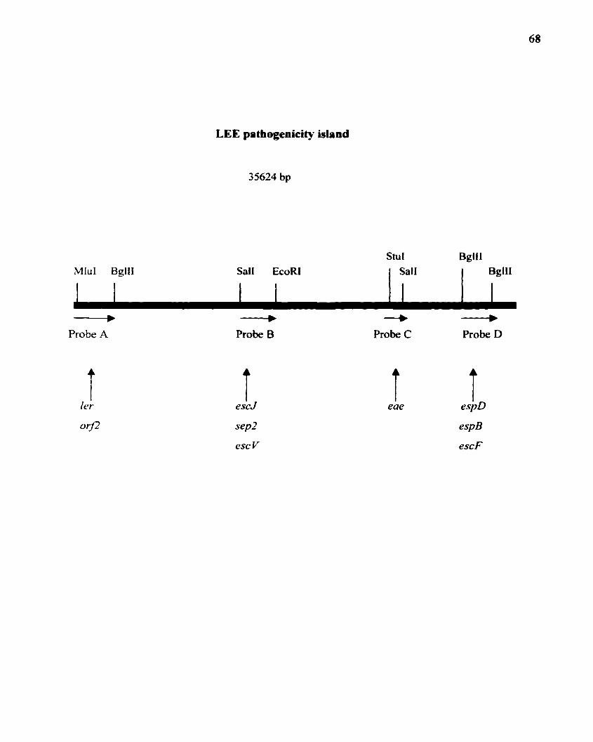
LEE pathogenicity island
35624 bp
Stut BglilMluI BglII Sail EcoRJ lJSaIl L.LII
... ... -+ III'
Probe A Probe B Probe C Probe D
i i i iler escJ eae espD
or!2 sep2 espB
escV escF
68

69
each of the 4 probes was hybridized to DNA from isolated colonies of strain B2F1 by colony
blot analysis. Enterohemorrhagic E. coli 0157:H7 and enteropathogenic E. coli 0127:H6
strains that encode the LEE locus were used as the positive controls and the laboratory strains
DH5a and S17-1 ('Apir) served as negative controls. The positive control strains hybridized
with all four probes while the negative control strains, DH5a and S 17-1 ('Apir) did not nor did
B2FI (Figures 6 and 7). Thus, B2FI does not encode any of the 4 regions of the LEE locus
detected by probes A-D.
II. In vitro model of adherence of STEC to human colonic
epithelial cells.
Humap colonic epithelial cell Une used to optimize the in vitro
adherence assay. The HEp-2 epithelial cell line that was derived from human larynx has
been used extensively to study the localized adherence (LA) of intimin-producing EPEe and
EHEC. The localized adherence pattern involves the accumulation of filamentous actin at the
site of bacterial attachmenL and this actin accumulation is detected by fluorescein-conjugated
phalloidin staining ofactin in HEp-2 cells (Cravioto et aI., 1979; Knutton et aI., 1989; Jerse,
et aL 1990). EHEC require intimin to adhere to HEp-2 cells in vitro and to produce the FAS
phenotype. (McKee et al.. 1995). However~ STEe group 1 and 2 isolates do not encode the
gene for intimin. Indeed McKee at aI. (1995) showed that strain B2F1 does not adhere well to
HEp-2 cells. However, 82Ft was originally isolated from the feces ofa patient with HUS, and
therefore~

70
Figure 6. Colony blot hybridization ofSTEC strain B2F1 with restricted DNA fragments
derived from the locus ofenterocyte effacement (LEE)~ probes A and B. Probe specifications
are described in detail in Figure 5. Positive control strains known to encode the LEE are EHEC
strain 86-24 in position 1-2 and EPEe strain E2348/69 in positions 5-6. Probe A (position 13
14) and probe B (position 15-16) are positive controls for probe reactivity. Negative controls
are two laboratory strains ofE. coli. DH5a and S I7-1 (lpir) (positions 7-10). B2FI is always
in positions II and 12. Position 3 and 4 were not used.

11

72
Figure 7. Colony blot hybridization ofSTEC strain 82Ft with restricted DNA fragments
derived from the locus ofenterocyte effacement (LEE)~ probes C and D. Probe specifications
are described in detail in Figure 5. Positive control strains kno\W to encode the LEE are EHEC
strain 86-24 in position 1-2 and EPEC strain E2348/69 in positions 5-6. Probe C (position 15
16) and probe D (position 15-16) are positive controls for probe reactivity. Negative controls
are two laboratory strains ofE. coli.. DH5a and S17-1(Apir) (positions 7-10). 82Fl is always
in positions 11 and 12. Positions 3 and 4 were not used.

73

74
must have persisted in the human gastrointestinal tract for about 8 days (mean time to
development ofHUS from time of infection). Therefore, we hypothesized that 82Ft has the
capacity to adhere to human colonic epithelial cells by an intimin-independent mechanism. We
proposed to test our hypothesis by conducting our B2F1 adherence assay on an epithelial cell
line of human colonic origin.
The T84 human colonic adenocarcinoma cell line is one of the very few cell lines that
resembles in vitro the native intestinal cell from which it was derived, the colonic crypt cell
(Dharmsathaphom and Madara, 1990). T84 cells can undergo polarization and fonn tall
columnar epithelial monolayers followed by the fonnation of intercellular occluding junctions
(tight junctions) and desmosomes. T84 cells mature both structurally and functionally with the
formation of transepithelial monolayer resistance.. a barrier to paracellular flow and nonnal
distribution of various proteins to apical and basolateral compartments. Thus, T84 cells when
grown in vitro may resemble the in situ microvilli of the intestine (Madara and
Dhannathaphom., 1985). Furthennore, T84 cells are not sensitive to killing by Shiga toxin
because they lack the toxin binding receptor (Philpott et al.. 1997). For these reasons., T84 cells
were used in this study to establish an in vitro model ofadherence to human colonic epithelial
cells by STEC.
EHEC and STEC strains used to optimize tbe adberence assay. Table
4 describes the clinical isolates and control E. coli strains used to establish the adherence assay.
The clinical isolates that were chosen as positive controls included three intimin-encoding
isolates. EHEC 0157:H7 strain 933 and EHEC 01S7:H7 strain 86-24, and EHEC

75
Footnotes for Table 4.
• Strain 933 and 86-24 are both EHEC group I serotype 0157:H7 clinical isolates.
933cu is the plasmid-cured derivative of933 and 86-24eae~lO is an eae deletion mutant of86
24. H30 is an EHEC group 2 member~ serotype 026:Hll. Clinical isolate 82Fl belongs to
STEC group 1., is intimin negative. encodes two copies ofStx2d (Stx2dl and 2d2) and serotype
091 :H2I. HS is a nonpathogenic intestinal flora isolate ofE. coli ofserotype 09:H4 and DH5a
is a rough K-12 laboratory strains ofE. coli.
b The presence of the -90 kb plasmid was detennined by hybridization with the 3.4 kb
HindUI fragment derived from pOl57 carried by EHEC 0157:H7~ and is designated as probe
CVD419 (Levine et al .. 1987).
C The toxin phenotype was determined by neutralization ofVero cell cytotoxicity with
anti-Shiga toxin I(Stx I) or anti-Stx2 antibodies. Stx 2dl and Stx 2d2 are variant forms of
Shiga toxin 2 that can be detected with antibodies against Stx 2 and these toxins can be
activated by human intestinal mucous (Melton-Celsa et al., 1996).
d He. hemorrhagic colitis; HUS~ hemolytic uremic SYndrome; N/A not applicable.

76
Table 4. Characteristics of bacterial strains used to optimize the in vitro
model of adherence to T84 cells by STEC organisms
Strain • (-90) kd Produces Sa type C Clinical
Plasmid present b intimin produced findings d
933 + + I and 2 He
933cu + I and 2 N/A
86-24 + + 2 only HC
85-24eae ill 0 + 2 only N/A
H30 + + I only HC
82Fl + 2d HUS
HS none N/A
DH5a none N/A

77
026:HII strain H30. Strain 82Ft served as the intimin-negative STEC strain. A nonpathogenic
E. coli K-12~ strain DH5a.. was used in most experiments as a strain to defme the
background level ofbacterial adherence in this system. HS~ an intestinal flora isolate ofE. coli,
was included in an initial assay (Levine et a1., 1978). Mannose blocks type 1 fimbrial binding
of K-12 strains to epithelial cells in vitro (Ofek et a1., 1977) and was included in each adherence
assay so that type I fimbrial-independent binding of pathogenic E. coli to 184 cells could be
assessed.
Qualitative analysis ofSTEC adherence to T84 cells. To test the adherence
assay a trial run was perfonned with HEp-2 cells as described previously (McKee et. a1., 1995)
and B2F I failed to bind to HEp-2 cells as had been shown by Dr. McKee (McKee et al.., 1995).
Also. as previously demonstrated (McKee et al., 1995), the intimin-positive strain 86-24 fonned
small microcolonies on HEp-2 cells and was FAS positive (Figure 8). These confinnatory
findings substantiated the validity of the adherence assay. We next sought to apply the assay
to T84 cells.
Semi-confluent cultures ofT84 cells were inoculated with B2Fl, H30 or DH5a and
incubated for periods of one. three and five hours. Within three hours~ the difference in the
amount of adherence of 82Ft compared to DH5a and H30 was readily apparent by phase
contrast light microscopy. Isolate H30 required five hours of incubation (Figure 9) before it
attached to T84 cells at levels greater than the DH5a, but B2F I adhered well even after 3 hours
of incubation. All strains, H30, B2Ft and DH5a., were seen in high numbers attached to the
plastic between cells (data not shown).

78
Figure 8. Phase contrast (panels A and C) and fluorescent (panels B and D) micrographs
of HEp-2 cells after a six hoW" incubation with B2Ft (panels A and B) and 86-24 (panels C and
D). Strain 86-24 attached to and fonned microcolonies with the accumulation of filamentous
actin (arrow panel C and panel D) on HEp-2 cells. B2F I did not bind to HEp-2 cells as
assessed visually. Fluorescein isothiocyanate (FITC)-eonjugated to phalloidin was used to stain
polymerized filamentous actin.

79

80
Figure 9. Non-confluent human colonic epithelial T84 cells five hours post-inoculation
with 82Ft (panel A). and H30 (panel B)'!' or DH5a (panel C). B2FI is intimin-negative'!' H30
is intimin-positive. and DH5a is the poorly adherent control strain.

81

82
The level ofadherence ofall isolates to confluent monolayers ofT84 cells after one hour
of incubation was insufficient for microscopic detection. Three hours of incubation was
required before it was apparent that a substantial number ofB2Fl bacteria had bound to T84
cells. but after three hours very few H30 and 86-24 bacteria could be detected (data not shown).
The intimin-positive strains took longer to form colonies but after five hours appeared to adhere
well to T84 cells. After 5 hours of incubation. 82Ft. 86-24 and H30 adhered to confluent
cultures ofT84 cells (Figure 10). In contrast. following three or five hours of incubation the eae
deletion mutant 86-24eae~10 did not bind well to T84 cells.. and only an occasional DHSa
bacteriwn was seen attached to the monolayers (Figure 10). When confluent monolayers were
used. the amount of non-specific binding appeared to be confined to the plastic slide not
covered by the T84 cell monolayer; this area was typically located at the extreme periphery of
the T84 monolayer. From these findings we concluded that the binding ofwild-type 82FI to
1'84 cells should be visually distinguishable from the binding ofadherence-deficient mutants
ofB2FI after three hours of incubation.
To determine if the incubation period ofB2FI on T84 cells could be shortened to one
hour. the size of the bacterial inoculum was increased from a ratio ofone-hundred bacteria to
one T84 cell (l 00: 1) to a ratio of 1000: 1. At this 1000: I ratio., the eukaryotic cell sheet was
destroyed within the one-hour incubation period whether 82Fl or DH5a were used as the
infecting organisms. Therefore.. a one hour incubation period was not feasible as a means of
screening mini-Tn5phoACmr mutants for adherence defects. We also concluded that a ratio of
100 bacteria to one T84 cell would be the standard inoculum for aU subsequent adherence
assays.

83
Figu re 10. Confluent T84 cells infected for five hours with B2F I and intimin-positive
STEC. Strains tested were: 82Ft (panel A)~ EHEC 0157:H7 strain., 86-24 (panel B)., and
EHEC 026:H 11 strain H30 (panel C) or 86-24eaeA lOan eae deletion mutant of 86-24 (panel
D). and E.coli DH5a (panel E).

84

85
Phenotypic comparisog oftbe capacity ofB2FJ to adbere to polarized venus
non-polarized T84 cells. Next we sought to determine if the kinetics and relative degree
of binding ofwild-type 82Ft to T84 cells would be the same for non-polarized and polarized
T84 cells. For this purpose, polarized T84 cells that had transepithelial resistance values greater
than or equal to 500 n were used in the adherence assay and compared to non-polarized T84
cells that had been grown to confluency on plastic chamber slides. Three strains were tested
for binding: 82Ft. 86-24, and DH5a. Following a five hour incubation period, phase contrast
light microscopy was used to evaluate the adherence ofthese organisms to polarized T84 cells.
B2F1. by microscopic analysis'!' appeared to bind to polarized and non-polarized T84 cells in
similar numbers and in the same pattern of large disorganized macrocolonies (Figure 11 panel
A nonpolarized.. and panel B polarized T84 cells incubated with B2F I). However., 86-24
bacteria inoculated onto polarized T84 cells formed microcolonies that were highly organized
with bacteria tightly associated to each other (Figure II panel D). This phenotype was different
from the larger less organized and less compressed 86-24 colonies associated with non
polarized T84 cells (Figure II panel C). Because 82FI attaches to non-polarized and polarized
T84 cells with a similar phenotype, we concluded that non-polarized T84 cells, which are easier
to maintain and can be grown in plastic chamber slides., could be used in the initial screen for
adherence mutants. We also decided that the more physiologically relevant polarized T84 cells
would be used to confirm the reduced adherence phenotype ofany mutants generated.

86
Figure 11. Polarized (panels B. D., F) and non-polarized (panels A. C. E) T84 cells
inoculated with B2Fl(panels A and B). 86-24 (panels C and D) or DH5a (panels E and F) and
incubated for 5 hours. Arrows indicate location ofadherent bacteria.

87

88
The non-pathogenic strain of intestinal flora ofE. coli. HS. was included in the above assay (not
shown). Isolate HS avidly bound to polarized and non-polarized T84 cells in an aggregative
like fashion (data not shown). The aggregative binding pattern ofHS was significantly different
from that of B2F1 and ofEHEC bacteria. The results suggest that T84 cells are an appropriate
cell line for in vitro adherence analysis of intestinally adherent organisms.
Quantitative analysis of HZP. adherence to T84 eells. To confirm the
microscopic data on B2F1 binding to T84 cells, a quantitative adherence assay was developed.
At the end of one. three. and five hours of incubation, non-polarized T84 cells with attached
82F1 or DH5a bacteria were removed with trypsin from 8-cell chamber slides. The bacterial
eukaryotic cell suspension was serially diluted and plated on L8 agar to obtain viable colony
counts. Each time point was done in triplicate for both strains of bacteria. The geometric mean
adherent colony fonning units (CFU) was calculated and plotted for both of the experimental
strains used (Figure 12). Differences between adherence ofB2FI and DH5a strains that were
observed microscopically were reflected as differences in the magnitude ofthe CFUs recovered
in the representative quantitative assay. Adherence levels of STEC strain 82FI were greater
than that of the control DH5a. for all three time points tested. DH5a values did not exceed 5 x
105 adherent CFU/ml at any time, whereas B2Ft adherent bacteria yielded a 5 hour value of3.5
x 108 CFU/ml. As previously noted, DH5a bacteria were typically seen attached only to the
plastic slides and at the periphery ofthe monolayer, a finding that could account for the number
of DH5a bacteria recovered.

89
Figure 12. Quantitative adherence assay comparing the amounts ofB2FI bacteria and
E. coli K-12 strain. DH5a attached to non-polarized T84 cells. The kinetics ofattachment was
compared for both strains at I. 3. and 5 hours. Geometric mean values from triplicate samples
are shown for each time point.

E- 107:::>u..0c:«SCD
106EU.1::-CDE0 105CD
C!)
104
I~~~~i{f~il
.t}~J~~~
~I_~DH5a B2F1
Time (hours)
_ 1 Hour
~ 3 Hours
5 Hours
90

91
A difference in adherence kinetics of intimin-encoding strains 86-24 and H30 to T84
cells and adherence by B2Fl to these cells had been observed by phase contrast microscopy.
We theorized that if the visual data correlated with the quantitative results.. mutants that were
slower to bind to T84 cells could be detected. A quantitative assay with non-polarized T84 cells
was done to test whether the lag phase noted for H30 and 86-24 would correlate with the
number ofCFU/ml recovered following a three and five hour adherence assay.
After three hours of incubation. 3 x lOs CFU/ml were recovered for H30 and 86-24
strains (Figure 13). In contrast, three hours post infection.. the amount ofB2Fl recovered was
two logs higher at 2 x 10' CFU/ml. Microscopic analysis supported the quantitative results of
the three hour post infection cultures and (photomicrographs are not shown for this time point)
indicated that the levels of B2F1 bacteria bound to T84 cells were greater than levels of86-24
and H30. After five hours of incubation with T84 cells (Figure 13)~ adherence levels ofB2FI
were similar to that of intimin-positive strains 86-24 and H30 (3 x 108,8 X 10', and 1.9 x 10⁸
adherent CFU/mL respectively). The number of DH5a and 86-24eaeL110 recovered (-lOs
CFU/ml) were 2 to 3 logs lower than that for the STEC strains at three and five hours after
infection. These results indicate that the quantitative assay would be useful to detect reduced
adherence mutants of B2F1 and to detect mutants that were slower to adhere to T84 cells~ i.e.
those mutants that adhered with different kinetics.
Quantitative results using polarized T84 cells (experiment was done in triplicate) were
similar to those obtained when non-polarized T84 cells were used. B2Ft adherence values were
7 x 10⁷ CFU/ml after 5 hours in contrast to 103 CFU/ml ofDH5a (Figure 14). Because the
polarized monolayer ofT84 cells extended completely across the collagen-coated membrane

92
Figure 13. Comparison of the number of intimin-encoding STEC (86-24 and H30) and
intimin-negative 82Ft adherent to non-polarized T84 cells three and five hours post-infection.
Geometric mean values from triplicate samples are shown for each time point.

93
108
~
108
IE I-::;)LL(.) 107 -c::: 3 Houra:sQ)
E 5 Hour(J..- 106Q) H
E0(I)
(!)
105
86-24eae41 ODHSa H30 86-24 B2F1
Strains tested

94
Figure 14. Quantitative adherence assay that compared the number of B2F 1 bacteria
adherent to polarized T84 cells versus the poorly adherent E.co/i K-12 strain DH5a. The
amount ofbacteria recovered was compared I., 3., and 5 hours post-inoculation. Geometric mean
values of triplicate samples are shown for each time point.

95
I;,~~;
£~~~~i~iJ~~::-:::,<;"::
DH5a B2Fl
Time (hours)
107 - 1 Hour
~106
3 Hours
5 Hours
105
E-....:::::>LLocasCDE(,)...-CDEoCD
C!J

96
with no discernible gaps~ the amount ofnon-specitic binding of DH5a was reduced. However.,
the CFU/ml of bacteria recovered for 82Fl were comparable to those obtained when non
polarized monolayers were used. 8y comparison ofthe quantitative data obtained with polarized
versus non-polarized T84 cells.. we concluded that the easy to use non-polarized T84 cells could
be used to confirm quantitatively the microscopic evaluation of differences in adherence of
mini-Tn5phoACmr isolates and wild-type 82FI to T84 cells but that the polarized T84 cells
would be used as the ultimate standard to more precisely define quantitative differences in
adherence to T84 cells between 82F I and putative adherence mutants. Therefore we elected
to use the scheme outlined in Figure 15 to screen and test putative adherence mutants of82FI.
III. The role of the large -90 kb plasmid in the adherence to
human colonic epithelial T84 cells by STEC 82Ft.
The experiments described below were designed to determine if the large plasmid
encoded adherence factors~ or if its presence was necessary for adherence of STEC bacteria to
human intestinal epithelial cells.
Transformation ofa poorly-adherent laboratory E. coli strain PUlOS
with the large plasmid isolated from aD jsogenje mutant ofstrajn 82Ft. An
isogenic mini-Tn5phoACmr mutant of82FI was isolated in this laboratory prior to this study
and was designated as strain 283. Strain 283 contained a mini-Tn5phoACmr insertion within
the large -90 kb plasmid. Because hemolysin is encoded on the large -90 kb plasmid of STEC
bacteria~ strain 283 was hemolytic on washed sheep red blood cells. Strain 283 was also

97
Figure 15. Schematic of the in vitro adherence assay used to evaluate attachment of
STEC strains and mini-Tn5phoACmr mutants. The parameters of the assay are discussed in
detai I in the text.

Screen Mini-Tn5phoACmr mutants in a 3 hour adherenceassay with non-polarized T84 cells. Assess visually.
Re-evaluate putative adherence mutants forcapacity to adhere to non-polarized T84 cells
after 5 hours of incubation. Assess visually.
1Retest adherence-defective mini-Tn5phoACmr
mutants in a 3 hour quantitative adherence assaywith non-POlarized T84 cells.
Confirm reduced adherence phenotype ofmini-Tn5phoACmr
mutants in a 3 hour quantitative adherenceassay with polarized T84 cells.
98
99
chloramphenicol resistant by virtue of the presence of the mini-Tn5phoACmr insertion within
the large plasmid. The large plasmid (p283) was purified from 283 and electroporated into a
non-pathogenic~ E. coli laboratory strain., DH I08. Chloramphenicol-resistant transfonnants
were tested for the presence ofthe -90 kb plasmid by colony hybridization with probe CVD419
(Figure 16). One probe-positive transfonnant DHIOB(P283) was selected for further study.
Strains DHIOB. DHtOB(p2B3)" 2B3 and wild-type 82Ft were then tested in the standard three
and five hour qualitative adherence assays to assess whether they attach to T84 cells. Strain
283. attached to T84 cells at levels comparable to its parent 82Ft. (5 hour results shown in
Figure 17 panel A and 8" respectively) and as expected., the laboratory strain DHI08 did not
adhere to T84 cells (Figure 17 panel D). Because the isogenic mutant 283 adhered as well to
T84 cells as did wild-type. it was presumed that the transposon had not inserted within a gene
that encoded factors essential for adherence (Figure 17 panel A). The transfonnant.
DHI0B(p2B3).. adhered poorly to T84 cells; similar to the parental DHI08 strain (Figure 17
panel C and D). In conclusion" this experiment indicates that the -90 kb plasmid derived from
STEC 283 is not in itselfsufficient to impart the wild-type adherence phenotype to a previously
poorly adherent laboratory strain ofE. coli. DH 108.
Capacity of a plasmid-cured derivative of 82Ft to bind to colonic
epithelial T84 cells. A derivative strain 82Ft devoid of the large plasmid was isolated
previously by others in this laboratory. This plasmid-cured derivative was designated as S II.,
and. due to the absence of the large plasmid., it was hemolysis negative on washed sheep red
blood cells.

100
Figure 16. E. coli DHIOB(p2B3) transfonnants tested by colony blot hybridization with
probe CVD419. Probe CVD419 consists ofa 3.4 kb HindUI fragment derived from plasmid
pO 157 carried by STEC strain 933. Negative control strains included the parental strain DH I OB~
933cu~ EPEC~ E. coli K-12 strains DH5a and SI7-1 (Apir). Positive controls consisted ofstrains
86-24. 82Fl and probe CVD419 DNA.

101

102
Figure 17. Human colonic epithelial T84 cells five hours post-infection with strain 2B3
(panel A). 82Ft (panel B)~ transfonnant DHIOB(p2B3) (panel C) and the parental strain
DHIOB (panel D).

103

104
The absence of the plasmid was confirmed by colony blot analysis with the plasmid sPeCific
probe CVD419 (Figure (8). Colonies ofEHEC 0157:H7 (86-24 and 933) and parental strain
B2F1 served as positive controls~ while E. coli strain DH IOB and the plasmid-cured derivative
01'933 were the negative control strains used in this colony blot.
After five hours the plasmid-cured derivative~ SII, attached to T84 cells as well as the
parental Vtild-type strain (Figure 19). Quantitative analysis was done using non-polarized T84
cells and indicated that there was no significant difference in adherence to T84 cells between
B2F1 and S I 1 (data not shown).
IV. Attempt to construct by PI transductioD aD alkaline
phosphatase null mutation in 82Ft.
Wild-type 82FI is naturally phosphatase positive and produced blue colonies when
grown on LB agar that contained XP. Hence~ generalized PI transduction was used to try
to transfer a non-functional phoA deletion mutation encoded by the E. coli strain SH210 into
wild-type 82Ft. The phoA mutation in SH210 is linked to TnlO that encodes the tetracycline
resistance gene. The transduction procedure that was used is detailed in Materials and Methods.
Bacteriophage PI ::Tn9 (SH2lOl encodes a temperature-sensitive repressor that at 42°C becomes
inactive. A shift in temperature from 37°C to 42°C induced the bacteriophage to become lytic.
The Tn9 insertion within this PI phage carries a chloramphenicol resistance gene that serves
as a selectable marker. A high titer lysate (-I x 1011 PFU/ml) of PI::Tn9 (SHIO) was preparedand
was used in an attempt to infect 82Fl. Simultaneously, a control laboratory strain C600 was
transduced with the same amount and sample preparation oflysate. Forty per cent ofthe isolates

105
Figure t8. Colony hybridization of the plasmid-cured derivative of B2F1~ designated as SII
with probe CVD419. EHEC strains 933~ 86-24JB~ 86-24m~ 86-24, H30, and STEC strain
B2F 1 contained the -90 kb plasmid and served as the positive controls. Strains 933cu and
DH5a. were the negative controls.

106

107
Figure 19. Comparison ofwild-type 82FI and its plasmid~ured derivative SII to adhere
to T84 cells five hours after infection. Strains tested: S11 (panel A), B2Ft (panel B). and
DH5a (panel C).

108

109
were cotransduced and contained both the phoA mutation and the Tn}0 encoded tetracycline
resistance gene from strain SH210. However~ PI::Tn9 could not infect B2Ft even when used
at a multiplicity of infection of one-hundred bacteriophage to one bacterium (no
chloramphenicol or tetracycline resistant colonies were isolated). Therefore. an alternate
approach was used to facilitate discrimination between wild-tyPe B2FI and putative in frame.
blue mini-Tn5phoACm r mutants of that organism.
v. Construction and screening of the mini-Tn5phoACm' mutant
bank prepared from wild-type strain 82Ft.
Isolation of mini-TnSphoACm' mutants. When 80 Jlg/ml ofXP was added
to LB agar. the blue phenotype of wild-tyPe B2FI took longer than 18 hours to develop. In
contrast. isolates ofB2FI with in-frame insertions ofmini-Tn5phoACmr within genes encoding
exported or surface exposed proteins could be detected within 13 to 15 hours after plating.
Because it took longer for wild-type colonies to tum blue. mutants with phoA fusions could be
differentiated from wild-type if the blue color ofcolonies were scored within 13 to 15 hours.
After this initial screen. mutants were re-streaked for isolation~ frozen" and tested for the blue
phenotype on LB agar that contained forty micrograms per milliliter ofXP.
After numerous matings between the mini-Tn5phoACmr host strain S17-I(Apir) and
82Ft. 22.555 transconjugates were isolated. Because mating was allowed to occur overnight
in some cases" some of the transconjugates that were isolated may have been siblings. There
were 253 isolates that produced blue colonies faster than wild-type (within 15 hours) and that
were a darker blue than wild-type. These isolates were presumed to contain in-frame insertions

110
in genes that encoded exported proteins and represented approximately one percent of the total
transconjugates isolated (Table 5). All isolates that were blue on XP-LB agar (253 total) were
screened for adherence to T84 cells. Only 5 of the 253 blue isolates (2%) were ampicillin
resistant: the other 248 colonies were ampicillin sensitive.
Isolation of three adherenee-deficent mutants ofSTEC 091 :821 strain
82Fl. Three mutants designated as 34.3~ 34.7 and 30.10a were blue on XP-LB agar (see
Figure 20). chloramphenicol resistant, and ampicillin sensitive. Thus, they were presumed to
be cured of the donor plasmid that carried the mini-Tn5phoACmr cassette. These three mutants
grew at rates similar to wild-type B2F1 when grown in Knutton tissue culture adherence assay
media at 37°C (Figure 21). Three ofthe 253 isolates that contained the transposon were reduced
in their capacity to bind to T84 cells (Figures 22 and 23).
The isolates. 34.3. 34.7 and 30.10~ were tested in the standard three and five hour
qualitative adherence assay with nonpolarized T84 cells. Assays were done in triplicate on
more than three separate occasions, and several isolates of each mutant were tested.
Microscopic evaluation of mutants 34.3 and 34.7 (Figure 22 panel B and C results of a
representative 5 hour assay) indicated that both strains failed to adhere well to T84 cells.
Mutant 30.1 Oa did not adhere well to T84 cells, (Figure 22 panel A) and the pattern of
the limited number of adherent organisms was distinct from wild-type. Specifically, strain
30.10a attached as individual bacteria with several bacteria attached to and suspended above
the cell sheet of non-polarized T84 cells (Figure 19 panel A) or polarized (not shown). In
contrast, B2F1 adhered in large colonies to T84 cells (Figure 22 panel E). The total CFU/ml
recovered three hours after infection of polarized T84 cells were compared for strains 34.3~

III
Table 5. Results of conjugal matings for transfer of miniTn5phoACm r to create a mutant bank: ofSTEC091 :H21 strain B2F 1
Duration of mating No. transconjugates No. of blueisolated colonies (phoA+)
one hour 7,592 94
18 hours 14,963 159
22,555 253

112
Figure 20. Appearance ofmini-Tn5phoACmr mutants designated as 34.3,34.7 and 30.IOa
and parental strain 82FI grown on XP agar. Strains were grown for 15 hours on agar
supplemented with 80 J.1g1mJ of the alkaline phosphatase substrate XP.

113
114
Figure 21. Growth rates of three adherence mutants~ 34.3~ 34.7 and 30.1~ compared with
parental strain B2Fl when grown in Kutton tissue culture adherence assay media at 37°C.
Cultures were incubated for a total of8 hours with shaking and the optical density 600 om was
recorded every hour. The initial reading through 6 hours are shown on the graph.

1.50
1.35
1.20• wildtype
1.05Er::: 0.90 30.10a00 0.75 •CO 34.3
C 0.600 ,. -. 34.7
0.45
0.30•
0.15 •.. -0.00
0 2 4 6 8
Time in hours
115

116
Figure 22. Capacity ofmini-Tn5phoACmT mutants 30. lOa (panel A), 34.3 (panel B)~ 34.7
(panel C) to adhere to T84 cells after 5 hours of incubation as compared to wild-type 82Ft
(panel E). Poorly adherent control strain DH5a is shown in panel D. Arrow in panel A points
to single adherent bacterium; arrow in panel E indicates a colony of organisms.

117

118
Figure 23. The number of bacteria adherent to polarized T84 cells after three hours of
incubation detennined for three mini-Tn5phoACm fmutants~ 34.3~ 34.7 and 30.10a and
compared to wild-type B2FI. DH5a was included as a poorly adherent control. Geometric
mean values for triplicate samples are depicted for each time point.

-E........::JLL.U 101
c:asCDEu.-'-i 106
EoCDC'
__ Jlllllll34.3 34.7 30.10a DH5a B2F1
Strains tested (3 hour incubation)
119

120
34.7. and 30.10a to those of the parental strain B2FI(Figure 23) [quantitative results ofa three
hour assay with nonpolarized T84 cells is not shown]. Approximately lOS CFU ofeach strain,
34.3. 34.7 and 30.1 Oa, were recovered as compared to 106 DH5a and approximately 107 of
wi Id-type. These results suggest at least a IDO-fold difference in the capacity ofeach of these
three mutants to adhere to T84 cells as compared to the wild-type.
Following a five hour adherence assay, mutant 30. lOa destroyed the T84 cell monolayer.
This destruction did not always occur and appeared to depend on the age of the monolayer that
was used (10-12 day old monolayers were most susceptible) [data not shown]. Mutants 34.3
and 34.7 caused no discernible. or only moderate damage, five hours after inoculation when 10
12 day old non-polarized T84 monolayers were used (data not shown).
Characterization ofadhereace mutauts. Table 6 summarizes the phenotypes
examined for each mutant. The procedures used to assess each ofthese characteristics has been
described in the Materials and Methods section of this thesis.
Antiserum against serotype 091 bacteria agglutinated strains 34.3, 34.7 and 30.10a as
well as the parental strain B2Fl; hence the mutants were all smooth and of the 091 serogroup.
The adherence mutants, like wild-type, produced capsule when grown on Maloney media and
were not auxotrophic since they grew well on minimal M9 media Strains 34.3, 34.7 and 30.1Oa
grew well on MacConkey media and were not sensitive to the bile present in this medium.
Strains 34.3. 34.7 and 30.10a secreted Stx2d in amounts similar to those secreted by
B2Fl(quantitation ofStx2d not shown) and were all motile at 37°C. However, 34.3 and 34.7
were not motile in soft-agar when grown at 30°C whereas the parent 82Ft and mutant 30.IOa
were motile (data not shown). Note that the motility findings at 30°C were made subsequent

Table 6. Characteristics of adherence nlutants 34.3, 34.7, and30.IOa compared to the parental strain B2F I
Stx
Strains 091 Motility Growth Growth on Alnpicillin detected Hemolysin
tested serogroup Capsule onM9 MacConkey sensitive In produced30°C 37°C filtrates
Wildtype + + + + + + + + +
34.3 + + - + + + + + +++
34.7 + + - + + + + + +++
30.10a + + + + + + + + ++
-N-

122
to the initial phenotypic characterization of the mutants. In addition.. all of the adherence
mutants (34.3. 34.7 and 30.1 Oa) produced large clear zones of hemolysis on agar plates that
contained washed (Figure 24) or unwashed (not shown) sheep red blood cells (SRBC). The
phenotype exhibited by the mutants was in contrast to that ofB2FI which produced small turbid
zones of hemolysis visible when the SRBCs were washed before incorporation into the agar.
One possible reason for the hyper-hemolytic phenotype ofthe 34.3, 34.7 and 30.1 Oa was
that the outer membranes ofthese mutants were leaky. Such a leaky phenotype might explain
the damage to T84 cells sometimes seen when these mutants were tested in the five hour
adherence assay. Therefore, two tests were done to determine if the membranes of the mutants
exhibited nonnal function e.g. maintained barrier function and if they differed in release of
proteases. Strains 30.1Oa, 34.3 and 34.7 were grown separately in the presence ofcrystal violet
and on LB agar plates that contained 3% instant non-fat dry milk (source ofcasein). Both the
wild-type and the mutant strains were able to exclude the crystal violet and to grow up to the
crystal violet-impregnated filter (data not shown). Similarly, none of the colonies.. mutants or
wild-type.. produced zones of clearance on agar plates that contained casein (Figure 25).
Therefore. no differences in protease leakage/export was evident among the strains. These
findings taken together with the capacity ofthe mutants to grow in the presence ofbile salts in
MacConkey agar indicated that the outer membranes of isolates 34.3.. 34.7 and 30.1 Oa probably
function nonnalIy. Therefore, the reason for the large, clear zones of hemolysis on SRBC agar
was due to another factor(s).
VI. Restriction mapping of the mini-Tn5phoACmr insertion
present in adherence mutants 34.3, 34.7 and 30.10a.

Figure 24. Comparison of the hemolytic phenotypes ofwild-type 82Ft. its isogenic mini-
Tn5phoACm r adherence-defective mutants.. and the plasmid~ured strain S11. Because S11 no
longer carries the large plasmid.. it serves as the negative control for hemolysis. Agar plates that
contained 50/0 washed sheep red blood cells. with 10 mM CaCI added were incubated at 37°C
in a 5% CO:! atmosphere for 24 hours. The next day.. after a short 2 hour incubation at 4°C.. the
hemolysis phenotype was scored.
124
125
Figure 25. Comparison of the growth of 82Ft and adherence mutants 34.3~ 34.7 and
30.10a on LB agar that contained 3% instant nonfat dry milk. Plates were incubated overnight
at 37°C.

126

127
Southern hybridization analysis of HindllJ-restricted total DNA isolated from mutant
34.3 and from mutant 34.7 revealed a 6 kb size fragment that hybridized to the chloramphenicol
resistance gene (Cm1 (Figure 26). Because the same size restriction fragment hybridized to the
chloramphenicol probe.. the possibility existed that both mutants contained the transPOson
insertion within the same gene. Therefore, a second set of Southern analyses was done with
Sail-and Pstl-singly and doubly-restricted total DNA from 34.3 and 34.7. The blots were again
probed with the Cmr (Figure 27). B2Ft served as the negative control in these experiments
because it does not contain the Cmr• The DNA hybridization pattern was the same for both
mutants. Also.. an identical 1.4 kb fragment was detected whether DNA was restricted with Pstl
alone or in combination with Sail, a rmding that indicated that the Pstl fragment was inside the
Sail fragment for both mutants. Lastly.. the size of the Sa/l DNA fragment that hybridized with
the probe was 9 kb in both samples. Adherence mutants 34.3 and 34.7 were isolated from the
same conjugal mating mixture. and had the same Southern hybridization pattern.. thus, we
concluded that 34.3 and 34.7 contained the insertion of the transposon in the same gene and
were probably of the same clonal derivative.
By contrast.. we concluded that mutant 30.1 Oa contained the mini-Tn5phoACmr insertion
at a site distinct from that of 34.3 and 34.7 based on Southern analysis (Figure 26 and 27).
DNA restricted with HindUI hybridized with the Cmr probe to yield a 12 kb size fragment.
Restriction of30.1 Oa DNA with a combination ofSall and Pstl hybridized to the probe to yield
a 5 kb fragment.. and restriction with Pstl alone yielded an 8 kb fragment. There is a fragment
that is >23 kb present on both the HindUI and Sail Southern blot figures and in both cases this
fragment probably represents incompletely cut DNA.

128
Figure 26. Southern hybridization of Hindill-restricted total DNA from B2FI mini-
Tn5phoACmr adherence-defective mutants 34.3,34.7. 30. lOa The chloramphenicol resistance
gene from mini-Tn5phoACmr was labeled with ECL-horseradish-peroxidase and used as the
probe in the analysis. Position of molecular weight size markers (kb) are indicated.

23_
10 _
8 _
6 -
-5
3 _
1.6 _
1.0 _
HindII I
129

130
Figure 27. Southern hybridization ofSall-digested (lanes 2~ 5~ 8~ and 11), PSII-digested
(lanes 1. 4. 7. and 10) or both SaIl and Pstl-digested (lanes 3~ 6, 9.. and 12) total DNA from
82Fl rnini-Tn5phoACmr adherence-defective mutants 34.3 (lanes 1-3),34.7 (lanes 4-6) and
30.IOa (lanes 10-12); and the parental strain 82Fl(lanes 7-9). The probe used in the analysis
was the ECL-horseradish-peroxidase-Iabeled chloramphenicol resistance gene from mini
Tn5phoACm f DNA size markers (kb) are indicated.

GiJ I 34.7 1 I B2Fli
123456789
130.108 I10 II 12
131
23-
9 -5 -3 -2 -
1_

132
Southern hybridintion analysis of tbe large plasmid encoded by tbe
adherence mutants fgr tbe presence of the mjni-Tn5,phoACmr insertioQ. The
-90 kb plasmid that is present in B2FI and 77-81%ofnon-OI57:H7 STEe (Levine., et a1•., 1987)
was purified from the parental wild-type B2Ft and mini-Tn5phoACmf mutants., 283., 34.3., 34.7
and 30. lOa. EHEC 0157:H7 strain 933 was used as a positive control and the plasmid-cured
derivative of933. 933cu., served as a negative control. The plasmids were isolated by the Kado
and Liu method (Kado and Liu., 1981) and were virtually free of chromosomal DNA. The
plasmid isolated from each strain was electrophoretically separated in a 0.6% agarose gel and
transterred to a nitrocellulose membrane for Southern hybridization analysis. The DNA
transfers described above were probed with eVD419 (Levine., et at.. 1987)., to prove that
plasmid DNA had been isolated from each of the E.coli strains. The nicked (higher band) and
super-coiled (lower band) forms ofthe large plasmid were detected by the probe. Plasmid preps
from each of the STEC strains that carried the -90 kb plasmid hybridized with probe eVD419
(Figure 28 panel A.. detected as two bands). DNA isolated from 283 was denatured during
processing and ran as a smear. As expected., the plasmid-cured derivative 933cu did not react
with eVD419.
The same membrane was then re-probed (after the plasmid-specific probe was removed)
with the emf gene from mini-Tn5phoACmfto determine ifthe transposon had inserted into the
large plasmid encoded by the mutants (Figure 28 panel B). The -90 kb plasmids isolated from
mutants 30.1 Oa and 2B3 both contain a mini-Tn5phoACmf insertion and hybridized with the

133
Figure 28. Southern hybridization analysis of unrestricted plasmid DNA isolated from
mini-Tn5phoACmr adherence-defective strains 34.3~ 34.7, 30.IO~ and controls B2Ft, 933c~
933. and 2B3. The probe used in the analysis in panel A was the ECL-horseradish-peroxidase
labeled 3.4 kb Hindlll restriction fragment from plasmid pOIS7 called CVD419. The probe
used in the analysis in panel B was the ECL-horseradish-peroxidase-labeled chloramphenicol
resistance gene from mini-Tn5phoACmr• Position ofmolecular weights size markers (kb) are
shown.

- ,-.Io IN
- ,-.Io !.oJ
II II34.3
34.7 34.7
30.IOu 30.IOa
933cu 933cu
U2fl 933
~" ~~.j~i·'~·'18 2 f J' • 'S. I- ,f.'~ ~ • :l .t1Jo,"'.. !2B3 < ' ~ ,,- ." .' ..•_1./'t1..'..... ' ~"''''''.- t\ i J~ ) I~')~~t'~,~
, 'ok. ". 283
w~

135
emf gene probe. Hence., the mini-Tnp5hoACmr had inserted onto the large plasmid in strain
30.10a but inserted into the chromosome ofmutants 34.3 and 34.7 (Figure 28 panel B).
VII. Strategy used to clone the mmi-TnSphoACmr disrupted gene
from strain 34.7.
The main goal of this investigation was to identifY factor(s) important for the adherence
of intimin-negative STEC to human epithelial cells. To achieve this goal., an adherence mutant
was identified and the gene that contained the mini-Tn5phoACmr was cloned and characterized.
The strategy used to clone the gene., and identifY the cloned gene is discussed below.
Three mutants were isolated by transposon mutagenesis that were unable to bind to T84
cells in the same manner as wild-type. In strain 30.1O~ the transposon had inserted within a
gene encoded by the large plasmid. Because the plasmid-eured derivative ofB2Fl., called S11,
adhered to T84 cells with a wild-type phenotype., this indicated that the plasmid did not encode
adhesins or regulators needed for adherence to human colonic epithelial cells (T84 cells).
Therefore. mutant 30.10a was not chosen for further study. Strains 34.3 and 34.7 were shown
to contain the transposon insertion within the same region of DNA and were presumed to
contain the same insertion. Mutant 34.7 did not appear to damage T84 cells during a standard
3 hour adherence assay and was selected as the candidate from which to clone and identifY the
disrupted gene.
The gene was cloned directly from the genomic DNA of 34.7 (Figure 29). Hindlll
digestion of total DNA resulted in fragments that were 6 kb in size and that hybridized to the
transposon encoded emf probe. The Hindlll enzyme cut within the mini-Tn5phoACmr coding

136
Figure 29. Strategy used to clone the gene that contained the mini-Tn5phoACmr insertion
from the adherence-deficient mutant 34.7.
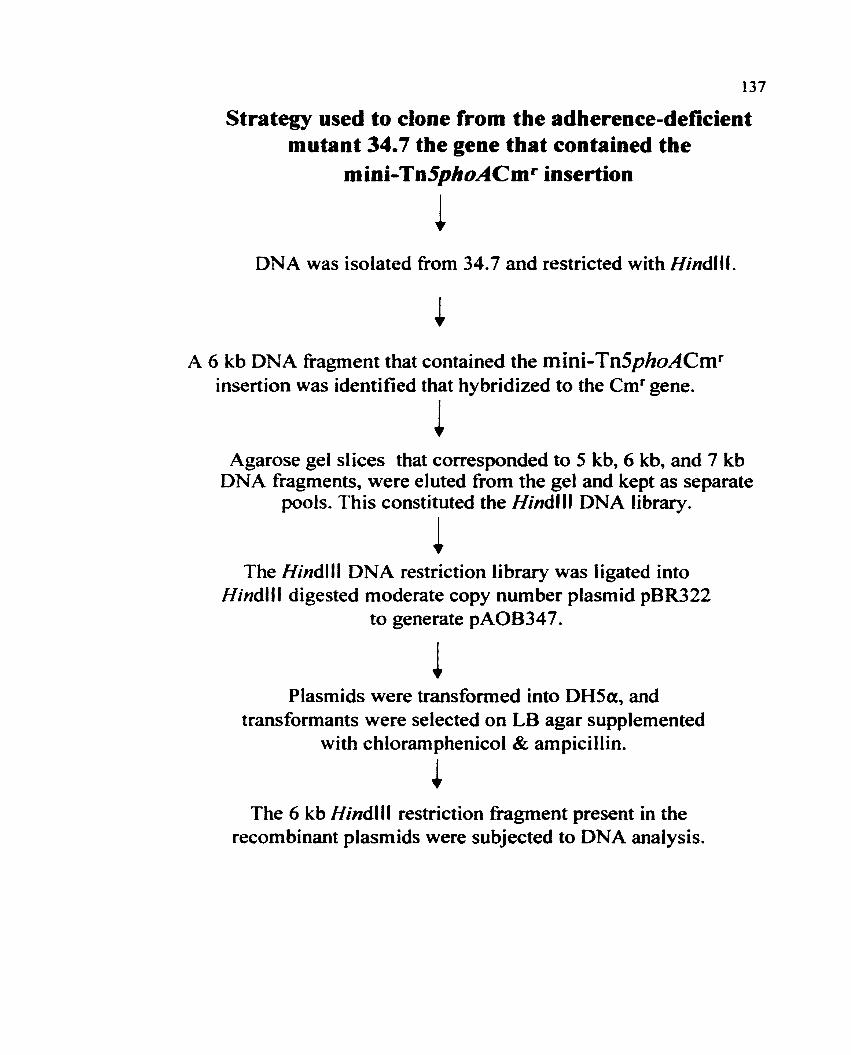
137
Strategy used to clone from the adherence-deficientmutant 34.7 the gene that contained the
mini-Tn5phoACm r insertion
~DNA was isolated from 34.7 and restricted with Hindili.
A 6 kb DNA fragment that contained the mini-Tn5phoACmr
insertion was identified that hybridized to the emf gene.
~Agarose gel slices that corresponded to 5 kb, 6 kb, and 7 kb
DNA fragments, were eluted from the gel and kept as separatepools. This constituted the HindUI DNA library.
~The Hindlll DNA restriction library was ligated into
Hindlll digested moderate copy number plasmid pBR322to generate pAOB347.
Plasmids were transformed into DH5a, andtransformants were selected on LB agar supplemented
with chloramphenicol & ampicillin.
~The 6 kb HindUI restriction fragment present in the
recombinant plasmids were subjected to DNA analysis.

138
sequence but left the chloramphenicol gene marker intact. Because the HindUI fragment was 6
kb in size. we hoped that this fragment would contain some portion of the 3 I' end of the mini
Tn5phoACmr disrupted gene. Gel slices were made above (7 kb), below (5 kb) and at the
position of the 6 kb fragment to improve the chance that the fragment of DNA that contained
the mini-Tn5phoACm f insertion would be isolated. Hindill-restricted DNA fragments were
separated on a 0.7% agarose gel (Figure 30 shows only the 6 kb pool used for ligation) and
separate pools were made that consisted of DNA fragments in the size ranges of5, 6 and 7 kb.
A bank was constructed of the three different pools of Hindi II-restricted DNA by ligating these
fragments into the HindUI site of the pBR322 cloning vector. The resultant plasmids were
transformed into the laboratory strain DH5a. Recombinants that harbored the plasmid and that
contained the 6 kb fragment of interest were selected on LB agar with chloramphenicol and
ampicillin added. Two chloramphenicol and ampicillin resistant colonies were recovered and
both plasmids isolated from these transformants designated pAOB347.1 and pAOB347.2,
contained a 6 kb insert of the predicted size (Figure 31 is a diagram of pAOB347.I).
VIII. Identification of the gene tbat contained tbe transposon
insertion.
To determine the sequence ofa portion of the DNA adjacent to the mini-Tn5phoACmf
insertion present on the 6 kb fragment that was cloned from 34.7 onto the recombinant plasmids
pAOB347.l and pAOB347.2.. primer AMIl was used that hybridized to transPOson DNA
located at the distal 3 I' end of mini-Tn5phoACm f (Figure 32). Automated sequence analysis
revealed approximately 300 bp ofDNA encoded by mutant 34.7 that was located at the fusion

139
Figure 30. Agarose gel electrophoresis of the 6 kb HindUI DNA fragment pool derived
from mutant 34.7. In panel A lane 4 contains the 6 kb fragment that was used to generate the
recombinant plasmid pAOB347. Panel B (lanes 4 and 5) are the Hindlll digested recombinant
plasmid pAOB347.1 and pAOB347.2., respectively., that contain the 6 kb fragment isolated from
the DNA of34.7. Lane 3 panel B contains spillover from lane 4 Le. pAOB347.1. Lanes marked
as (M) in both panels A and B contain DNA molecular weight markers (kb).

23
6
4
2
236421
M M 6KB1 2 3 4
AOB347M M .1 .1 .2
1 2 3 4 5
140

141
Figure 31. Drawing ofplasmid pAOB347 that carries the 6 kb Hindlll DNA fragment cloned
from the mini-Tn5phoACmf adherence mutant 34.7. The 6 kb fragment is represented by the
grey region that encodes the chloramphenicol resistance gene derived from the mini
Tn5phoACmf~ and the black region that represents the gene adjacent to the 3' end of the
chloramphenicol resistance gene't and the hatched portion that symbolizes the rest of the DNA
cloned from mutant 34.7 at the 3' end ofthe chloramphenicol resistance gene. The 6 kb HindllJ
DNA fragment was ligated into the Hindill-digested moderate copy number cloning vector
pBR322 that encodes tetracycline and ampicillin resistance. Map drawn to scale with
CloneMap program version 2.11 from CGC Scientific.. inc.

Hindlll 29EcoRV 188
BamHI376
Hindlll 4388
10363 bp
Ndel2299
142
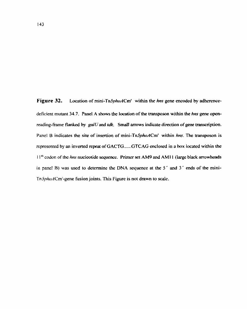
143
Figure 32. Location of mini-Tn5phoACm f within the hns gene encoded by adherence
deficient mutant 34.7. Panel A shows the location of the transposon within the hns gene open
reading-frame flanked by ga/Vand tdk. Small arrows indicate direction ofgene transcription.
Panel B indicates the site of insertion of mini-Tn5phoACm f within hns. The transposon is
represented by an inverted repeat ofGACTG......GTCAG enclosed in a box located within the
11 th codon of the hns nucleotide sequence. Primer set AM9 and AM11 (large black arrowheads
in panel B) was used to determine the DNA sequence at the 5' and 3' ends of the mini
Tn5phoACm f -gene fusion joints. This Figure is not drawn to scale.

144
Transposon
413 bp
IIns.......-_5---+-----........;...-------'---+--- .
lbp
A
AM9 AMI Ir ~.~ATG AGe GAA GCA crr AAA AIT CTG AAC ACC AI GACTG ........ GTCAG ~C CGT........
1234567
M SEA L K I
H-NS-AMINO ACIDS
8 9 10
L N TMini-Tn5phoAsite of insertionwithin hns
BI...-- ---...I

145
joint between mini-Tn5phoACmr and the gene into which the transposon had inserted. The
BasicLocal Alignment Search Iool (BLASn was used to match the 300 bp of DNA cloned
from 34.7 to relevant gene sequences present in the nucleic acid database maintained by the
National Center for Biotechnology Information. The 300 bp of DNA derived from mutant 34.7
matched the E. coli hns gene.
IX. The site of insertion of the transposon within the hns gene.
Sequencing of the 6 kb fragment at the 3' end of the mini-Tn5phoACmr gene fusion
in hns indicated that the insertion occurred out-of-frame between the first and the second
nucleotide of the eleventh codon (isoleucine) of hns (Figure 32). Because the out-of-frame
insertion ofmini-Tn5phoACmr occurred early in the hns sequence neither a functional H-NS
nor mini-Tn5phoACmr phosphatase protein could have been made.
Because~ mutant 34.7 produced blue colonies on XP medi~ we had initially assumed
that the mutant contained an in-frame insertion within a gene that encoded an exported or
surface exposed protein. We now speculated that the absence ofH-NS in 34.7 may have de
repressed the transcription ofalkaline phosphatase and this could have caused the blue colonies
detected when 34.7 was grown on XP media. Other investigators have reported similar findings
when H-NS is inactivated (Bertin et al.~ 1994; Harrison et al... 1994 and Woolwine et at., 1998).
X. Sequence analysis of hns encoded by 34.7, an hns knockout
and adherence-deficent mutant of 82Ft.
Primer AM9, transposon specific primer, was used to obtain the sequence information
at the 5' end of the hnj'::mini-Tn5phoACmr fusion joint (Figure 32). The complete hns

146
sequence derived from 34.7 was then aligned with the hns encoded by K-12" a non-pathogenic
E.coli. with the aid ofthe 8LAST program. The Kohara cosmid clone (clone #250" accession:
090759) was used for the alignment. This clone corresponds to the 27.6 through the 28 minute
region ofthe linkage map prepared by Oshima et al.~ in 1996. The results indicated that the hns
gene encoded by B2Ft is almost identical to that encoded by E.coli K-12(Figure 33). In 82Fl
the tenth codon has been replaced by threonine (bolded and underlined in Figure 33); which is
an asparagine in E.coli K-12. Furthennore~ the orientation of the transcriptional unit of hns
encoded by 82Ft was the same as in K-t2 E. coli (Figure 32). The hns gene encoded by 82Ft
and K-12 E. coli is transcribed (5" to 3") counter clockwise and is located between the ga/U
and Idk genes. Both ga/U and tdk are transcribed (5" to 3") clockwise in the opposite
orientation of the hns gene. Intervening non-coding regions of DNA separate the three genes.
XI. Isolation of the wild-type hns gene from 82Ft.
Based on the sequence ofhns from B2F I, primers (MS4 and MS5) were designed for
(peR) amplification ofhns and flankingga/U and tdksequences directly from the chromosome
of wild-tyPe 82Fl. Figure 34 shows the location of the primers used for PCR amplification.
BamHI restriction sites were engineered into the primers so that the PCR DNA product would
contain this restriction site at both its 5" and 3" ends. The DNA product obtained after PCR
amplification was purified by extraction from an agarose gel with the Geneclean spin system
and then ligated into the cloning vector pCR2.1 (Clark" 1988 and Mead et aI.., 1991) to generate
pAOBIOO.. Figure 35. The pCR2.1 cloning vector contains 3"-T overhangs for direct ligation
of Taq amplified PCR products. The recombinant plasmids were transformed into DH5a, and

147
Figure 33. Nucleic acid sequence ofhns encoded by STEC 091 :H21 strain B2F1.

148
Nucleic acid sequence ofhns encoded by
STEC 091:H21 strain B2Fl
ATGAGCGAAACACTTAAAATTCTGAACACCATCCGTACTCTTCGTGCGCA
51 GGCAAGAGAATGTACACTTGAAACGCTGGAAGAAATGCTGGAAAAATTAG
101 AAGTTGTTGTTAACGAACGTCGCGACGAAGAAAGCGCGGCTGCTGCTGAA
151 GTTGAAGAGCGCACTCGTAAACTGCAGCAATATCGCGAAATGCTGATCGC
201 TGACGGTATTGACCCGAACGAACTGCTGAATAGCCTTGCTGCCGTTAAAT
251 CTGGCACCAAAGCTAAACGTGCTCAGCGTCCGGCAAAATATAGCTACGTT
301 GACGAAAACGGCGAAACTAAAACCTGGACTGGCCAAGGCCGTACTCCAGC
351 TGTTATCAAAAAAGCAATGGATGAGCAAGGTAAATCCCTCGACGATTTCC
401 TGATCCAGCAATA

149
Figure 34. Location of the primers used to amplify and to sequence the 1882 bp DNA
PCR product from the chromosome ofwild-type strain B2Ft. The large grey arrow shows the
location ofhns and direction of transcription ofthe gene. The small black arrows at the distal
5'" and 3'" ends are the location ofprimers MS4 and MS5 that were used to amplify the 1.9 kb
DNA product from the chromosome ofB2Fl. Location of the BamHI (B) sites engineered
onto the ends of the peR fragment are indicated. The primers used to sequence the 1.9 kb
region overlapped each other (large black arrows with locations of the start ofeach primer is
designated in base pairs) and hybridized to sequences on the top-strand (5' -3') of the 1.9 kb
PCR product. The location of the start sites ofgalU and /dk are shown. Drawn to scale with
the CloneMap program.

ISO
1882 bp peR product
B
hnsMS4.••••••••••~~~I-.J..__-J_---'~======:--_....._-_...._~i=~
---.IOObp

151
Figure 35. Diagram of plasmid pAOBIOO that carries the 1882 bp region of DNA cloned
from the chromosome of wild-type B2F I. The 1.9 kb region encodes hilS and flanking galU
and tdk gene sequences. Primer set used for PCR amplification was designated as MS4 and
MSS (position ofprimers are shown in Figure 32). The 1.9 kb PCR product (black region) was
ligated into the multiple cloning site ofvector pCR2.l to generate pAOBI00. This plasmid
conters kanamycin (hatched) and ampicillin (grey) resistance.

EcoRI
152

153
transfonnants were selected on media that contained ampicillin. Purified plasmid was isolated
from one of the selected transfonnants. and the 1.9 kb insert was sequenced. The location of
the primers that were used to sequence the insert that contained hns and a portion ofgalU and
tdk are shown in Figure 34.
XII. Analysis of Shiga-toxin and STEC hemolysin production in
the hns null mutant 34.7 deficient in adherence to colonic
epithelial T84 cells.
Shiga toxin production by hns mutant 34.7. The relevance ofH-NS as a
regulator of the expression of Shiga toxin in STEe has not been investigated. Because
inactivation ofH-NS has been shown to affect the expression ofother virulence genes encoded
by various strains of pathogenic E. coli (Colonna et al.~ 1995 and White- Ziegler et aI.., 1998);
we were interested in whether inactivation ofhns in 34.7 had any effect on the expression of
Stx2d. Although T84 cells are not sensitive to Stx2d production by B2Ft., there was a
possibility that if the levels ofStx expressed by mutant 34.7 were increased., T84 cells could
be damaged enough to prevent adherence of34.7 to them. For this reason., the levels of Shiga
toxin produced by the hns mutant 34.7 was compared to the levels of toxin produced by wild
type B2F 1. Because H-NS has been shown., in some cases., to exert its effect at 300 e on genes
that encode virulence factors. Stx2d production by B2Ft and 34.7 were compared at 30°C and
37°C (Figures 36 and 37., respectively). The Vero cell cytotoxic assay was used to detennine
the level of toxin produced by these bacteria.

154
Figure 36 Shiga toxin levels produced by the hns::mini-Tn5phoACmr adherence mutant
strain. 34.7 compared to the levels of Shiga toxin produced by the wild-type strain., B2Ft at
30°C. Filtrates ofsonic extracts ofstationery phase cultures were prepared and tested on Vero
cells to determine the cytotoxicity of each preparation. The cytotoxic titer per milliliter of
culture was expressed as the reciprocal of the highest dilution required to kill 50% of the cells
in a given well (CDso/ml). The geometric mean ofthree separate tests are shown. Not shown.,
Iysates of non-toxin-producing negative control strain., DH5a.. were less than t01 CD50/ml.

155
4/28/984/22/984/15/9810°
E........
1040LO
C0r: 103 _ 34.7 lysateasQ)
E ~ WTlysate() 102'-....CDE
1010CDC!l
Preparations Tested

156
Figure 37. Shiga toxin levels produced by the hns::mini-Tn5phoACmr adherence mutant
34.7 compared to the levels of Shiga toxin produced by the wild-tyPe strain.. 82Ft at 37°C.
Filtrates ofsonic extracts of stationery phase cultures were prepared and tested on Vero cells
to detennine the cytotoxicity ofeach preparation. The cytotoxic titer per milliliter ofculture
was expressed as the reciprocal of the highest dilution required to kill 50% of the cells in a
given well (CDsclml). The geometric mean of three separate tests are shown. Not sho\\tn,
Iysates of non-toxin-producing negative control strain.. DH5a, were less than 101 CDSO/ml.

105 .
.....G)
Eo 101 :G)
Cl
4/15/98 4/22/98 4128/98
Preparations Tested
• 34.7 lysate
~ WTlysate
157

158
Shiga toxin is a periplasmic protein that is produced during logarithmic phase ofgrowth,
and growth at 3~C is optimal for toxin production (reviewed by O~8rien et al... 1992). Hence~
B2FI and 34.7 were grown to stationery phase at 30°C and 37°C for comparison of Stx2d
production. Filtrates of the sonic extracts were tested on Vero cells to determine the
C}10toxicity. The C)10toxic titer per milliliter ofculture was expressed as the reciprocal of the
highest dilution required to kill 50010 of the cells in a given well (CDsJrnl). Three separate tests
were done. in duplicate.. at both temperatures.. and the CDso/mt (titers) were compared. Titers
ranged from greater than 103 CDsot'ml to approximately lOs CDsot'ml and there were no
pronounced differences in the amounts of Stx2d produced by 82Ft and 34.7. Nor did the
temperature at which the bacteria were grown appear to be a factor in the levels of toxin
produced (see Figures 36 and 37). In conclusion.. hns does not appear to have a role in the
regulation of Stx2d toxin production by STEC strain B2F1. Hence.. the reduced adherence of
34.7 can not be explained by increased levels of Stx2d interfering with bacterial-host cell
interaction.
Titer of STEC-bemolysjn secreted by the h ns mutant and wild-type
82Ft. Strain 34.7.. ahns null mutant ofB2Fl, produced large zones ofhemolysis on agar that
contained washed SRBCs and these zones were in contrast to wild-type 82F1 that produced
small turbid zones ofhemolysis. Typically hemolytic activity was not detected in the cell free
filtrates of wild-type 82Ft or other STEC (Schmidt et aI... 1995). Taken together.. these results
suggested that the mutant 34.7 might secrete hemolysin and this hemolysin might reduce
adherence to T84 cells. As a first step to test this theory.. the secreted hemolYtic activity of34.7
was quantified. A hemolysis tube assay was used based on the method of Welch et al. (Welch

t59
et al.. 1983). Briefly, five milliliter cultures at an 00600 of 1.0 were centrifuged, the supernatants
were harvested and filtered. The hemolytic titer was determined and expressed as the reciprocal
of the dilution where the lysis of the erythrocytes was evident after two hours incubation at
37°C. Figure 38 shows the 00s.w reading obtained from undiluted samples. This representative
assay indicated that strain 34.7 secreted hemolysin (the titer was 8), whereas, the wild-type
strain had no detectable hemolytic titer by tube assay. Mutant 34.3 also secreted hemolysin (the
titer was 4). The negative control. S11 (the plasmid-eured derivative of 82Ft) secreted no
detectable hemolysin. These results indicate that the hns adherence mutant 34.7 secretes an
abnonnal amount of hemolysin.
Comparison of the leyel of STEC hemolysin produced by 34.' and
wild-type alE) by SOS-PAGE and Western blot analysis.. Protein profiles of
34.7 and 82Ft were compared by SDS-PAGE electrophoresis to detennine the relative amounts
of hemolysin produced by each strain; the molecular weight ofSTEC hemolysin corresponded
to a band of 107-kDa. The samples for the SDS-PAGE gel included the supernatant (S), the
pellet (P) and the total lysate with culture supernatant (T) from strains 82FI, S11 and 34.7. In
panel A of Figure 39 and in the lanes marked as 34.7 a heavier band at I07-kDa (denoted by
an arrowhead) was present when compared to lanes that contain 82Fl samples (Figure 39).
SII. the plasmid-cured derivative of 82Ft, does not produce STEC hemolysin and served as
the negative control. The band corresponding to the size ofSTEC hemolysin from 34.7 was
primarily found in the supernatant. Note that protein present in these samples were
trichloroacetic acid precipitated prior to SOS-PAGE analysis.

160
Figure 38. Comparison ofthe extracellular STEC-hemolysin activity detected in the culture
supernatants of adherence mutant 34.7, 34.3~ 82Fl, DH5a (hemolysin negative) and Sil
(hemolysin negative). A representative assay is shown and only undiluted samples were
graphed for comparison. The maximum lysis (SRBCs plus water) was 1.5 ODs-w and the blank
(LB broth plus SRBCs) was 0.04 OD540•

..-.. 0.70.Ec:o 0.60·VIt) 0 50C -o"-" 0.40:m--m~ .o 0.30
E~ 0.20
UCO 0.10a::CIJ 0.00 'f[[,,11
811 B2F1 DH5a 34.3 34.7
Strains tested
161

162
Figure 39. Comparison ofthe amount ofSTEC hemolysin protein produced by adherence
mutant 34.7. 34.3 and wild-type~ 82Ft by 80S-PAGE (panel A) and Western blot analysis
(panel B).
Panel A represents the protein profiles of 34.7. 82Ft., and 811 present in the culture
supernatant (8), pellet (P) and total sonic extract (n fractions ofstationery phase cultures. The
proteins in each fraction reflect equivalent colony fonning units per milliliter. The position of
the I07-kDa band that represents the STEC hemolysin is marked by a small black dot in the
supernatant fractions for both 82FI and 34.7.
Panel B is a Western blot done on a different day (samples in this gel do not conespond
to the samples shown above). Sonic extracts (includes the culture supernatant) of34.3 and 34.7.
30.10a. B2FI and SII were prepared at the same time from cultures that had reached OD 600
of 1.0. Strain P8F4000 carries a low copy number plasmid that encodes the a-hemolysin
against which the anti-serum used in the assay was prePared. The position of the I07-kDa band
present in 34.3 and 34.7 samples is marked by a small black arrowhead. Position ofmolecular
weight markers (kb) are labeled in both panels.

34.7 B2FI SII
163
S P T SPT SPT200 kb
135 kb-
42 kbl.---
31 kbk--
A0
cu 0..... 0 0~
f'. ("'f"') ..... ~........ . . <:> ~..... N ~ ~ enen co ("fi ("'f"') ("f"')Q..
104 kb:--_
82k.......b-
48kb--
B

164
That this 107-kDa band was in fact STEC hemolysin was demonstrated with a
polyclonal antiserum prepared against E. coli a-hemolysin as the probe. Fortunately, this anti-a
hemolysin serum(gift from Dr. Rodney Welch, Dept. Medical Microbiology, Univeristy of
Wisconsin) had been previously shown by Welch and colleagues (personal communication) to
cross-react with STEC hemolysin. The 107-kDa hemolysin band of protein was apparent in
samples 34.7. 34.3 but not in 82Ft, S11 or mutant 30.10a samples (Figure 39 panel B). In
sample pSF4000, the 110-kDa a-hemolysin band was present [pSF4000 is an a-hemolysin
expressing clone in E. coli WAFt 00 (Bauer et aI., 1996)]. Adherence mutants deficient in
H-NS. 34.7, 34.3, and the reference strain WAFIOO (pSF4000) produce greater amounts of
hemolysin than was produced by wild-type B2F1 (protein samples were not trichloroacetic acid
precipitated). Because the proteins ofthe whole cell extracts were not precipitated before use,
there probably wasn't a sufficient amount of STEC hemolysin in the B2FI sample to be
detected by the cross-reactive a-hemolysin antibody. In conclusion, the SDS-PAGE, and
Western blot analysis, taken with the hemolytic tube assay results, indicated that STEC
hemolysin production was deregulated in the hns mutant, 34.7, and in mutant 34.3.
XIII. H-NS regulates attachment of STEC 091:821 to T84 cells
and hemolysin production.
When hns. carried on a moderate copy number plasmid, pTHK113, (kindly provided
by Dr. Thomas Kawula, at the University ofNorth Carolina) was introduced into mutant 34.7
(shown in panel B of Figure 40) the adherence phenotype was restored to that of wild-type
(Figure 40 panel F). The 34.7 mutant transformed with the p8R322 vector was not adherent

165
Figure 40. Restoration of the adherence phenotype of mutant 34.7 to that of the parent
isolate B2F I in the presence of wild-type H-NS. Mutant 34.7 was transformed with plasmid
pTHK I 13 (moderate copy number plasmid) and a three hour adherence assay on polarized T84
cells was done. Assay results are presented for each strain as, panel A, B2Ft; panel C,
B2Fl(pBR322)~panel E, B2FI(THK.t13); and panel B, 34.7; panel D, 34.7(pBR322); panel F,
34.7(THKlI3). Black arrows indicate clumps ofadherent bacteria.

166

167
(Figure 40 panel D).. and wild-type B2Ft adherence was not adversely affected by the presence
of plasmids pBR322 or pTHKl13 [Figure 40 panel A, 82Ft alone; panel C.. B2FI (pBR322);
and panel E.. B2Fl(pTHKlI3)]. These findings confinn that the hns mutation in 34.7 was
responsible for the defect in adherence of that strain. Similarly, when mutant 34.7 was
transformed with hns (carned on pTHKl13) the hemolytic wild-type phenotype was restored..
Le. small turbid zones ofhemolysis on SRBC agar (data not shown). Therefore, the presence
ofH-NS conferred production of wild-type levels of hemolysin on mutant 34.7.
XIV. Construction of an hns mutation in the hemolysin-
negative strain, S11.
We were still concerned that excess STEC hemolysin production by 34.7 may have
caused damage to T84 epithelial cells that was not readily discernible during the adherence
assay and that this damage could be directly responsible for the reduced adherence phenotype
of34.7. Consequently.. we sought to introduce the hns mutation from 34.7 into SIL a plasmid
cured. and hence hemolysin-negative variant ofB2Fl.
For this purpose, primer set MS4 and MS5 was used to clone DNA from mutant 34.7
that included the mini-Tn5phoACmr insertion within hns and portions of the flanking galU and
tdk genes (Figure 41). The PCR product generated was 4.3 kb in size.. included all of the
hns::mini-Tn5phoACmf and a portion ofthe galU (609 bp) and tdk (107 bp) genes that flanked
the hns gene. In Figure 39 small grey arrowheads represent the primers used to amplify the 4.3
kb region of DNA from the 34.7 chromosome. The 4.3 kb amplified DNA product was ligated
into the cloning vector pCRl.I to generate pCR121} (Figure 42). Subsequently, the 4.3 kb

168
Figure 41. Mini-Tn5phoACmr insertion within the hns gene encoded by adherence mutant
34.7 and the primers used to amplify the 4.3 kb fragment from its chromosome. The dashed
line drawn between primers MS4 and MS5 (grey arrowheads) represent the internal fragment
(4.3 kb) that was ligated into the pCRl.l cloning vector to generate pCR1211. The double line
drawn between primers MS6 and MS2 (large black arrows) indicate the predicted external PCR
fragment (-4.5) obtained from mutant 34.7.

mini-Tn5phoA Cmf
~·6=============================================-!i
~···············i··················"~~ M~
4.3 kb peR product was ligated into cloning vectorpCRl.l to generate the recombinant plasmid pCR1211
169

170
Figure 42. Plasmid pCR1211. This plasmid contains the 4.3 kb PCR fragment cloned from
mutant 34.7. The 4.3 kb fragment that contained the mini-Tn5phoACm f insertion within hns
is represented on pCRI211 as a black region interrupted by a white region. The location of the
disrupted hns gene within the 4.3 kb fragment is shown (hns I086-hns 3966). Plasmid
peR1211 encodes kanamycin and ampicillin resistance. Map is drawn to scale.

BamHI294
hns3966
mini-Tn5phoACmr
171

172
insert was moved from pCRl.} into the BamHI site of the suicide vector pSTAMP (Melton-
Celsa. this study) the recombinant plasmid was designated as pMS1211 (Figure 43). Plasmid
pSTAMP. a derivative ofpMAK705. (Hamilton et al.. 1989) contained a temperature sensitive
(ts) origin of replication" and encoded ampicillin resistance. Since plasmid pMSI2II is a
derivative of pSTAMP. it also contained a ts origin of replication and as a consequence"
pMS 1211 replicated at 30°C but did not replicate well at 44°C. The temperature-sensitive
plasmid. pMS 1211, was electroporated into S11 and transfonnants that were ampicillin and
chloramphenicol resistant were selected at 30°C. Then. stationery phase cultures of ten
S11(pMS 1211) transfonnants were diluted (1 O-fold), plated onto media containing
chloramphenicol and ampicillin. and incubated overnight at 44 ac. Eight putative cointegrates
that grew well at 44 °C on media supplemented with chloramphenicol and ampicillin were
recovered. These colonies were presumed to be cointegrates that had undergone homologous
recombination (single cross-over occurred between ga/V and tdk genes that flank hns on the
chromosome and the partial ga/Vand tdk that flanked hns::mini-Tn5phoACmr on the suicide
plasmid). Single cross-over homologous recombination resulted in the insertion of the whole
pMS 1211 plasmid into the chromosome ofS11 (Figure 44 is a schematic representation of the
cointegrate). The putative cointegrates were screened by PCR with primers MS6 and AMII,
and four of the eight cointegrates yielded the predicted 1.4 kb DNA PCR product (data not
shown). To obtain a double crossover" the four cointegrates were passaged ten times in LB
broth that contained ampicillin at 30°C. Approximately one-thousand colonies were replica
plated, screened on media containing ampicillin or chloramphenicol, and eight possible double

173
Figure 43. Suicide plasmid pMS1211. Plasmid pMS1211 was used for allelic exchange of
wild-type hns for mutant hns in the plasmid--cured derivative of B2F I., designated as S11. This
plasmid contains the BamHI 4.3 kb fragment cloned from pCR1211 and then ligated into the
BamHI site ofthe temperature sensitive suicide plasmid pSTAMP. Plasmid pMS1211 encodes
ampicillin resistance. Map is drawn to scale.

EcoRI 160
BamHI183
174

175
Figure 44. AUelic exchange following single-crossover between hns and flanking ga/Uand
tdk sequences~ cointegration of pMS12 I I into the S 11 chromosome~ and resolution to yield
mutant S11366.15. Panel A represents the single-crossover event. Panel B shows cointegration
ofthe plasmid pMS 121 t that resulted in two chromosomal copies ofhns with the distal 5' copy
disrupted by the mini-Tn5phoACmr insertion. Primer set MS6 and AMI I (large black arrow
heads) were used to confirm that cointegration of the plasmid had occurred. In panel C MS6
and MS2 primers (large grey arrow heads) were used to confirm allelic exchange of the wild
type hns tor hns that contained the transposon~ in the resultant hns mutant S11G3C5. Black
regions indicate hns. The hatched areas represent the ampicillin resistance gene. The clear areas
show the mini-Tn5phoACmr• Figures are not drawn to scale.

A
pMS1211'. " 9293 bp
\\"epA\ '
\. Apr~I.. hn--
III•.t.$all!
Mini~Tn5phoA
B....MS6
mini-Tn5phoACmr
+hns -2.5 kb 1m'!-..AMII
IdleApr
5 kb
galU
wthn.'1
hI'S
tIms
604 bp Illie
,,:;,;;;1"'~;,1
MS6 mini-Tn5phoACmrMS2
C, I
~

177
recombinants that were chloramphenicol resistant but ampicillin sensitive were isolated. Of the
eight putative double recombinants isolated. only two strains (SIIG3C5~ SIIG3A2) were
confirmed to contain the predicted 4.5 kb fragment after PCR amplification (Figure 45. panel A)
with the external primers MS6 and MS2 [Please note: two colonies of strains S11G3C5~
S II G3A2 and S II G354 were tested simultaneously with both primer sets]. Strain S II G354
encodes wild-type hns on its chromosome and carries the suicide plasmid pMS 1211. A 1.9 kb
PCR product was predicted for SIIG354 (wt copy) following PCR with primers MS4IMS5~ but
is not present, but the -4.3 kb fragment amplified from the moderate copy number plasmid
pMS 1211 can be seen. One isolated colony ofS II G3C5, SII G3A2. G3~ E2, S II and 34.7 were
grown overnight, the DNA was isolated.. restricted with Hindlll, and then probed with hns by
Southern blot analysis (Figure 45 panel B). Two fragments of DNA hybridized with the hns
probe in the lanes that contained DNA from the two double recombinants (Figure 45 marked as
S II G3C5 and S11 G3A2 on panel B). However, only the 6.5 kb fragment had been predicted
and was consistent with the 6.5 kb fragment obtained when restricted DNA from the original
adherence mutant 34.7 was probed simultaneously. The DNA isolated from the LB broth
cultures of the recombinants contained a mixed population of wild-type (9 kb band) and mutant
(6.5 kb) because the hns probe hybridized to a 9 kb DNA fragment, and this 9 kb fragment
corresponded to the size fragment noted when wild-type restricted DNA was probed with hns
(Figure 45 panel B). Cointegrate restricted DNA (designated as G3 and E2) was included as a
control and contained the predicted 6.5 kb and 4.3 kb size fragments. The 2 kb fragment in the
lanes marked as G3 and E2 (cointegrates) corresponds to the Hind II I restriction fragment
generated from plasmid pMS1211 that contained hns::mini-Tn5phoACmf• The Southern data

178
Figure 45. PCR and Southern hybridization analysis of two putative allelic exchange hns
mutants S II 03C5 and S II 03A2.
Panel A: Ethidium bromide stained agarose gel electrophoresis of PCR fragments
generated from strains S1103C5~ SII03A2.. SIIG354.. SII(wt) and 34.7 with the external
primer set MS6IMS2 (designated as a) and the internal primer set MS4~1S5 (designated as b)
[see Figure 41]. Primers MS6 and MS2 generate a 1.9 kb product from the parental strain
S I 1(wt).. -4.5 kb product from mutants 34.7.. S II G3C5.. and SII 03A2. Primers MS4 and MS5
generate -4.3 kb product from strains that carry pMS121 1. Strain S110354 served as the primer
controL was grown only at 30°C.. encodes wild-type hns on its chromosome~ and carries plasmid
pMSI211 (Please note: wild-type hns represented by a faint 1.9 kb band was present on the
original electrophoresis geL lanes marked as b under S II G354.. but did not reproduce in the
picture).
Panel B: Southern hybridization of Hindill-restricted total DNA from hns::mini
Tn5phoACmr mutants 34.7.. SI IG3C5.. SIIG3A2.. parental SII and cointegrates 03'!' E2. The
wild-type hns gene cloned from B2F1 was labeled with ECL-horseradish-peroxidase and used
as the probe in the analysis. Position ofmolecular weight size (kb) markers are indicated in both
panels A and B.

4321
SllG3C5 IlG3A2 SllG354 34.7abababab aa bb aaba
A
179
23 __--...,
9
6
4
-n NU -<M M0 0r--: -("l"")
N -~ 0 tJJrJ:J rJ:JM
B
180
along with the PCR results suggested that double recombination had occurred and that the wild-
type copy had been replaced by hns::mini-Tn5phoACmf in some of the organisms within the
population. However~ the broth cultures contained some wild-type S11 DNA that was detectable
by Southern analysis. The wild-type 9 kb band noted on Southern blots after overnight grovv-th
of SII G3C5 and SIIG3A2. may represent isolates that deleted the mini-TniphoACm f cassette
from hns to yield a wild-type revertant. Instability of the insenion within hns encoded by the
double recombinants S 11G3C5 and S11 G3A2 could be a reason for the discrepancy between the
PCR and Southern data.
Mutants. SIIG3C5 and SIIG3A2~ all ofwhich grew well at 44 °c on chloramphenicol~
were passaged more than 35 times in media without ampicillin. After these passages a sparse
amount ofgrowth of S 11G3C5 and SIIG3A2 was still evident in the first quadrant ofagar that
contained ampicillin. We speculate that during resolution of the plasmid from the cointegrate
the \\'ild-type copy ofhns replaced the hns::mini-Tn5phoACm f on the pSTAMP plasmid and was
not cured. Indeed. isolates of the mutants that no longer grew on agar supplemented with
ampicillin amplified (using primers AMP1 and AMP2 specific for the ampicillin resistance gene)
a peR DNA product that corresponded to the predicted 550 bp product derived from the
ampicillin resistance gene (Figure 46). Wild-type S II did not encode the ampicillin resistance
gene and was always negative by PCR. Ultimately~ approximately 200 isolated colonies of the
mutants were tested by PCR and none could be found that did not contain the ampicillin
resistance gene encoded by the plasmid. However~ when plasmid preparations were made ofthe
mutants. no plasmid was detected upon inspection by agarose gel electrophoresis nor did we

Figure 46.
lSI
Ethidium bromide-stained agarose gel electrophoresis of PCR fragments
generated from colonies of hns::mini-Tn5phoACmr strains SIIG3C5 and SlIG3A2 with
ampicillin specific primers AMP I and AMP2. Lane I contained molecular weight markers, lane
2-5 and lane 6-9 included colonies ofSIIG3C5 and SlIG3A2, respectively. Lane 10 contained
strain S11 and lane 11 contained plasmid pSTAMP that encoded the ampicillin resistance gene.

3-
2 __
1-
0.5--
1 2 3 4 5 6 7 8 9 10 II
182

183
detect the wild-type hns 1.9 kb fragment by PCR with MS6IMS2 primers. We decided that the
results from growth ofSI IG3C5 and SIIG3A2 on ampicillin plates and the plasmid isolation
results were less conclusive than the PCR findings with primers AMPlIAMP2. Therefore, we
sunnised but could not conclusively prove that we had not cured S IlIhns::mini-Tn5phoACm f
(designated as SIIG3C5 and SIIG3A2) of the pSTAMP plasmid with wild type hns.
Partial inactivation o(hns following insertioQ ofa suicide plasmid. The
construction of the temperature-sensitive suicide plasmid, pMS358, used to generate the hns
mutation in SI I. is detailed briefly below. PCR was used to amplifY from the wild-type strain,
S 11 a 358 bp DNA product ofhns that did not contain the promoter and was also missing 35
nucleotides from the 5" end and 19 nucleotides from the 3" end. PCR primers MS3 and MS9
were used and are shown in Figure 47. This truncated hns PCR product (358 bp) was ligated into
the vector pCR2.1 to generate pCR358 (Figure 48). This DNA fragment was subsequently
ligated into the EcoRI site of the temperature-sensitive suicide plasmid pSTAMP to generate
plasmid pMS358(Figure 49). Plasmid pMS358 replicated well at 30°C but poorly at 44°C. The
plasmid pMS358 was electroporated into S I I and transfonnants that were ampicillin resistant
were recovered at 30°C. One SII (PMS358) colony was grown at 30°C, then stationery cultures
were diluted (1 O-fold), plated and incubated overnight at 44°C on agar containing ampicillin.
Nineteen putative cointegrates that were resistant to ampicillin at 44°C were isolated and
screened by PCR to confirm that integration of the plasmid had occurred via a single crossover
between the wi Id-type hns on the chromosome, and the hns fragment present on plasmid
pMS358. Primers (MS10 and MS II) were used that hybridized to sequences on the chromosome
external to the 5" and 3 ' ends ofthe hns coding region to determine if homologous recombi-

184
Figure 47. Primer set used to generate the 358 bp internal hns peR product. The large black
arrows marked MS3 and MS9 are the location of the primers that were used to amplify from
wild-type S II the hns 358 bp DNA fragment. The large white arrows are external primers
(MS I0 and MS I 1) that were used to confirm integration ofthe suicide vector that contained the
358 bp hns fragment into the SII chromosome. Drawn to scale with the Clonemap program.

358 bp hns PCR product ligatedinto cloning vector pCR2.1to generate pCR358
185
MS3 1 MS9100 bp
5' .. ,.J
MSIOMSII

186
Figure 48. Plasmid map ofpCR358. This plasmid contains the 358 bp product (small black
region marked on the plasmid) used to construct the suicide plasmid pMS358. The vector
pCR358 encodes kanamycin and ampicillin resistance.

pCR3584265 bp
Kan
MCS
Kpn 1245
Hindlll235
EcoR 1659EcoRV671
187

188
Figure 49. Plasmid pMS358. The small grey area represents the 358bp EcoRI fragment
derived from pCR358 and ligated into the EcoRf site of pSTAMP to generate pMS358. The
suicide plasmid contained a temperature sensitive origin of replication (repA)~ conferred
ampicillin resistance~ and was used to construct an insenional mutation within the hns gene of
S11. Map is dravm to scale.

Sma 14068
Ndel3063
pMS3585262 bp
EcoRJ 522
KpnlSma 1
Hindlll
SSpl 1617
189

190
nation had occurred within hns (Figure 47). Four of the nineteen ampicillin-resistant colonies
isolated at 44°C were confinned to contain the plasmid insertion as evidenced by the presence
ofa 5.3 kb peR product as compared to the 464 bp PCR product amplified from the wild-type
S 11 (Figure 50. Bands present in lanes 8-10 were faint and did not show up well in the picture).
Southern blot analysis (Figure 51) of Sail-restricted total DNA isolated from cointegrates
S I 1366.15 and S11366.) 9 hybridized to two fragments of size 6.8 kb and 4.0 kb~ and BgllI
restricted DNA hybridized to a fragment of 13 kb when probed with hns. Total DNA of the
wild-type S11 was restricted with Sail and Bg/lI~ probed with hns. and hybridized to fragments
that were 5.6 kb and 7.9 kb~ respectively. The sizes of the fragments of restricted DNA from
S11366.15 and S11366.19 that hybridized with hns probe correlated with the expected shift in
size following the insertion of the 5.2 kb suicide plasmid within the hns gene.
Theoretically~ insertion of the plasmid into the wild-type hns should generate two
incomplete copies of the gene as illustrated in Figure 52. The copy of hns located 5" of the
inserted plasmid encoded the native promoter but was missing 19 nucleotides from its 3" end
(6 amino acids). The defective hns located 3" ofthe plasmid was missing 35 nucleotides from
its 5 ' end (12 amino acids) and could not produce protein because it did not encode a promoter.
However. because the hns that contained the native promoter was only missing 6 amino acids"
we were concerned that some H-NS activity remained. Therefore, in addition to the adherence
phenotype of the two mutants, the motility and alkaline phosphatase phenotypes (known to be
affected by mutations in hns) were assessed.

191
Figure 50. PCR analysis of putative mutants of S II that contain an insertion of the suicide
plasmid pMS358 within hns. Ethidium bromide-stained agarose electrophoresis gel contained
in Lanes L 2.. II and 13 molecular weight markers; lane 3~ strain SII(wt); lane 4~ 34.7; lanes 8
10 and 14-15 strains that were ampicillin resistant at 44°C. External primer pair MS IOIMS II
(Figure 47) was used to generate PCR products. Position of molecular weight (kb) markers are
indicated.

643
2
1
0.5__
I 2 3 4 5 6 7 8 9 10 II 12 13 14 15
192

193
Figure 51. Southern hybridization ofSall-digested and Bg/ll-digested total DNA isolated
from hns mutants SI1366.15 and SI1366.19. Thehns::mini-Tn5phoACmf mutant34.7 and wild
type strain S11 were included as controls. Lanes that contain sample are: Lanes 2 and 9~ parental
S 11: lanes 3 and 1O~ mutant 34.7; lanes 4 and 11, mutant S11366.15; lanes 5 and 12, mutant
S11366.19. Plasmid pMS358~ lanes 6 and 13. The wild-type hns gene cloned from B2Fl was
labeled with ECL-horseradish-peroxidase and used as the probe in the analysis. The positions
of the molecular weight (kb) markers~ lanes 1 and 8~ are indicated. Lane 7 is empty.

Sail Bglil
194
23--
10-
8--
6--
4

195
Figure 52. Illustration of the hns gene encoded by 811366.15 following insertion of the
plasmid pM8358. Mutant 811366.15 encodes two incomplete copies of hns that has been
disrupted by the 5.2 kb plasmid that contains the ampicillin gene (black regions). Primer set
MS 10 and MS 11 hybridize to DNA sequences that are located outside of the hns gene ( black
small arrows) and were used to confinn that conintegration ofthe plasmid had occurred. The size
of the peR product predicted after insertion is -5.6 kb and the predicted size for wild-type hns
is -500 bp. The grey regions represents the truncated hns. The hatched areas indicate wild-type
hns. Panel A illustrates the single-crossover event between wild-type hns and the partial hns.
Single-cross over between homologous hns sequences resulted in cointegration of the plasmid.,
sho'Wn in panel B.

Ndef
196
5:" -
Ssp
a/V 144 b
wild-typehns
mal
_.;;;...;.....;~__t.;._dk 3'
A'-- ......
MSIO...5~ - --
MSll... 3'--
20 bp deletion
I(393 bp hns)
"6 amino acid deletion
3S bp deletion
I(378 bp hns)
"promoterless with12 amino acid deletion
BL-...---------- ---'

197
xv. The effects of partial inactivation of H-NS in strain SII.
The in vitro growth rate of811366.15 was similar to wild·type 811 when grown for eight
hours at 37°C in LB broth (Figure 53). However~ motility in semi-soft agar at 30°C was
somewhat less than that of the parental 811 strain when the swarm diameters were compared
(Figure 54 panel A). At 30°C in semi-soft agar~ the diameter of the swann ring of 8 1I was 16
mm as compared to 6 mm for mutant 811366.15 (Table 7). The original adherence mutant 34.7
that contained a transposon insertion within the hns gene., devoid of H-N8 activity~ was
completely non-motile at 30°C in semi-solid agar. These results indicated that the activity of H
N8 in mutant 811366.15 was not completely ablated but had been reduced. That alkaline
phosphatase activity had been de-repressed when H-N8 activity was reduced in mutant
81 1366. I5 as indicated by the appearance ofcolonies on XP agar. Mutant 8 I 1366.15 was blue
on media with XP after 18 hours of growth at 37°C as compared to wild-type 8 II which was
only faintly blue. However" the blue color of811366.15 on XP agar was not as dark as the blue
color noted for mutant 34.7 [mutant 34.7 which has no functional H·N8 activity] (Figure 55).
In a standard three hour adherence assay~ the capacity of 8 11366.15 and 811366.19 to
bind to non-polarized T84 cells was reduced (Photomicrographs in Figure 56). Panels A and B
show 811 with and without the suicide vector (pM8358)., respectively. Mutant 811366.15 is
shown in Figure 56 panel C and mutant 34.7 is shown in panel D. There was approximately a
lO-fold difference in the number ofadherent 811366.15 and 811366.19 organisms recovered
(--lOsCFU/ml) as comPared to -I06CFU/ml bacteria recovered for wild·type 811 (Figure 57)
following a 3.5 hour adherence assay on non-polarized T84 cells. In conclusion., the insertion
of the suicide plasmid into 811 to generate S11366.15 and S11366.19 resulted in the reduced

198
Figure 53. Growth rates ofhns mutant SIl366.1S and 34.7 compared with parental strains S11
and B2F1. Cultures were grown for 8 hours with shaking in LB broth at 37°C.

10
.~. • 511E .-c:/-0
( 34.70 1<0
//~
/-en82F1
c: •CD/
,-C
as 0.1-~ • 511366.15
0-;::Q.0 .,
I0.01 .-
3 4 5 60 1 2
Time (hours)
199

200
Figure 54. The motility of hns mutants S11366.15 and 34.7 compared to wild-type strains
82FI and SII. Cultures were grown in semi-soft agar at 30°C for IS hours. Panel B includes
S11366.15(pTHK116) designated as 48.

S II SII366.I5 34.7
201
A
82FI S II
BS11366.15 48 34.7

202
Footnote for Table 7.
;l Parental strains are STEC 091 :H21 B2F1 and its plasmid-cured derivative S11. The
isogenic mutant of B2F1 is 34.7. Mutant S11366.15 is the isogenic mutant of S11 and
S11366.15.48 contains plasmid pTHKl16-7 that encodes a functional hilS gene.
b Data are the mean values of three independent experiments done in triplicate.
C Values in parentheses represent the relative rate of motility of the isogenic mutant,
S11366.15 and S11366.15.48 when compared to its parental strain. Motility rates were
detennined by comparing the swarm diameters of the hns mutant strains to those of the wild
type strains after 15 hours of growth at 30°C.

TABLE 7: Motility of parental strainsB2FI andS I I compared to isogenic mutants
203
Straina Relevant genotypeDiameter of motilityswarm ring (mm)b
B2FI Wild-type hns 19.0
S 11 Wild-type hns 16.390 kb plasmid cured
34.7 hns: :TnphoA 0
S 11366.15 hns::Apr 6.0 (0.37)C
S 11366.15.48 hns::Apr/pTHKl16-7 6.6 (0.41)

204
Figure 55. Appearance of hns mutants 34.7 and S11366.15 and wild-type strain B2F1
grown on XP agar. Strains were grown for 15 hours on agar that contained 80 ~g/ml of the
alkaline phosphatase substrate XP 37°C.

34.7 hns::Tn5phoACmr
reduced adherence mutant
Wild-type B2F1
!
S 11366. 15 partial inactivation ofhnsreduced adherence mutant
!
205

206
Figure 56. Adherence of hns mutants S11366.15 and 34.7 and the plasmid-cured wild-type
strain S11 to T84 cells three hours post-infection. Panels A and B were infected with S11 and
S1I(pMS358). respectively. Mutant strains S 11366.15 and 34.7 are shown in panels C and D,
respectively. Black arrows mark clumps ofadherent bacteria.

207

208
Figure 57. Non-polarized T84 cells infected with three hns mutants S11366.15. S 11366.19
and 34.7 and wild-type strains SII and B2FI. (Bars labeled as 15 and 19 represent mutants
S 11366.15 and S11366.19. respectively.)The relative number of bacteria adherent to non
polarized T84 cells after three hours of incubation was detennined. Geometric mean values are
plotted for each time point.

E--;:)u..oc:: 108caCDECo)0;:-CDEoCD
"
209
15 19 34.7 B2F1 811
Strains tested (3 hr incubation)

210
activity ofthe H-NS protein. The reduction in H-NS activity" indicated by a reduction in motility
and an elevation ofalkaline phosphatase. was sufficient to decrease the capacity ofS11366.15
and S 11366.19 to bind to T84 cells. Thus,!, the full function of H-NS is required for adherence
ofSTEC 091 :H21 strain SII to T84 cells.
Presence of H-NS restores the wild-type adherence phenotype and the
regulation ofalka'ine phosphatase to mutant SII366,.5. The wild-type Sil strain
adhered well to T84 cells but mutant S11366.15 did not (Figure 58 panels A and B, respectively).
Microscopic observation revealed that when hns. carried on plasmid pTHKI13 was introduced
into mutant 34.7 the wild-type adherence phenotype was restored. Likewise, when S11366.15
was transformed with plasmid pTHKlI6-7!, plasmid pACYC184 that encoded normal H-NS
protein (gift from Dr. Tom Kawula.. University ofSouth Carolina). the adherence phenotype was
restored to wild-type (Figure 58 panel F). Strain S I 1366.15 transformed with the pACYC184
vector alone was not adherent (panel D). SI I adherence was not adversely affected by the
presence of plasmid of pACYC184 or pTHK116-7 (Figure 58 panels C and E, respectively).
Mutant S11366.15 that contained plasmid pTHK116-7 was no longer blue on XP media
and. like wild-type, remained faint blue after 18 hours of incubation at 37°C (Figure 59). But,
full motility was not restored to S 11366.15 even in the presence of wild-type H-NS (Figure 54
panel B marked as 48 at white arrow). This Partial reversion could be due to an hns gene dosage
effect. A critical parameter might be the copy number ofhns. Also, the partially inactivated form
of the H-NS protein produced by mutant S11366.15 may form dimers with wild-type H-NS and
lead to a decrease in H-NS activity resulting in a dominant-negative phenotype.

211
Figure 58. Restoration of the adherence phenotype of mutant S11366.15 to that of the
parent strain 5 II in the presence of wild-type H-N5. The photomicrograph shows: mutant
S11366.15 alone. panel B; SI1366.15(pACYC184)!t panel 0 and 511366.15 transformed with
plasmid pTHKlI6-7!t panel F. SI I is shown in Panel A; Sll(pACYC184)!t Panel C and
S I I(pTHK 116-7)~ panel E.
212

213
Figure 59. The presence of wild-type H-N8 restores regulation ofalkaline phosphatase
in the hns mutant SI1366.15. Strain 811366.15.48 contains pTHKI16-7. Strains were grown
for 15 hours at 37°C on XP agar.

S 11366. 15.48Wild-type
82Fl
214
S 11366.15

215
In conclusion.. mutation of hns in a hemolysin-negative background showed that the
absence of H-NS function. not the presence of increased STEC hemolysin.. was responsible for
the reduction in the adherence of mutant 34.7 to T84 cells. Therefore.. the totality ofevidence
indicates that H-NS regulates the expression ofan adherence factor (s) needed by 82Ft to bind
to human colonic epithelial T84 cells in vitro.

Discussion
The initial aim ofthis project was to identifY intestinal cell adhesins ofthe non-OI57:H7~
intimin negative 091 :H21 STEC strain B2Fl. Although that goal was not accomplished~ three
novel findings were generated from this dissertation project. Firs~ we discovered that the H-NS
protein positively regulates adherence of B2F1 to T84 human colonic epithelial cells. Second~
we used a plasmid-cured strain ofB2Fl. S 11 ~ to show that the large plasmid is not required tor
the organisms to adhere to T84 cells at levels comparable to the parent 82F1. Third~ we
demonstrated that H-NS negatively regulates the plasmid-encoded hemolysin that is produced
by B2F I. In the sections below~ I discuss details of the areas of investigation that led to these
three conclusions.
I. Adherence oLSIEC to buman colonic epitb.dia.L.cdJs in
culture.
T84 cells were used to assess the capacity ofSTEC strain B2Ft to bind to human colonic
epithelial cells. When B2F 1 was applied to T84 cells~ three hours were required before the
number ofadherent B2Fl was substantially higher (-2-3 logs) than the numbers ofeither control
strain K-12 E. coli DH5a or 86-4eae& lOused. Therefore~ the three hour period may be
necessary, following environmental cues~ to permit the expression by B2F I of H-NS-mediated
adherence factors. By comparison the EHEC strains tested (86-24 and H30) required a total of
five hours incubation with T84 cells before the numbers ofadherent bacteria present were greater
than the poorly-adherent control strains. This observation is consistent with a previous report that
216

217
E. coli 0157:H7 required at least five to six hours of incubation before the adherence levels to
HCT-8 (human ileocecal) or HEp-2 cells (human larynx) became statistically different from
those ofthe E. coli K-t2 strain (McKee and O~Brien. 1995). In that report the authors suggested
that de novo protein SYnthesis or up-regulation of E. coli 0157:H7 bacterial proteins in the
presence ofthe eukaryotic cell may account for this lag phase. Clearly the kinetics ofadherence
to human colonic epithelial T84 cells by intimin-positive and intimin-negative STEC are
dissimilar and may be indicative of the different modalities by which these bacteria adhere to
cells.
In this investigation, the intimin-positive E. coli strain 86-24 attached to polarized T84
cells in tight rounded clusters.. and this phenotype was distinctly different from the irregular
microcolonies noted on non-polarized cells. In contrast, the intimin-negative strain, B2FL
adhered in large irregularly shaped colonies to both polarized and non-polarized T84 cells. The
marked difference between the adherence patterns of B2F t and 86-24 to polarized versus non
polarized T84 cells could reflect the use by B2FI and 86-24 ofdifferent intestinal cell adhesins.
However. the intestinal epithelial cell receptor for intimin has not been identified. Evidence
indicate that the EPEC intimin receptor may be an EPEC bacterial protein, named Tir
(translocated intimin receptor), that is tyrosine-phosphorylated following insertion into the
eukaryotic cell membrane (Rosenshine et aI., 1996). Tir is also encoded by 0157:H7 but the Tir
protein produced by 0157:H7 has not been shown to be phosphorylated following insertion into
epithelial cells (Ismaili et al., 1998). Furthermore.. purified intimin and purified intimin-maltose
binding protein fusions can adhere to epithelial cells in the absence ofTir (Kaper et al.. 1998).
In addition, reports by Frankel et al. show that EPEC and EHEC intimin can bind to a subset of

218
~ I integrins (Frankel et al.~ 1996). Therefore~ the Tit protein may not be the only receptor
employed for 0157:H7 (e.g. strain 86-24 used in this study) adherence to epithelial cells and
0157:H7 may well utilize eukaryotic encoded receptors. Thus.. the change in the phenotype of
86-24 on polarized T84 cells may reflect are-distribution of the intimin-specific receptors that
occur after polarization. A similar re-distribution of B2F I-specific receptors does not appear to
occur.
The qualitative adherence data usually supported the quantitative data for all strains
tested. For example~ the number of E. coli DH5a recovered in a quantitative assay probably
reflected the amount of bacteria attached to the plastic slide since bacteria were seen by light
microscopy to be attached to plastic and only rarely to the monolayer. This basal level of
adherence was detected for poorly adherent strains whenever quantitative analysis was done,!, and
it was considered to reflect non-specific attachment of bacteria. In contrast.. although wild-type
B2F1 attached to the plastic slides they also adhered to T84 cells in large clusters unlike DHSa.
The number ofadherent EHEC and STEC recovered after 5 hours ofincubation were comparable
and microscopic assessment ofthe adherence capacity ofwild-type revealed clusters of bacteria
adherent to T84 cells.
The plasmid pOl57 encoded by E. coli OIS7:H7 strain 933 has been the subject of
numerous investigations on its role in adherence (Karch et al.~ 1987; Tzipori et at'!' 1987; Toth
et a1.. 1990). Because none of the previous published reports dealt with STEC intirnin-negative
strains~ we sought to determine if the large plasmid played a role in the adherence ofB2FI to
T84 human colonic cells. In our first experiment, the data indicated that the large plasmid alone
was not sufficient to impart the wild-type adherence phenotype to a non-adherent laboratory

219
strain ofE.c:oli DHIOB. In a second experiment, a plasmid-cured derivative ofB2FI designated
as S11. when assessed by quantitative and microscopic analysis~ adhered to T84 cells as well as
wild-tyPe B2FI. Together these two experiments indicate that the large plasmid encoded by
STEC does not encode all the factors necessary for adherence of these bacteria to intestinal
epithelial cells in vitro. However, these experiments did not establish whether or not the -90 kb
plasmid is important for in vivo colonization of STEC in a suitable animal model. The role of
the large plasmid in pathogenicity or virulence has not been clearly defined in large part because
of a lack of an appropriate animal model. The available models address particular aspects of
STEC-associated disease. For example. the streptomycin-treated mouse model has been used
to show that both E. coli 0157:H7 strain 933 and its plasmid-cured derivative 933-cu are
maintained at a stable level when they are individually fed to mice (Wadolkowski et al.~ 1991).
But when the two strains are fed simultaneously, 933-cu is unable to compete with the parental
strain in two-thirds of the mice tested (Wadolkowski et al.~ 1991). In 1993.. Lindgren et al.
reponed that STEC B2F I(Stt) could colonize the large intestine oforally infected streptomycin
treated mice as well as EHEC 933 (Str'). In addition. the plasmid-cured derivative ofB2Fl~Sil.
also colonized as well and was as virulent as the parent strain, B2F1 in this streptomycin-treated
mouse model of infection (personal communication from Dr. Angela Melton-Celsa). Co
colonization studies in this model with strains B2FI and S11 have not been done.. so it is not
known whether the absence of the large plasmid would reduce the capacity ofS II to maintain
colonization in the presence of the parent. B2Fl is highly virulent in the streptomyocin-treated
mouse model and virulence is largely due to the production of Stx2d toxin (Lindgren et aI..
1993). Therefore co-colonization of two strains (S 11 and B2F1) capable of producing similar

220
amounts ofactivatable toxin may not be feasible because the host could be killed so quickly that
colonization can not be adequately assessed.
The gnotobiotic pig model was used extensively to establish the essential role of the eae
gene in AlE lesions of E. coli 0157:H7. In that modeL the large plasmid carried by 0157:H7
strain 933 does not contribute to the colonizing capacity of the strain (Tzipori 1987). An initial
report of infection ofgnotobiotic pigs with B2F I indicated that it did not colonize those animals
or cause disease. By contrast, intimin-positive E. coli 0 157:H7 strains were shown to colonize
gnotobiotic pigs. Strain 86-24eae~10'!' that contained an internal deletion in eae, did not colonize
and persist when fed to gnotobiotic pigs (McKee et aI.., 1995). However., when strain 86
24eae~I0 (no functional intimin is produced) was fed to streptomycin-treated mice the eae
deletion mutant still colonized these animals. Thus~ the streptomycin-treated mouse model could
not be used to assess colonization ofSTEC strains that utilize intimin as their primary adherence
factor. In fact. E. coli K-12 strains adhere to and colonize the mouse intestinal tract but at lower
levels than does 86-24. A preliminary experiment (Dr. EvelYn Dean-Nystrom., USDA., personal
communication) with caesarean--derived-colostrum-deprived (COCO) piglets revealed that B2Ft
did colonize the large intestine of piglets. As predicted., the 0157:H7 strains., but not 82FI.,
caused AlE lesions. Since intimin-negative STEC strain,!, 82FI'!' can colonize COCD piglets., this
may be a more relevant model than the streptomycin-treated mouse model to assess colonization
of the large intestine by STEC.
II. Comparison of important characteristics ex~
adberence mutants or strain 8llL

221
Only three adherence deficient mutants (34.3~ 34.7 and 30.1Oa) were isolated as a result
oftransposon mutagenesis ofB2FI and none of the three adherence mutants that were isolated
contained the transposon insertion within the structural gene of the adhesin encoded by B2FI.
We reasoned that ifB2F1 encoded more than one adhesin it would have been difficult. using the
current method.. to isolate a null adherence mutant of B2F1. Mutants that exhibited an obvious
reduction in their capacity to adhere to T84 cells were chosen for further study. Therefore~ the
criteria used to choose mutants may have been too stringent and eliminated other candidates.
Southern hybridization analysis of the three mutants revealed that 30.1 Oa contained the
insertion in a gene that was carried on the -90 kb plasmid. Because the large plasmid did not
appear to encode genes essential for adherence ofB2FI to T84 cells (see above) we sought an
explanation for the poor adherence phenotype of that mutant and the fact that in a 5 hour
adherence assay the T84 monolayer was destroyed. Therefore.. the STEC hemolysin phenotype
of mutant 30.1 Oa was tested. Abnonnally large zones of lysis around colonies were produced
when the isolate was grown on agar supplemented with SRBCs. However~ when the protein
profile ofmutant 30. IDa was analyzed by SDS-PAGE, the amount ofSTEC hemolysin protein
produced was not elevated. Separate reports by Schmidt et al. (1994) and Bauer et aI (1996)
support the theory that in mutant 30.1Oa export of STEC hemolysin into the supernatant may
have occurred because the mini-Tn5phoACmr disrupted a gene or cis-regulatory element (not yet
described) on the large plasmid that regulates the STEC hemolysin translocation genes hlyBD.
Specifically, Schmidt et al.~ (1994) reported that plasmid pE040 (that contained a 11.9 kb
BamHI fragment that encoded the entire STEC hlyCABD operon cloned from the -90 kb plasmid
pO157 onlo pUC8) exhibited strong hemolytic activity when expressed in an E. coli laboratory

222
strain C600 (140-fold higher than when expressed by EDL933). When the entire -90 kb plasmid
was transfonned into strain C600. no hemolysin activity was detected in the supernatant (Bauer
and Welch.. 1996)~ and the difference in expression was partially overcome by adding the
translocator genes hlyBD in trans. In sum.. we propose that the mini-Tn5phoACm f insertion into
the plasmid ofmutant 30.1 Oa altered transport ofSTEC hemolysin which affected adherence of
the organism to T84 cells.
STEC hemolysin is translocated by two membrane-associated proteins (HlyBO) that are
normally synthesized at a lower level than cytosolic HIye and the precursor fonn of HlyA
(Koronakis and Hughes.. 1996). HlyB is an inner membrane protein which has an integral
membrane domain that consists of six helical transmembrane segments and HlyO contains a
transmembrane segment that spans the inner membrane of the cell (Koronakis et a1.~ 1995). In
addition to HlyBD.. an unlinked chromosomally encoded outer membrane protein., ToIC, is
required for translocation ofSTEC hemolysin and is believed to form a porin-like structure to
create a periplasmic bridge to components of the translocation HlyBO complex (Koronakis et
a1. 1997). We reasoned that if the concentration ofHlyBO and TolC proteins are substantially
increased due to a defect in the regulation of these proteins, the increased expression of HlyBD
and TolC could affect the normal concentration and distribution of other proteins (such as an
adhesin) that are normally exported to the outer membrane of the cell. Indeed.. the adhesin
expressed by 30.1 Oa may not be able to assemble correctly on the outer membrane of the cell
when abnormally high levels ofHlyBD and TolC are expressed. Consequently, 30.10a does not
adhere to T84 cells with a wild-type phenotype but binds inefficiently as single bacterium to T84
cells.

223
Two other adherence mutants~ 34.7 and 34.3~ that did not adhere to T84 cells were
characterized~and Southern restriction mapping and hybridization with the mini-Tn5phoACmr
specific chloramphenicol resistance gene probe revealed that these two mutants were clonally
related. Therefore. we selected 34.7 for further study. Subsequently~ the gene that contained the
transposon insertion was cloned and identified as the histone-like nucleoid-structuring gene
(hns). In 34.7 the disruption of hns by mini-Tn5phoACmr occurred early in the nucleic acid
sequence ofthe gene~ and eliminated production ofH-NS. The hns gene encodes a cytoplasmic
protein that has been previously shown by other investigators to be important in the regulation
of the expression ofa variety of unrelated genes encoded by E. coli. (reviewed by Atlung and
Ingmer.. 1997). As mentioned earlier, mutant 34.7 was blue within 15 hours when grown on LB
agar that contained XP and had been presumed to contain a positive PhoA fusion. However~
sequence data revealed that the transPOson insertion occurred out-of-frame within the hns gene;
therefore. no functional PhoA could have been expressed from the hns promoter. One
explanation for the observation that the 34.7 hns mutant was blue at 15 hours on XP media is that
H-NS.. known in most cases to act as a repressor, may downreguJate endogenous levels ofPhoA.
Indeed.. other investigators have inadvertently isolated mutants that contained TnphoA insertions
within the hns gene because these mutants were blue on media that contained XP and were
believed to contain positive PhoA gene fusions (Harrison et al.~ 1994; Ciacci-Woolwine et aI.,
1998). In one such example Harrison et aI., (1994) isolated an out-of-frame, blue-on-XP agar
hns112::TnphoA mutant ofSalmonella typhimur;um strain C5 that was attenuated for virulence
in a mouse model.

224
The hns mutant 34.7~ like the 30.1Oa mutant was altered in its hemolysin phenotype. In
contrast. the total amount of hemolysin produced by 34.7 was increased relative to levels
produced by the parent strain B2Ft. Nevertheless, 34.7 did not cause discernible damage to T84
cell monolayers after five hours of incubation, in contrast to 30.1Oa Even though 34.7 secreted
STEC hemolysin, hemolytic activity was low with an average titer of 2 detected in culture
supernatants 5 hours after infection ofT84 cells. So, the amount ofSTEC hemolysin in solution
may not have been sufficient to cause overt damage to T84 cells during the 5 hour adherence
assay. Indeed. studies by Bauer and Welch (1996) with E. coli 0157:H7 hemolysin concluded
that the amount ofconcentrated STEC hemolysin needed to effectively kill bovine BL-3 cells
(30%) was --70 ng. Hence, it is unlikely that 70 ng of STEC hemolysin were present in the
supernatants recovered 5 hours post infection ofT84 cells by mutant 34.7. The concentration of
STEC hemolysin in the supernatants was not measured. However, when supernatants recovered
from T84 cells infected for 5 hours with 34.7 were tested for hemolytic activity by tube assay
with SRBCs as the indicator cells, no SRBC lysis was evident. Moreover, hns encoded by the
hemolysin negative strain S 11 (plasmid-cured derivative ofB2Fl) was partially inactivated by
insertion of a suicide plasmid into the hns gene. Consequently, this hns mutant (S 11366.15)
produces an H-NS protein missing 6 amino acids at its carboxy-terminus, a region that contains
domains important in both DNA-binding (Dorman et al., 1999; Bertin et aI., 1999) and protein
protein interactions (Donato et al., 1999). Indeed, the hns mutation in S11366.15 rendered the
non-hemolytic strain unable to bind to T84 cells. This finding directly indicates that H-NS
serves as a positive regulator for STEC adherence to T84 cells in culture, and indirectly shows

225
that undetectable hemolysin-mediated damage to T84 cells by mutant 34.7 was not responsible
for the poor adherence of that mutant.
[n addition. the motility ofmutant 34.7 was examined because previous observations by
other groups indicate that H-NS is involved in the biogenesis of flagella in E. coli (Bertin et al..,
1994: Hinton et aI.., 1992). Specifically, H-NS affects expression of flagella biosynthesis at the
level of the flagellum-chemotaxis master regulon encoded by jlhCD (Bertin.. et al., 1994).
Subsequently, we found that mutant 34.7 was nonmotile at 30°C, whereas the Parent, B2FL was
motile at 30°C. Mutant S 11366.15. that expresses a Partially inactivated form ofH-NS, exhibited
a defect in its capacity to swarm when grown at 30°C. In addition, clinical strains of E. coli
01 57:H·(Gunzer. elal., 1992) as well as STEC OX3:H- (Bonnetet aI.., 1998) that do not express
flagella have been isolated from the stools of patients with HUS. Clonal analysis revealed that
the non-motile strain ofserolype OX3:H- is related to 0X3:H21, and that both isolates belong
to STEC group 1 (Whittam, 1998; Bonnet et aI.., 1998). Hence, flagella-negative strains ofEHEC
and STEC that have ··silenced" the expression of flagella are fully capable of colonizing and
maintaining infection ofthe human intestine. Therefore., the nonmotile status ofmutant 34.7 is
probably not the cause for its reduced adherence to human colonic T84 cells.
III. Implications oUbe current investigation apd...fiu:tber studies
warraptecL
Mutations in hns are pleiotropic., affecting the synthesis ofan array of gene products
involved in numerous biological processes in E. coli (Laurent-Winter et al.., 1997; Atlung and
Ingmer, 1997; Donato and Kawula.. 1999;). For example, H-NS acts as a repressor offimB

226
(encodes the type 1 funbrial subunit protein) expression by directly binding to the promoter
region and inhibiting transcription (Donato et aI... 1997). In hns mutant strains that carry fimB
lacZ operon fusions.. expression ofl3-galactosidase is 30-fold higher relative to that of the wild
type H-NS expressing strains (Donatoand Kawula., 1999). Similarly~ results ofthe present study
indicate that H-NS represses the level ofSTEC hemolysin that is typically produced and secreted
by wild-type B2F I since mini-Tn5phoACmr disruption ofhns in 34.7 resulted in an increase in
hemolysin production. In the same mutan~ adherence to T84 cells was reduced at 37°e and no
swarming was evident at 30°C. These three phenotypes appear to be linked to the hns null
mutation that is present in mutant 34.7 and suggests that H-NS coreguiates expression ofgene(s)
important in adherence as well as motility and hemolysin production in STEC.
We theorize that for STEC to adhere to colonic epithelial cells~ expression of hemolysin
is repressed or tightly controlled. In this study~ 5 hours post-infection ofT84 cells with wild-type
82Ft (at 37°C) no epithelial damage was apparent. Thus, STEC hemolysin produce~ regulated,
and delivered from wild-type 82Ft does not kill T84 cells in vitro, in contrast to the amounts of
hemolysin translocated from the bacterium 30.1Oa. Whether such tight regulation of hemolysin
occurs in the infected human host remains to be seen. Some STEC hemolysin is produced in
humans as assessed by the documented antibody response of 19 of the 20 serum samples from
patients with 0157-associated HUS that reacted with STEC hemolysin (Schmidt et aI.~ 1995).
What role this hemolysin plays in the pathogenesis of STEC remains to be determined.
The related a-hemolysin produced and secreted by uropathogenic E. coli is known to have a wide
spectrum of cytocidal activity for erythrocytes~ granulocytes (Bhakdi et al., 1989; Gadegerg et
at... 1984), monocytes (Bhadki et aI., 1990), endothelial cells (Suttorp et aI., 1990) and renal

227
epithelial cells ofmice., ruminants., and primates (Keane et aI... 1987; Mobley et aI... 1990). STEC
hemolysin lyses sheep and human erthrocytes and kills bovine leukocytes (albeit with a specific
activity more than 20-fold lower than that ofa-hemolysin), but unlike
a-hemolysin., STEC hemolysin has virtually no activity against human leukocytes (Bauer et aI..,
1996). Thus.. STEC hemolysin may produce non-cytocidal effects on specific human target cells.
that have not yet been defined and may affect normal cell functions in various ways. For
instance. it has been reported that when sublytic amounts ofa-hemolysin are injected into mice.
it initiates a cytokine response similar to that seen in vitro and elevates the levels of IL-l and
TNF in serum (May et at., 1996). This ability to stimulate cytokine response may aid in the
pathogenesis of uropathogenic E. coli by redirecting the immune response or by activation of
host cell signal transduction that may be advantageous to the bacteria. Another example is
intimin-positive STEC that utilize host cell signal transduction that results in intimate attachment
ofthese bacteria to the host epithelial cell. Whether or not STEC-hemolysin serves to upregulate
or downregulate receptors on human epithelial cells is not known, but expression of STEC
hemolysin appears to be regulated by H-NS in vitro.
H-NS typically serves as a repressor ofgene expression for those genes that it regulates.
The thermoregulation ofE. colipap (pyelonephritis-associated pili) transcription by H-NS is one
such system. The expression of Pap pili that facilitate the attachment ofE. coli to uroepithelial
cells is shut offoutside the host at temperatures below 26°C; but these pili are expressed at 37°C
and playa critical role in the colonization of host tissues in upper urinary tract infections (Welch
et aL 1981; Hagberg et al., 1983; Lindberg et aI., 1984). This type of H-NS-controlled
thermoregulatory response has been shown to affect virulence factor expression in many bacterial

228
genera including Escherichia (Roosendaal et aI., 1986~ Jordi et aI., 1992) and Shigella (Maurelli
et a1.. 1984). Whether adherence factor(s) expression by B2Ft is thennoregulated by H-NS
remains to be determined.
Flagella biogenesis is one ofthe few examples ofa well documented system in which H
NS actually serves as a positive regulator (Bertin et aI., 1994; Bertin et al., 1999). Recently, a
LEE-encoded regulator (Ler) was localized to orjI on the LEE pathogenicity island (Mellies et
a1. 1999). Ler is a positive regulator that controls the expression of genes of the EPEC LEE
pathogenicity island. Ler shares amino acid homology with the DNA structural protein and
transcriptional regulator H-NS (24% identity and 44% similarity to H-NS of Salmonella;
McDaniel. 1986; Elliot et al.• 1998; Mees et al., 1999). Ler is also present in E. coli 0157:H7
and is an important transcriptional activator of0157:H7 virulence genes. Intimin-negative STEC
do not encode the LEE pathogenicity island or the Ler gene but H-NS encoded by B2FI may
coordinately activate STEC adherence and motility but repress the amount ofSTEC hemolysin
produced. This latter finding, taken with the global virulence regulator effects of the 0157:H7
H-NS-related Ler. suggest that H-NS or H-NS-Iike molecules may be important as global
regulators ofSTEC in a manner similar to Per (which is not in E. coli 0157:H7) and Ler control
of EPEe genes of the LEE pathogenicity island.
Whether H-NS is first in the hierarchy ofa regulatory cascade for B2Ft or ifit is the only
global regulator involved in B2Ft adherence remains unknown. Because the structural gene (s)
or regulatorf genes (except H-NS) involved in adherence were not identified we could not
detennine if regulation by H-NS occurred by promoter-DNA binding or via protein-protein
contact. STEe hemolysin is not a proven virulence factor of STEe bacteria and regulation of

229
its expression in STEC or 0157:H7 has not been previously described. Coordinated regulation
of hemolysin activity and adherence in STEC suggests (but does not prove) that hemolysin plays
a role in virulence. Furthermore~ regulation of genes encoded by the large plasmid by
chromosomal gene products (i.e. H-NS) has not previously been reported in STEC.
Future experiments that attempt to isolate the structural gene involved in adherence
should be done in hemolysin-negative derivative SII. To avoid isolation of mutations that de
repress or activate the levels ofendogenous alkaline phosphatase~an alkaline phosphatase null
mutant should be generated ifTnphoA mutagenesis is used. Other translational fusion systems
are available to generate protein-fusions to outer membrane., periplasmic or secreted proteins and
may be considered in lieu of TnphoA (Le. beta-Iactamase). Alternatively, a transcriptional
luciferase-operon-fusion-cosmid bank (or chloramphenicol acetyltransferase system) could be
prepared for both mutant 34.7 and wild-type 82Ft. A comParison ofthe genes activated by H
NS in both backgrounds could be made and those that are missing in 34.7 could be recovered
from wild-type and studied further. Fortunately, H-NS appears to activate expression of
adherence factors in B2F1 and analysis of SOS-PAGE gel protein profiles may show the
diminution of or absence of a protein in mutant 34.7 that is present in wild-type. Reverse
genetics (though cwnbersome and risky) could be used to clone the gene and determine if it is
important in adherence. Differential display ofprokaryotic messenger RNAs, pioneered by Liang
and Pardee in 1992 for use in eukaryotic systems.. could be exploited to isolate genes not present
in non-pathogenic E. coli, that are present in STEC and that are expressed during adherence to
T84 cells. Because the E. coli K-12 stain DH5a is poorly adherent to T84 cells, gene transfer
ofcosmid bank gene clusters from wild type STEC into DH5a could identitY the STEC genes

230
that encode the adhesin (s). The exact mechanism by which H-NS regulates STEC hemolysin
may itselfbe important to explore and may lead to discovery ofan intermediate regulatory gene
important in coordinate regulation ofthe expression of hemolysin and adherence factor(s).

Bibliography
Akerley, B. J., P. A. Cotter, and J. F. Miller. 1995. Ectopic expression of the flagellar
regulon alters development of the Bordetella-host interaction. Cell 80:611-620.
Akerley, B. J., and J. F. Miller. 1993. The bvgAS locus negatively controls motility and
flagella synthesis in Bordetella bronchiseptica. J. Bacteriol. 174:980-990.
Albert, M. J., S. M. Faruque, M. Ansaruzzaman, M. M.lslam, K. Haider, K. Alam, I.
Kabir, and R. Robins-Browne. 1992. Sharing of virulence-associated properties at the
phenotypic and genetic levels between enteropathogenic Escherichia coli and Hafnia alvei.
J. Med. Microbiol. 37:310-314.
Allerberger, F., D. Rossbotb, M. P. Dierieb, S. Aleksic, H. Schmidt, and H. Karch. 1996.
Prevalence and clinical manifestations ofShiga toxin-producing Escherichia coli infections
in Austrian children. Eur. J. Clin. Microbiol. Infect. Dis. 15:545-550.
Arnqvist, A., A. Olsen, and S. Normark. 1994. cr-dependent growth-phase induction ofthe
csgBA promoter in Escherichia coli can be achieved in vivo by (J70 in the absence of the
nucleoid-associated protein H-NS. Mol. Microbiol. 13:1021-1032.
231

232
Ashkenazi, S•., L. May, M. LaRocco, E. L. Lo~ and T. G. Cleary. 1991. The effect of
postnatal age on the adherence ofenterohemorrhagic Escherichia coli to rabbit intestinal cells.
Pediatr. Res. 29:14-19.
Atlung ,T., and H. Ingmer. 1997. H-NS: a modulator ofenvironmentally regulated gene
expression. Mol. Microbiol. 24:7-17.
Ausbel, F. M•., R. Brent, R. E. Kingston., D. D. Moore, J. G. Seidman, J. A. Smith. (ed.)
1990. Current protocols in molecular biology. Greene Publishing Associates and Wiley
Interscience~ New York.
Baldini, M. M., J. B. Kaper, M. M. Levine, D. C. Candy, and H. W. Moon. 1983.
Plasmid-mediated adhesion in enteropathogenic Escherichia coli. J. Pediatr. Gastroenterol.
Nutr. 2:534-538.
Banafvala, N., M. Debeukelaer, P. GrifTm, T. Barrett, K. Greene, J. Green and J. Wells.
1996. Shiga-like toxin-producing Escherichia coli 0111 and associated hemolytic-uremic
syndrome: a family outbreak. Pediatr. Infect. Dis. J. IS: 1008-1011.
Barth, M., C. Marschall, A. Mumer, D. Fischer, and :R. Henggearonis. 1995. Role for
the histone-like protein H-NS in growth phase..<fependent and osmotic regulation ofas and
many as dependent genes in Escherichia coli. J. Bacteriol. 177:3455-3464.

233
Bauer, M., and R. A. Welch. 1996. Characterization of an RTX toxin from
enterohemorrhagic Escherichia coli 0157:H7. Infect. Immun. 64: 167-1 75.
Benjelloun-Touimi, z., P. J. Sansonetti, and C. Panot. 1995. SepA, the major
extracellular protein of Shigella jlexneri: autonomous secretion and involvement in tissue
invasion. Mol. MicrobioL 17:123-135.
Benz, I., and M. A. Schmidt. 1989. Cloning and expression of an adhesion (AIDA-I)
involved in diffuse adherence ofenteropathogenic Escherichia coli. Infect. Immun. 57: 1506
1511.
Bertin, P., E. Terao, E. H. Lee, P. Lejeune, C. Colson, A. Danchin, and E. Collatz. 1994.
The H-NS protein is involved in the biogenesis of flagella in Escherichia coli. J. Bacteriol.
176:5537-5540.
Bertin, P., P. Lejeune, C. Laurent-Winther, and A. Danchin. 1990. Mutations in bglY,
the structural gene for the DNA-binding protein HI, affect expression of several Escherichia
coli genes. Biochimie 72:889-891.
Bertin, P., N. Henhabiles, E. )(rin, C. Laurent-Winter, C. Tendenl, E. Turlin, A.
Thomas, A. Danchin and R. Brasseur. 1999. The structural and functional organization

234
ofH-NS-like proteins is evolutionarily conserved in Gram-negative bacteria Mol. Microbiol.
31:319-329.
Beutin, L., M. A. Montenegro, I. 0nkov, F. 0nkov, J. Prada, S. Zimmermann, and R.
Stephan. 1989. Close association of verotoxin (Shiga-like toxin) production with
enterohemolysin production in strains ofEscherichia coli. J. Clin. Microbiol. 27:2559-2564.
Bhakdi, S., S. Greulich, M. Mubly, B. Ebenpacber, H. Becker, a. Thiele, and F. Hugo.
1989. Potent leukocidal action ofEscherichia coli hemolysin mediated by permeabilization
of target cell membranes. J. Exp. Med. 169:737-754.
Bhakdi, S., M. Muhly, S. Korom, and G. Schmidt. 1990. Effects of Escherichia coli
hemolysin on human monocytes: cytocidal action and stimulation of interleukin I release.
J. Clin. Invest. 85: 1746-1753.
Bilge, S. S., C. R. Clausen, W. Lau, and S. L. Moseley. 1989. Molecular characterization
of a frimbrial adhesin. F1845., mediating diffuse adherence of diarrhea-associated.
Escherichia coli to HEp-2. J. Bacteriol. 171:4281-4289.
Bimboin, H. C., and J. A. Doly. 1979. A rapid alkaline extraction procedure for screening
recombinant plasmid DNA. Nucleic Acids Res. 7:1513-1523.

235
Blattner, F. R.., G. I. Plunkett, C. A. Bloch., N. T. Perna., V. Burland, M. Riley, J.
Collado-Vides, J. D. Glasner, C. K. Rode. G. F. Mayhew, J. Gregor, N. W. Davis, H. A.
Kirkpatrick, M. Goeden, D. J. Rose. B. Mau, and Y. Shao. 1997. The complete genome
sequence of Escherichia coli K-12. Science 277:1453-1462.
Bokete, T. N., T. S. Whittam, R.. A. Wilson. C. R. Clausen, C. M. O'Callahan, S. L.
Moseley, T. R. Fritsche, and P. I. Tarr. 1997. Genetic and phenotypic analysis of
Escherichia coli with enteropathogenic characteristics isolated from Seattle children. J. Infec.
Dis. 175:1382-1389.
Bolivar, F., R.. L. Rodriguez, P. J. Greene, M. C. Betlach, H. L. Heyneker, and H. W.
Boyer. 1977. Construction and characterization ofnew cloning vehicles. ll. A multipurpose
cloning system. Gene 2:85-113.
Bonnet, H., B. Souweine, G. Gauthier, C. Rich, V. LivreUi, J. Sirot, B. Joly, and C.
Forestier. 1998. Non-0157:H7 Stx2-producing Escherichia coli strains associated with
sporadic cases ofhemolytic-uremic syndrome in adults. J. Clin. Microbiol. 36: 1777-1780.
Bopp, C. A., K. D. Greene, F. P. Downes, E. G. Sowers, J. G. Wells, and I. K.
Wachsmuth. 1987. UnusuaJ Verotoxin-producing Escherichia coli associated with
hemorrhagic colitis. J. Clin. Microbiol. 25: 1486-1489.

236
Bray, J. 1945. Isolation ofantigenically homogeneous strains of Bact. coli Neapo/itanum
from summer diarrhoea of infants. J. PathoL BacterioL ~7:239-247.
Brewster, D. H., M. I. Brown., D. robertson, G. L. Houghton, J. Bimson., and J. C. Sharp.
1994. An outbreak of Escherichia coli 0157 associated with a children's paddling pool.
Epidemiol. Infect- 112:44I-447.
Brinton, C. C. 1959. Non-flagellar appendages of bacteria. Nature 183:782-786.
Bronder, W., H. Schmidt, and H. Karch. 1996. KatP, a novel catalase-peroxidase encoded
by the large plasmid of enterohemorrhagic Escherichia coli OI57:H7. Microb. 142:3305-
3315.
Bronder, W., H. Schmidt., and H. Karch. 1997. EspP.. a novel extracellular serine protease
ofenterohaemorrhagic Escherichia coli 0157:H7 cleaves human coagulation factor V. Mol.
Microbiol. 24:767-778.
Cantey, J. R., and R. K.. Blake. 1977. Diarrhea due to Escherichia coli in the rabbit: a novel
mechanism. J. Infect. Dis. 13~:454-462.
Chapman, P. A., C. A. Siddons., P.M. Zadik, and L. Jewes. 1991. An improved selective
medium for the isolation ofEscherichia coli 0157. J. Med. Microbiol. 3~:107-110.
237
Chomczynski, P. 1992. One-hour alkaline capillary transfer for blotting ofDNA and RNA.
..<\nal. Biochem. 201:134-139.
Church, G. M. and W. Gilbert. 1984. Genomic sequencing. Proc. Nat!. Acad. Sci. USA.
81 : 1991-1 995.
Ciacci-Woolwine, F., I. C. Blomfield, S. H. Richardson, and S. B. Mizel. 1998.
Salmonella flagellin induces tumor necrosis factor alpha in a human promonocytic cell line.
Infect. Immun. 66: 1127-1134.
Cimolai, N., S. Basalyga, D. G. Mah, B. J. Morrison, and J. E. Carter. 1993. A
continuing assessment of risk factors for the development of Escherichia coli 0157:H7
associated hemolytic-uremic syndrome. Clin. NephroL 42:85-89.
Cimolai, N., B. J. Morrison, and J. E. Carter. 1992. Risk factors for the central nervous
system manifestations of gastroenteritis-associated hemolytic uremic syndrome. Pediatrics
90:616-621.
Clar~ J. M. 1988. Novel non-templated nucleotide addition reactions catalyzed by
procaryotic and eucaryotic DNA polyrnerases. Nucleic Acids Res. 16:9677-9686.

238
Colonna, B., M. Casalino, P. A. Fradiani, C. z.pglia, S. Naitza, L. Leoni, G. Prossed..
A. Coppo, P. Gbetardini, and M. Nieoletti. 1995. H-NS regulation of virulence gene
expression in enteroinvasive Escherichia coli harboring the virulence plasmid integrated into
the host chromosome. J. BacterioL 177:4703-4712.
Coonel, H., W. Agaee, P. Klemm, M. Schembri, S. Marild, and C. Svanborg. 1996. Type
I fimbrial expression enhances Escherichia coli virulence for the urinary tract. Proc. Natl.
Acad. Sci. USA 93:9827-9832.
Cotter, P. A., and J. F. Miller. 1994. BVgAS-mediated signal transduction: analysis of
phase-locked regulatory mutants of Bordetella bronchiseptica in a rabbit model. Infect.
Immun. 62:3381-3390.
Cotter, P. A., and Miller, J. F. 1997. A mutation in the Bordetella bronchiseptica bvgS
gene results in reduced virulence and increased resistance to starvation~ and identifies a new
class of Bvg-regulated antigens. Mol. Microbiol. 24:673-687.
Cransberg, K., J. H. Van DenKerkhof, J. R. Binffer, C. Stijnen, K. Wernan, N. C. Van
De Kar, J. Nauta, and E. D. Wolff. 1996. Four cases ofhemolytic uremic syndrome-source
contaminated swimming water? 46:45-49.
239
Cravioto, A., R. J. Gross, S. M. Scotland, and B. Rowe. 1979. An adhesive factor found
in strains of E. coli belonging to the traditional infantile enteropathogenic serotypes Curro
Microbiol. 3:95-99.
Dagberg, B., and B. E. Ublin. 1992. Regulation ofvirulence-associated plasmid genes in
enteroinvasive Escherichia coli. J. Bacteriol. 174:7606-7612.
DeFez, R., and M. Defelice. 1981. Cryptic operon for P-glucosidase metabolism in
Escherichia coli K-12: genetic evidence for a regulatory protein. Genetics 97:11-25.
Deibel, C., S. Kramer, T. Cbakraborty, and F. EbeL 1998. EspE, a novel secreted protein
of attaching and effacing bacteria, is directly translocated into infected host cells where it
appears as a tyrosine-phosphorylated 90 kDa protein. Mol. Microbiol., 28:463-474.
DeLorenzo, V., M. Herrero, U. Jakubzik., and K. Timmis. 1990. Mini-Tn5 transposon
derivatives for insertion mutagenesis, promoter probing, and chromosomal insertion ofcloned
DNA in Gram-negative Eubacteria. J. Bacteriol. 172:6568-6572.
Devinney, R., M. Stein, D. Reinscheid, A. Abe, S. Ruschkows~ and B. B. Finlay. 1999.
Enterohemorrhagjc Escherichia coli 0157:H7 produces Tir, which is translocated to the host
cell membrane but is not tyrosine phosphorylated. Infect. Immun. 67:2389-98.

240
Dharmsathapbom. K.. aDd J. L. Madara. 1990. Established intestinal cell lines as model
systems for electrolyte transport studies. Methods Enzymol. 192:354-389.
Donato, G. M., aDd T. H. Kawula. 1999. Phenotypic analysis of random hns mutations
differentiate DNA-binding activity from properties ofjimA promoter inversion modulation
and bacterial motility. J. Bacteriol. 181:941-948.
Donato, G. M., M. J. Lelwelt. and T. H. Kawula. 1997. Promoter-specific repression of
jimB expression by the Escherichia coli nucleoid-associated protein H-NS. J. Bacteriol.
179:6618-6625.
Donnenberg, M. S., J. A. GirOn, J. P. Nataro. and J. B. KaPer. 1992. A plasmid-encoded
type IV fimbrial gene of enteropathogenic Escherichia coli associated with localized
adherence. Mol. Microbiol. 6:3427-3437.
Donohue-Rolfe, A., M. Jacewiez, and G. T. Keuseb. 1989. Isolation and characterization
of functional Shiga toxin subunits and renatured holotoxins. Mol. Microbiol. 3: 1231-1236.
Donohue-Rolfe, A., G. T. Keuseh, C. Edson, D. Thorley-Lawson, and M. Jacewiez. 1984.
Pathogenesis ofShigella diarrhea. IX. Simplified high yield purification ofShigella toxin and
characterization of subunit composition and function by the use of subunit-specific
monoclonal and polyclonal antibodies. J. Exp. Med. 160:1767-1781.

241
Dorman, C. J., J. C. Hinton, and A. Free. 1999. Domain organization and oligomerization
among H-NS-like nucleoid-associated proteins in bacteria. Trends Microbiol. 7: 124-128.
Dower, W., W. Miller, and C. Ragsdale. 1988. High efficiency transformation of E. coli
byelectroporation. Nucleic Acids Res. 16:6127-6145.
Drolet, R. J. J. M. Fairbrotber, J. Harel, and P. Helie. 1994. Attaching and effacing and
enterotoxigenic Escherichia coli associated with enteric colibacillosis in the dog. Can. J. Vet.
Res. 58:87-92.
Dupont, H. L., S. B. Formal, R. B. Homick. M. J. Snyder, J. P Libonati, D. G. Sheaban,
E. H. LaBree, and J. P. Kalas. 1971. Pathogenesis of Escherichia coli diarrhea. N. Eng.
J. Med. 285: 1-9.
Durno, c. R., R. Soni, and P. Sherman. 1989. Adherence ofvero cytotoxin-producing
Escherichia coli serotype 0157:H7 to isolated epithelial cells and brush border membranes
in vitro: role of type 1 fimbriae (pili) as bacterial adhesin expressed by strain CL-49. Clin.
Invest. Merl. 12: 194-200.
Edwards, A. C., A. R. Melton-Cels., L. Arbutbnott, J. R. Stinson, C. K. Schmitt, H. C.
Wong, and A. D. O'Brien. 1998. Vero cell neutralization and mouse protective efficacy of
humanized monoclonal antibodies against Escherichia coli toxins StxI and Stx2, p. 388-392.

242
In J. B. Kaper~ A. D. O"Brien (ed.}, Escherichia coli 0157:H7 and other Shiga toxin-
producing E. coli strains. ASM Press~ Washington~D. C.
Elliott, S. J., L. A. Wainwright. T. Mc:Oaniel, B. Mac:Namara., M. Donnenberg, and J.
B. Kaper. 1998. The complete sequence of the locus ofenterocyte effacement (LEE) from
enteropathogenic Escherichia coli E2348/69. Mol. Microbiol. 28:1-4.
Elliott, S. J., J. Yu, and J. B. Kaper. 1999. The cloned locus ofenterocyte effacement from
enterohemorrhagic Escherichia coli 0/57:H7 is unable to confer the attaching and effacing
phenotype upon E. coli K-12. Infect. Immun. 1999 67:4260-4263.
Fagan, P. 1(., M. A. Homitlky, I(. A. Bettelheim, and S. P. Djordjevie. 1999. Detection
of Shiga-like toxin (sIXI and sIX2). intimin (eaeA)~ and enterohemorrhagic Escherichia coli
(EHEC) hemolysin (EHEC h/yA) genes in animal feces by multiplex PCR. Appl. Environ.
Microbiol. 65:868-72.
Fasano, A., B. A. Kay, R. G. Russell, D. R. Maneval, and M. M. Levine. 1990.
Enterotoxin and cytotoxin production by enteroinvasive Escherichia coli. Infect. Immun.
58:3717-3723.
Fcng, P. K. A. Lampel, H. Kanh, and T. S. Whittam. 1998. Genotypic and phenotypic
changes in the emergence of Escherichia coli 0157:H7. J. Infec. Dis. 177:1750-1753.

243
Fiscber, J. C. Maddox., R. Modey, D. Kinden., and M. Miller. 1994. Pathogenicity ofa
bovine attaching effacing Escherichia coli isolate lacking Shiga-like toxins. Am. J. Vet. Res.
55:991-999.
Frankel, G•., O. Lider, R. Henhkoviz, A. P. Mould, S. G. Kacbalsky., D. C. A. Candy, L.
Cabalon, M. J. Humpbries, and G. Dougan. 1996. The cell-binding domain of intimins
from enteropathogenic Escherichia coli binds to ~I integrins. J. BioI. Chern. 271 :20359
20364.
Fratamico, P. M., S. Bbaduri, and R. L. Buchanan. 1993. Studies on Escherichia coli
serotype 0157:H7 strains containing a60-MDa plasmid and on 60-MDa plasmid-cured
derivatives. J. Med. Microbiol. 39:371-381.
Free, A., and C. J. Dorman. 1995. Coupling of Escherichia coli hns mRNA levels to DNA
synthesis by autoregulation: implications for growth-phase control. Mol. Microbiol. 18:101
113.
Gadeberg, O. V., and I. 0rskov. 1984. In vitro cytotoxic effect ofa-hemolytic Escherichia
coli on human blood granulocytes. Infect. Immun. 45:255-260.

244
Gentry, M. K., and J. M. Dalrymple. 1980. Quantitative microliter cytotoxicity assay for
Shigella toxin. J. clin. Microbiol. 12:361-366.
Giron, J. A., A. S. Ho, and G. K. Sehoolnik. 1991. An inducible bundle-fonning pilus of
enteropathogenic Escherichia coli. Science 254:710-713.
Giron, J. A., M. M. Levine, and J. B. Kaper. 1994. Longus: along pilus ultrastructure
produced by human enterotoxigenic Escherichia coli. Mol. MicrobioL 12:71-82.
Giron, J. A., A. S. Do, and G. K. Sehoolnik. 1993. Characterization of fimbriae produced
by enteropathogenic Escherichia coli. J. Bacteriol. 175:7391-7403.
Glaser, P., H. Sakamoto, J. Bellalou., A. Ullmann, and A. DanchiD. 1988. Secretion of
cytolysin~ the calmodulin sensitive adenylate cyclase-hemolysin bifunctional protein of
Bordetella pertuss. EMBO J. 7:3997-4004.
Goransson, M., B. Sonden., P. Nilsson, B. Dagberg, D. Forsman, K. Emanuelsson, and
B. E. Uhlin. 1990. Transcriptional silencing and thermoregulation of gene expression in
Escherichia coli. Nature (London). 344:682-685.

245
Grant, S. G•., J. J. Bloom. and D. Hanahan. 1990. Differential plasmid rescue from
transgenic mouse DNAs into Escherichia coli methylation-restriction mutants. Proc. Natl.
Acad. USA 12:4645-4649.
Griffin, P.M., S.M. Ostroff, R.V Tauxe, KeD Greene, J.G. Wells, J.H. Lewis, and P.A.
Blake. 1988. Illnesses associated with Escherichia coli 0157:H7 infections. Ann. Intern.
~ed. 109:705-712.
Griffin., P. M. 1995. Escherichia coli 0157:H7 and other enterohemorrhagic Escherichia
coli~ p. 739-761. In M. J. Blaser~ P. D. Smith~ J. I. Ravdin.. H. B. Greenberg.. and R. L.
Guerrant (ed.)" Infections of the gastrointestinal tract. Center for Disease Control. U. S.
Department of Health and Human Services.
Gross., R. J., S. M. Scotland, and B. Rowe. 1976. Enterotoxin testing of Escherichia coli
causing epidemic infantile enteritis in the U. K. Lancet 1:629-631.
GUDzer, F., H. Bohm, H.Russmann, M. Bitzan, S. Aleksic, and H. Karch. 1992.
Molecular detection ofsorbitol-fennenting Escherichia coli 0157 in patients with hemolytic
uremic syndrome. J. Clin. Microbiol. 30: 1807-1810.

246
Gunzer, F., and H. Kareb. 1983. Expression ofA and B subunits ofShiga-like toxin II as
fusions with gluthathione s-transferase and their potential for use in seroepidemiology. J.
Clin. Microbiol. 31:2604-2610.
Gyles, C., R. Jobnson, A. Gao, K. Ziebell, D. Pierard, S. AJeksie, and P. Doerlin. 1998.
Association ofenterohemorrhagic Escherichia coli hemolysin with serotypes of Shiga-like
toxin-producing Escherichia coli of human and bovine origins. Appl. Environ. Microbiol.
64:4134-4 I41.
Hagberg, L., a, S. Hull, S. Falkow, R. Freter, and C. Svanborg-Eden. 1983
Contribution ofadhesion and bacterial persistence in the mouse urinary tract. Infect. Immun.
40:265-272.
Hale, T. L., E. V. Oaks, and S. B. Formal. 1985. Identification and antigenic
characterization of virulence-associated!, plasmid-coded proteins of Shigella spp. and
enteroinvasive Escherichia coli. Infect. Immun. 50:620-629.
Hamilton, C. M., M. Aida, D. K. Washburn, P. Dabitzke, and S. R. Kusbner. 1989.
New method for generating deletions and gene replacements in Escherichia coli. J. Bacteriol.
t 71 :4617-4622.

247
Harrison, J. A., D. Pickard, C. F. Higgins, A. Khan, S. N. Cbatfield, T. Ali, C. J.
Dorman, C. E. Hormaeebe, and G. Dougan. 1994. Role ofhns in the virulence phenotype
of pathogenic salmonellae. Mol. Microbiol. 13: 133-140.
Hayes, P. S., K. BlolD, P. Feng, J. Lewis, N. A. Strockbine, and B. Swaminatban. 1995.
Isolation and characterization of a P-D-glucuronidase-producing strain of Escherchia coli
serotype OI57:H7 in the United States. J. Clin. Microbiol. 33:3347-3348.
Higgins, C. F., C. J. Dorman, D. A. Stirling, L. Waddel~L. R. Bootb, G. May, and E.
Bremer. 1988. A physiological role for DNA supercoiling in the osmotic regulation ofgene
expression in S. typhimurium and E.coli. Cell 52:569-584.
Hinton, J. C., D. S. Santos, A. Seirafi, C. S. Hulton, G. D. Pavitt, and C. F. Higgins.
1992. Expression and mutational analysis of the nucleoid-associated protein H-NS of
Salmonella typhimurium. Mol. Microbiol. 6:2327-2337.
Ito, H., A. Terai, H. Kurazono, Y. Takeda, and M. Nishibuchi. 1990. Cloning and
nucleotide sequencing ofVero toxin 2 variant genes from Escherichia coli 091 :H21 isolated
from a patient with the hemolytic uremic syndrome. Microbiol. Pathog. 8: 47-60.
Ismaili, A., D. Philpott, D. M. McKay, M. H. Perdue, and P. M. Sberman. 1998.
Epithelial cell responses to Shiga toxin-producing Escherichia coli infection, p.213-225. In

248
J. B. Kaper. A. D. O'Brien (ed.), Escherichia coli 0157:H7 and other Shiga toxin-producing
E. coli strains. ASM Press. Washington. D. C.
Izumiya, H., J. Terajima, A. Was&, Y.lnagald, K.ltoh, K. Tamura, and H. Watanabe..
1977. Molecular typing ofEscherichia coli 0157:H7 isolates in Japan by using pulsed-field
gel electrophoresis. J. Clin. MicrobioL 35: 1675-1680.
Jacewicz, M. S., D. W. Acheson, D. G. Binion, G. A. West, L.. L. Lineieome., C. Fioeehi,
and G. T. Keuseh.. 1999. Responses ofhuman intestinal microvascular endothelial cells to
Shiga toxins I and 2 and pathogenesis ofhemorrhagic colitis. Infect. Immun. 67:1439-44.
Jarvis, K. G., and J. B. Kaper. 1996. Secretion of extracellular proteins by
enterohemorrhagic Escherichia coli via a putative type III secretion system. Infect. lmmun.
64:4826-4829.
Jarvis, K.. G., J. a. Giron, A. E. Jene, T. K. MeDaniel, M .. S. Donnenberg, and J .. B.
Kaper. 1995. Enteropathogenic Escherichia coli contains a specialized secretion system
necessary for the export ofproteins involved in attaching and effacing lesion fonnation. Proc.
Natl. Acad. Sci. USA 92:7996-8000.

249
Jerse, A. E., J. Vu, B. D. TaU, and J. B. Kaper. 1990. A genetic locus ofenteropathogenic
Escherichia coli necessary for the production of attaching and effacing lesions on tissue
culture cells. Proc. Natl. Acad. Sci. USA 86:7839-7843.
Johnson, R.. P., R. C. Clarke, J. B. Wilson, S. Read, K. Rahn, s. Renwick, K. Sandbu, D.
Alves, M. Karmali, H. Lior, S. McEwen, J. Spika. and C. Gyles. 1996. Gro\\ling concerns
and recent outbreaks involving non-OI57:H7 serotyPes ofverotoxigenic Escherichia coli. J.
Food Prot. 59: 1112-1122.
Jordi, B. J., B. Dagberg, L. A. deHaan, A. M. Hamen, B. A. van der Zeijst, W. Gaastra,
and B. E. Uhlin. 1992. The positive regulator CfaD overcomes the repression mediated by
histone-like protein H-NS (HI) in the CFAll fimbrial operon of Escherichia coli. EMBO J.
t 1:2627-2632.
Kado, C. I., and S. T. Liu. 1981. Rapid procedure for detection and isolation of large and
small plasmids. J. Bacteriol. 145:1365-1373.
Kaper, J. B., Gansheroff, L. J., M. R.. Wachtel, and Alison D. O'Brien~ 1998. Intimin
mediated adherence of Shiga toxin-producing Escherichia coli and attaching-and-effacing
pathogens, p. 148-156. In J. B. Kaper, A. D. O'Brien (ed.), Escherichia coli 0157:H7 and
other Shiga toxin-producing E.coli strains. ASM Press, Washington, D. C.

250
Karch, H., J. Heesemann') R. Laufs, A. D. 09Brien, C. O. Tacket, and M. M. Levine.
1987. A plasmid ofenterohemorrhagic Escherichia coli 0 157:H7 is required for expression
ofa new fimbrial antigen for adhesion to epithelial cells. Infect. Immun. 55:455-461.
Karch, H., N. Stockbine, and A. O'Brien. 1986. Growth of Escherichia coli in the
presence oftrimethoprim-sulfamethoxazole facilitates detection ofShiga-like toxin producing
strains by colony blot assay. FEMS Microbiol. Lett. 35:141-145.
Karch, H., H. Schmidt, and W. Brander. 1998. Plasmid-encoded detenninants of
Escherichia coli 0157:H7. p. 183-194. In J. B. Kaper, and A. D. O'Brien (ed.), Escherichia
coli 0157:H7 and other Shiga toxin-producing E.co/i strains. ASM Press, Washington.. D. C.
Karmali, M. A., B. T. Steele, M. Petrie and C. Lim. 1983. Sporadic cases ofhaemolytic
uraemic syndrome associated with faecal cytotoxin and cytotoxin-producing Escherichia coli
in stools. Lancet i:619-620.
Karmali, M. A., M. Petrie, C. Lim, P. C. Fleming, G. S. Arbus, and H. Lior. 1985. The
association between idiopathic hemolytic uremic syndrome and infection by verotoxin
producing Escherichia coli. J. Infect. Dis. 151:775-782.
Kawula, T. H., and P. E. Omdorff. 1991. Rapid site-specific DNA inversion in
Escherichia coli mutants lacking the histonelike protein H-NS. J. Bacteriol. 173:4116-4123.

251
Keane, W. F., R. A. Welch, G. Gekker, and P. K. Peterson. 1987. Mechanism of
Escherichia coli a-hemolysin induced injury to isolated renal tubular cells. Am. J. Pathol.
126:350-357.
Keith, B. R., P. A. Maurer, P. A. Spears, and P. E. Orndorff. 1986. Receptor-binding
function of type I pili affects bladder colonization by a clinical isolate of Escherichia coli.
Infect. Immun. 53:693-696.
Kenny, B. 1999. Phosphorylation of tyrosine 474 ofthe enteropathogenic Escherichia coli
(EPEC) Tir receptor molecule is essential for actin nucleating activity and is preceded by
additional host modifications. Mol. Microbiol. 31: 1229-1241.
Kenny, B., and B. B. Finlay. 1997. Intimin-dependent binding of enteropathogenic
Escherichia coli to host cells triggers novel signaling events.. including tyrosine
phosphorylation of phospholipase Cyl. Infect. Immun. 65:2528-2536.
Kenny, B., and B. B. Finlay. 1995. Protein secretion by enteropathgenic Escherichia coli
is essential for transducing signals to epithelial cells. Proc Natl. Acada. Sci. USA 92:7991
7995.
Kidd, V. J. 1983. In-situ hybridization to agarose gels. Bethesda Research Laboratories
Focus 6:3-4.

252
Knutton, S., T. Baldwin, P. H. Williams., and A. S. MeNeisb. 1989. Actin accumulation
at sites of bacterial adhesion to tissue culture cells: basis of a new diagnostic test for
enteropathogenic and enterohemorrhagic Escherichia coli. Infect. Immun. 57: 1290-1298.
Kolter, R., M. Inuuka, and D. R. Helinski. 1978. Transcomplementation-dependent
replication ofa low molecular weight origin fragment from plasmid R6K. Cell 15:1199-1208.
Konowalehuk J., J. L. Speir, and S. Stavrie. 1977. Vero response to a cytotoxin of
Escherichia coli. (nfect. Immun. 18:775-779.
Laemmli, U. K. 1970. Cleavage of structural proteins during assembly of the head of the
bacteriophage T4. Nature 227:680-685.
Laurent-Winter, C•., S. Ngo, A. Danebin, and P. Bertin. 1997. Role of Escherichia coli
histone-like nucleoid-structuring protein in bacterial metabolism and stress response
identification oftargets by two-dimensional electrophoresis. Eur. J. Biochem. 244:767-773.
Laurent-Winter, C., P. Lejeune, and A. DanehiD. 1995. The Escherichia coli DNA
binding protein H-NS is one ofthe first proteins to be sYnthesized after a nutritional upshift.
Res. Microbiol. 146:5-16.
Levine, M. M. 1987. Escherichia coli that cause diarrhea: enterotoxigenic~ enteropathogenic~

253
enteroinvasive~ enterohemorrhagic, and enteroadherent. J. Infect. Dis. 155:377-389.
Levine, M. M., E. J. Bergquist, D. R., D. H. Waterman, R. B. Hornick, C. R. Young, S.
Sotman, and B. Rowe. 1978. Escherichia coli strains that cause diarrhea but do not produce
heat-labile or heat-stable enterotoxins and are non-invasive. Lancet i: 1119-1122.
Levine, M. M., J. Xu, J. B. Kaper, H. Lior, V. Prado, B. Tall, J. Nataro, H. Karch, and
K. Wachsmuth. 1987. A DNA probe to identifY enterohemorrhagic Escherichia coli of
0157:H7 and other serotypes that cause hemorrhagic colitis and hemolytic uremic syndrome.
J. Infect. Dis. 156:175-182.
Liang, P.., and A. B. Pardee. 1992. Differential display ofeukaryotic messenger RNA by
means of the polymerase chain reaction. Science 257:967-973.
Lindberg, F. P., B. Lund, and S. Nonnark. 1984. Genes of pyelonephritogenic E.coli
required for digalactosidase-specific agglutination ofhuman cells. EMBO J. 3:1167-1173.
Lindgren, S. W., A. R. Melton-Celsa, and A. D. O'Brien. 1993. Virulence of
enterohemorrhagic Escherichia coli 091 :H21 clinical isolates in an orally infected mouse
model. Infect. Immun. 61 :3832-3842.

254
Lior, H. 1994. Escherichia coli 0157:H7 and verotoxigenic Escherichia coli (VTEC). Dairy
Food Environ. Sanit. 14:378-382.
Louise, C. D., and T. G. Obrig. 1995. Specific interaction of Escherichia coli 0157:H7
derived Shiga-like toxin II with human renal endothelial cells. J. Infec. Dis. 172:1397-1401.
Lucht, J. M., P. Deneh, B. Kempf, and E. Bremer. 1994. Interactions of the nucleoid
associated DNA-binding protein H-NS with the regulatory region of the osmotically
controlled proU operon of Escherichia coli. J. BioI. Chern. 269:6578-6586.
Madara, J. L., and K. Dharmsatbapborn. 1985. Occluding junction structure-function
relationships in a cultured monolayer. J. Cell BioI. 101:2124-2133.
Makino, K., K.lsbii, T. Yuanaga, M. Hattori, K. Yokoyama, C. H. Yutsudo, Y. Kubota,
Y. Yamaichi, T. lida, K. Yamamoto, T. Honda, C. Han, E. Obtsubo, M. Kasamatsu, T.
Hayasbi, S. Kubara, and H. Sbinagawa. 1998. Complete nucleotide sequences of 93-kb
and 3.3-kb plasmids ofan enterohemorrhagic Escherichia coli 0157:H7 derived from Sakai
outbreak. DNA Res. 5:1-5.
Maloney, P. C. H. Scbneider, and B. L. Brandt. 1972. Production and degradation of
serogroup B Neisseria meningilidis polysaccharide. Infect. Immun. 6:657-661.

255
Maniatis, T., E. F. Fritseh, and J. Sambrook. 1989. Molecular cloning: a laboratory
manuaL Cold Spring Harbor Laboratory~Cold Spring Harbor.. N. Y.
Mareh, S. B., and S. Ratnam. 1986: Sorbitol-MacConkey medium for detection of
Escherichia coli 0157:H7 associated with hemorrhagic colitits. J. Clin. Microbiol. 23:869
872.
Mathewson, J. J., R. A. Oberhelman, H. L. Dupon4 F. Javier de la Cabaea, and E. V.
Garibay. 1987. Enteroadherent Escherichia coli as a cause ofdiarrhea among children in
Mexico. J. Clin. Microbiol. 25:1913-1919.
Maurelli, A. T., and P. J. Sansonetti. 1988. Identification of a chromosomal gene
controlling temperature-regulated expression ofShigella virulence. Proc. Natl. Acad. Sci.
USA 85:2820-2824.
May, G., P. Deneh, M. Haanlt, A. Middendorf, and E. Bremer. 1990. The osmZ (bglY)
gene encodes the DNA-binding protein H-NS (Hla)~ a component ofthe Escherichia coli K12
nucleoid. Mol. Gen. Genet. 224:81-90.
May, A. K., R. G. Sawyer, T. Gleason, A. Whitworth, and T. L. Pruett. 1996. In vivo
cytokine response to Escherichia coli alpha-hemolysin determined with genetically

256
engineered hemolytic and nonhemolytic Escherichia coli variants. Infect Immwt. 64:2167-2171.
McCormick, B. A., D. P. Franklin, D. C. Laus, and P. S. Cohen. 1989. Type 1 pili are
not necessary for colonization of the streptomycin-treated mouse large intestine by type-I
piliated Escherichia coli F-18 and E.co/i K-12. Infect.lmmun. 57:3022-3029.
McDaniel, T. K., K. G. Jarvis, M. S. DonneDberg, and J. B. Kaper. 1995. A genetic locus
of enterocyte effacement conserved among diverse enterobacterial pathogens. Proc. Natl.
Acad. Sci. USA 92: 1664-1668.
McDaniel, T. K., and J. B. Kaper. 1997. A cloned pathogenicity island from
enteropathogenic Escherichia coli confers the attaching and effacing phenotype on E.coli K
12. Mol. Microbiol. 23:399-407.
McKee, M.L., A. R. MeltoD-Celsa, R. A. Mosley, D. H. Francis, and A. D. O'Brien.
1995. Enterohemorrhagic Escherichia coli 0157:H7 requires intimin to colonize the
gnotobiotic pig intestine and to adhere to HEp-2 cells. Infect. Immun 63:2225-2233.
Mead, D. A., N. J. Pey, C. Hernstadt, R. A. Marcil, and L. M. Smith. 1991. A universal
method for the direct cloning of peR amplified nucleic acid. Bioffechnology 9:657-663.

257
Mellies, J. L., S. J. Elliott., V. Sperandio, M. S. Donnenbel"l, and J. B. Kaper. 1999. The
Per regulon ofenteropathogenic Escherichia coli: identification ofa regulatory cascade and
a novel transcriptional activator, the locus ofenterocyte effacement (LEE)-encoded regulator
(Ler). Mol. Microbiol. 32:296-306.
Mendel, M., and A. Higa. 1970. Calcium dependent bacteriophage DNA infection. J. MoL
BioL 53: 159-162.
Meng J., S. Zhao, M. P. Doyle. 1998. Virulence genes of Shiga toxin-producing
Escherichia coli isolated from food, animals and humans. Int. J. Food Microbiol. 22:229-35.
Miller, V. L., and J. J. Mekalanos. 1988. A novel suicide vector and its use in construction
of insertion mutations: osmoregulation of outer membrane proteins and virulence
determinants in Vibrio cholerae requires toxR. J. Bacteriol. 170:2575-2583.
Mobley, H. L., D. M. Green, A. L. Trifillis, D. E. Johnson, G. R. Chppendale, C. v.
Lockatell, B. D. Jones, and J. W. Warren. 1990. Pyelonephritogenic Escherichia coli and
killing ofcultured human renal proximal tubular epithelial cells: role ofhemolysin in some
strains. Infect. Immun. 58: 1281-1289.

258
Moon, H. W., S. C. Whipp, R. A. Argenzio, M. M. Levine., and R. A. GiaDDeUa. 1983.
Attaching and effacing activities of rabbit and human enteropathogenic Escherichia coli in
pig and rabbit intestines. Infect. Immun. 41:1340-1351.
Nataro, J., and J. B. Kaper. 1998. Diarrheagenic Escherichia coli. Clin. Microbiol. Rev.
11: 142-210.
Nataro, J. P., Y. Deng. D. R. Maneval, A. L. German, W. C. Martin, and M. M. Levine.
1992. Aggregative adherence fimbriae I of enteroaggregative Escherichia coli mediate
adherence to HEp-2 cells and hemagglutination of human erythrocytes. Infect. Immun.
60:2297-2304.
Newland, J. W., N. A. Strockbine, S. F. MiUer, A. D. O'Brien, and R. K. Holmes. 1985.
Cloning of Shiga-like toxin structural genes from a toxin converting phage of Escherichia
coli. Science 230: 179-181.
Ofek, (., D. Mirelman, and N. Sharon. 1977. Adherence of Escherichia coli to human
mucosal cells mediated by mannose receptors. Nature (London) 265:623-625.
Oshima, T., K. Ito, H. Kabayama, and V. Nakamura. 1995. Regulation of /rp gene
expression by H-NS and Lrp proteins in Escherichia coli: dominant negative mutations in lrp.
Mol. Gen. Genet. 247:521-528.

259
Ostroff, S. M., J. M. Kobayasbi. and J. H. Lewis. 1989. Infections with Escherichia coli
o157:H7 in Washington State; the first year of statewide disease surveillance., lAMA
262:355-359.
O'Brien, A. D., and G. D. LaVeek, M. R. Tbompson, and S. B. FormaL 1982. Production
ofShigella dysenteriae type I-like cytotoxin by Escherichia coli. l. Infect. Dis. 146:763-769.
O'Brien., A. D., T. a. Lively, M. Cben, S. W. Rothman, and S. B. Formal. 1983.
Escherichia coli 0157:H7 strains associated with haemorrhagic colitis in the United States
produce a Shigella dysenteriae 1 (Shiga) like cytotoxin. Lancet i:702.
O'Brien, A. d., V. L. Tesb, A. Donobue-Rolfe, M. P. Jaekson, S. Olsnes, K. Sandvig, A.
A. Lindberg., and G. t. Keuseb. 1992. Shiga toxin: biochemistry., genetics., mode ofaction.,
and role in pathogenesis., p. 66-94. In P. l. Sansonetti (ed.)., Pathogenesis of Shigellosis.
Springer-Verlag KG.. Berlin-Heidelberg, Germany.
O'Brien, A. D., J. W. Newland, S. F. Miller, R. K. Holmes, H. W. Smith, and S. B.
Formal. 1984. Shiga-like toxin-converting phages from Escherichia coli. Science 226:694
696.
O'Brien, A. D., and G. D. LaVeek.. 1983. Purification and characterization ofa shigella
dysenteriae I-like toxin produced by Escherichia coli. Infect. Immun. 40:675-683.

260
O'Brien, A. D., M. R. Thompson, J. R. CaDtey, and S. B. Formal. 1977. Production of
a Shigella dysenleriae-like toxin by pathogenic Escherichia coli, abstr. 8-103, p. 32. In
Abstracts of the 77th Annual Meeting of the American Society for Microbiology 1977.
American Society for Microbiology, Washington, D. C.
Perera., L. P., L. R. Marques., and A. D. O'BrieD. 1988. Isolation and characterization of
monoclonal antibodies to Shiga-like toxin II ofenterohemorrhagic Escherichia coli and use
of the monoclonal antibodies in a colony enzyme-linked immunosorbent assay. J. Clin.
Microbiol. 26:2127-2131.
Perna, N. T., G. F. Mayhew, G. Poslal, S. J. El6ott, M. S. Donnenberg, J. B. Kaper, and
F. R. Blattner. 1998. Molecular evolution ofa pathogenicity island from enterohemorrhagic
Escherichia coli 0157:H7. Infect.lmmun. 66:3810-3817.
Philpott, D. J., C. A. Ackerley, a. J. Kiliaan, M. A. Karmali, M. H. Perdue, and P. M.
Sherman. 1997. Translocation of verotoxin-l across T84 monolayers: mechanism of
bacterial toxin pentration ofepithelium. Am. J. Physiol. 273:01349-G1358.
Pon, C. L., R. A. Calogero, and C. O. Gualeni. 1988. Identification, cloning, nucleotide
sequence and chromosomal map location of hns, the structural gene for Escherichia coli
DNA-binding protein H-NS. Mol. Gen. Genet. 212:199-202.

261
Provence, D. L., and R. Curtiss III. 1994. Isolation and characterization ofa gene involved
in hemagglutination by an avian pathogenic Escherchia coli strain. Infect. Immun. 61: 1369
1380.
Raleigh, E. A., N. E. Murray, H. Revel, R. M. Blumentbal, D. Westaway, A. D. Reitb,
P. W. Rigby, J. Elbai, and D. Hanahan. 1988. McrA and McrB restriction phenotypes of
some E. coli strains and implications for gene cloning. Nucleic Acids Res. 16: 1563-1575.
Reilly, A. 1998. Prevention and Control ofenterohaemorrhagic Escherichia coli (EHEC)
infections: Memorandum from a WHO meeting. Bulletin of the World Health Organization.
76:245-255.
Remuzzi, G., and P. Ruggenenti. 1998. The hemolytic uremic syndrome. Kidney Int.
53:54-57.
Renwic~ S. A., J. B. Wilson, and R. C. Clarke. 1993. Evidence ofdirect transmission of
EscherichiacoliOl57:H7 infection between calves and a human. J. Infect. Dis. 68:792-793.
Renz, M., and C. KUrL 1984. A colorimetric method for DNA hybridization. Nucleic
Acids Res. 12:3435-3444.

262
Riley, L. W., R. S. Remis, S. D. Helgerson, H. B. MeGee, J. G. WeDs, B. R. Davis, R. J.
Hebert, E. S. Ol~ott, L. M. Johnson, N. T. Hargrett, P. A. Blake,. and M. L. Cohen. 1983.
Hemorrhagic colitis associated with a rare Escherichia coli serotype. N. Engl. J. Med.
308:681-685.
Robins-Browne, R. M. 1987. Traditional enteropathogenic Escherichia coli of infantile
diarrhea. Rev. Infect. Dis. 9:28-53.
Roosendaal, B., P. M. vanBergenen-Henegouwen, and F. K. deGraff. 1986. Subcellular
localization of K99 fimbrial subunits and effect of temperature on subunit synthesis and
assembly. J. Bacteriol. 165: 1029-1032.
Rosensbine, I., S. Ruscbowski, M. Stein, D. Reinscbeid, S. D. Mills, and B. B. Finlay.
1996. A pathogenic bacterium triggers epithelial signals to form a functional bacterial
receptor that mediates actin pseudopod formation. EMBO J. 15:2613-2624.
Rothbaum, R., A. J. M~adams, R. Giannella, and J. C. Partin. 1982. A clinico
pathological study ofenterocyte-adherent Escherichia coli: a cause of protracted diarrhea in
infants. Gastroenterology 83:441-454.
Samadpour, M., L. Grimm, B. Desai, D. Alfi, J. E. Ongerth, and P. I Tarr. 1993.
Molecular epidemiology ofEscherichia coli 0 157:H7 strains using bacteriophage A.-restriction

263
fragment length polymorphism analysis: application to a multistate foodbome outbreak and
a day care center cluster. J. Clin. Microbiol. 31:3179-3183.
Sanger, F. S., Nieklen, S., and Coulson, A. R. 1977. DNA sequencing with chain
tenninating inhibitors. Proc. Natl. Acad. Sci. USA 74:5463-5467.
Savarino, S. J., P. Fox, D. Vikang, and J. P. Nataro. 1994. Identification and
characterization ofa gene cluster mediating enteroaggregative adherence fimbria 1 biogenesis.
J. Bacteriol. 176:4949-4957.
Savarino S. J., A. Fasano, J. Watson, B. M. Martin, M. M. Levine, S. GuandaUni, and
P. Guerry. 1993. Enteroaggregative Escherichia coli heat-stable enterotoxin 1 represents
another subfamily of E.coli heat-stable toxin. Proc. Natl. Acad. Sci. USA 90:3093-3097.
Savarino, S. J., A. Fasano, D. C. Robertson, and M. M. Levine. 1991. Enteroaggregative
Escherichia coli elaborate a heat-stable enterotosin demonstrable in an in vitro rabbit
intestinal model. J. Clin. Invest. 87: 1450-1455.
Schaeffer, A. J., W. R. Sehwan, S. J. Hultgren, and J. L. Dunan. 1987. Relationship of
type I pilus expression in Escherichia coli to ascending urinary tract infections in mice.
Infect. Immun. 55:373-380.

264
Schauer, D. B., and S. Falkow. 1993. The eae gene ofCitrobacterfreundii biotype 4280
is necessary for colonization in transmissible murine colonic hyperplasia. Infect. Immun.
61 :4654-4661.
Schmidt, H., C. Geitz., P. I. Tarr, M. Froseh, and H. Kareh. 1999. Non-OI57:H7
pathogenic Shiga toxin-producing Escherichia coli: phenotypic and genetic profiling of
virulence traits and evidence for clonality. J. lofec. Dis. 179:115-123.
Schmidt, H., L. BeuM, and H. Kareh. 1995. Molecular analysis of the plasmid-encoded
hemolysin of Escherichia coli 0157:H7 strain EDL 933. 1995. Infect. Immun. 63: 1055
1061.
Schmidt, H., C. Kernbaeb, and H. Karcb. 1996. Analysis of the EHEC hly operon and its
location in the physical map of the large plasmid of enterohemorrhagic Escherichia coli
0157:H7. Microbiol. 142:907-914.
Schmidt, H., B. Henkel, H. Karch. 1997. A gene cluster closely related to type II secretion
pathway operons of Gram-negative bacteria is located on the large plasmid of
enterohemorrhagic Escherichia coli 0157 strains. FEMS Microbiol. Lett. 148:265-272.

265
Schmidt, H., H. ROssmann, and H. Karch. 1993. Virulence determinants in
nontoxinogenic Escherichia coli 0157 strains that cause infantile diarrhea Infect. Immun.
61 :4894-4898.
Schmidt, H., H. Karch, and L. Beutin. 1994. The large-sized plasmids of
enterohemorrhagic Escherichia coli 0157 strains encode hemolysins which are presumably
members of the E. coli alpha·hemolysin family. FEMS Microbiol. Lett. 117: 189-196.
Schmitt, C. 1<., M. L. McKee, and A. D. O'Brien. 1991. Two copies ofShiga-like toxin
II-related genes common in enterohemorrhagic Escherichia coli strains are responsible for the
antigenic heterogeneity of0157:H- strain E32511. Infect. Immun. 59:1065-1073.
Schmitt, C. K., S. C. Darnell, V. L. Tesh, B. A. Stocker, and A.D. O'Brien. 1994.
Mutation of jlgM attenuates virulence of Salmonella ryphimurium, and mutation of jliA
represses the attenuated phenotyPe. J. Bacteriol. 176:368-377.
Schmitt, C. K., S. C. Damell, and A. D. O'Brien. 1996. The attenuated phenotype of a
Salmonella typhimuriumjlgM mutant is related to expression of FliC flagellin. J. Bacteriol.
178:2911-2915.

266
Schweizer H., and W. Boos. 1983. Transfer of the delta (arg F-Iac) U169 mutation between
Escherichia coli strains by selection for a closely linked TnlO insertion. Mol. Gen. Genet.
192:293-295.
Scotland') S. M., H. R. Smitb, and B. Rowe. 1985. Two distinct toxins active on Vero cells
from Escherichia coli 0157:H7. Lancet 2:885-886.
Selander') R. 1(.') D. A. Caugant, H. Ocbman, J. M. Musser, M. H. Gilmour, and T. S.
Whittam. 1986. Methods of multilocus enzyme electrophoresis for bacterial population
genetics and systematics. Appl. Environ. Microbiol. 51 :873-884.
Sbi, X., B. C. Waasdorp, and G. N. Bennett. 1993. Modulation ofacid-induced amino acid
decarboxylase gene expression by hns in Escherichia coli. J. Bacteriol. 175: 1182-1186.
Sbindo, H., T. Iwaki, R. leda, H. Kurumizaka, C. Uegucbi, T. Mizuno, S. Morikawa, H.
Nakamura, and H. Kuboniwa. 1995. Solution structure of the DNA binding domain ofa
nucleoid-associated protein~ H-NS. from Escherichia coli. FEBS Lett. 360: 125-131.
Silbavy, T. J., M. L. Berman, and L. W. Enquist. 1984. Experiments with gene fusions.
Cold Spring Harbor Laboratory. Cold Spring Harbor. New York.

267
Simon, R., U. Prierer, and A. Pibler. 1983. A broad host range mobilization system for
in vivo genetic engineering: transposen mutagenesis in Gram-negative bacteria. Biotechnology
1:784-791
Slutsker, L., A. Ria, Greene, 1<., Wells, J., HUNaper, L., Grifrm, P. 1997. Escherichia
coli OJ57:H7 diarrhea in the Uoited States: clinical and epidemiologic features. Ann. Intern.
Merl. 126:505-513.
Sokurenko, E. V., H. S. Courtney, D. E. Obman, P. Klemm, and D. L. Hasty. 1994.
FimH family of type 1 fimbrial adhesins: functional heterogeneity due to minor sequence
variations amongfimH genes. J. Bacteriol. 176:748-755.
Soloaga, A., M. P. Veiga, L. M. Garcia-Segura, H. Ostolaza, R. Brasseur and F. M. GoaL
1999. Insertion of Escherichia coli a-hemolysin in lipid bilayers as a non-transmembrane
integral protein: prediction and experiment. Mol. Microbiol. 31:1013-1024.
Sonden, B., and B. E. Ublin. 1996. Coordinated and differential expression ofhistone-like
proteins in Escherichia coli: regulation and function of the H-NS analog StpA. EMBO J
15:4970-4980.
Southem, E. M. 1975. Detection of specific sequences among DNA fragments separated
by gel electrophoresis. J. Mol. BioI. 98:503-517.

268
Spassky, S. Rimsky, H. Garreau, and H. Due. 1984. Hla. an E.co/i DNA-binding protein
which accumulates in stationary phase., strongly compacts DNA in vitro. Nucleic Acids Res.
12:5321-5340.
Spurio, R.., M. Falconi, A. Brandi, C. L. Pon, and C. o. Gualeni. 1997. The oligomeric
structure ofnucleoid protein H...NS is necessary for recognition of intrinsically curved DNA
and for DNA bending. EMBO J. 16: 1795-1805.
Staley, T. E., E. W. Jones, and L. D. Corley. 1969. Attachment and penetration of
Escherichia coli into the intestinal epithelium of the ileum in newborn pigs. Am. J. Pathol.
56:371-392.
Stanley, P., V. Koronakis, and C. Hughes. 1998. Acylation ofEscherichia coli hemolysin:
a unique protein lipidation mechanism underlYing toxin function. Microbiol. Mol. BioI. Rev.
62:309-333.
Stein, M., D. Kenny, M. A. Stein, and B. D. Finlay. 1996. Characterization of EspC, a
IIO-kilodalton protein secreted by enteropathogenic Escherichia coli which is homologous
to members of the immunoglobulin a protease-like family of secreted proteins. J. Bacteriol.
178:6546-6554.

269
Stein, P. E., A. Boodhoo, G. T. Tyrell, J. Brunton, and R. J. Read. 1992. Crystal
structure of the cell-binding B oligomerofverotoxin-l from E.co/i. Nature 355:748-750.
Strathdee, C. a., and R. V. Lo. 1987. Extensive homology between the leukotoxin of
Pasteurella haemo/ytica Al and the alpha-hemolysin of Escherichia coli. Infect. lmmun.
55:3222-3236.
Strockbine, N., K. Sowen, K. Greene, P. Hayes, P. Griffin, and J. Welh. 1997.
Characterization ofShiga toxin-producing non-O157 Escherichia coli from the United States.
1983-1997. abstr. V234/llt p.67. In 3rd International Symposium and Workshop on Shiga
toxin (Veroc}1otoxin)-Producing Escherichia coli Infections.
Strockbine, N. A., L. R. Marques, R. K. Holmes, and A. D. O'Brien. 1985.
Characterization of monoclonal antibodies against Shiga-like toxin from Escherichia coli.
Infect. Immun. 50:695-700.
Suttorp, N., M. Fuhrmann, S. Tannertotto, F. Grimmmger, and S. Bhakdi. 1993. Pore
fonning bacterial toxins potently induce release of nitric oxide in porcine endothelial cells.
J. Exp. Med. 178:337-341.
Suzuki, T., C. Ueguchi, and T. Mizuno. 1996. H-NS regulates OmpF expression throug
micF antisense RNA in Escherichia coli. J. Bacteriol. 178:3650-3653.

270
Tanaka, K., S. Muramatsu, H. Vamada, and T. Mizuno. 1991. Systematic
characterization ofcurved DNA segments randomly cloned from Escherichia coli and their
functional significance. Mol. Gen. Genet. 226:367-376.
Tarr, P. I. 1994. Escherichia coli OI57:H7: clinical. diagnostic. and epidemiological aspects
ofhuman infection. Clin. Infect. Dis. 20: 1-10.
Tesb, V. L., J. A. Burris, J. W. Owens, V. M. Gordon, E. A. Wadolkowski, A. D.
O'Brien, and J. E. Samuel. 1993. Comparison ofthe relative toxicities ofShiga-like toxins
type [ and type II for mice. Infect. Immun. 61:3392-3402.
Tippner, D., and R. Wagner. 1995. Fluorescence analysis of the Escherichia coli
transcription regulator H-NS reveals two distinguishable complexes dependent on binding to
specific or nonspecific DNA sites. J. BioI. Chern. 270:22243-22247.
Tippner, D., H. AfDerbaeh, C. Bradaezek, and R. Wagner. 1994. Evidence for a
regulatory function of the histone-like Escherichia coli protein H-NS in ribosomal RNA
synthesis. Mol. Microbiol. 11:589-604.
Tobe, T., M. Voshikawa, T. Mizuno, and C. SURkaWa. 1993. Transcriptional control of
the invasion regulatory gene virB ofShige/lajlexneri: activation by VirF and repression by
H-NS. J. Bacteriol. 175:6142-6149.

271
Totb., I., M. L. Coben, H. S. Rumseblag, L. W. Riley, E. H. Wbite, J. H. Carr, W. W.
Bond, and I. K. Waebsmutb. 1990. Influence of the 6O-megadalton plasmid on adherence
of Escherichia coli 0 157:H7 and genetic derivatives. Infect. Immun. 58: 1223-1231.
Towbin, H., T. Staebelin, and J. Gordon. 1979. Electrophoretic transfer of proteins from
polyacrylamide gels to nitrocellulose sheets: procedure and some applications. Proc. Nail.
Acad. Sci. USA 76:4350-4354.
Tupper, A. E., T. A. Owen-Hugbes, D. W. Ussery, D. S. Santos, D. J. Ferguson, J. M.
Sidebotham, J. C. Hinton, and C. F. Higgins. 1994. The chromatin-associated protein H
NS alters DNA topology in vitro. EMBO J. 13:258-268.
Tuttle, J., T. Gomez. M. P. Doyle, J. G. Wells, T. Zhao, R. V. Tauxe, P.M. Griff'm. 1999.
Lessons from a large outbreak of Escherichia coli 0157:H7 infections: insights into the
infectious dose and method of widespread contamination of hamburger patties. Epidemiol.
Infect. 122: 185-192.
Tzipori, S., R. M. Robins-Browne, G. Gonis, J. Hayes, M. Witbers, and E. MeCartney.
1985. Enteropathogenic Escherichia coli enteritis: evaluation of the gnotobiotic piglet as a
model ofhuman infection. Gut 26:570-578.

272
Tzipo~ S., H. Kareh, K.I. Waehsmuth, R. M. Robins-Browne, A. D. O'Brien, Hermy
Lior, M. L. Cohen, J. Smithers, and M. M. Levine. 1987. Role of a 60-megadalton
plasmid and Shiga-like toxins in the pathogenesis of infection caused by enterohemorrhagic
Escherichia coli 0157:H7 in gnotobiotic piglets. 55:3117-3125.
Ueguchi. C., T. Suzuki., T. Yoshida, K. Tanaka, and T. Mizuno. 1996. Systematic
mutational analysis revealing the functional domain organization ofEscherichia coli nucleoid
protein H-NS. J. Mol. BioI. 263: 149-162.
Ueguchi, C., C. Seto, T. Suzuki, and T. Mizuno. 1997. Clarification of the dimerization
domain and its functional significance for the Escherichia coli nucleoid protein H-NS. J.
Mol. BioI. 274:145-151.
Vegucbi, C., and T. Mizuno. 1993. The Escherichia coli nucleoid protein H-NS functions
directly as a transcriptional repressor. EMBO J. 12: 1039-1046.
Vlshen, M. H., and J. L. Rollo. 1980. Pathogenesis of Escherichia coli gastroenteritis in
man: another mechanism. N. Engl. J. Med. 302:99-101.
Ussery, D. W., J. C. D. Hinton, B. J. A. M. Jordi, P. E. Granum, A. Seirafi, R. J.
Stephen, a. E. Tupper, G. Berridge, J. M. Sidebotham, and C. F. Higgins. 1994. The
chromatin-associated protein H-NS. Biochimie. 76:968-980.

273
Wadolkowski., E. A., L. M. Sung, J. A. Burris, J. E. Samuel, and A. D. O"Brien. 1990.
Acute renal tubular necrosis and death ofmice orally infected with Escherichia coli strains
that produce Shiga-like toxin type U. Infect. Immun. 58:3959-3965.
Wadolkowski, E. A., J. A. Burris. and A. D. O'Brien. 1990. Mouse model for
colonization and disease caused by enterohemorrhagic Excherichia coli 0157:H7. Infect.
Immun. 58:2438-2445.
Wandersman, C., and P. Delepelaire. 1990. ToIC~ an Escherichia coli outer membrane
protein required for hemolysin secretion. Proc. Nat!' Acad. Sci. USA 87:4776-4780
Weikel, C. S., K. M. Tiemens, S. L. Moseley. 1988. Species specificity and lack of
production of STB enterotoxin by Escherichia coli strains isolated from humans with
diarrheal illness. Infect. Immun. 56:323-325.
Weikel, C. S., H. N. Nellans, and R. L. Guerrant. 1986. In vivo and in vitro effects ofa
novel enterotosin. STb, produced by Escherichia coli. J.lnfect. Dis. 153:893-901.
Welch, R. A. 1987. Identification of two different hemolysin determinants in uropathogenic
Proteus isolates. Infect. Immun. 55:2183-2190.
274
Welch, R. A., R. Hull, and S. Falkow. 1983. Molecular cloning and physical
characterization ofa chromosomal hemolysin from Escherichia coli. Infect. Immun. 42: 178
186.
Welch, R. A., E. P. Dellinger, B. Minshew, and S. Falkow. 1981. Hemolysin contributes
to virulence ofextraintestinal E.co/i infections. Nature (London) 294:665-667.
Welch, R. A., and S. Pellett. 1988. Transcriptional organization of the Escherichia coli
hemolysin. J. Bacteriol. 170:1622-1630.
Wells, J. G., B. R. Davis, I. K. Wachsmuth., L. W. Riley, R. S. Remis, R. Sokolow, and
G. K.. Morris. 1983. Laboratory investigation of hemorrhagic colitis outbreaks associated
with a rare Escherichia coli serotype. J. Clin. Microbiol. 18:512-520.
White-Ziegler, C. A., M. L. Hill, B. A. Braaten, M. W. van der Woude, and D. A. Low.
1998. Themoregulation of Escherichia coli pap transcription: H-NS is a temperature
dependent DNA methylation blocking factor. Mol. Microbiol. 28:1121-1137.
Whittam, T. S. 1998. Evolution of Escherichia coli 0157:H7 and other Shiga toxin
producing E.co/i strains~ p. 195-209. In J. B. Kaper, A. D. O~Brien (00.), Escherichia coli
0157:H7 and other Shiga toxin-producing E.coli strains. ASM Press~ Washington~D. C.

275
Whittam, T. S., I. K. Wachsmuth, and R. A. Wilsoo. 1988. Genetic evidence ofclonal
descent of Escherichia coli 0157:H7 associated with hemorrhagic and hemolytic uremic
syndrome. J. Infect. Dis. 157:1124-1133.
Whittam, T. S., M. L. Wolfe, I. K. Wachsmuth, F. Onkov, I. Onkov, and R. A. Wilson.
1993. Clonal relationships among Escherichia coli strains that cause hemorrhagic colitis and
infantile diarrhea. Infect. 1010100. 61: 1619-1629.
Wieler, L. H., T. K. McDaniel, T. S. Whittam, and J. B. Kaper. 1997. Insertion site of
the locus of enterocyte effacement in enteropathogenic and enterohemorrhagic Escherichia
coli differs in relation to the clonal phylogeny of the strains. FEMS Microbiol. Lett. 156:49-
53.
Williams, R. M., Rimsky, and H. Rue. 1996. Probing the structure!> function!> and
interactions of the Excherichia coli H-NS and StpA proteins by using dominant negative
derivatives. J. Bacteriol. t 78:4335-4343.
Willshaw, G. A., S. M. Scotland, H. R. Smith, T. Cheasty, A. Thomas, and B. Rowe.
1994. Hybridization ofstrains of Escherichia coli 0157 with probes derived from the eaeA
gene ofenteropathogenic E.co/i and the eaeA homolog from a vero cytotoxin-producing strain
of E.coli 0157. J. Clin. Microbiol. 32:897-902.

276
Winsor, D. 1<., Jr., S. AshkeDazi, R. Chiovetti, aDd T. G. Cleary. 1992. Adherence of
enterohemorrhagic Escherichia coli strains to a human colonic epithelial cell line (T84).
lnfecl.lmmun. 60:1613-1617.
Woolwine-Ciacci, F., I. C. Blomfield, S. H. Richardson, and S. B. Mael. 1998.
Salmonella flagellin induces tumor necrosis factor alpha in a human promonocytic cell line.
Infect. Immun. 66: 1127-1134.
Yam, W. C., D. N. Tsang, T. L., Que, M. Peiris., W. H. Seto, and I<. Y. Yuen. 1998.
A unique strain of Escherichia coli 0157:H7 that produces low verocytotoxin levels not
detected by use ofa commerical enzyme immunoassay kit. Clin. Infect. Dis. 27:905-906.
Yamashino, T., M. Kakeda, G. Ueguebi, and T. Mizuno. 1994. An analogue of the dna!
molecular chaperone whose expression is controlled by <T during the stationary phase and
phosphate starvation in Escherichia coli. Mol. MicrobiaL 13:475-483.
Zadik, P. M., P. A. ChapmaD, and C. A. Siddons. 1993. Use oftellurite for the selection
of verocytotoxigenic Escherichia coli. J. Med. Microbiol. 39: 155-158.
Zhang, A., S. Rimsky, M. E. Reaban, H. Bue, and M. Belfort. 1996. Escherichia coli
protein analogs StpA and H-NS: regulatory loops, similar and disparate effects on nucleic acid
dynamics. EMBO J 15: 1340-1349.

277
Zuber, F., D. Kotlan, S. Rimsky, and H., Due. 1994. Modulated expression promoters
containing upstream curved DNA sequences by the Escherichia coli nucleoid protein H-NS.
Mol. Microbiol. 12:23-240.
Zhu, C., J. Harel, M. Jacques, C. Desautels, M. S. Donnenberg, M. Beaudry, and J. M.
Fairbrother. 1994. Virulence properties and attaehing-effacing activity of Escherichia coli
045 from swine postweaning diarrhea. Infect. Immun. 62:4153-4159.