Embed Size (px)
Citation preview

EYE STRUCTURE & FUNCTION6-7 January 2015
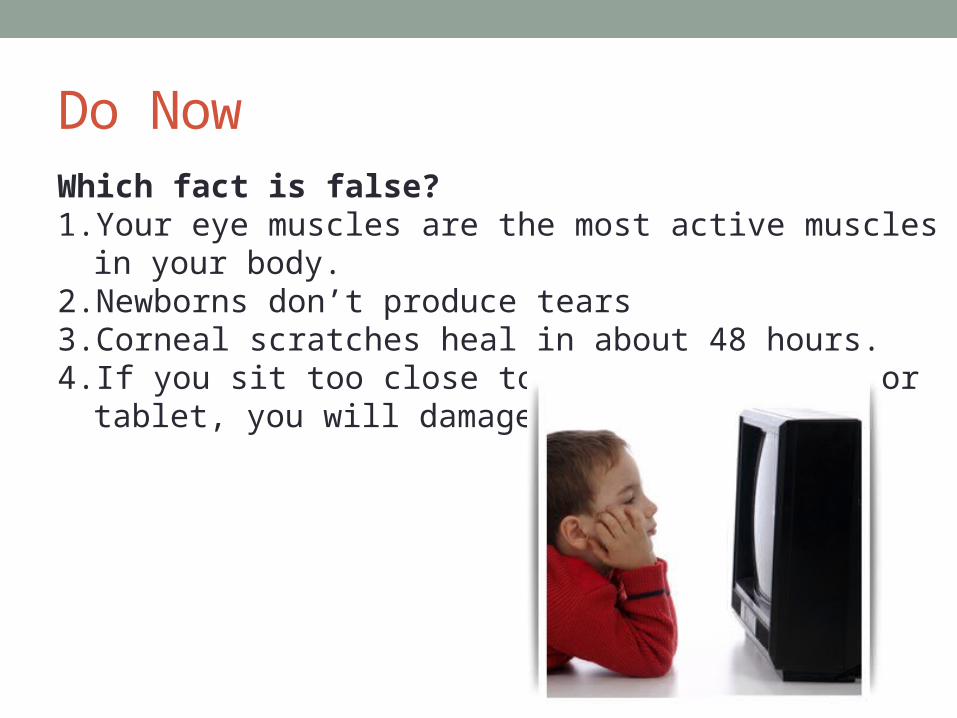
Do NowWhich fact is false?1. Your eye muscles are the most active muscles in your body.2. Newborns don’t produce tears3. Corneal scratches heal in about 48 hours.4. If you sit too close to a TV , computer, or tablet, you will
damage your eyes.

External Eye & Accessory StructuresLacrimal refers to tears. Tears cleanse and lubricate the eyes, and also fight bacteria.

External Eye & Accessory StructuresThe lacrimal ducts empty into the nasal cavity this is why nose and eye irritation is often linked.
• you get the sniffles if cry and • you get watery eyes if congested

External Eye & Accessory StructuresConjunctiva are the membranes the line the eyelid and eyeball.
Conjunctivitis inflammation of these membranes, caused by irritants, allergies, or infection (e.g. “pink eye”).

Extrinsic eye muscles
Control movement of the eyes.
Locations and functions can be reasoned out!
Remember: rectus = straight, oblique = slanting
Name Action
Lateral rectus
Medial rectus
Superior rectus
Inferior rectus
Inferior oblique
Superior oblique

Extrinsic eye muscles
Control movement of the eyes.
Locations and functions can be reasoned out!
Remember: rectus = straight, oblique = slanting
Name Action
Lateral rectus Moves eye laterally
Medial rectus Moves eye medially
Superior rectus Moves eye up
Inferior rectus Moves eye down
Inferior oblique Moves eye up and laterally
Superior oblique
Moves eye down and laterally

EyeballThe eye has three tunics, or coats.
• Sclera – “whites of the eye” , outermost, thick connective tissue. • Choroid – has blood vessels, middle layer• Retina – contains the photoreceptors (rods & cones), inner layer

EyeballThe eye is divided into two fluid-filled chambers:
• The anterior chamber is filled with aqueous humor• The posterior chamber is filled with vitreous humor• Both fluids maintain eye pressure, and the aqueous humor nourishes the
cornea and lens.
Glaucoma occurs when the aqueous humor doesn’t drain properly, resulting in increased eye pressure and blindness.

Pathway of light1. Light enters the eye at the cornea – a clear, hard part of the sclera.
Functions: protects eye and focuses light
Fun fact: the cornea is responsible for ~70% of the eye’s
focusing ability

Pathway of light2. Light passes through the pupil which is the opening in front of the
lens. • The size of the pupil is controlled by the muscles of the iris
(the colored part of the eye).• The pupil dilates or contracts to vary the amount of light hitting the
retina.

Pathway of light3. The light passes through the lens, which focuses the light onto the
retina.• The ciliary body are muscles which change the shape of the
lens to
focus on nearby items, a process called accomodation.

Pathway of light4. The light passes through the vitreous humor to land on the retina,
which contains the photoreceptors.
There are no photoreceptors on the optic disc, which is where the optic nerve exits the eye – this causes a small blind spot.

PhotoreceptorsRods
• more abundant • sensitive to low levels of light• do not discriminate colors
Cones • 3 types, each sensitive to a different wavelengths• triggering of more than one cone is
interpreted by brain as different colors
e.g. if both red and green are activated,
the brain will interpret the light as yellow
or orange
• greater resolution than cones • mostly found in fovea centralis
Responsible for night and peripheral vision – that’s why colors seem to be lost in the dark
Responsible for color and fine detail vision – including reading

Color BlindnessColor blindness is usually caused by the absence of one or more cones.
• Occurs in ~5% of population• X-linked trait … much more common in men

Color Blindness

Color Blindness

Color Blindness

Color Blindness

Refraction & AccomodationLight is bent – or refracted – by nearly every eye structure that it passes through on the way to the retina.
However the lens is the only structure that can vary how much the light is bent in order to allow us to focus on different objects – a process called accomodation.
• At rest, our eyes naturally focus on far-away objects. • However, by contracting the ciliary body muscles, we can make the lens bulge so that it has greater refractive ability – allowing us to focus on close items.
As we get older, our lens loses elasticity – making it harder to focus on nearby items.
This condition is called presbyopia (old eyes)

Fun fact:Refraction flips and reverses the light rays, forming an upside down and reversed image on the retina … but the brain learns to interpret visual information correctly.
Watch me!

• What were our objectives, and what did you learn about them.
• What was our learner profile trait and how did we exemplify it?
• How does what we did today address our unit question?
Closure

Exit Ticket1. Nine children attending the same day-care center developed
red, inflamed eyes and eye lids. What is the most likely cause and name of this condition?
2. Name two structures that help us to see in low light conditions.
3. Why do you often have to blow your nose after crying?
4. Name 4 substances that refract light? Which refracts light the most? Which is responsible for accomodation?