Embed Size (px)
Citation preview
EYE ON PACIFIC
INSIDE
-Uses of autofluorescence in clinical practice: p. 3
-The Vision Therapy Service has been remodeled: p. 4
-Don’t ignore decreased best corrected visual acuity: p. 4 pacificu.edu
News for local referring providers Fall | 2014
Pacific University College of Optometry
Corneal Topography in Optometric Practice As vision subspecialties continue to grow, we can ensure that patients continue to get the care they deserve.
Corneal topography has
revolutionized our understanding
of corneal disease diagnosis and
progression. Corneal topography
has numerous applications in
refractive surgery, and the
implications for contact lens design
and management continue to
evolve as information is gleaned
from the modern corneal
topographer.
The modern corneal topographer
has the ability to interpret the
videokeratoscopic image in several
ways. In optometric practice, three
display modes rise to the forefront:
axial, tangential, and elevation
displays.
MATT LAMPA, OD, FAAO | CONTACT LENS SERVICE CHIEF
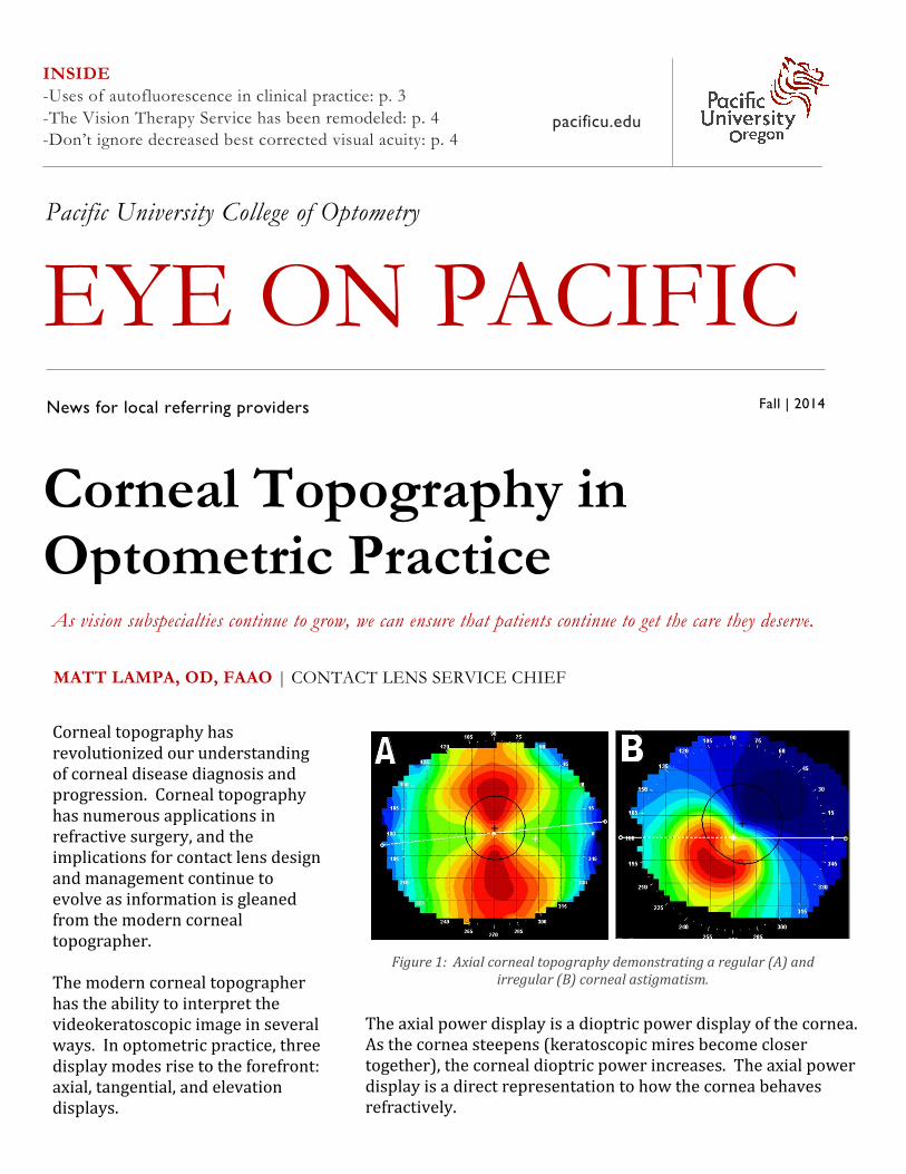
Figure 1: Axial corneal topography demonstrating a regular (A) and
irregular (B) corneal astigmatism.
The axial power display is a dioptric power display of the cornea.
As the cornea steepens (keratoscopic mires become closer
together), the corneal dioptric power increases. The axial power
display is a direct representation to how the cornea behaves
refractively.
Corneal Topography (continued)
another, i.e., how the slope or shape changes with
distance across the cornea. The tangential display
gives us an understanding of position of corneal
change. This is particularly important to the
contact lens practice when analyzing the position
of the contact lens with orthokeratology (See
Figure 2).
The elevation display attempts to describe the
cornea in terms of height rather than curvature.
This has direct implications as it relates to contact
lenses. The corneal topographer assumes a
reference surface and theoretically overlies this
on the cornea. From this reference surface the
The axial display is useful in
monitoring changes in corneal
power following refractive surgery
or orthokeratology, as well as
determining the amount and type
(regular or irregular) of
astigmatism over the pupil (See
Figure 1). This allows us to predict
how a patient may see optimally
with spectacles after careful
manifest refraction and when a
non-flexing rigid contact lens may
be indicated to improve the final
visual outcome for the patient (See
Figure 1).
A tangential display is more of a
smoothing of the corneal profile
and attempts to compare how one
point on the cornea relates to
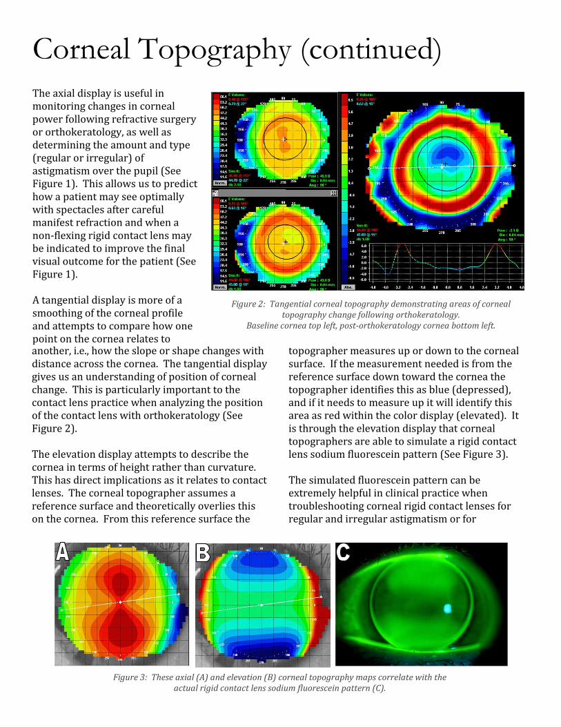
Figure 2: Tangential corneal topography demonstrating areas of corneal
topography change following orthokeratology.
Baseline cornea top left, post-orthokeratology cornea bottom left.
topographer measures up or down to the corneal
surface. If the measurement needed is from the
reference surface down toward the cornea the
topographer identifies this as blue (depressed),
and if it needs to measure up it will identify this
area as red within the color display (elevated). It
is through the elevation display that corneal
topographers are able to simulate a rigid contact
lens sodium fluorescein pattern (See Figure 3).
The simulated fluorescein pattern can be
extremely helpful in clinical practice when
troubleshooting corneal rigid contact lenses for
regular and irregular astigmatism or for
Figure 3: These axial (A) and elevation (B) corneal topography maps correlate with the
actual rigid contact lens sodium fluorescein pattern (C).
Corneal Topography (continued)
orthokeratology. Some corneal topographers
even attempt to simulate scleral lens designs
within their topography software.
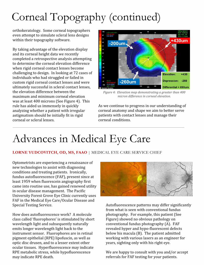
By taking advantage of the elevation display
and its corneal height data we recently
completed a retrospective analysis attempting
to determine the corneal elevation difference
when rigid corneal contact lenses become
challenging to design. In looking at 72 cases of
individuals who had struggled or failed in
custom rigid corneal contact lenses and were
ultimately successful in scleral contact lenses,
the elevation difference between the
maximum and minimum corneal elevation
was at least 400 microns (See Figure 4). This
rule has aided us immensely in quickly
analyzing whether a patient with irregular
astigmatism should be initially fit in rigid
corneal or scleral lenses.
Figure 4: Elevation map demonstrating a greater than 400
micron difference in corneal elevation.
As we continue to progress in our understanding of
corneal anatomy and shape we aim to better serve
patients with contact lenses and manage their
corneal conditions.
Advances in Medical Eye Care LORNE YUDCOVITCH, OD, MS, FAAO | MEDICAL EYE CARE SERVICE CHIEF
Optometrists are experiencing a renaissance of
new technologies to assist with diagnosing
conditions and treating patients. Ironically,
fundus autofluorescence (FAF), present since at
least 1959 when fluorescein angiography first
came into routine use, has gained renewed utility
in ocular disease management. The Pacific
University Forest Grove Eye Clinic currently uses
FAF in the Medical Eye Care/Ocular Disease and
Special Testing Service.
How does autofluorescence work? A molecule
class called ‘fluorophores’ is stimulated by short
wavelength light and subsequently naturally
emits longer wavelength light back to the
instrument sensor. Fluorophores are in retinal
pigment epithelial (RPE) lipofuscin, as well as
optic disc drusen, and to a lesser extent other
ocular tissues. Hyperfluorescence may indicate
RPE metabolic stress, while hypofluorescence
may indicate RPE death.
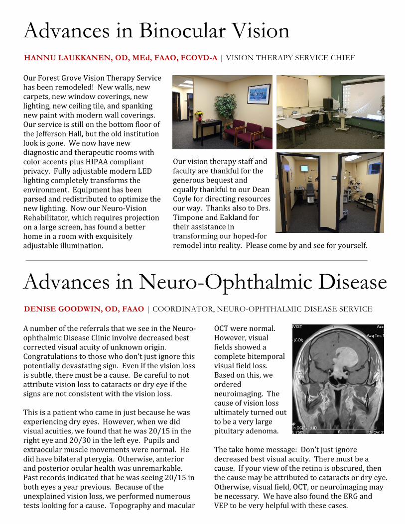
Autofluorescence patterns may differ significantly
from what is seen with conventional fundus
photography. For example, this patient (See
Figure) showed no obvious pathology on
conventional fundus photography (A). FAF
revealed hyper and hypo-fluorescent defects
below his macula (B). The patient admitted
working with various lasers as an engineer for
years, sighting only with his right eye.
We are happy to consult with you and/or accept
referrals for FAF testing for your patients.
Advances in Neuro-Ophthalmic Disease DENISE GOODWIN, OD, FAAO | COORDINATOR, NEURO-OPHTHALMIC DISEASE SERVICE
A number of the referrals that we see in the Neuro-
ophthalmic Disease Clinic involve decreased best
corrected visual acuity of unknown origin.
Congratulations to those who don’t just ignore this
potentially devastating sign. Even if the vision loss
is subtle, there must be a cause. Be careful to not
attribute vision loss to cataracts or dry eye if the
signs are not consistent with the vision loss.
This is a patient who came in just because he was
experiencing dry eyes. However, when we did
visual acuities, we found that he was 20/15 in the
right eye and 20/30 in the left eye. Pupils and
extraocular muscle movements were normal. He
did have bilateral pterygia. Otherwise, anterior
and posterior ocular health was unremarkable.
Past records indicated that he was seeing 20/15 in
both eyes a year previous. Because of the
unexplained vision loss, we performed numerous
tests looking for a cause. Topography and macular
The take home message: Don’t just ignore
decreased best visual acuity. There must be a
cause. If your view of the retina is obscured, then
the cause may be attributed to cataracts or dry eye.
Otherwise, visual field, OCT, or neuroimaging may
be necessary. We have also found the ERG and
VEP to be very helpful with these cases.
Advances in Binocular Vision HANNU LAUKKANEN, OD, MEd, FAAO, FCOVD-A | VISION THERAPY SERVICE CHIEF
Our Forest Grove Vision Therapy Service
has been remodeled! New walls, new
carpets, new window coverings, new
lighting, new ceiling tile, and spanking
new paint with modern wall coverings.
Our service is still on the bottom floor of
the Jefferson Hall, but the old institution
look is gone. We now have new
diagnostic and therapeutic rooms with
color accents plus HIPAA compliant
privacy. Fully adjustable modern LED
lighting completely transforms the
environment. Equipment has been
parsed and redistributed to optimize the
new lighting. Now our Neuro-Vision
Rehabilitator, which requires projection
on a large screen, has found a better
home in a room with exquisitely
adjustable illumination.
Our vision therapy staff and
faculty are thankful for the
generous bequest and
equally thankful to our Dean
Coyle for directing resources
our way. Thanks also to Drs.
Timpone and Eakland for
their assistance in
transforming our hoped-for
remodel into reality. Please
OCT were normal.
However, visual
fields showed a
complete bitemporal
visual field loss.
Based on this, we
ordered
neuroimaging. The
cause of vision loss
ultimately turned out
to be a very large
pituitary adenoma.
come by and see for yourself.
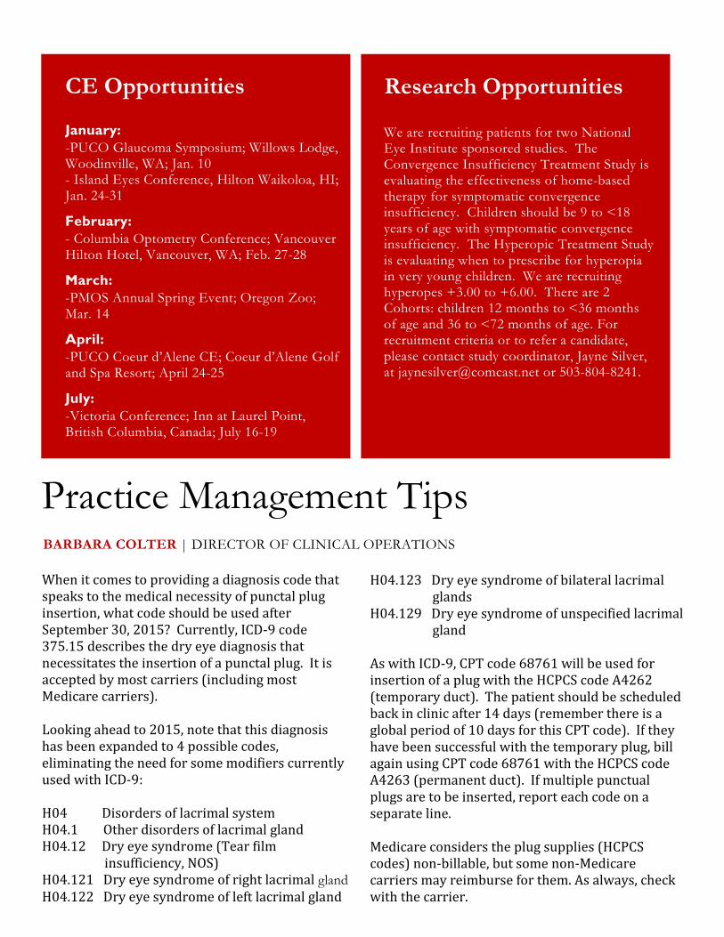
Practice Management Tips BARBARA COLTER | DIRECTOR OF CLINICAL OPERATIONS
When it comes to providing a diagnosis code that
speaks to the medical necessity of punctal plug
insertion, what code should be used after
September 30, 2015? Currently, ICD-9 code
375.15 describes the dry eye diagnosis that
necessitates the insertion of a punctal plug. It is
accepted by most carriers (including most
Medicare carriers).
Looking ahead to 2015, note that this diagnosis
has been expanded to 4 possible codes,
eliminating the need for some modifiers currently
used with ICD-9:
H04 Disorders of lacrimal system
H04.1 Other disorders of lacrimal gland
H04.12 Dry eye syndrome (Tear film
insufficiency, NOS)
H04.121 Dry eye syndrome of right lacrimal gland
H04.122 Dry eye syndrome of left lacrimal gland
H04.123 Dry eye syndrome of bilateral lacrimal
glands
H04.129 Dry eye syndrome of unspecified lacrimal
gland
As with ICD-9, CPT code 68761 will be used for
insertion of a plug with the HCPCS code A4262
(temporary duct). The patient should be scheduled
back in clinic after 14 days (remember there is a
global period of 10 days for this CPT code). If they
have been successful with the temporary plug, bill
again using CPT code 68761 with the HCPCS code
A4263 (permanent duct). If multiple punctual
plugs are to be inserted, report each code on a
separate line.
Medicare considers the plug supplies (HCPCS
codes) non-billable, but some non-Medicare
carriers may reimburse for them. As always, check
with the carrier.
January: -PUCO Glaucoma Symposium; Willows Lodge, Woodinville, WA; Jan. 10 - Island Eyes Conference, Hilton Waikoloa, HI; Jan. 24-31
February: - Columbia Optometry Conference; Vancouver Hilton Hotel, Vancouver, WA; Feb. 27-28
March: -PMOS Annual Spring Event; Oregon Zoo; Mar. 14
April: -PUCO Coeur d’Alene CE; Coeur d’Alene Golf and Spa Resort; April 24-25
July: -Victoria Conference; Inn at Laurel Point, British Columbia, Canada; July 16-19
CE Opportunities
We are recruiting patients for two National Eye Institute sponsored studies. The Convergence Insufficiency Treatment Study is evaluating the effectiveness of home-based therapy for symptomatic convergence insufficiency. Children should be 9 to <18 years of age with symptomatic convergence insufficiency. The Hyperopic Treatment Study is evaluating when to prescribe for hyperopia in very young children. We are recruiting hyperopes +3.00 to +6.00. There are 2 Cohorts: children 12 months to <36 months of age and 36 to <72 months of age. For recruitment criteria or to refer a candidate, please contact study coordinator, Jayne Silver, at [email protected] or 503-804-8241.
Research Opportunities
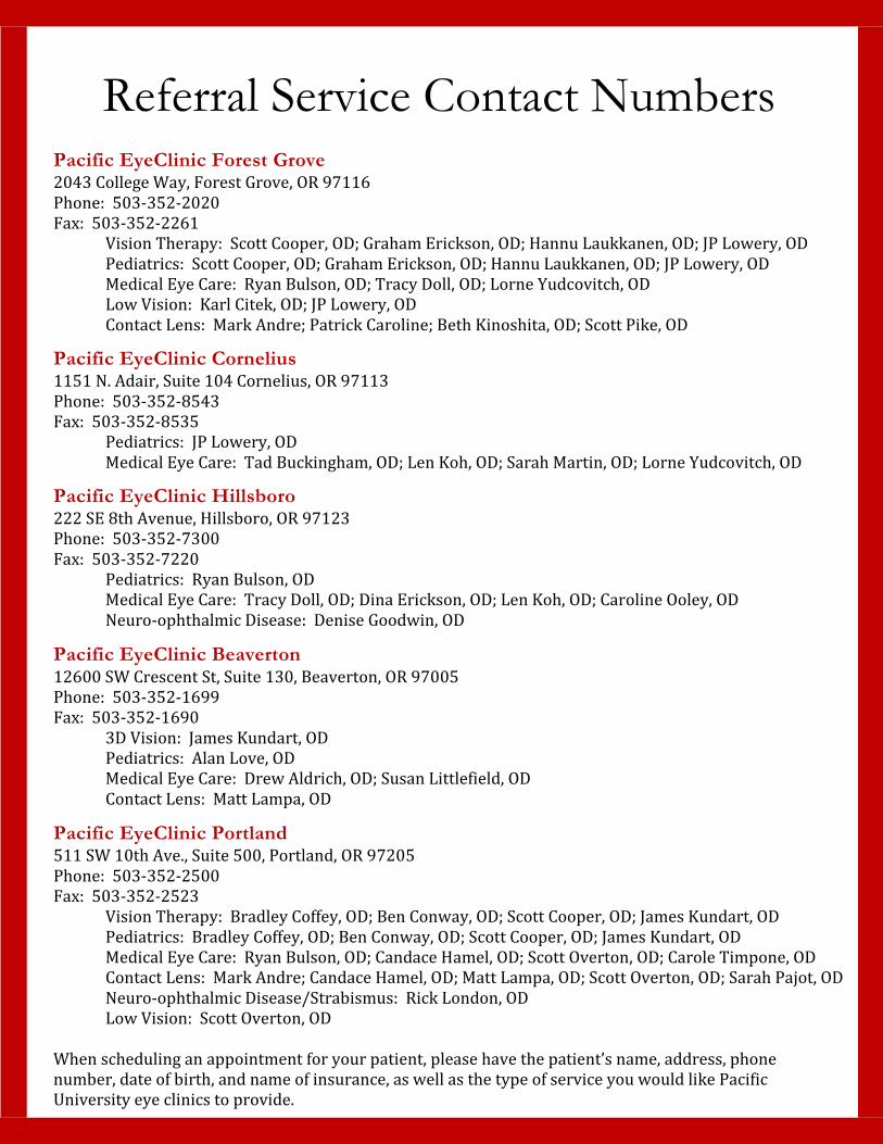
Referral Service Contact Numbers
Pacific EyeClinic Forest Grove 2043 College Way, Forest Grove, OR 97116
Phone: 503-352-2020
Fax: 503-352-2261
Vision Therapy: Scott Cooper, OD; Graham Erickson, OD; Hannu Laukkanen, OD; JP Lowery, OD
Pediatrics: Scott Cooper, OD; Graham Erickson, OD; Hannu Laukkanen, OD; JP Lowery, OD
Medical Eye Care: Ryan Bulson, OD; Tracy Doll, OD; Lorne Yudcovitch, OD
Low Vision: Karl Citek, OD; JP Lowery, OD
Contact Lens: Mark Andre; Patrick Caroline; Beth Kinoshita, OD; Scott Pike, OD
Pacific EyeClinic Cornelius 1151 N. Adair, Suite 104 Cornelius, OR 97113
Phone: 503-352-8543
Fax: 503-352-8535
Pediatrics: JP Lowery, OD
Medical Eye Care: Tad Buckingham, OD; Len Koh, OD; Sarah Martin, OD; Lorne Yudcovitch, OD
Pacific EyeClinic Hillsboro 222 SE 8th Avenue, Hillsboro, OR 97123
Phone: 503-352-7300
Fax: 503-352-7220
Pediatrics: Ryan Bulson, OD
Medical Eye Care: Tracy Doll, OD; Dina Erickson, OD; Len Koh, OD; Caroline Ooley, OD
Neuro-ophthalmic Disease: Denise Goodwin, OD
Pacific EyeClinic Beaverton 12600 SW Crescent St, Suite 130, Beaverton, OR 97005
Phone: 503-352-1699
Fax: 503-352-1690
3D Vision: James Kundart, OD
Pediatrics: Alan Love, OD
Medical Eye Care: Drew Aldrich, OD; Susan Littlefield, OD
Contact Lens: Matt Lampa, OD
Pacific EyeClinic Portland 511 SW 10th Ave., Suite 500, Portland, OR 97205
Phone: 503-352-2500
Fax: 503-352-2523
Vision Therapy: Bradley Coffey, OD; Ben Conway, OD; Scott Cooper, OD; James Kundart, OD
Pediatrics: Bradley Coffey, OD; Ben Conway, OD; Scott Cooper, OD; James Kundart, OD
Medical Eye Care: Ryan Bulson, OD; Candace Hamel, OD; Scott Overton, OD; Carole Timpone, OD
Contact Lens: Mark Andre; Candace Hamel, OD; Matt Lampa, OD; Scott Overton, OD; Sarah Pajot, OD
Neuro-ophthalmic Disease/Strabismus: Rick London, OD
Low Vision: Scott Overton, OD
When scheduling an appointment for your patient, please have the patient’s name, address, phone
number, date of birth, and name of insurance, as well as the type of service you would like Pacific
University eye clinics to provide.