Embed Size (px)
Citation preview

J. exp. Biol. (1975), 6a, 175-187 175Uth 1 plate and 10 figures
WinInted in Great Britain
EYE MOVEMENTS IN DAPHNIA PULEX (DE GEER)
BY B. J. FROST
Department of Psychology, Queen's University, Kingston, Ontario, Canada
(Received 31 July 1974)
SUMMARY
1. The various types of eye movement exhibited by the cyclopean eye ofDaphnia pulex were studied using high speed motion photography.
2. This rudimentary eye, which consists of only 22 ommatidia, can movethrough approximately 150° in the sagittal plane and 6o° in the horizontalplane.
3. Four classes of eye movement were found: (1) a high speed tremor at16 Hz with an amplitude of 3-4°, which resembles physiological nystagmus,(2) a slow rhythmic scanning movement at 4 Hz, and 5-60 amplitude, (3)large fast eye movements similar to saccadic eye movements and (4) opto-kinetic nystagmus produced by moving striped patterns.
4. Where the fast tremor occurred concurrently with the slow rhythmicscan, a Fourier analysis revealed that the former was the fourth harmonic ofthe latter.
INTRODUCTION
The classification of different patterns of eye movement and the description of theirfunctional significance have resulted mainly from observations of simple camera-typeeyes of vertebrates. However, the compound and simple eyes of many invertebratesare also movable, and detailed studies of several species have recently been published.For example, Gregory, Moray & Ross (1964), Moray (1973) and Downing (1973)have shown that the single ommatidium which constitutes the lateral eye of themarine Copepod Copilia is scanned back and forth in the focal plane of its anteriorlens. Land (1969) has shown that the antero-median eyes of the jumping spidersPhidippus johnsoni and Metaphidipus aeneolus produce several different patterns ofeye movement, some of which are related to the identification of certain environ-mental stimuli. Also Horridge & Burrows (1968 a, b, c) have described optokineticnystagmus, saccadic and compensatory eye movements in the compound sessile eyeof the crab Carcinus.
It has long been known that many members of the Cladoceran order have movableeyes. Daphnia in particular has been shown to produce large eye movements especiallyin the sagittal plane, and indeed Scheffer, Robert & Medioni (1958) have used thisresponse very effectively to determine the spectral luminosity curve for Daphnia pulex(De Geer). Early investigations revealed that the Daphnid eye appears to maintaina fixed direction with respect to a light beam (Radl, 1910; Ewald, 1913), and von Frisch& Kupelwieser (1913) demonstrated that this 'fixation' is an important element in thedorsal light reaction, and therefore possibly involved in the normal orientation

176 B. J. FROST
behaviour of the organism. The purpose of this investigation was to determine the g r ^optical properties of the Cyclopean compound eye of Daphnia pulex and to describetheir full range of eye movements. Informal observations have revealed that manydifferent patterns of eye movements are produced, and an attempt has been made tospecify the stimulus conditions which elicit specific movements so that inferences canbe made about their functional significance.
MATERIAL AND METHODS
A culture of Daphnia pulex (De Geer) was kept in swamp water maintained at roomtemperature, and fed occasionally with a timothy hay broth. For histology, seven largefemales 2-3 mm long were fixed in Bouin's solution, dehydrated in ethyl alcohol, andembedded in paraffin wax. Serial coronal sections were cut at 5 /im intervals from thehead region of four Daphnia, while 10 /on serial sagittal sections were cut from theremaining 3 specimens. All sections were then mounted, stained with haematoxylinand eosin, made into permanent slides and examined under a binocular compoundmicroscope. Measurements of the various parameters of the cyclopean compound eye,such as radius or curvature of ommatidial lenses, rhabdom size, etc., were made withthe aid of a micrometer scale attachment mounted in one of the oculars of the micro-scope.
Records of eye movements were obtained by filming the cyclopean eye through oneeyepiece of a binocular dissecting microscope, with an Araflex 16 mm single lensreflex movie camera using Kodak Tri X film. The film was shot at either 25 or50 frames per second. A frame by frame determination of eye position was made withthe aid of a 16 mm projector fitted with a single frame advancement mechanism andframe counter. The procedure involved making an initial tracing of the outline of theeye, carapace, and prominent landmarks such as the one or two ommatidial lensesreflecting light, together with the frame limits of the first frame. This designated thearbitrary zero position of the eye. Then in subsequent frames the angular position ofthe eye was determined by superimposing the tracing of the eye onto the projectedimage of the eye and measuring the angular displacement between the traced andprojected frame lines. Generally sections of film where no body movement occurredwere used, but slight movement could be compensated for by making a new tracingof the changed position.
Informal observations had revealed that Daphnia exhibit a variety of different eyemovements. Consequently an attempt was made to describe the specific stimulusconditions which give rise to particular types of eye movement. The stimulus condi-tions that were used in formal experiments were as follows:
(1) Diffuse white light. Under this condition the substage illumination of the micro-scope was passed through a diffusing screen immediately below the slide containingthe preparation. This in effect produced a 'ganzfeld' for the Daphnia, and providedan opportunity to observe spontaneous eye movements when very few visual contourswere present in the visual field.
(2) A moving spot of light. During preliminary observations it was noted that largesaccadic eye movements could be produced by moving a light across the visual fieldof Daphnia. Consequently under this condition the substage microscope light was

Eye movements in Daphnia pulex (De Geer)
s
OF,
177
Fig. 1. Apparatus for presenting stimuli in two alternate positions in Daphnia's visual field.Light from a tungsten light source (6 V automobile headlamp) was collimated by a short focallength lens L, and directed on to two flexible fibre optic cables OFj and OF& Daphnia werepositioned on their side in the bottom of a well W. The two optic fibres were inserted 900 apartthrough the plexiglass block so that their ends came in close proximity to the wall of the well.A sliding shutter S made it possible to illuminate either GFj or OF% alternately.
focused to form an intense spot 2 mm in diameter on the ground glass screen immedi-ately below the Daphnia. This spot was then moved back and forth across the visualfield of the Daphnia by manipulating the microscope mirror.
(3) A light spot flashed alternately between two positions. This stimulus arrangementwas used to observe eye movements generated by change in position without con-tinuous movement through the intervening space. To produce such stimuli the simpleoptical device illustrated in Fig. 1 was constructed. Daphnia were positioned on theirsides facing midway between the two light spots and then pinned to a thin layer ofwax in the bottom of the well. Thus the two spots of light, which subtended approxi-mately 150 of visual angle, could be alternated between 2 positions 900 apart in thesagittal plane of the Daphnia.
(4) Moving striped patterns with velocity and spatial frequency varied. Optokineticnystagmus was investigated by placing Daphnia in a fixed position in the centre of acylinder around which various patterns of stripes were rotated. Fig. 2 shows aschematic diagram of the apparatus used to make these observations. The apparatus,which was driven by a Cole-Parmer constant torque, variable speed drive unit, pro-duced a visual field of moving vertical stripes which extended approximately 1800 inthe horizontal plane and 700 in the vertical plane.
Several different stimulus cylinders were constructed which produced: (a) Blackand white (clear) stripes subtending visual angles to the Daphnia of 450, 22-5°, 150,
. 10°, and 50; (b) a single chromatic stripe 22-5° in visual angle, produced by inserting12 EXB 62

i 7 8 B. J. FROST
Fig. 2. Apparatus for presenting moving grating patterns to Daphnia to elicit optokineticnystagmus. A plexiglass cylinder A, was divided into two hemicylinders by a plexiglasspartition B. Daphnia were gently wedged into a sawcut in the upper surface of block C so thatthey faced the other hemicylinder which was filled with water. Various stimulus patterns wereattached to marginally larger plexiglass cylinders D, which could be rotated in either directionby a friction drive wheel E. A constant torque variable speed drive unit connected to the drivewheel permitted velocity adjustments over a wide range of values. A diffusing screen F backilluminated by a i oo W tungsten bulb produced an evenly illuminated background against whichthe various patterns of stripe were viewed by the Daphnia.
a two layers thick strip of yellow filter transmitting wavelengths centred around590 nm (Edmonds No. 815) between the ends of a blue filter (Edmonds No. 855)transmitting wavelengths centred around 445 nm (with this arrangement the blueand yellow regions of the pattern transmitted approximately equal intensities of light);(c) a single polarized stripe, also 22-5° in visual angle, produced by inserting a sectionof Polaroid filter with the e-vector rotated 900 from the Polaroid filter making up theremainder of the cylinder. Angular velocity of these stimuli were varied over therange of 5-7200 sec"1 in either a clockwise or anticlockwise direction.
RESULTS
The cyclopean compound eye
The histological observations indicate that the Daphnia eye consists of a cyclopeancompound structure containing 22 ommatidia arranged with their axes approxi-mately 380 apart around the surface of a nearly hemispherical structure. The meanradius of curvature of ommatidial corneal lenses was 16-5 /tm while the mean thicknessof the lenses was 41 /tm.
Twenty-two bundles of optic nerve fibres each containing 8 axons, presumablyfrom the same ommatidium, leave the eye and terminate in the first optic ganglion
Eye movements in Daphnia pulex (De Geer)
5°
179
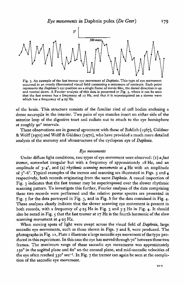
Fig. 3. An example of the fast tremor eye movement of Daphnia. This type of eye movementoccurred in an evenly illuminated visual field containing a minimum of contours. Each pointrepresents the Daphnia's eye position on a single frame of movie film, the dorsal direction is upand ventral down. A Fourier analysis of this data is presented in Fig. 5, where it can be seenthat the fast tremor has a frequency of 17 Hz, and that it is superimposed on a slower wavewhich has a frequency of 4-25 Hz.
of the brain. This structure consists of the familiar rind of cell bodies enclosing adense neuropile in the interior. Two pairs of eye muscles insert on either side of theanterior loop of the digestive tract and radiate out to attach to the eye hemisphereat roughly 900 intervals.
These observations are in general agreement with those of Rohlich (1967), Giildner& Wolff (1970) and Wolff & Giildner (1970), who have provided a much more detailedanalysis of the anatomy and ultrastructure of the cyclopean eye of Daphnia.
Eye movements
Under diffuse light conditions, two types of eye movement were observed: (1) a fasttremor, somewhat irregular but with a frequency of approximately 18 Hz, and anamplitude of 3-40, and (2) rhythmic scanning movements at 4 Hz with an amplitudeof 5°-6°. Typical examples of the tremor and scanning are illustrated in Figs. 3 and 4respectively, both records originating from the same Daphnia. A casual inspection ofFig. 3 indicates that the fast tremor may be superimposed over the slower rhythmicscanning pattern. To investigate this further, Fourier analyses of the data comprisingthese two records were performed and the relative power spectra are presented inFig. 5 for the data portrayed in Fig. 3, and in Fig. 6 for the data contained in Fig. 4.These analyses clearly indicate that the slower scanning eye movement is present inboth records, with a frequency of 4-25 Hz in Fig. 3 and 3-5 Hz in Fig. 4. It shouldalso be noted in Fig. 5 that the fast tremor at 17 Hz is the fourth harmonic of the slowscanning movement at 4-25 Hz.
When moving spots of light were swept across the visual field of Daphnia, largesaccadic eye movements, such as those shown in Figs. 7 and 8, were produced. Thephotographs in Fig. 11, Plate 1 illustrate a large saccadic eye movement of the type pro-duced in this experiment. In this case the eye has moved through 700 between these twoframes. The maximum range of these saccadic eye movements was approximately1500 in the sagittal plane and 60° on the coronal plane, and mid-saccadic velocities ofthe eye often reached 3300 sec"1. In Fig. 7 the tremor can again be seen at the comple-tion of the saccadic eye movement.

i8o B. J. FROST
300 msec
Fig. 4. An example of the rhythmic scanning type of eye movement. This pattern also tends tooccur in an evenly illuminated visual field containing few contours. The Fourier analysis forthis section of record is illustrated in Fig. 6 where it can be seen that the most power occursat 3-5 Hz.
150
100
50
Power spectrum
10 15Frequency (Hz)
20 25
Fig. 5. Fourier analysis of the fast tremor eye movements presented in Fig. 3. An inspection ofFig. 3 indicates that the slow rhythmic scan illustrated in Fig. 4 may also be present. Thisconclusion is confirmed by the Fourier analysis, which indicates that there is considerablepower around 4 Hz. It should also be noted that the frequency of the fast tremor at 17 Hz isthe fourth harmonic of the slow rhythmic scan at 4-25 Hz.

60
50
30
20
10
Eye movements in Daphnia pulex (De Geer)
Power spectrum
181
10 15Frequency (Hz)
20 25
Fig. 6. Fourier analysis of the slow rhythmic scan presented in Fig. 4, indicating that themajor frequency component is at 3*5 Hz.
40°
300 msec
• - V
Fig. 7. Large spontaneous saccadic eye movement in the sagittal plane. This movementoccurred under diffuse illumination. Note that the fast tremor appears to be superimposed onthe saccade and is clearly visible when the eye comes to rest at the end of the record.
Observations taken with the two light spots alternated in positions 900 apart yieldessentially the same results as produced under the moving stimulus conditions. Anexample of the saccadic-like eye movements produced by this condition is shown inFig. 9.
Optokinetic nystagmus was produced by moving a striped cylinder around Daphnia,using the apparatus described above. Although different widths of stripe ranging from
|45° to 50 of visual angle were used, stripes of less than 150 failed to elicit any measurable

182 B. J. FROST
20°
200 msec
Fig. 8. Saccadic eye movements produced by a moving stimulus. The substage illumination wasfocused to form a bright spot 2 mm in diameter on a ground glass screen below the Daphnia,and in this case was moved dorsally and then ventrally. The eye position can be seen to followthis movement first in a dorsal direction and then in a ventral direction.
10°
200 msec
Fig. 9. Saccadic eye movement produced by displacement of a light spot. See text for details.
optokinetic nystagmus. The optimum width of stripe was approximately 22-5°, andthe ability of the cyclopean eye to follow different velocities of this stimulus wasinvestigated. At slower velocities (A) of the stimulus, eye velocity does not alwaysmatch stimulus velocity, but rather the eye appears to track first in the direction ofstimulus movement, and then remain stationary for a period of time before returningwith a saccadic movement. However, at medium stimulus velocities (B), the slowerfollowing or pursuit phase can be clearly differentiated from the fast saccadic returns.Again, at higher velocities eye velocity fails to match stimulus velocity, as can be seenin the latter part of record (C), until it eventually disappears at a stimulus velocity ofapproximately 2200 sec"1.

Eye movements in Daphnia pulex (De Geer) 183
f\
30°
1 sec
>••/
30°
1 sec
V'*
10°
400 msec
Fig. 10. Optokinetic nystagmus produced by a grating pattern moved at various velocities. Theapparatus illustrated in Fig. 2 was used to rotate the grating pattern, consisting of 22-5° blackand white stripes, around Daphnia. The stimulus velocity was 400 sec"1 in A, 75° sec"1 in B,and 1500 sec"1 in C.
Since optokinetic nystagmus provides a useful index of visual resolution it wasdecided to see if stripes formed by e-vector rotation and wavelength differences wouldalso elicit eye following responses. However, it appears that only luminance differenceswill produce optokinetic nystagmus, since none of these other stimulus patternsproduced any discernible response.
DISCUSSION
The results of this series of experiments indicate that Daphnia exhibit a variety ofdifferent types of eye movements, many of which bear a close resemblance to thosedescribed for higher organisms.
(1) The fast tremor. This type of eye movement has a frequency in the range of15-20 Hz and sometimes occurs in conjunction with other types of movement. Thefast tremor appears to be similar to physiological nystagmus in man, which has beenshown by stabilized image experiments (Ditchburn & Ginsborg, 1952; Iarbus, 1967)to be essential for reducing adaptation and the fading of visual images. It has alsobeen suggested by Autrum (1950) and Mazokhin-Porshnyakov (1969) that the irregularjerky flight patterns produced by many insects with fixed compound eyes mightachieve the same effect as eye movements by producing rapid transients in the visualinput. Because of the rather coarse visual resolution of the cyclopean eye of Daphniathese continual tremoring movements would increase the probability of changing theluminous flux reaching a particular ommatidium. The fact that the amplitude of these

184 B. J. FROST
movements in only 3°-4°, whereas ommatidia are spaced approximately 35indicates that the most significant changes in luminous flux might take place at thesubommatidial level. Optical calculations made on the basis of measurements of theommatidial lenses, together with theoretical calculations of the static resolving powerof single ommatidia (Mazokhin-Porshnyakov, 1969), indicate that subommatidialresolution will be poor. However, both empirical observations by Burtt & Catton(1954, 1956, i960) and theoretical considerations by Mazokhin-Porshnyakov (1969)indicate that a compound eye in motion is able to resolve image detail which wouldgo unnoticed in the absence of motion. It would therefore seem likely that the fasttremor type of eye movement found in Daphnia could serve two possible functions,one concerned with preventing loss of information through adaptation and the otherto increase acuity.
(2) Rhythmic scanning movements. This pattern of movement, which consists of slowsinusoidal oscillations of the eye at approximately 4 Hz with an amplitude of 5-60,tends to occur more frequently when Daphnia are placed in a diffusely lit environmentwith a minimum of contours. On some occasions the scanning occurs in the absenceof any obvious tremor. The possible function this type of eye movement could beperforming is not altogether clear. However, Moray (1973) has argued that therelatively slow scan found in Copilia denticulata increases the amount of informationthat can be transmitted about the visual field. If this is generally true, then the scanningmovements of Daphnia might possibly be concerned with a search for visual detail.This interpretation is consistent with the fact that this type of movement seems tooccur more often in a diffusely illuminated field than in one containing many visualcontours. Land (1969) has reported that similar scanning movements are producedby the antero-medial eyes of jumping spiders, but in this case the characteristics ofthe scan are clearly determined by the stimulus configuration presented to the spider.In fact, it appears that the scanning movements in jumping spiders are intimatelyassociated with object identification.
An interesting relationship was observed between the frequencies of the fast tremorand the rhythmic scanning movements when they occurred concurrently. A Fourieranalysis of these data reveals that the tremor frequency was in fact the fourth harmonicof the rhythmic scan, which suggests that a single neural time base might be re-sponsible for generating both patterns of movement. Many different neuronal modelscan be generated to provide the sort of neural economy suggested by these eye move-ments but it would be premature to specify these at this stage in the absence of anyelectrophysiological data.
(3) Saccadic eye movements. These fast and often very large eye movements havebeen observed to extend to a remarkable 1500 in the sagittal plane. Experiments withmoving spots of light and spots changed in position indicate that stimulus displace-ment is a sufficient condition to produce this type of movement. However, Daphniaplaced in a fixed position in a stationary visual field in which several contours arepresent will also exhibit spontaneous saccadic eye movements. In vertebrate speciespossessing a fovea the function of saccadic eye movements is clear, that is they bringthe image of an object of interest to the organism onto the region of most acute vision(Walls, 1967). Also, other types of areal specialization, such as the colour fields incertain avian retinae, would require the appropriate alignment of the eyes with

Eye movements in Daphnia pulex (De Geer) 185
Hccadic-like movements. However, there is no evidence available at present to suggestareal specialization in the compound eye of Daphnia, although subsequent studies ofthe microstructure of ommatidia in different locations on the eye might reveal suchdifferentiation. In fact, since the ommatidia are approximately equally spaced aroundthe nearly hemispherical structure of the eye, it seems unlikely that saccadic move-ments can be functioning to extend the visual field of the organism, because the visualfield is essentially limited by parts of the Daphnia's own anatomy. In animals possessingnon-foveate eyes, such as most species of fish, Walls (1967) has suggested that eyemovements serve the function of compensating for both voluntary and involuntarybody movements. Several attempts were made to film free-swimming Daphnia whilethey were confined to a very thin transparent tank in the hope of being able to correlateeye movements with self-induced changes in position. However, with the equipmentavailable it was not possible to produce film in which both eye and body position couldbe clearly determined.
(4) Optokinetic nystagmus. The series of experiments in which various stripedpatterns were moved through the visual field of Daphnia confirm that its visual resolu-tion is poor, since stripes of less than 150 visual angle and velocities of movementfaster than 2200 sec 1 do not elicit an optokinetic response. As in other species,optokinetic nystagmus in Daphnia is characterized by a slower pursuit phase, where theeye matches the angular velocity of the moving stripes in order to stabilize its visualworld, and a faster return or saccadic phase.
It might be argued that rotation of the striped pattern could produce flickeringillumination at the Daphnia's eye. If each individual flash evoked an eye movement, it ispossible this could be mistaken for optokinetic nystagmus. However there are severalfacts which make this interpretation of the data unlikely. In the first instance, theapparatus and stimuli were designed so that the mean luminance of the striped hemi-cylinder viewed by Daphnia remained constant during movement of the patterns.Secondly, under optimum conditions of stimulus velocity and spatial frequency theeye movements could be clearly differentiated into a following phase, where the eyematched the direction and angular velocity of the stripes, and a faster reset, or return,saccadic phase. Finally, the frequency of eye movements did not increase in frequencywith increased stimulus velocity as would be predicted from the flicker hypothesis.
An analysis of the colour dances of Daphnia by Baylor & Smith (1957) and thedetermination of their spectral luminosity curve by Scheffer et al. (1958) indicatesthat these organisms possess at least a primitive form of colour vision. Daphnia alsoorient orthogonally to the e-vector of plane polarized light (Baylor & Smith, 1953;Hazen & Baylor, 1962), and electron microscopy studies (Eguchi & Waterman, 1966)have revealed that rhabdom microvilli are oriented only in two planes 900 apart. Thisindicates that the Daphnia visual system is specialized for differentiating the e-vectorof incident light. Consequently, stripes constituted of wavelength differences ande-vector differences were used, but under no conditions were optokinetic responsesproduced by these stimuli. Thus, it would seem that optokinetic nystagmus can onlybe produced by moving patterns of luminance differences within the visual field.
When the surface of water is still, then underwater animals possess an 'arealwindow' (Walls, 1967) in their visual field, which is an optical effect produced by thetotal reflexions of rays that strike the surface at angles greater than the critical angle

186 B. J. FROST
of incidence. In the natural habitat oiDaphnia this would provide a visual field whiolconsisted of a bright circular window centred above the organism, surrounded by adarker field constituted of reflexions from the bottom of the swamp or pond. Goodman(1965) has suggested that locusts regulate their flight attitude by orienting their headsso that the horizon is horizontal and the upper half of their visual field is brighter thanthe lower half, then reflexly align their bodies with their heads. As von Frisch &Kuplewieser (1913) and Harris,' 1953) have suggested, such an 'areal window' couldpossibly provide a similar orientational landmark for Daphnia, wherein any departurefrom their maintained orientation would result in a reflex change in eye positionfollowed by compensatory movements of their setae to regain their appropriateorientation.
I am grateful to Professor Gerald Westheimer and Dr Michael Land for theirhelpful suggestions during the course of this research. This work was mainly carriedout in the Department of Physiology-Anatomy, at the University of California,Berkeley, and was in part supported by a grant (NB 03154) from the National Instituteof Neurological Diseases U.S. Public Health Services to Professor Westheimer, andin part by a National Research Council of Canada Grant No. AO 353 to Barrie J.Frost.
REFERENCES
AUTRUM, H. (1950). Die Belichtungspotentiale und das Sehen der Insecten. Z. vergl. Physiol. 33, 176.BAYLOR, E. R. & SMITH, F. E. (1957). Diurnal migration of plankton crustaceans. In Recent Advances
in Invertebrate Physiology (ed. B. T. Scheer). Eugene: University of Oregon Press.BAYLOR, E. R. & SMITH, F. E. (1953). The orientation of Cladocera to polarised light. Am. Nat. 87,
97-101.BURTT, E. T. & CATTON, W. T. (1954). Visual perception of movement in the locust. J. Physiol., Lond.
125, 566-80.BURTT, E. T. & CATTON, W. T. (1956). Electrical responses to visual stimulation in the optic lobes of
the locust and certain other insects. J. Physiol., Lond. 133, 68-88.BURTT, E. T. & CATTON, W. T. (i960). Is the mosaic theory of insect vision true? Proc. XI int. ent.
Congr. 1, 670-3.DITCHBURN, R. W. & GINSBORG, B. L. (1952). Vision with a stabilized retinal image. Nature, Lond. 170,
36.DOWNING, A. C. (1973). Optical scanning in the lateral eyes of the copepod Copilia. Perception, vol. 1,
no. 3, pp. 245-367.EGUCHI, E. & WATERMAN, T. H. (1966). Fine structure patterns in crustacean rhabdoms. In The
Functional Organisation of the Compound Eye (ed. C. G. Bernhard). Oxford: Pergamon Press.EWALD, W. F. (1913). The applicability of the photochemical energy-law to light reactions in animals.
Science, N. Y. 38, 236-7.GOODMAN, L. J. (1965). The role of certain optomotor reactions in regulating stability in the rolling
plane during flight in the desert locust, Schistocerca gregaria. J. exp. Biol. 42, 385-407.GREGORY, R. L., MORAY, N. & Ross, H. (1964). The curious eye of Copilia. Nature, Lond. 201, 1166-8.GULDNER, P. H. & WOLFF, J. (1970). Uber das Ultrastruktur des Komplexauges des Wasserflohs Daphnia
pulex. Z. Zellforsch. 104, 259-74.HARRIS, J. E. (1953). Physical factors involved in the diurnal migration of plankton. Quart. Jl microsc.
Sci. 94, 537-50.HAZEN, W. E. & BAYLOR, E. R. (1962). Behaviour of Daphnia in polarised light. Biol. Bull. mar. biol.
Lab., Woods Hole 123, 243-52.HORRIDGE, G. A. & BURROWS, M. (1968a). Tonic and phasic systems in parallel in the eyecup responses
of the crab Carcinus. J. exp. Biol. 49, 269-84.HORRIDGE, G. A. & BURROWS, M. (19686). The onset of the fast phase in the optokinetic response of
the crab Carcinus. J. exp. Biol. 49, 299-313.HORRIDGE, G. A. & BURROWS, M. (1968c). Efferent copy and voluntary eyecup movement in the crab
Carcinus. J. exp. Biol. 49, 315-24.IARBUS, A. L. (1967). Eye Movements in Vision. New York: Plenum Press.


Journal of Experimental Biology, Vol. 62, No. 1 Fig. 11, Plate
B. J. FROST (Facing p. 187)

t Eye movements in Daphnia pulex (De Geer) 187
ND, M. F. (1699). Movements of the retinae of jumping spiders in response to visual stimuli. J. exp.Biol. 51, 471-93.
MAZOKHIN-PORSHNYAKOV, G. A. (1969). Insect Vision. New York: Plenum Press.MORAY, N. (1973). Visual mechanisms in the copepod Copilia. Perception, vol. 1, no. 2, pp. 193-207.RADL, E. (1910). Uber den Phototropismus einiger Arthropoden. Biol. Zbl. 21, 75-86.ROHLICH, P. (1967). Fine structural changes induced in photoreceptors by light and prolonged dark.
In Symposium on Neurobiology of Invertebrates. Budapest: Publishing House of the HungarianAcademy of Sciences.
SCHEFFER, D., ROBERT, P. & MEDIONI, J. (1958). Reactions oculo-motrices de la Daphnie (Daphniapulex De Geer) en response a des lumieres monochromatique d'egale 6nergie. Sensibility visuelle etdermo-optique. C. r. Se"anc. Soc. Biol. (Strasbourg) 152, 1000-3.
VON FRISCH, K. & KUPELWIESER, H. (1913). Uber den Einfluss der Lichtfarbe auf die photoaktischenReaktionen niederer Krebse. Biol. Zbl. 33, 517-52.
WALLS, G. L. (1967). The Vertebrate Eye and its Adaptive Radiations. New York: Hafner.WOLFF, J. R. & GOLDNER, P. H. (1970). Uber die Ultrastruktur des' Nervus Opticus' und des Ganglion
Opticum I von Daphnia pulex. Z. Zellforsch. 103, 526-43.
EXPLANATION OF PLATE
Photographs of cyclopean compound eye of Daphnia pulex showing a ventral saccadic eye movementof 70°. Some of the 22 bundles of optic nerve fibres can be seen leaving the eye and converging on thefirst optic ganglion of the brain. Two of the four eye muscles can also be seen to be attached to dorsal-lateral and ventro-lateral positions of the eye and to insert close to the forward loop of the gut.