Embed Size (px)
Citation preview
Eye Examination and Vision Screening in Infants, Children, and
PEDIATRICS Vol. 98 No. I July 1996 153
Young Adults
Committee on Practice and Ambulatory Medicine, Section on Ophthalmology
Vision screening and eye examination are vital forthe detection of conditions that distort or suppress the
normal visual image, which may lead to inadequateschool performance or, at worst, blindness in children.Retinal abnormalities, cataracts, glaucoma, retinoblas-toma, eye muscle imbalances, and systemic diseasewith ocular manifestations may all be identified bycareful examination. Examination of the eyes can be
performed at any age, beginning in the newborn pe-riod, and should be done at all well infant and well
child visits. Vision screening should be performed for achild at the earliest age that is practical, because a smallchild rarely complains that one eye is not seeing prop-
erly. Conditions that interfere with vision are of ex-treme importance, because visual stimuli are critical to
the development of normal vision. Normal visual de-velopment requires the brain to receive equally clean,focused images from both eyes simultaneously for vi-sual pathways to develop properly.
Vision screening should be carried out as part ofthe regular plan for continuing care beginning at 3
years of age. Vision screening guidelines have beenendorsed by the American Academy of Pediatrics(AAP), the American Association for Pediatric Oph-
thalmology and Stnabismus (AAPOS), and the Amer-ican Academy of Ophthalmology (AAO). To achieve
the most accurate testing possible, the most sophis-ticated test that the child is capable of performingshould be used (see “Appendix 1”).l
As with other specialty areas, it is important for
the pediatrician to establish contact with an ophthal-mologist who is experienced in treating children’seye problems and who practices in the same geo-graphic area. A close working relationship with such
a specialist will clarify questions about proceduresfor eye screening as well as indications for referralfor specialized eye examinations.
TIMING OF EXAMINATION AND SCREENING
Children should have age-appropriate assess-
ments for eye problems in the newborn period and atall subsequent health supervision visits. Infants atrisk for eye problems, such as retinopathy of prema-
turity, or those with family histories of congenitalcataracts, retinoblastoma, and metabolic and geneticdiseases should have ophthalmologic examinations
The recommendations in this statement do not indicate an exclusive course
of treatment or serve as a standard of medical care. Variations, taking into
account individual circumstances, may be appropriate.
PEDIATRICS (ISSN 0031 4005). Copyright 10 1996 by the American Acad-
emy of Pediatrics.
in the nursery. All infants should be examined by 6months of age to evaluate fixation preference, ocular
alignment, and the presence of any eye disease.These infants should continue to be checked until 3
or 4 years of age, when visual acuity in children canbe evaluated more easily. Formal vision screeningevaluations should begin at 3 years of age.
PROCEDURES FOR EYE EVALUATION
Before objective testing, an adequate historyshould be obtained to elicit evidence of any visualdifficulties. Appropriate questions that might be
asked initially would include: “Does your child seemto see well?” “Does your child hold objects unusuallyclose to his or her face when trying to focus?” “Dothe eyes appear straight?” “Do the eyes seem to
cross?” It is important to listen carefully to parentswho note that their children may have problems withtheir eyes or vision, because parents’ observationsoften prove correct. Relevant family histories regard-ing eye disorders or early use of glasses always
should be explored.Eye evaluation in the physician’s office should
include the following:
I . External inspection of the eyes;2. Tests for visual acuity on an age-appropriate
basis;3. Tests for ocular muscle motility and eye muscle
imbalances; and4. Ophthalmoscopic examination.
The child should be comfortable and in goodhealth at the time of the examination and, if at all
possible, should have some preparation for the test-ing situation. Particularly for younger children, par-
ents should demonstrate the anticipated testing pro-cedures. It is often convenient for the younger childto sit on the parent’s lap during the procedures.
Children who have eyeglasses generally shouldhave their vision tested while wearing the eye-
glasses. However, eyeglasses prescribed for usewhile reading should not be worn when distanceacuity is being tested.
Various tests are available to the pediatrician for
assessing vision in children at various ages. Differentpicture tests, such as the LH test and Allen cards, can
be used for children 3 to 4 years of age. Tests forchildren olden than 4 years include wall charts con-
taming Snellen letters, Snellen numbers, the tum-bling E test, and the HOTV test (a letter-matching
test involving these four letters). Consideration must
by guest on September 6, 2018www.aappublications.org/newsDownloaded from
154 EYE EXAMINATION AND VISION SCREENING
be given to obtaining proper occlusion of the un-
tested eye; cardboard and paddle occluders havebeen found to be inadequate for covering the eye.There are commercially available occluder patchesthat provide positive occlusion for appropriate test-ing (see comment 4 in “Appendix 1”).I The distance
for all vision testing except for that in which Allencards are used should be 10 ft. Vision testing should
be performed in a well-lit area. Pediatricians canachieve equally acceptable results using differenttechniques. A recent study of 102 pediatric practices
revealed that 53% of them use screening machines.2Because screening machines may be difficult forsome children 3 and 4 years of age, pediatriciansshould have picture cards and wall charts on handfor testing these patients.
BIRTH TO 2 YEARS OF AGE
An eye evaluation for infants and children frombirth to 2 years of age should include:
1 . Eyelids and orbits;
2. External examination;3. Motility;4. Eye muscle balance;
5. Pupils; and6. Red reflex.
Examination of the eyelids and orbits consists ofevaluating the structures for symmetry and function,
such as the ability to open both eyes. Neonates andyoung infants generally will open their eyes whenheld upright or leaned slightly forward. The orbitsmay be evaluated by looking for asymmetrical prom-inence of one eye compared with the other, the pres-
ence of masses such as hemangiomas, or craniofacialabnormalities involving the orbital bones.
External examination of the eyes consists of a pen-
light evaluation of the conjunctiva, sclera, cornea,and iris. A cloudy or asymmetrically enlarged cor-nea, for example, may be a sign of congenital glau-coma. The pupils should be equal, round, and reac-tive to light on both sides.
The examination of ocular motility, muscle bal-
ance, and visual acuity commonly may be performedtogether. Although young infants may not com-monly fix on a target, such as a toy, and follow it
until they are at least 3 months of age, older infantsshould do so readily. A penlight may be used to
evaluate the light reflection from the cornea, knownas the cornea! light reflex. These light reflectionsshould present symmetrically on both corneas inrelation to the anterior segment structures, such asthe pupil. Asymmetry of the appearance of the con-neal light reflex may be an indication of an eye
muscle imbalance. The unilateral cover test, as de-scnibed in “Appendix 2,” also may be used for thispurpose but may be more difficult to perform in this
age group.’The unilateral cover test is useful only in infants and
children who are able to fixate on a target. By using aninteresting toy as a target and moving it up, down, and
from side to side, it is possible to determine whetherthe eyes see together. If possible, the examiner should
cover one eye with his or her hand and continue tomove the toy to see if each eye individually is able to fix
on and follow the object. A sign of poor vision in oneeye would be the child’s objection to the other eyebeing covered. If the child wifi follow an object happilywith the right eye covered but strongly objects andmoves his on her head away when the examiner at-
tempts to cover the left eye for the same purpose, poorvisual acuity in the right eye may be suspected. Al-though very small infants may seem uninterested inlooking at a toy, they commonly will follow a human
face at close range.The red reflex test is used to perform a screening
evaluation for abnormalities of the back of the eye(posterior segment) and opacities in the visual axis,such as a cataract or cornea! opacity. An ophthalmo-scope focused on the pupil is used to view the eyes12 to 18 in away. The red reflex should be symmet-nical. Dark spots in the red reflex, a blunted red reflex
on one side, the lack of a red reflex, or the presence
of a white reflex are all indications for referral.
FROM 2 TO 4 YEARS OF AGE
Children older than 2 years should have the sameeye evaluation as described previously for those frombirth through 2 years of age; two additional measures
also should be included. As children get older, visiontesting and ophthalmoscopy become possible. The very
earliest that vision testing is possible with picture cardsis at approximately 2#{189}years of age. Vision testing is
recommended for all children starting at 3 years of age.In the event that the child is unable to cooperate withvision testing at 3 years of age, a second attempt should
be made in 4 to 6 months.3 Children who, after re-
peated attempts, cannot be tested should be referred to
ophthalmologists experienced in the care of childrenfor eye evaluations. “Appendix 2” provides a detailedexplanation of the techniques for vision testing appli-cable to this age group.
Ophthalmoscopy may be possible in very cooper-ative 4-yean-olds who are willing to fixate on a toywhile the ophthalmoscope is used to evaluate theoptic nerve and retinal vasculature in the posteriorpole of the eye.
AT 5 YEARS AND OLDER
Eye evaluation for children 5 years and older
should include the previously described componentsof the eye evaluation for younger children; virtually
all children should be able to undergo vision testingby this time, and most children should be sufficientlycooperative for ophthalmoscopy. As for eye exami-nations in the age groups of birth to 2 years of ageand 2 through 4 years, the frequency of examinationsis in accordance with the AAP “Recommendations
for Preventive Pediatric Health Care.”3 Any childunable to be tested after two attempts or in whom anabnormality is detected should be referred for an
initial eye evaluation by an ophthalmologist experi-enced in the care of children.
MUSCLE IMBALANCE TESTING
The assessment of ocular alignment in the pre-school and early school-aged child is of considerable
by guest on September 6, 2018www.aappublications.org/newsDownloaded from
AMERICAN ACADEMY OF PEDIATRICS 155
importance. The development of ocular muscle im-balance may occur at any age in children and mayrepresent not only simple strabismus but also seriousorbital, intraocular, and intracranial disease. The cor-neal light reflex test and either the unilateral covertest at near and at distance or the random-dot-E
stereo test for stereoacuity (depth perception) shouldbe carried out. The latter two tests are more likely todetect lesser degrees of eye muscle imbalance thatmay have significant consequences for the child’svisual ability. Some children may have prominent lid
folds that cover the medial portion of the sclera onboth sides, presenting the impression of crossed eyes
(esotropia). Corneal reflex testing, the cover test, andthe random-dot-E stereo test are useful in differenti-
ating true esotropia from pseudoesotropia. Detectionof eye muscle imbalances or the inability to differen-tiate true strabismus from pseudostrabismus neces-
sitates referral. “Appendix 2” describes how bothtests for eye muscle imbalance are administered andinterpreted.
REFRACTIVE ERRORS
Refractive errors requiring the use of eyeglasses
exist in nearly 20% of the pediatric population beforethe late teenage years. The most common clinically
significant refractive error is myopia (nearsighted-ness), usually seen in school-aged children and cor-rectable with eyeglasses. Hyperopia (farsightedness)
can cause problems in performing close work butusually does not necessitate correction in children
unless it is sufficient to cause crossed eyes or reducedvision. Astigmatism (unequal curvature of the refrac-tive surfaces of the eye) necessitates corrective eye-glasses if it causes significantly decreased vision or is
of such severity to contribute to the development ofamblyopia (lazy eye). In addition, unequal amounts
of refractive error in the two eyes (anisometropia)also may lead to amblyopia and may require a pre-scniption for corrective eyeglasses. The detection ofamblyopia at an early age is an important aspect ofthe routine eye examination in the pediatric popula-tion. Left undetected and untreated, amblyopia maylead to irreversible visual deficit.
RECOMMENDATIONS
The pediatrician and others in the office shouldbecome expert at vision testing of young children.Although this is a difficult group to test, there can bevery serious sequelae when a problem with visualacuity, ocular alignment, or another abnormality ofthe eyes is not identified. All newborns should bescreened for risk factors involving visual problems,
and all children should have their visual statusesevaluated on a regular and periodic basis.1 The re-sults of the vision screening and eye evaluation,along with instructions for follow-up care, should beclearly communicated to parents.2 All pediatriciansand other providers of care to children should be
familiar with the screening guidelines of the AAPOS,AAO, and AAP. Every effort should be made to
ensure that vision screening is performed using ap-propriate testing conditions, instruments, and tech-
niques.
COMMITTEE ON PRACTICE AND AMBULATORY
MEDICINE, 1995 TO 1996Peter D. Rappo, MD, ChairpersonEdward 0. Cox, MDJohn L. Green, MDJames W. Herbert, MDE. Susan Hodgson, MDJames Lustig, MDThomas C. Olsen, MDJack T. Swanson, MD
AAP SEcTIoN LiAisoNsA. D. Jacobson, MD
Provisional Section on Administration and PracticeManagement
Robert Sayers, MDSection on Uniformed Services
Julia Richerson Atkins, MDResident Section
LIAIsoN REPRESENTATIVES
Todd Davis, MDAmbulatory Pediatric Association
Michael O’Neill, MDCanadian Pediatric Society
SEcTIoN ON OPHTHALMOLOGY EXECUTIVE COMMITTEE,1995 TO 1996
Walter M. Fierson, MD, ChairpersonHarold P. Koller, MD (Chair-Elect)Robert D. Gross, MBA, MD, Ex-officio, Immediate
Past ChairpersonSusan H. Day, MDGary T. Denslow, MDAllan M. Eisenbaum, MDHoward L. Freedman, MD
APPENDIX 1
Vision Screening Guidelines
Vision screening represents one of the most sensi-tive techniques for the detection of eye abnormalitiesin children. Pediatricians, family physicians, schoolnurses, and public health vision-screening personnel
have used a variety of criteria in determining which
children require comprehensive eye evaluation byophthalmologists. The AAP Section on Ophthalmol-ogy, in cooperation with AAPOS and AAO, havedeveloped guidelines for use by all pediatric visionscreening professionals to standardize the process ofscreening and to detect children with eye abnormal-ities who might be overlooked by less-stringent or
inconsistent guidelines. These guidelines (Table) rep-resent a first effort at a national standard for visionscreening to be used by physicians, nurses, educa-tional institutions, public health departments, andother childhood vision evaluation services.
APPENDIX 2
Testing Procedures for Assessing Visual Acuity
When performing screening, test the child’s right
eye first by covering the left. A child who has con-rective eyeglasses should be screened wearing the
eyeglasses. Tell the child to keep both eyes openduring testing. If the child fails the practice line,move up the chart to the next larger line. If the childfails this line, continue up the chart until a line isfound that the child can pass. Then move down thechart again until the child fails to read a line. After
by guest on September 6, 2018www.aappublications.org/newsDownloaded from
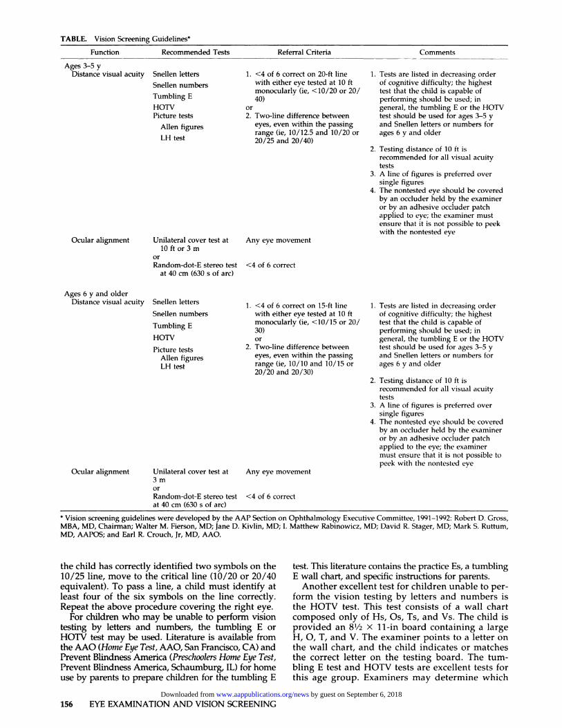
Ages 3-5 y
Distance visual acuity Snellen letters
Snellen numbers
Tumbling E
HOTV
Picture tests
Allen figures
LH test
Any eye movement
<4 of 6 correct
Snellen letters
Snellen numbers
Any eye movement
<4 of 6 correct
* Vision screening guidelines were developed by the AAP Section on Ophthalmology Executive Committee, 1991-1992: Robert D. Gross,
MBA, MD, Chairman; Walter M. Fierson, MD; Jane D. Kivlin, MD; I. Matthew Rabinowicz, MD; David R. Stager, MD; Mark S. Ruttum,MD, AAPOS; and Earl R. Crouch, Jr, MD, AAO.
156 EYE EXAMINATION AND VISION SCREENING
TABLE. Vision Screening Guidelines*
Function Recommended Tests
Ocular alignment Unilateral cover test at
10 ft or 3 m
or
Random-dot-E stereo test
at 40 cm (630 s of arc)
Referral Criteria
1. <4 of 6 correct on 20-ft line
with either eye tested at 10 ft
monocularly (ie, <10/20 or 20/
40)
or
2. Two-line difference between
eyes, even within the passing
range (ie,10/12.5 and 10/20 or20/25 and 20/40)
Comments
1. Tests are listed in decreasing order
of cognitive difficulty; the highest
test that the child is capable of
performing should be used; in
general, the tumbling E or the HOTV
test should be used for ages 3-5 y
and Snellen letters or numbers for
ages 6 y and older
2. Testing distance of 10 ft is
recommended for all visual acuity
tests
3, A line of figures is preferred over
single figures
4. The nontested eye should be covered
by an occiuder held by the examiner
or by an adhesive occluder patch
applied to eye; the examiner must
ensure that it is not possible to peekwith the nontested eye
Ages 6 y and older
Distance visual acuity
Tumbling E
HOTV
Picture tests
Allen figures
LH test
Ocular alignment Unilateral cover test at
3m
orRandom-dot-E stereo test
at 40 cm (630 5 of arc)
1. <4 of 6 correct on 15-ft line
with either eye tested at 10 ft
monocularly (ie, <10/15 or 20/
30)
or
2. Two-line difference between
eyes, even within the passing
range (ie, 10/10 and 10/15 or
20/20 and 20/30)
I . Tests are listed in decreasing order
of cognitive difficulty; the highest
test that the child is capable of
performing should be used; in
general, the tumbling E or the HOTV
test should be used for ages 3-5 y
and Snellen letters or numbers for
ages 6 y and older
2. Testing distance of 10 ft is
recommended for all visual acuity
tests
3. A line of figures is preferred over
single figures
4. The nontested eye should be covered
by an occiuder held by the examiner
or by an adhesive occluder patch
applied to the eye; the examiner
must ensure that it is not possible to
peek with the nontested eye
the child has correctly identified two symbols on the10/25 line, move to the critical line (10/20 or 20/40
equivalent). To pass a line, a child must identify atleast four of the six symbols on the line correctly.Repeat the above procedure covering the right eye.
For children who may be unable to perform vision
testing by letters and numbers, the tumbling E orHOlY test may be used. Literature is available from
the AAO (Home Eye Test, AAO, San Francisco, CA) andPrevent Blindness America (Preschoolers Home Eye Test,
Prevent Blindness America, Schaumburg, IL) for home
use by parents to prepare children for the tumbling E
test. This literature contains the practice Es, a tumblingE wall chart, and specific instructions for parents.
Another excellent test for children unable to per-
form the vision testing by letters and numbers isthe HOTV test. This test consists of a wall chartcomposed only of Hs, Os, Ts, and Vs. The child is
provided an 8V2 X 11-in board containing a largeH, 0, T, and V. The examiner points to a letter on
the wall chart, and the child indicates or matchesthe correct letter on the testing board. The tum-bling E test and HOTV tests are excellent tests for
this age group. Examiners may determine which
by guest on September 6, 2018www.aappublications.org/newsDownloaded from
AMERICAN ACADEMY OF PEDIATRICS 157
test is most useful in their practices and use thattest preferentially.
If a child is not able to perform the tumbling E orHOTV test, the LH symbol test or Allen card test may
be used. The Allen card test is the older of the twoand is well known for its commonly used figures,consisting of a schematic truck, house, birthday cake,bear, telephone, horse, and tree. The Allen card testhas four flash cards containing the seven figures. It isimportant that a child identify either verbally or bymatching all seven pictures before actual vision test-ing. In this case, testing should be performed withthe remaining figures.
Once it is established that the child can identify thefigures, perform initial testing with the child havingboth eyes open; testing for each eye individually
should then be performed. Begin walking backwardvery slowly, flipping through the cards and present-
ing different pictures to the child. Continue to move
backward as the child correctly calls out the figurespresented. When the child begins to miss the figures
being presented consistently, move forward severalfeet to confirm that the child is able to identify thefigures at this point. All Allen figures are 20/30 sizefigures. The farthest distance at which the child is
able to identify the pictures accurately becomes thenumerator, and 30 becomes the denominator. There-
fore, if the child were able to identify the picturesaccurately at 15 ft. the visual acuity would be re-
corded as 15/30. This would be equivalent to 20/40
or 10/20. A matching panel of all of the Allen figuresmay be prepared on a copy machine and used in the
same way as for HOTV testing, if necessary, fortesting very young children or for practice at home.
The LH symbol test is slightly different from theAllen card test in that it is made up of flash cards heldtogether by a spiral binding. The flash cards containlarge examples of a house, apple, circle, and square;these should be presented to the child before visiontesting. Unlike the Allen cards, the LH symbol test
contains flash cards with more than one figure per cardand with smaller figure sizes, so that testing may be
performed at 10 ft. Each card contains the symbol sizeand visual acuity value for a 10-ft testing distance. Thevisual acuity is determined by the smallest symbols thechild is able to identify accurately at 10 ft. For example,if the child is able to identify a symbol at 10 ft for which
the visual acuity value on the card reads 10/15, thechild’s visual acuity is 10/15 or 20/30. If it is not pos-sible to perform all testing at 10 ft. use a similar tech-nique to Allen card testing and present the pictures tothe child one at a time by covering all but one pictureon the card and moving backward at 10 ft. At this
point, proceed down in size to the smallest figures thechild is consistently able to identify correctly. A match-ing panel is provided with the LH test and may behelpful in testing very young children. At least three of
four figures should be identified for each size or dis-tance.
Allen cards and wall charts are available frommany medical supply houses. When ordering wallcharts, be sure to indicate that a 10-ft testing distancewill be used.
Testing Procedures for Assessing Ocular Alignment
The two tests to detect ocular misalignment are the
unilateral cover test and the random-dot-E stereotest. Either test should be performed to determinewhether strabismus (ocular misalignment) is present.
To perform the unilateral cover test, have the childlook straight ahead at an object 10 ft (3 m) away. An
eye chart is commonly used to test children older
than 3 years. For younger children, it is helpful to usea colorful noise-making toy.
A child who has eyeglasses should be screenedwearing the eyeglasses. As the child looks at a distantobject, cover the left eye with a occiuder, and look formovement of the uncovered right eye. If the right eye
does not move, there is no apparent misalignment ofthat eye. If the right eye moves outward, the eye isesotropic (crossed). if the right eye moves inward, it is
exotropic (out-turned). Any movement is the criterion
for referral to an ophthalmologist.After testing the right eye, test the left eye for
movement. Occlude the right eye and look for move-ment of the uncovered left eye. If the left eye does not
move, there is no apparent misalignment of that eye.If the left eye moves outward or inward, this meets
the criterion for referral to an ophthalmologist.To perform the random-dot-E stereo test, the cards
should be held 16 in (40 cm) from the child’s eyes.
Explain the game to the child. Show the child the grayside of the card that says “model” on it. Hold the modelE in the direction at which the child can read it con-
rectly. Have the child touch the model E to understandbetter that the picture will stand out. A child may showwhich direction the legs are pointing. Let the child
know that is correct, but it is more important that the
child knows that the picture will stick out.Place the stereo glasses on the child. If the child is
wearing corrective eyeglasses, place the stereoglasses over the glasses. Make sure the glasses stay
on the child and the child is looking straight ahead.The child should be shown both the stereo blank card
and the raised and recessed E card simultaneously.Hold each card so you can read the back. The blank
card should be held so you can read it. The E cardshould be held so you can read the word “raised.”Both cards must be held straight. Do not tilt the cardstoward the floor or the ceiling-this will cause dank-
ness and glare. Ask the child to look at both cardsand to point to or touch the card with the picture ofthe E. The E must be presented randomly, switchingfrom side to side. Use the following order to present
the E card to the child: right, left, down, right, up,and left. The child is shown the cards up to six times.To pass the test, a child must identify the E correctlyin four of six attempts.
REFERENCES
I . American Academy of Pediatrics, Section on Ophthalmology. Vision
screening guidelines. AAP News. 1995;1 1:25
2. Wasserman RC, Croft CA, Brotherton SE. Preschool vision screening in
pediatric practice: a study from the Pediatric Research in Office Setting
(PROS) network. Pediatrics. 1992;89:834-838
3. American Academy of Pediatrics, Committee on Practice and Ambula-
tory Medicine. Recommendations for preventive pediatric health care.
Pediatrics. 1995;96:373-374
by guest on September 6, 2018www.aappublications.org/newsDownloaded from
1996;98;153Pediatrics Committee on Practice and Ambulatory Medicine, Section on Ophthalmology
Eye Examination and Vision Screening in Infants, Children, and Young Adults
ServicesUpdated Information &
http://pediatrics.aappublications.org/content/98/1/153including high resolution figures, can be found at:
Permissions & Licensing
http://www.aappublications.org/site/misc/Permissions.xhtmlentirety can be found online at: Information about reproducing this article in parts (figures, tables) or in its
Reprintshttp://www.aappublications.org/site/misc/reprints.xhtmlInformation about ordering reprints can be found online:
by guest on September 6, 2018www.aappublications.org/newsDownloaded from
1996;98;153Pediatrics Committee on Practice and Ambulatory Medicine, Section on Ophthalmology
Eye Examination and Vision Screening in Infants, Children, and Young Adults
http://pediatrics.aappublications.org/content/98/1/153the World Wide Web at:
The online version of this article, along with updated information and services, is located on
Copyright © 1996 by the American Academy of Pediatrics. All rights reserved. Print ISSN: 1073-0397. American Academy of Pediatrics, 141 Northwest Point Boulevard, Elk Grove Village, Illinois, 60007.been published continuously since 1948. Pediatrics is owned, published, and trademarked by the Pediatrics is the official journal of the American Academy of Pediatrics. A monthly publication, it has
by guest on September 6, 2018www.aappublications.org/newsDownloaded from