Embed Size (px)
Citation preview
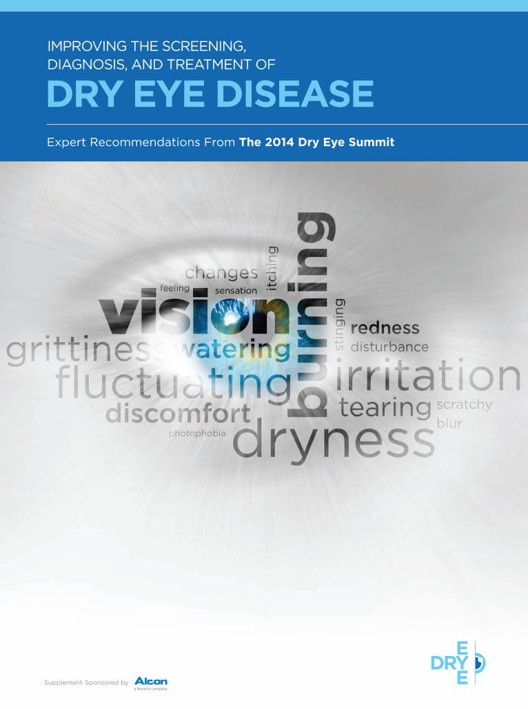
IMPROVING THE SCREENING, DIAGNOSIS, AND TREATMENT OF
DRY EYE DISEASEExpert Recommendations From The 2014 Dry Eye Summit
Supplement Sponsored by
0615_BioScience.indd 1 6/1/15 4:01 PM

IMPROVING THE SCREENING, DIAGNOSIS, AND TREATMENT OF
DRY EYE DISEASE
Marc Bloomenstein OD, FAAO Schwartz Laser Eye Center Scottsdale, Arizona
Derek Cunningham OD, FAAO Dell Laser Consultants Austin, Texas
Ian Benjamin Gaddie OD, FAAO Gaddie Eye Centers Louisville, Kentucky
Paul Karpecki OD, FAAO Koffler Vision Group Lexington, Kentucky
Scot Morris OD, FAAO Eye Consultants of Colorado Conifer, Colorado
Kelly Nichols OD, MPH, PhD, FAAO University of Alabama at Birmingham Birmingham, Alabama
Barbara Caffery, OD, PhD, FAAO
Doug Devries, OD
Mark Dunbar, OD, FAAO
S. Barry Eiden, OD, FAAO
Art Epstein, OD, FAAO, FABCO, FBCLA, DPNAP
David Geffen, OD, FAAO
Scott Hauswirth, OD
Milton Hom, OD, FAAO
Lyndon Jones, PhD, FCOptom, FAAO
Al Kabat, OD, FAAO
Tom Kislan, OD
Blair Lonsberry, OD, MEd, FAAO, ABO
Katherine Mastrota, OD, FAAO
Ron Melton, OD, FAAO
Jason Miller, OD, FAAO
Jason Nichols, OD, PhD, FAAO
Dominick Opitz, OD, FAAO
Jim Owen, OD, FAAO
C. Lisa Prokopich, OD, MSc
Thomas Quinn, OD, FAAO
John Rumpakis, OD, MBA
Jack Schaeffer, OD, FAAO
Joseph Shovlin, OD, FAAO
Kirk Smick, OD
Randall Thomas, OD, FAAO
William Townsend, OD
Gina Wesley, OD, FAAO
Walter Whitley, OD, FAAO
Despite our rapidly expanding knowledge around dry eye disease (DED), eye care professionals (ECPs) have encountered some challenges in translating this wealth of information into effective management strategies in general eye care practices. Significant gaps still exist in regard to disease prevalence and ECP awareness, diagnosis, and ultimately treatment of DED. In an effort to identify opportunities for improvement in screening, diagnosis, and treatment, more than 30 leaders in DED gathered in Dallas, Texas, on December 11–13, 2014, for the inaugural Dry Eye Summit. Joining the experts at this summit were representatives from 17 pharmaceutical and medical device companies who provided invaluable industry insights into the diagnostic tools and treatments available to ECPs, and how these tools are currently being used. The overall goal of the Summit was to create, through a consensus of the experts, practical recommendations that could easily be implemented and would have a substantial impact on the quality and consistency of care that patients with DED receive at the general practice level. Over the course of the meeting, the experts evaluated various screening, diagnostic, and treatment options, arriving at a consensus on baseline standards that can be used by all ECPs for clinical patient encounters. The summit participants also discussed strategies for integrating ocular surface wellness into optometric practices.
Expert Recommendations From The 2014 Dry Eye Summit
Distributed by Review of Optometry
PROGRAM CHAIRS EXPERT CONTRIBUTORS
ABSTRACT
1
0615_BioScience.indd 10615_BioScience.indd 1 6/1/15 4:03 PM6/1/15 4:03 PM

INTRODUCTION Dry eye disease (DED) is an ocular and public health issue that presents a conundrum for the eye care professional (ECP), with unique pathophysiologic challenges. In some ways, the level of attention to DED by the health care community has never been greater, as no fewer than 7 sets of guidelines have been published, by various groups, that are devoted to understanding its risk factors, diagnosis, and treatment.1-7 In addition, over the last decade, an influx of DED objective diagnostic tools and new treatment regimens have made it to the marketplace. Despite this focus and available resources, there are identifiable management gaps among ECPs, suggesting that clinicians are facing challenges in recognizing and utilizing published guidelines, as well as integrating effective diagnostic and treatment strategies into their practices.
There are a number of potential reasons why this body of research, developed by DED experts, has not gained traction and become standard practice for ECPs. These range from a simple lack of awareness that guidelines exist, to their perceived complexity, and to a failure on the part of experts to provide consistent education and de facto protocols to the ECPs who are seeing the majority of patients. In examining this issue, it has become clear that there is a pressing need not to create another set of scientific guidelines, but to develop a set of consensus-derived recommendations, drawing on the available information, that all ECPs can use to provide consistent, effective DED care to the mass population that suffers from DED but is currently not being diagnosed and treated.
Addressing this unmet need for minimum recommendations was the primary impetus for the inaugural Dry Eye Summit, held on December 11–13, 2014, in Dallas, Texas. More than 30 DED leaders attended the summit, with the overarching goal of making a substantial impact in the quality and consistency of care patients receive by creating baseline DED management recommendations that can be easily incorporated into daily practice. Representatives of 17 pharmaceutical and medical device companies, who offered their insights into the various diagnostic and treatment resources available to ECPs, joined these experts. The summit also built on previous expert consensus work by discussing some of the concepts of ocular surface wellness and how they relate to DED (see Sidebar, “Target: A Healthy Eye”). This supplement presents the key proceedings of the Dry Eye Summit and reviews the consensus recommendations developed for ECPs by the expert attendees.
Target: A Healthy Eye
As eye care practitioners (ECPs), we tend to focus on the vision of our patients and treating ocular disease, but an additional aspect of health care deserving our attention is ocular surface wellness. What is this? It is a proactive program of main-taining a healthy eye so that the risks of developing disease and vision problems are minimized. ECPs should be assessing each patient to see if he or she has a healthy eye that is defined as being white, with no feelings of irri-tation, and that delivers clear and consistent vision for the patient. Assessing the health of the ocular surface gives ECPs the opportunity to catch infections and irritations in their infancy and to provide recommendations on how to maintain or improve the eye’s health. Patients with dry eye disease have impaired ocular health and ECPs must be able to recognize the problem and prescribe an appropriate treatment until the patient again sees with a healthy eye.
2
0615_BioScience.indd 10615_BioScience.indd 1 6/1/15 4:03 PM6/1/15 4:03 PM

A number of groups have developed various definitions of DED that encompass concepts including tear dysfunction,2 increased osmolarity, discomfort and other symptoms, and visual disturbance.3 Common to all of these definitions is the understanding that DED is an inflammatory disease of the tears and ocular surface that impacts the eye’s ability to
refract correctly and that, if left untreated, can have a serious impact on functional vision, eye discomfort, and patient quality of life.
The simple and straightfor-ward term “dry eye disease” belies the complexity of a disorder that is associated with a wide range of caus-es that continue to expand. Earlier studies recognized the relationship between
DED and the tear film,8-12 skin diseases,13 and autoimmune mechanisms,14 while more recent research has identified ad-ditional risk factors that include age,15-17 eyelid and lacrimal gland damage,16-17 and contact lens wear.15,18-20 The latest re-ported risk factors implicate our reliance on technology and its impact on the evaporative and inflammatory mechanisms of DED; these risk factors include the use of mobile phones, tablets, computers, and other digital devices that are associ-ated with decreased blink rates (Table 1).15-28
A multifactorial disease associated with so many risk factors is bound to have a high prevalence rate, but how high? It is
difficult to quantify the epidemiology of DED given the multiple definitions that have been employed (reflecting the evolution of how DED is defined), varying understandings of disease severity, and differences in study design and patient populations. All of these have contributed to disparities in reported prevalence rates among different countries: for example, UK, 9.6%29; France, 21.9%30; Taiwan, 33.7%.31 In the United States, approximately 20 million Americans have at least early signs or symptoms of DED or more episodic manifestations of the disease.3
As high as these prevalence statistics are, they are likely to increase dramatically in the near future based on the growing impact of 3 risk factors: age, diabetes, and digital device use. There are currently over 100 million adults in the US over the age of 50, with another 10 million expected by the year 2020.32,33 Diabetes rates have been steadily rising for the last 40 years (2.8% of Americans in 1980, 4.5% in 2000, 9.3% in 2012) and there is no indication that this rate is going to decline, or even plateau, in the near future.34,35 Digital device use is increasingly creeping into every facet of our waking days, with new devices and expanding Wi-Fi networks making it easier to search the Internet, interact with social media, or watch videos anywhere and at any time.36-38 The coming age of wearable electronic devices will certainly magnify the current problem. In light of these trends, it is more important than ever that management of DED improves and that ECPs have the tools they need to ensure that they are identifying their DED sufferers, and when identified, that they are being managed appropriately.
3
DED is an inflammatory
disease of the tears and ocular
surface that impacts the eye’s
ability to refract correctly and
that, if left untreated, can have
a serious impact on functional
vision, eye discomfort, and
patient quality of life.
Common Risk Factors for Developing Dry Eye Disease15-28
Diseases
Diabetes Allergies Autoimmune diseases (e.g., Sjogren’s syndrome, thyroid disease, arthritis)
Contact lens wear
Medications
Antihistamines Decongestants Antidepressants Corticosteroids
Older age Digital device use
Ocular surgery
Table 1
During the Dry Eye Summit, the expert participants examined the myriad reasons why ECPs often face difficulties in diagnosing and treating DED, despite this disease being one of the leading causes of patient visits.39 A lack of concordance between the signs and symptoms of DED underscores the challenge faced by ECPs. For
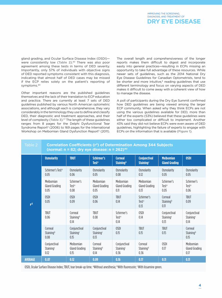
example, in a multisite analysis of 344 subjects in the U.S. and Europe that looked at correlations between multiple tests for DED, the only substantial correlation found was between corneal and conjunctival staining (r2 = 0.36). Correlations among these and other tests—osmolarity, tear break-up time (TBUT), Schirmer’s test, meibomian
DRY EYE DISEASE: THE MANAGEMENT CHALLENGES FOR THE ECP
THE SCOPE OF DRY EYE DISEASE
0615_BioScience.indd 10615_BioScience.indd 1 6/1/15 4:04 PM6/1/15 4:04 PM
IMPROVING THE SCREENING, DIAGNOSIS, AND TREATMENT OF
DRY EYE DISEASE
4
Correlation Coefficients (r2) of Determination Among 344 Subjects (normal: n = 82; dry eye disease: n = 262)40
r2
Osmolarity TBUT Schirmer’s Testa
Corneal Stainingb
Conjunctival Stainingc
Meibomian Gland Grading
OSDI
Schirmer’s Testa 0.05
Osmolarity 0.06
Osmolarity 0.05
Osmolarity 0.08
Osmolarity 0.12
Osmolarity 0.05
Osmolarity 0.05
Meibomian Gland Grading 0.05
Schirmer’s Testa 0.08
Meibomian Gland Grading 0.05
Meibomian Gland Grading 0.11
Meibomian Gland Grading 0.13
Schirmer’s Testa 0.05
Schirmer’s Testa 0.06
OSDI 0.05
OSDI 0.09
OSDI 0.06
TBUT 0.14
Schirmer’s Testa 0.13
Corneal Stainingb 0.11
TBUT 0.09
TBUT 0.06
Corneal Stainingb 0.14
TBUT 0.08
Schirmer’s Testa 0.14
OSDI 0.14
Conjunctival Stainingc 0.13
Conjunctival Stainingc 0.14
Corneal Stainingb 0.08
Conjunctival Stainingc 0.15
Conjunctival Stainingc 0.13
OSDI 0.15
TBUT 0.15
TBUT 0.15
Corneal Stainingb 0.15
Conjunctival Stainingc 0.12
Meibomian Gland Grading 0.15
Corneal Stainingb 0.14
Conjunctival Stainingc 0.36
Corneal Stainingb 0.36
OSDI 0.17
Meibomian Gland Grading 0.17
AVERAGE 0.07 0.12 0.09 0.16 0.17 0.11 0.11
OSDI, Ocular Surface Disease Index; TBUT, tear break-up time. aWithout anesthesia; bWith fluorescein; c With lissamine green.
Table 2
gland grading, and Ocular Surface Disease Index (OSDI)—were consistently low (Table 2).40 There was also poor agreement among these tests in terms of DED severity. Importantly, only 57% of individuals with objective signs of DED reported symptoms consistent with this diagnosis, indicating that almost half of DED cases may be missed if the ECP relies solely on the patient’s reporting of symptoms.40
Other important reasons are the published guidelines themselves and the lack of their translation to ECP education and practice. There are currently at least 7 sets of DED guidelines published by various North American optometric associations, and although each is comprehensive, they vary considerably in the terminology they use to define and classify DED, their diagnostic and treatment approaches, and their level of complexity (Table 3).1-7 The length of these guidelines ranges from 8 pages for the Delphi Dysfunctional Tear Syndrome Report2 (2006) to 169 pages for the International Workshop on Meibomian Gland Dysfunction Report5 (2011).
The overall length and comprehensiveness of the longer reports makes them difficult to digest and incorporate easily into general practices—resulting in ECPs missing an opportunity to take full advantage of these resources. While newer sets of guidelines, such as the 2014 National Dry Eye Disease Guidelines for Canadian Optometrists, tend to be shorter and more intuitive,6 reading guidelines that use different terminology and focus on varying aspects of DED makes it difficult to come away with a coherent view of how to manage the disease.
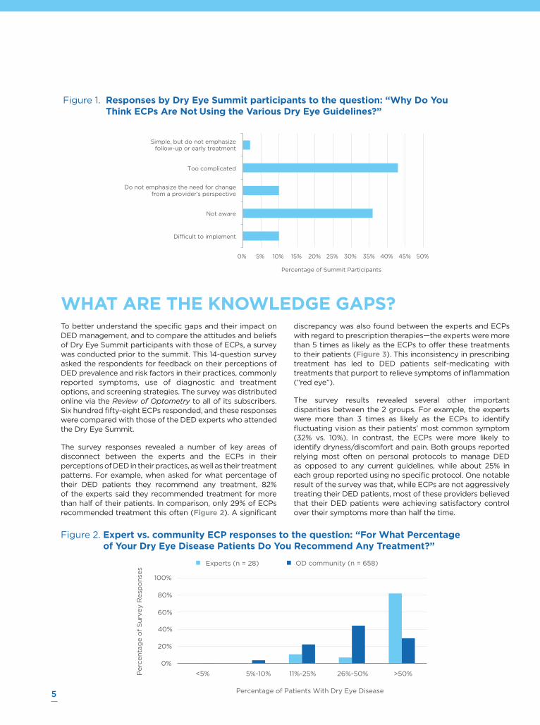
A poll of participants during the Dry Eye Summit confirmed how DED guidelines are being viewed among the larger ECP community. When asked why they think ECPs are not using the various guidelines available for DED, more than half of the experts (53%) believed that these guidelines were either too complicated or difficult to implement. Another 43% said they did not believe ECPs were even aware of DED guidelines, highlighting the failure of experts to engage with ECPs on the information that is available (Figure 1).
0615_BioScience.indd 10615_BioScience.indd 1 6/1/15 4:05 PM6/1/15 4:05 PM
5
To better understand the specific gaps and their impact on DED management, and to compare the attitudes and beliefs of Dry Eye Summit participants with those of ECPs, a survey was conducted prior to the summit. This 14-question survey asked the respondents for feedback on their perceptions of DED prevalence and risk factors in their practices, commonly reported symptoms, use of diagnostic and treatment options, and screening strategies. The survey was distributed online via the Review of Optometry to all of its subscribers. Six hundred fifty-eight ECPs responded, and these responses were compared with those of the DED experts who attended the Dry Eye Summit.
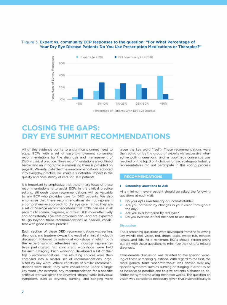
The survey responses revealed a number of key areas of disconnect between the experts and the ECPs in their perceptions of DED in their practices, as well as their treatment patterns. For example, when asked for what percentage of their DED patients they recommend any treatment, 82% of the experts said they recommended treatment for more than half of their patients. In comparison, only 29% of ECPs recommended treatment this often (Figure 2). A significant
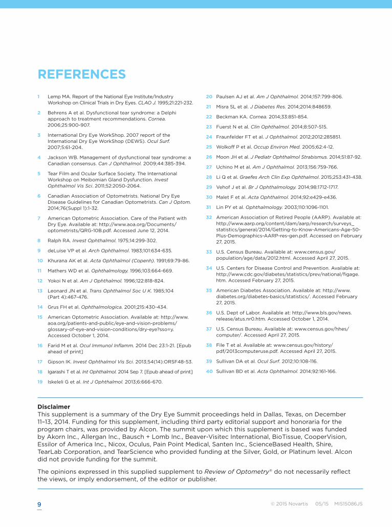
discrepancy was also found between the experts and ECPs with regard to prescription therapies—the experts were more than 5 times as likely as the ECPs to offer these treatments to their patients (Figure 3). This inconsistency in prescribing treatment has led to DED patients self-medicating with treatments that purport to relieve symptoms of inflammation (“red eye”).
The survey results revealed several other important disparities between the 2 groups. For example, the experts were more than 3 times as likely as the ECPs to identify fluctuating vision as their patients’ most common symptom (32% vs. 10%). In contrast, the ECPs were more likely to identify dryness/discomfort and pain. Both groups reported relying most often on personal protocols to manage DED as opposed to any current guidelines, while about 25% in each group reported using no specific protocol. One notable result of the survey was that, while ECPs are not aggressively treating their DED patients, most of these providers believed that their DED patients were achieving satisfactory control over their symptoms more than half the time.
WHAT ARE THE KNOWLEDGE GAPS?
Figure 1. Responses by Dry Eye Summit participants to the question: “Why Do You Think ECPs Are Not Using the Various Dry Eye Guidelines?”
0% 5% 10% 15% 20% 25% 30% 35% 40% 45% 50%
Difficult to implement
Not aware
Do not emphasize the need for changefrom a provider's perspective
Too complicated
Simple, but do not emphasizefollow-up or early treatment
Percentage of Summit Participants
Figure 2. Expert vs. community ECP responses to the question: “For What Percentage of Your Dry Eye Disease Patients Do You Recommend Any Treatment?”
0%
20%
40%
60%
80%
100%
<5% 5%-10% 11%-25% 26%-50% >50% Perc
en
tag
e o
f S
urv
ey R
esp
on
ses
Percentage of Patients With Dry Eye Disease
Experts (n = 28) OD community (n = 658)
0615_BioScience.indd 10615_BioScience.indd 1 6/1/15 4:05 PM6/1/15 4:05 PM

IMPROVING THE SCREENING, DIAGNOSIS, AND TREATMENT OF
DRY EYE DISEASE
Table 3 Overview of Current Guidelines for Dry Eye Disease1-7
Title Sponsoring Body PublicationYear
No. of Pages
Key Points
Report of the National Eye Institute/Industry Workshop on Clinical Trials in Dry Eyes
National Eye Institute (NEI)
1995 12 “Dry eye disorder” due to tear film deficiency or tear evaporation
Tests include validated questionnaire of symptoms and demonstration of ocular surface damage, tear instability, and tear hyperosmolarity
Dysfunctional Tear Syndrome: A Delphi Approach to Treatment Recommendations
Dysfunctional Tear Syndrome Study Group
2006 8 “Dysfunctional tear syndrome”
Treatment based primarily on patient symptoms and signs; diagnostic tests considered secondary in guiding therapy
2007 Report of the International Dry Eye WorkShop (DEWS)
Tear Film and Ocular Surface Society (TFOS)
2007 142 Dry eye is a multifactorial disease of the tears and ocular surface
Accompanied by increased osmolarity of tear film and inflammation of the ocular surface which lead to the cascade of visual degradation, epithelial cell damage, and discomfort
Management of Dysfunctional Tear Syndrome: A Canadian Consensus
University of Ottawa Eye Institute
2009 10 “Dysfunctional tear syndrome”
Management begins with a patient’s history, especially medication use
Treatment focuses on underlying inflammatory process and restoring normal tear film
The International Workshop on Meibomian Gland Dysfunction
Tear Film and Ocular Surface Society (TFOS)
2011 169 Proposed to develop a consensus understanding of the meibomian gland in health and disease
Subcommittee reports on: definition and classification, anatomy and pathophysiology, lipids, epidemiology, diagnosis and management, and clinical trials
National Dry Eye Disease Guidelines for Canadian Optometrists
Canadian Association of Optometrists (CAO)
2014 31 Clinical assessment defined as episodic, chronic, or recalcitrant
Begins with a screening process that includes key questions; a full workup is recommended to confirm diagnosis and identify comorbidities
Care of the Patient With Dry Eye
American Optometric Association (AOA)
2015 4 To be used in conjunction with the AOA Optometric Clinical Practice Guideline on Care of the Patient With Ocular Surface Disorder (revised April 2003)
Dry eye may result from disruption of any tear film component production, altered distribution of tears, or tear film layer disturbances
5 different presentations: aqueous-deficient dry eye, mucin-deficient dry eye, lipid abnormality dry eye, surfacing abnormalities, and epitheliopathies
Table 3
6
0615_BioScience.indd 10615_BioScience.indd 1 6/1/15 4:06 PM6/1/15 4:06 PM
7
All of this evidence points to a significant unmet need to equip ECPs with a set of easy-to-implement consensus recommendations for the diagnosis and management of DED in clinical practice. These recommendations are outlined below, and an infographic summarizing them is provided on page 10. We anticipate that these recommendations, adopted into everyday practice, will make a substantial impact in the quality and consistency of care for DED patients.
It is important to emphasize that the primary focus of these recommendations is to assist ECPs in the clinical practice setting, although these recommendations will be valuable to any ECP who provides care for DED patients. We also emphasize that these recommendations do not represent a comprehensive approach to dry eye care; rather, they are a set of baseline recommendations that ECPs can use in all patients to screen, diagnose, and treat DED more effectively and consistently. Eye care providers can—and are expected to—go beyond these recommendations as needed, consis-tent with good clinical practice.
Each section of these DED recommendations—screening, diagnosis, and treatment—was the result of an initial in-depth discussion, followed by individual workshops in which both the expert summit attendees and industry representa-tives participated. Six concurrent workshops were held for each category. Each workshop developed a list of their top 5 recommendations. The resulting choices were then compiled into a master set of recommendations, orga-nized by key word. Where variations of similar recommen-dations were made, they were consolidated under a single key word (for example, any recommendation for a specific artificial tear was given the keyword “drops,” while individual symptoms such as dryness, burning, and stinging were
given the key word “feel”). These recommendations were then voted on by the group of experts via successive inter-active polling questions, until a two-thirds consensus was reached on the top 3 or 4 choices for each category. Industry representatives did not participate in this voting process.
Recommendations
1 Screening Questions to Ask
At a minimum, every patient should be asked the following questions at each visit:
1 Do your eyes ever feel dry or uncomfortable?2 Are you bothered by changes in your vision throughout
the day?3 Are you ever bothered by red eyes?4 Do you ever use or feel the need to use drops?
Discussion
The 4 screening questions were developed from the following key words: feel, vision, red, drops, tasks, water, rub, contact lenses, and lids. At a minimum, ECPs should screen every patient with these questions to minimize the risk of a missed diagnosis.
Considerable discussion was devoted to the specific word-ing of these screening questions. With regard to the first, the more general term “uncomfortable” was chosen over any specific symptom such as burning or stinging in order to be as inclusive as possible and to give patients a chance to de-scribe the symptoms using their own words. The question on vision was considered necessary, given that vision difficulty is
CLOSING THE GAPS: DRY EYE SUMMIT RECOMMENDATIONS
Figure 3. Expert vs. community ECP responses to the question: “For What Percentage of Your Dry Eye Disease Patients Do You Use Prescription Medications or Therapies?”
0%
20%
40%
60%
Experts (n = 28) OD community (n = 658)
<5% 5%-10% 11%-25% 26%-50% >50%
Perc
en
tag
e o
f S
urv
ey R
esp
on
ses
Percentage of Patients With Dry Eye Disease
RECOMMENDATIONS
0615_BioScience.indd 10615_BioScience.indd 1 6/1/15 4:06 PM6/1/15 4:06 PM

IMPROVING THE SCREENING, DIAGNOSIS, AND TREATMENT OF
DRY EYE DISEASE
a prominent sign/symptom of DED, yet is often unrecognized as such. In this question, the word “change” was selected over “fluctuate,” as the latter may not be as well understood by patients. The question on red eyes was selected given the high prevalence of red eye as a symptom of DED and the importance of this symptom to patients in terms of cosmesis, as well as comfort. Because of the episodic nature of DED, the word “ever” was incorporated where appropriate. Finally, “drops” was preferred over “artificial tears” as it encompass-es the large variety of over-the-counter topical lubricants (as well as red eye relievers) that are available.
2 Diagnostic Tools to Use
At a minimum, the following diagnostic options should be considered in each patient for primary dry eye disease
1 Eyelid examination2 Staining3 Tear film instability
Discussion
Diagnostic tests were considered based on the following key words: lid, staining, TBUT/topography, osmolarity, volume, external, inflammation, and photography. “Eyelid examina-tion,” which includes the meibomian glands, was by far the most popular choice for diagnosis. (The participants consid-ered, but ultimately did not, separate out meibomian glands as a separate item.) The more generalized term “staining” was selected to include any form of corneal staining, at the ECP’s discretion. “Tear film instability” was selected to encom-pass a variety of tests related to vision, including TBUT and corneal topography, but could potentially include osmolarity testing. While other tests, such as blink analysis and meibog-raphy, were considered, it was felt that these tests were too new to recommend to ECPs. Although osmolarity testing was mentioned a number of times, the test was not recom-mended as a top 3 choice given that most optometrists do not currently own this technology. It and other advanced dry eye testing procedures were seen as a level to strive toward, especially for ECPs motivated to move beyond the baseline recommendations.
3 Basic Management Strategies
Basic management for dry eye disease includes:
1 For all patients a. Ocular lubrication b. Lid hygiene c. Nutrition2 Topical anti-inflammatories
Discussion
Treatments were considered based on the following key words: lubrication, anti-inflammatories, orals/nutraceuticals, meibomian gland/lid expression, lid hygiene, and advanced.
It was agreed that the first 3 treatment options should be a part of routine care for every DED patient. With regard to ocular lubrication, it is likely that most DED patients are already using some type of topical lubricant before they present to the ECP office, most likely a vasoconstrictor. The experts left it up to the individual ECP to choose the right class of lubricant (lipid-based or aqueous-based), rather than spec-ifying any particular type. Basic lid hygiene measures (e.g., hot compresses and lid cleanliness measures) were consid-ered an essential part of self-care; however, more complex forms of lid hygiene, such as mechanical meibomian gland expression or pulsation, should be performed only in office. Nutrition, which includes oral nutraceuti-cals and dietary interven-tions, was selected based on its benefits in terms of overall health and the fact that patients should be more willing to adopt this approach. Topical anti- inflammatories include any medication with an anti-inflammatory mech-anism of action, including topical corticosteroids and cyclosporine.
Additional discussion was devoted to the appro-priate patient follow-up interval. With regard to the initiation of treatment, the recommendation for follow-up was 3 to 4 weeks; with a longer follow-up interval, treatment compliance is likely to wane, while a shorter follow-up interval will likely not provide sufficient time to show improvement. For patients who are considered stable, most participants would wait no longer than 6 months, with a minority voting for a shorter (3 months) or longer (1 year) interval.
Conclusions
The consensus recommendations reached during this inau-gural Dry Eye Summit represent a huge opportunity for ECPs to make an impact on DED in their practices. Integrating them into community practices will enable ECPs to identify those DED patients who may be going undiagnosed, and help raise the overall standard of care. These recommenda-tions also are an opportunity to engage patients in discus-sions on ocular surface wellness by encouraging them to be aware of their overall eye health and to take an active role in becoming and staying healthy. We welcome feedback on these recommendations as you begin to incorporate them into practice. This is just the beginning of the discussion regarding the complex diagnosis and treatment algorithms that exist around DED. It is our hope that we will continue to empower clinicians with strategies to help patients maintain a healthy ocular surface, and to keep raising the bar a little higher each time.
8
These recommendations do not represent a comprehensive approach to dry eye care; rather, they represent a baseline approach that ECPs can use in all patients to screen, diagnose, and treat DED more effectively and consistently.
0615_BioScience.indd 10615_BioScience.indd 1 6/1/15 4:06 PM6/1/15 4:06 PM
© 2015 Novartis 05/15 MIS15086JS
1 Lemp MA. Report of the National Eye Institute/Industry Workshop on Clinical Trials in Dry Eyes. CLAO J. 1995;21:221-232.
2 Behrens A et al. Dysfunctional tear syndrome: a Delphi approach to treatment recommendations. Cornea. 2006;25:900-907.
3 International Dry Eye WorkShop. 2007 report of the International Dry Eye WorkShop (DEWS). Ocul Surf. 2007;5:61-204.
4 Jackson WB. Management of dysfunctional tear syndrome: a Canadian consensus. Can J Ophthalmol. 2009;44:385-394.
5 Tear Film and Ocular Surface Society. The International Workshop on Meibomian Gland Dysfunction. Invest Ophthalmol Vis Sci. 2011;52:2050-2064.
6 Canadian Association of Optometrists. National Dry Eye Disease Guidelines for Canadian Optometrists. Can J Optom. 2014;76(Suppl 1):1-32.
7 American Optometric Association. Care of the Patient with Dry Eye. Available at: http://www.aoa.org/Documents/optometrists/QRG-10B.pdf. Accessed June 12, 2014.
8 Ralph RA. Invest Ophthalmol. 1975;14:299-302.
9 deLuise VP et al. Arch Ophthalmol. 1983;101:634-635.
10 Khurana AK et al. Acta Ophthalmol (Copenh). 1991;69:79-86.
11 Mathers WD et al. Ophthalmology. 1996;103:664-669.
12 Yokoi N et al. Am J Ophthalmol. 1996;122:818-824.
13 Leonard JN et al. Trans Ophthalmol Soc U K. 1985;104 (Part 4):467-476.
14 Grus FH et al. Ophthalmologica. 2001;215:430-434.
15 American Optometric Association. Available at: http://www.aoa.org/patients-and-public/eye-and-vision-problems/glossary-of-eye-and-vision-conditions/dry-eye?sso=y. Accessed October 1, 2014.
16 Farid M et al. Ocul Immunol Inflamm. 2014 Dec 23:1-21. [Epub ahead of print]
17 Gipson IK. Invest Ophthalmol Vis Sci. 2013;54(14):ORSF48-53.
18 Igarashi T et al. Int Ophthalmol. 2014 Sep 7. [Epub ahead of print]
19 Iskeleli G et al. Int J Ophthalmol. 2013;6:666-670.
20 Paulsen AJ et al. Am J Ophthalmol. 2014;157:799-806.
21 Misra SL et al. J Diabetes Res. 2014;2014:848659.
22 Beckman KA. Cornea. 2014;33:851-854.
23 Fuerst N et al. Clin Ophthalmol. 2014;8:507-515.
24 Fraunfelder FT et al. J Ophthalmol. 2012;2012:285851.
25 Wolkoff P et al. Occup Environ Med. 2005;62:4-12.
26 Moon JH et al. J Pediatr Ophthalmol Strabismus. 2014;51:87-92.
27 Uchino M et al. Am J Ophthalmol. 2013;156:759-766.
28 Li Q et al. Graefes Arch Clin Exp Ophthalmol. 2015;253:431-438.
29 Vehof J et al. Br J Ophthalmology. 2014;98:1712-1717.
30 Malet F et al. Acta Ophthalmol. 2014;92:e429-e436.
31 Lin PY et al. Ophthalmology. 2003;110:1096-1101.
32 American Association of Retired People (AARP). Available at: http://www.aarp.org/content/dam/aarp/research/surveys_statistics/general/2014/Getting-to-Know-Americans-Age-50-Plus-Demographics-AARP-res-gen.pdf. Accessed on February 27, 2015.
33 U.S. Census Bureau. Available at: www.census.gov/population/age/data/2012.html. Accessed April 27, 2015.
34 U.S. Centers for Disease Control and Prevention. Available at: http://www.cdc.gov/diabetes/statistics/prev/national/figage.htm. Accessed February 27, 2015.
35 American Diabetes Association. Available at: http://www.diabetes.org/diabetes-basics/statistics/. Accessed February 27, 2015.
36 U.S. Dept of Labor. Available at: http://www.bls.gov/news.release/atus.nr0.htm. Accessed October 1, 2014.
37 U.S. Census Bureau. Available at: www.census.gov/hhes/computer/. Accessed April 27, 2015.
38 File T et al. Available at: www.census.gov/history/pdf/2013computeruse.pdf. Accessed April 27, 2015.
39 Sullivan DA et al. Ocul Surf. 2012;10:108-116.
40 Sullivan BD et al. Acta Ophthalmol. 2014;92:161-166.
REFERENCES
Disclaimer This supplement is a summary of the Dry Eye Summit proceedings held in Dallas, Texas, on December 11–13, 2014. Funding for this supplement, including third party editorial support and honoraria for the program chairs, was provided by Alcon. The summit upon which this supplement is based was funded by Akorn Inc., Allergan Inc., Bausch + Lomb Inc., Beaver-Visitec International, BioTissue, CooperVision, Essilor of America Inc., Nicox, Oculus, Pain Point Medical, Santen Inc., ScienceBased Health, Shire, TearLab Corporation, and TearScience who provided funding at the Silver, Gold, or Platinum level. Alcon did not provide funding for the summit.
The opinions expressed in this supplied supplement to Review of Optometry® do not necessarily reflect the views, or imply endorsement, of the editor or publisher.
9
0615_BioScience.indd 10615_BioScience.indd 1 6/1/15 4:07 PM6/1/15 4:07 PM
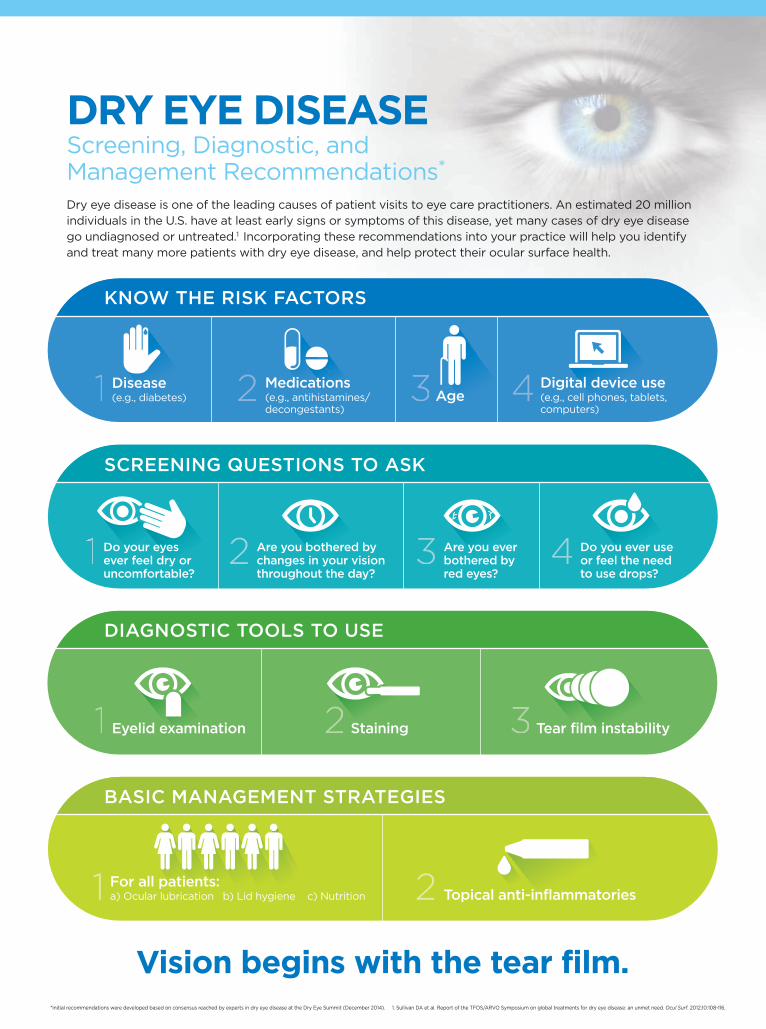
Do you ever use or feel the need to use drops?
Are you ever bothered by red eyes?
Are you bothered by changes in your vision throughout the day?
Do your eyes ever feel dry or uncomfortable?
2 3 4
SCREENING QUESTIONS TO ASK
Digital device use(e.g., cell phones, tablets,computers)
AgeMedications(e.g., antihistamines/decongestants)
Disease(e.g., diabetes)1 2 3 4
KNOW THE RISK FACTORS
Vision begins with the tear film.
Topical anti-inflammatories1 2
BASIC MANAGEMENT STRATEGIES
For all patients:a) Ocular lubrication b) Lid hygiene c) Nutrition
Eyelid examination1 Staining2 Tear film instability3
DIAGNOSTIC TOOLS TO USE
Screening, Diagnostic, and Management Recommendations*
Dry eye disease is one of the leading causes of patient visits to eye care practitioners. An estimated 20 million individuals in the U.S. have at least early signs or symptoms of this disease, yet many cases of dry eye disease go undiagnosed or untreated.1 Incorporating these recommendations into your practice will help you identify and treat many more patients with dry eye disease, and help protect their ocular surface health.
*Initial recommendations were developed based on consensus reached by experts in dry eye disease at the Dry Eye Summit (December 2014). 1. Sullivan DA et al. Report of the TFOS/ARVO Symposium on global treatments for dry eye disease: an unmet need. Ocul Surf. 2012;10:108-116.
DRY EYE DISEASE
0615_BioScience.indd 1 6/1/15 4:07 PM

DRY EYE CAN BE RELENTLESS
CALM THE STORM WITH LASTING RELIEF
For the 75% of dry eye patients worldwide with evaporative dry eye (MGD) symptoms1...
Your recommendation counts.
Make sure your patients
get the lasting symptom
relief they need by offering
them SYSTANE® BALANCE
Lubricant Eye Drops.2
Relief that lasts© 2014 Novartis 05/14 SYS14005JAD-B
SYSTANE® BALANCE Lubricant Eye Drops: Protecting the Ocular Surface by Increasing Lipid Layer Thickness (LLT)
LIPID LAYER
AQUEOUS LAYER
MUCIN LAYER
CORNEAL EPITHELIUM
MEIBOMIANGLAND
References: 1. Akpek EK, Smith RA. Overview of age-related ocular conditions. Am J Manag Care. 2013;19(5 suppl):S67-S75. 2. Korb DR, Blackie CA, Meadows DL, Christensen M, Tudor M. Evaluation of extended tear stability by two emulsion based artifi cial tears. Poster presented at: 6th International Conference on the Tear Film and OcularSurface: Basic Science and Clinical Relevance; September 22-25, 2010; Florence, Italy.
SYSTANE® Brand products are formulated for the temporary relief of burning and irritation due to dryness of the eye.
SYSTANE® BALANCE
Lubricant Eye Drops forms
a protective matrix that is
designed to replenish the lipid
layer for long-lasting relief from
the symptoms associated with
evaporative dry eye (MGD).
This unique formulation is
designed to work on all 3 layers
of the tear fi lm, specifi cally
increasing LLT. This helps create
a protective environment for the
ocular surface.2
0615_BioScience.indd 1 6/1/15 4:08 PM