Embed Size (px)
Citation preview
Eye and Associated Structures70% of all sensory receptors are in the eyeMost of the eye is protected by a cushion of
fat and the bony orbitAccessory structures include eyebrows,
eyelids, conjunctiva, lacrimal apparatus, and extrinsic eye muscles
EyebrowsCoarse hairs that overlie the supraorbital
marginsFunctions include:
Shading the eyePreventing perspiration from reaching the
eyeOrbicularis muscle – depresses the
eyebrowsCorrugator muscles – move the eyebrows
medially
Palpebrae (Eyelids)Protect the eye anteriorlyPalpebral fissure – separates eyelids Canthi – medial and lateral angles
(commissures)
Palpebrae (Eyelids)Lacrimal caruncle – contains glands that
secrete a whitish, oily secretion (Sandman’s eye sand)
Tarsal plates of connective tissue support the eyelids internally
Levator palpebrae superioris – gives the upper eyelid mobility
Palpebrae (Eyelids)Eyelashes
Project from the free margin of each eyelidInitiate reflex blinking
Lubricating glands associated with the eyelidsMeibomian glands and sebaceous glandsCiliary glands lie between the hair follicles

Palpebrae (Eyelids)
Figure 15.1b
ConjunctivaTransparent membrane that:
Lines the eyelids as the palpebral conjunctivaCovers the whites of the eyes as the ocular
conjunctivaLubricates and protects the eye
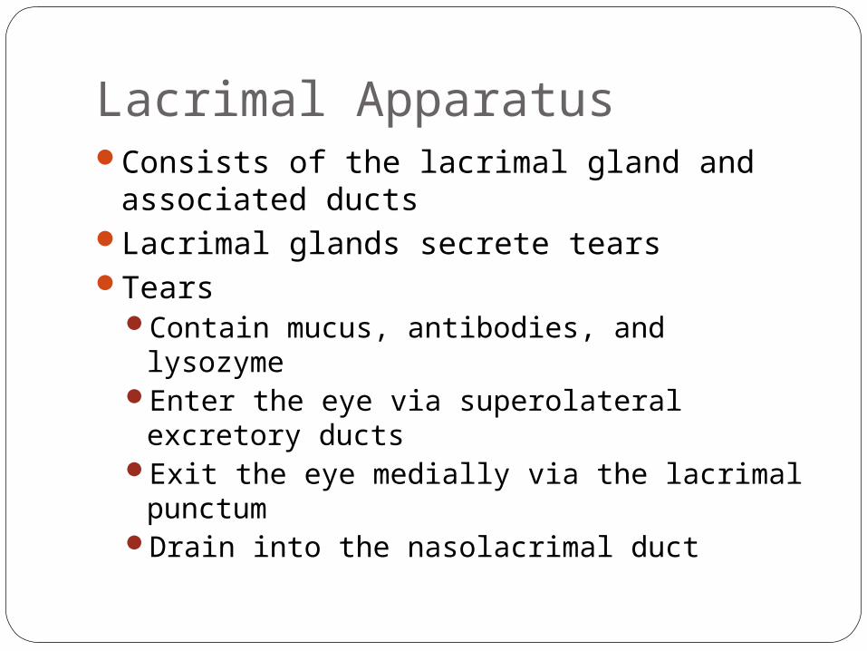
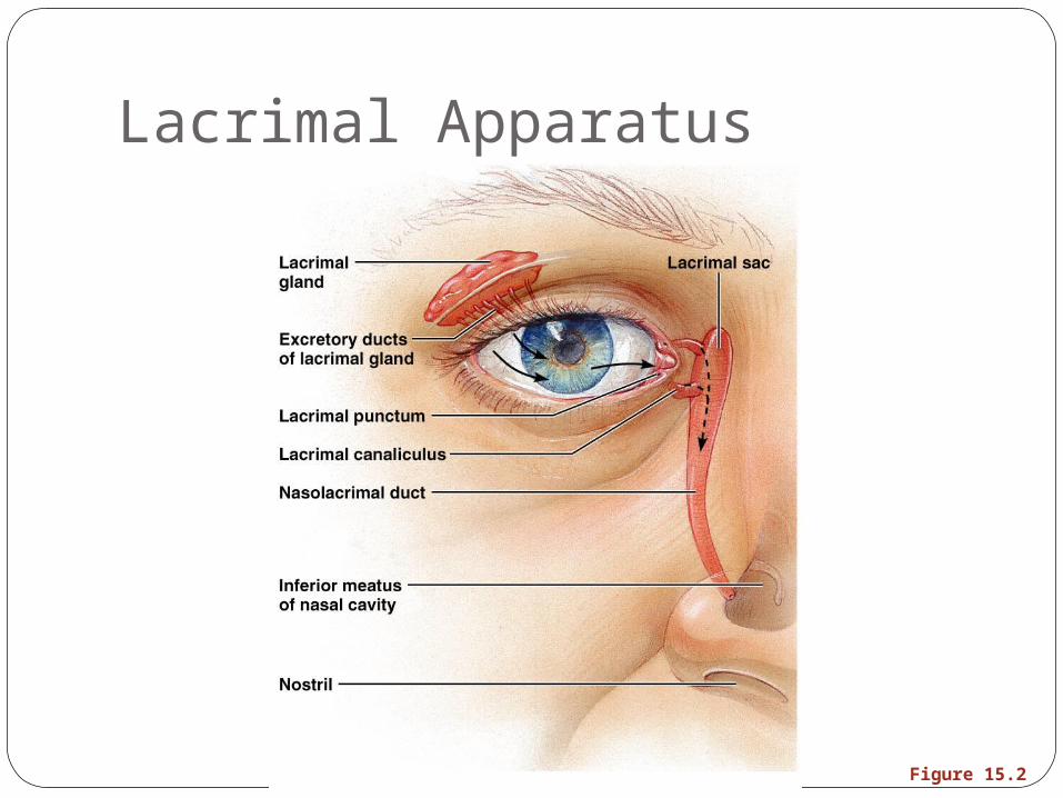
Lacrimal ApparatusConsists of the lacrimal gland and
associated ductsLacrimal glands secrete tears Tears
Contain mucus, antibodies, and lysozymeEnter the eye via superolateral excretory
ducts Exit the eye medially via the lacrimal
punctumDrain into the nasolacrimal duct
Lacrimal Apparatus
Figure 15.2
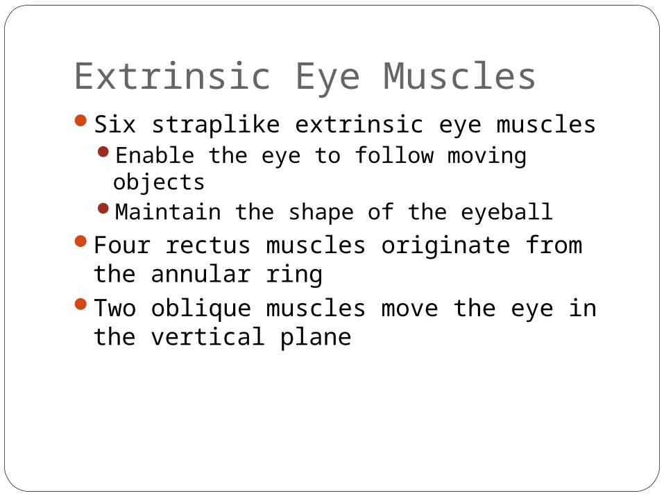
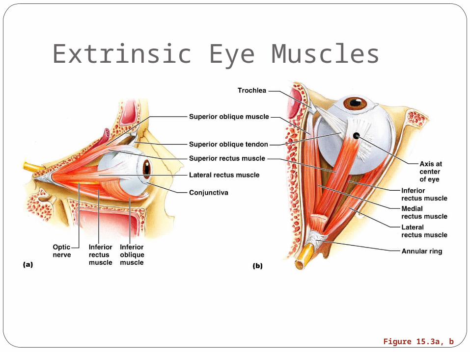
Extrinsic Eye MusclesSix straplike extrinsic eye muscles
Enable the eye to follow moving objectsMaintain the shape of the eyeball
Four rectus muscles originate from the annular ring
Two oblique muscles move the eye in the vertical plane
Extrinsic Eye Muscles
Figure 15.3a, b
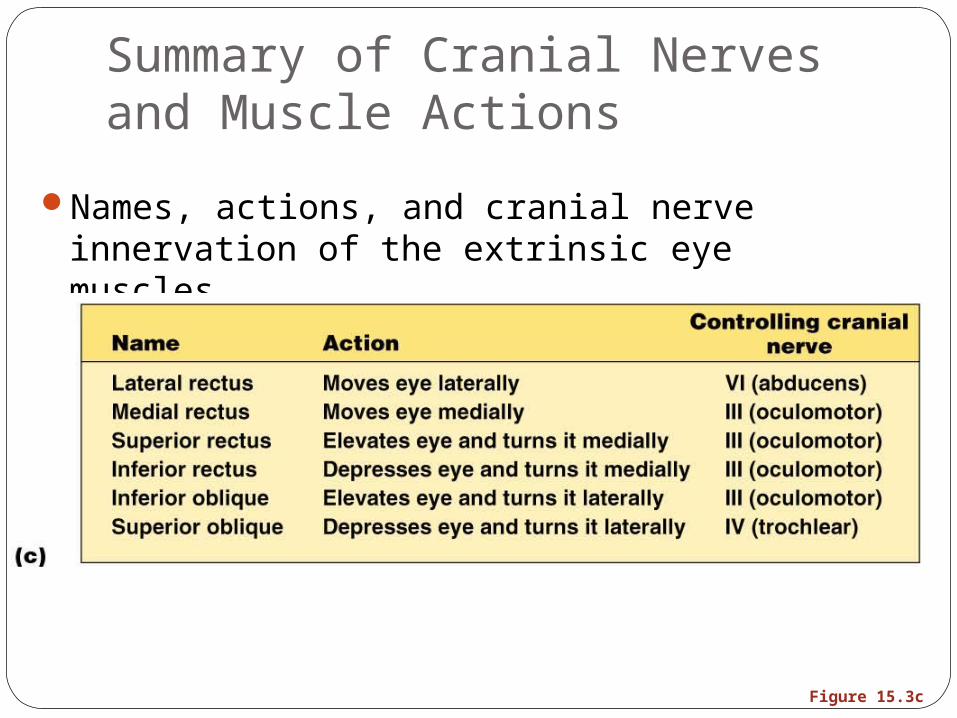
Figure 15.3c
Summary of Cranial Nerves and Muscle Actions
Names, actions, and cranial nerve innervation of the extrinsic eye muscles
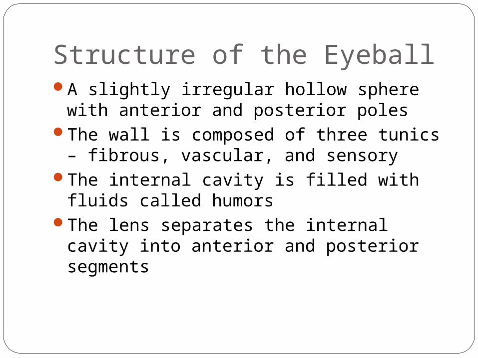
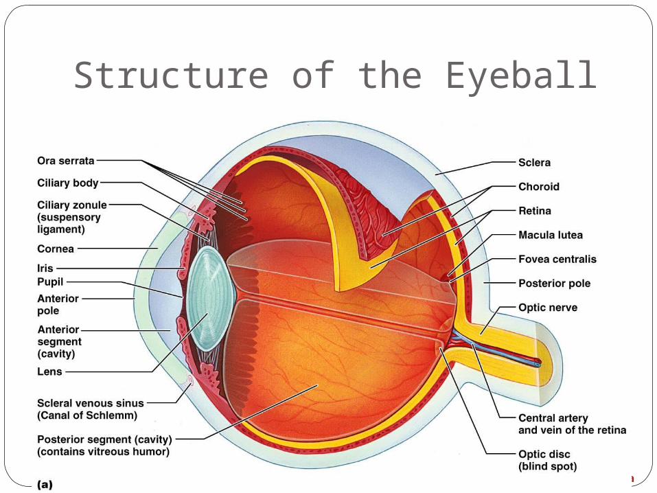
Structure of the EyeballA slightly irregular hollow sphere with
anterior and posterior polesThe wall is composed of three tunics –
fibrous, vascular, and sensoryThe internal cavity is filled with fluids called
humorsThe lens separates the internal cavity into
anterior and posterior segments
Structure of the Eyeball
Figure 15.4a
Fibrous TunicForms the outermost coat of the eye and is
composed of: Opaque sclera (posteriorly)Clear cornea (anteriorly)
The sclera protects the eye and anchors extrinsic muscles
The cornea lets light enter the eye
Vascular Tunic (Uvea): Choroid RegionHas three regions: choroid, ciliary body,
and irisChoroid region
A dark brown membrane that forms the posterior portion of the uvea
Supplies blood to all eye tunics
Vascular Tunic: Ciliary BodyA thickened ring of tissue surrounding the
lensComposed of smooth muscle bundles
(ciliary muscles)Anchors the suspensory ligament that
holds the lens in place
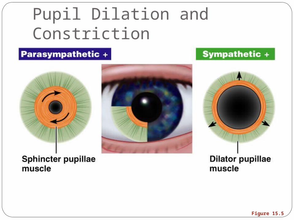
Vascular Tunic: IrisThe colored part of the eyePupil – central opening of the iris
Regulates the amount of light entering the eye during: Close vision and bright light – pupils constrictDistant vision and dim light – pupils dilateChanges in emotional state – pupils dilate when the
subject matter is appealing or requires problem-solving skills
Pupil Dilation and Constriction
Figure 15.5
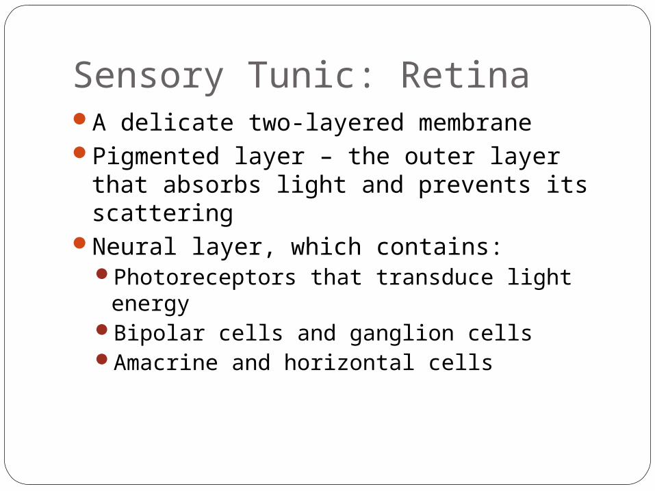
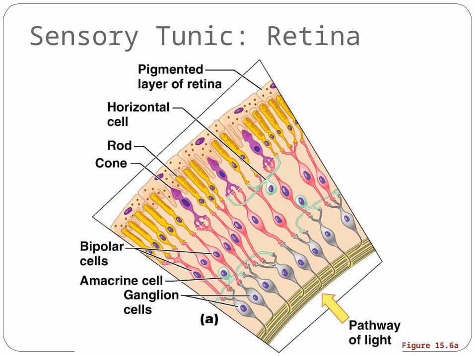
Sensory Tunic: RetinaA delicate two-layered membranePigmented layer – the outer layer that
absorbs light and prevents its scatteringNeural layer, which contains:
Photoreceptors that transduce light energyBipolar cells and ganglion cellsAmacrine and horizontal cells
Sensory Tunic: Retina
Figure 15.6a
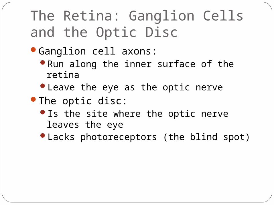
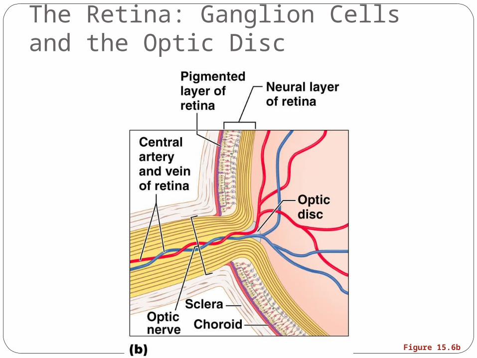
The Retina: Ganglion Cells and the Optic DiscGanglion cell axons:
Run along the inner surface of the retinaLeave the eye as the optic nerve
The optic disc:Is the site where the optic nerve leaves the
eyeLacks photoreceptors (the blind spot)
The Retina: Ganglion Cells and the Optic Disc
Figure 15.6b
The Retina: PhotoreceptorsRods:
Respond to dim lightAre used for peripheral vision
Cones:Respond to bright lightHave high-acuity color vision Are found in the macula lutea Are concentrated in the fovea centralis
Blood Supply to the RetinaThe neural retina receives its blood supply
from two sourcesThe outer third receives its blood from the
choroidThe inner two-thirds is served by the central
artery and veinSmall vessels radiate out from the optic
disc and can be seen with an ophthalmoscope
Inner Chambers and FluidsThe lens separates the internal eye into
anterior and posterior segmentsThe posterior segment is filled with a clear
gel called vitreous humor that:Transmits lightSupports the posterior surface of the lens Holds the neural retina firmly against the
pigmented layerContributes to intraocular pressure
Anterior SegmentComposed of two chambers
Anterior – between the cornea and the irisPosterior – between the iris and the lens
Aqueous humorA plasmalike fluid that fills the anterior
segmentDrains via the canal of Schlemm
Supports, nourishes, and removes wastes
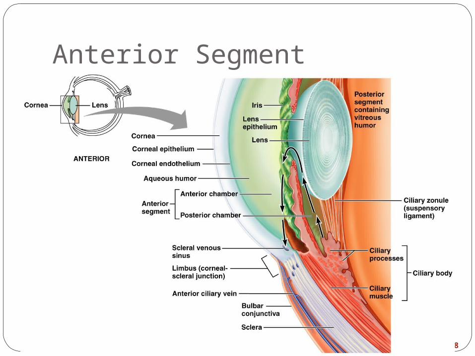
Anterior Segment
Figure 15.8
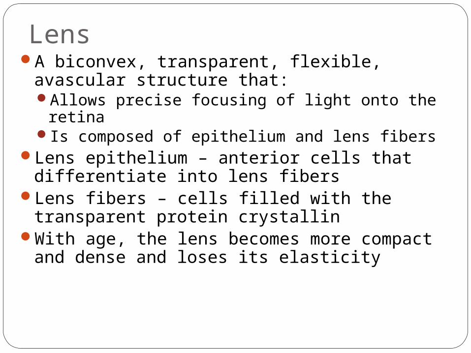
LensA biconvex, transparent, flexible, avascular
structure that:Allows precise focusing of light onto the retinaIs composed of epithelium and lens fibers
Lens epithelium – anterior cells that differentiate into lens fibers
Lens fibers – cells filled with the transparent protein crystallin
With age, the lens becomes more compact and dense and loses its elasticity
LightElectromagnetic radiation – all energy
waves from short gamma rays to long radio waves
Our eyes respond to a small portion of this spectrum called the visible spectrum
Different cones in the retina respond to different wavelengths of the visible spectrum
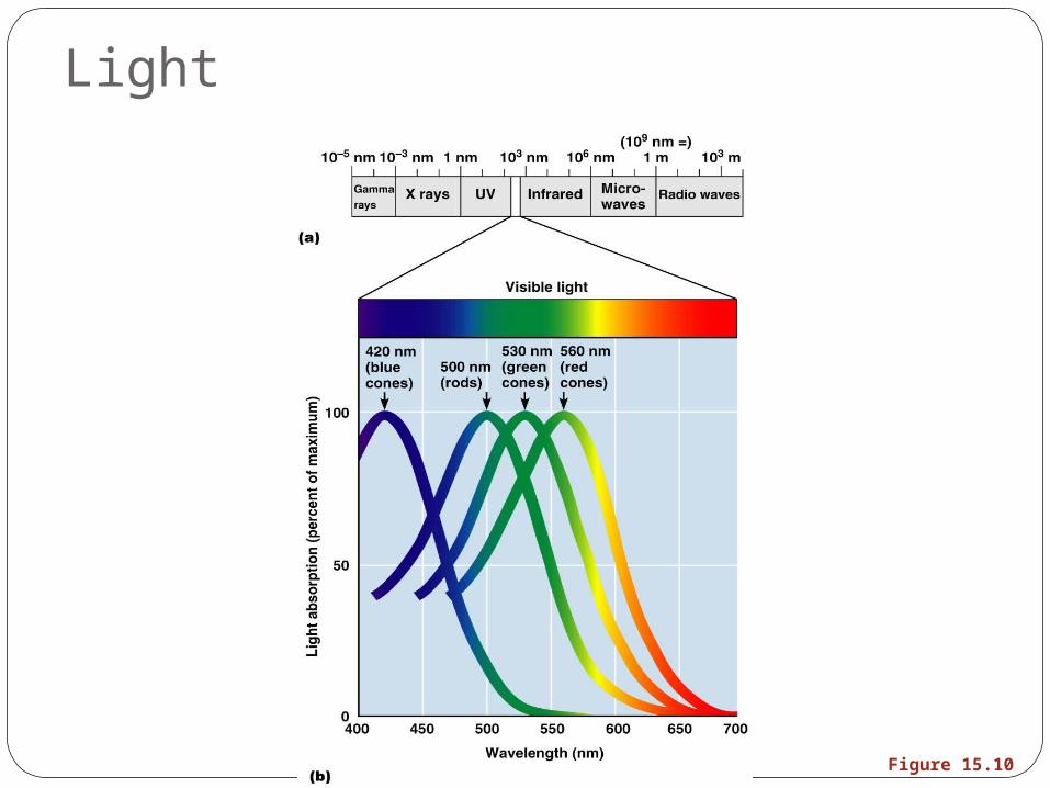
Light
Figure 15.10
Refraction and LensesWhen light passes from one transparent
medium to another its speed changes and it refracts (bends)
Light passing through a convex lens (as in the eye) is bent so that the rays converge to a focal point
When a convex lens forms an image, the image is upside down and reversed right to left
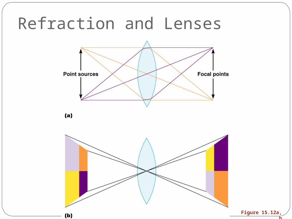
Refraction and Lenses
Figure 15.12a, b

Focusing Light on the RetinaPathway of light entering the eye: cornea,
aqueous humor, lens, vitreous humor, and the neural layer of the retina to the photoreceptors
Light is refracted:At the corneaEntering the lensLeaving the lens
The lens curvature and shape allow for fine focusing of an image
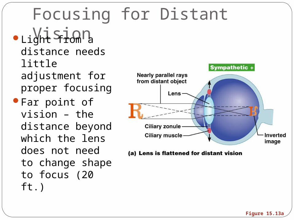
Focusing for Distant VisionLight from a distance needs little adjustment for proper focusing
Far point of vision – the distance beyond which the lens does not need to change shape to focus (20 ft.)
Figure 15.13a
Focusing for Close VisionClose vision requires:
Accommodation – changing the lens shape by ciliary muscles to increase refractory power
Constriction – the pupillary reflex constricts the pupils to prevent divergent light rays from entering the eye
Convergence – medial rotation of the eyeballs toward the object being viewed
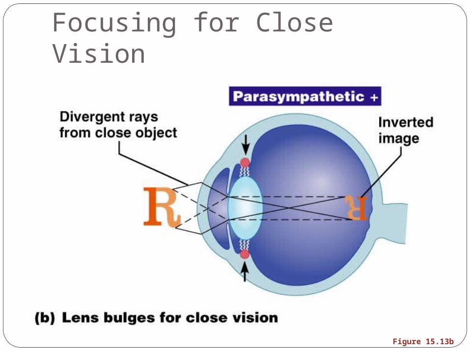
Focusing for Close Vision
Figure 15.13b
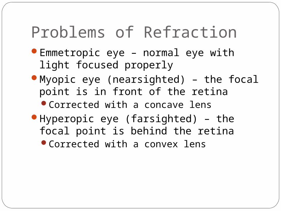
Problems of RefractionEmmetropic eye – normal eye with light
focused properlyMyopic eye (nearsighted) – the focal point
is in front of the retinaCorrected with a concave lens
Hyperopic eye (farsighted) – the focal point is behind the retinaCorrected with a convex lens
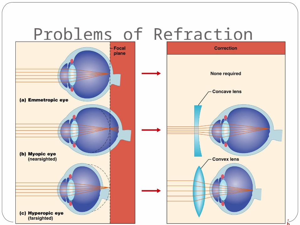
Problems of Refraction
Figure 15.14a, b
Photoreception: Functional Anatomy of Photoreceptors
Photoreception – process by which the eye detects light energy
Rods and cones contain visual pigments (photopigments) Arranged in a stack of disklike infoldings of the
plasma membrane that change shape as they absorb light
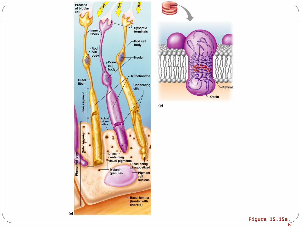
Figure 15.15a, b
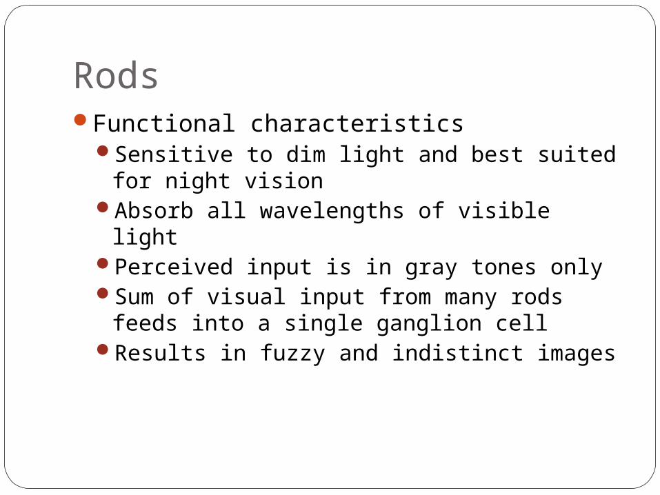
RodsFunctional characteristics
Sensitive to dim light and best suited for night vision
Absorb all wavelengths of visible lightPerceived input is in gray tones onlySum of visual input from many rods feeds
into a single ganglion cell Results in fuzzy and indistinct images
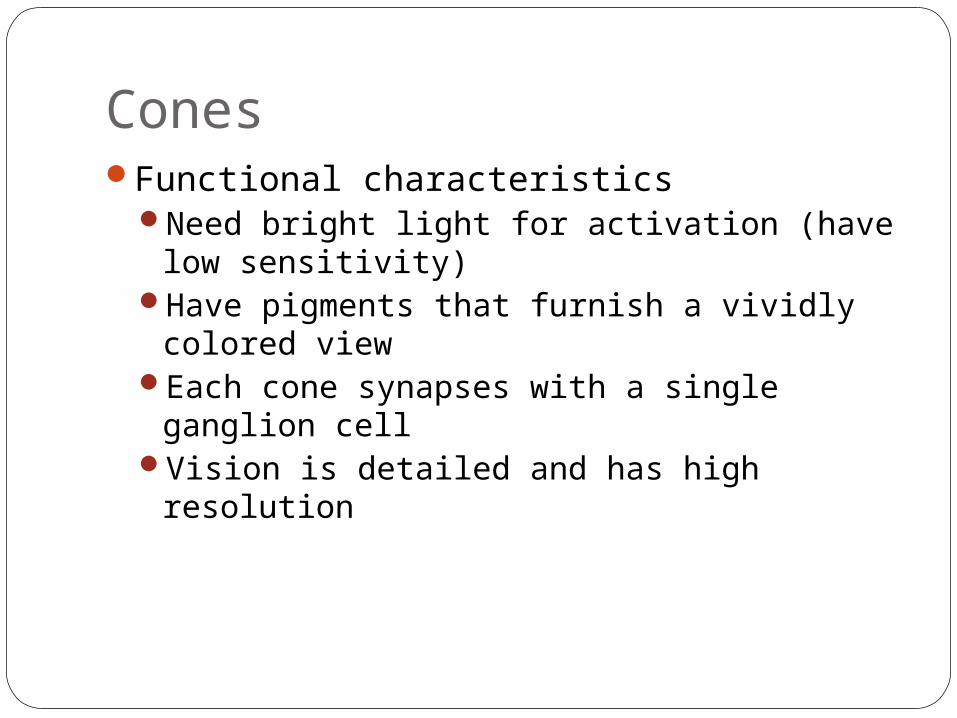
ConesFunctional characteristics
Need bright light for activation (have low sensitivity)
Have pigments that furnish a vividly colored view
Each cone synapses with a single ganglion cell
Vision is detailed and has high resolution
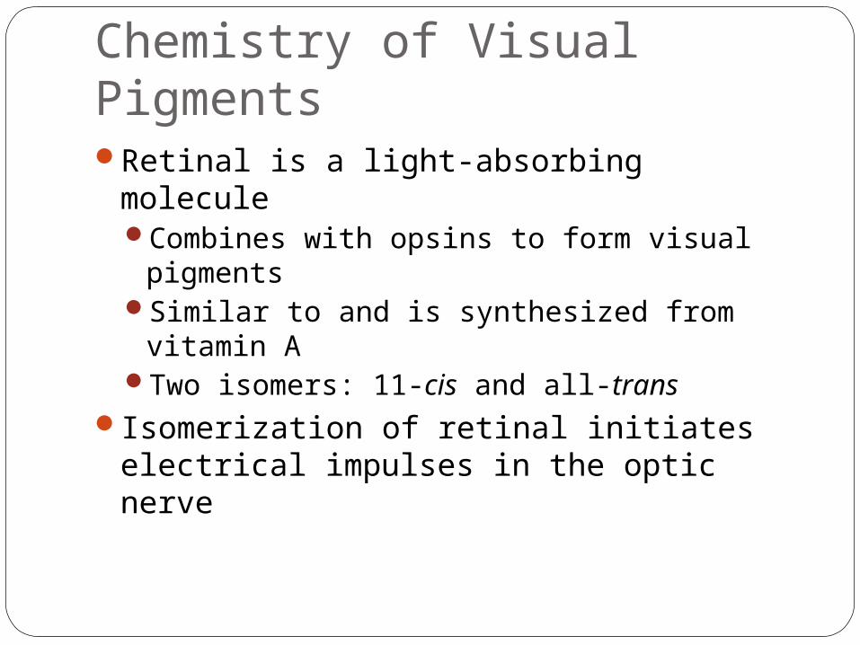
Chemistry of Visual PigmentsRetinal is a light-absorbing molecule
Combines with opsins to form visual pigments
Similar to and is synthesized from vitamin ATwo isomers: 11-cis and all-trans
Isomerization of retinal initiates electrical impulses in the optic nerve
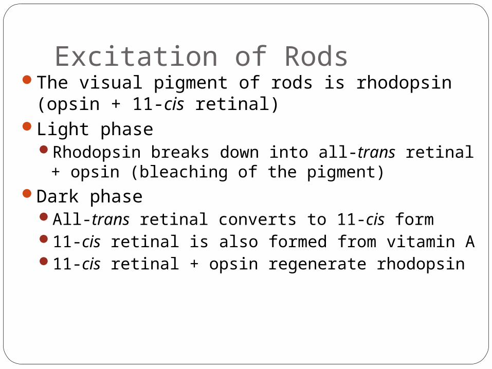
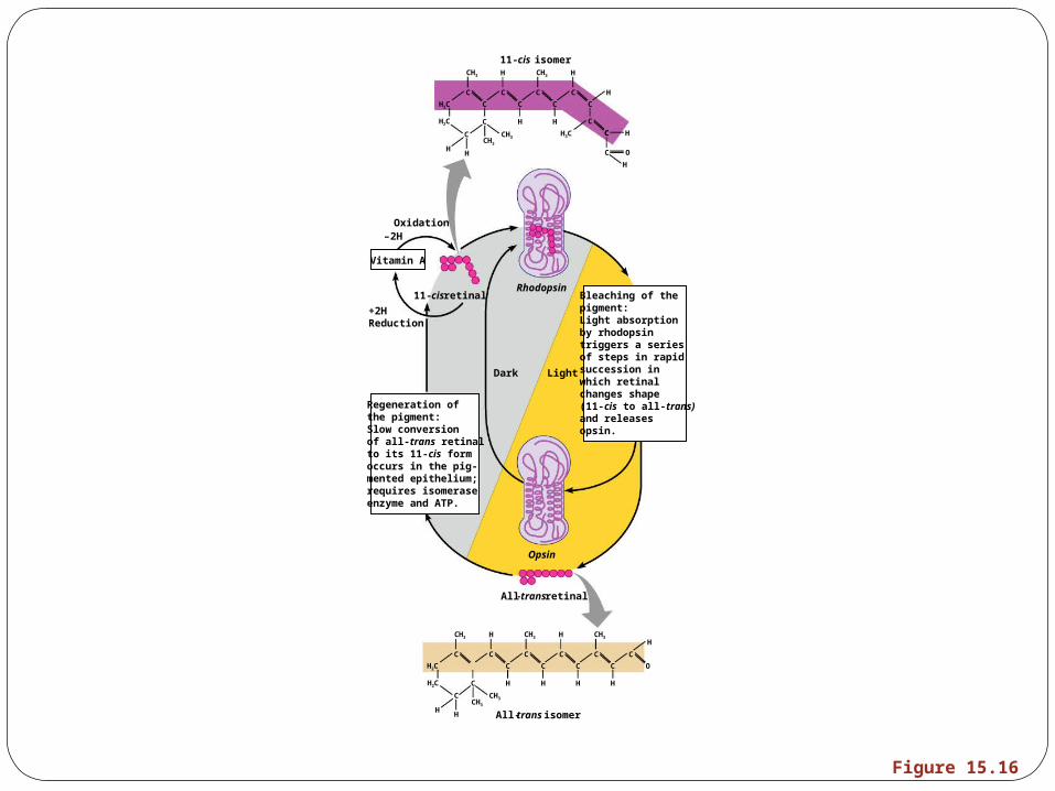
Excitation of RodsThe visual pigment of rods is rhodopsin
(opsin + 11-cis retinal)Light phase
Rhodopsin breaks down into all-trans retinal + opsin (bleaching of the pigment)
Dark phaseAll-trans retinal converts to 11-cis form 11-cis retinal is also formed from vitamin A11-cis retinal + opsin regenerate rhodopsin
CH3
C
C
HH
H2C
H2C C
C
CH3
H
CH3
C
H
C
CH3
C
H
C
H
C
H
C
CH3
C O
H
C
H
C
C
HH
H2C
H2C
H3C
C
C
C
CH3CH3
H
C
H
C
C O
C
H
C H
C
C
C HC
CH3 H CH3 H
Oxidation
Rhodopsin
Opsin
All-trans retinal
–2H
+2HReduction
Vitamin A
Regeneration ofthe pigment:Slow conversionof all-trans retinalto its 11-cis formoccurs in the pig-mented epithelium;requires isomeraseenzyme and ATP.
Dark Light
11-cis retinal
All-trans isomer
11-cis isomer
Bleaching of thepigment:Light absorptionby rhodopsintriggers a seriesof steps in rapidsuccession inwhich retinalchanges shape(11-cis to all-trans)and releasesopsin.
Figure 15.16
Excitation of ConesVisual pigments in cones are similar to rods
(retinal + opsins)There are three types of cones: blue,
green, and redIntermediate colors are perceived by
activation of more than one type of coneMethod of excitation is similar to rods
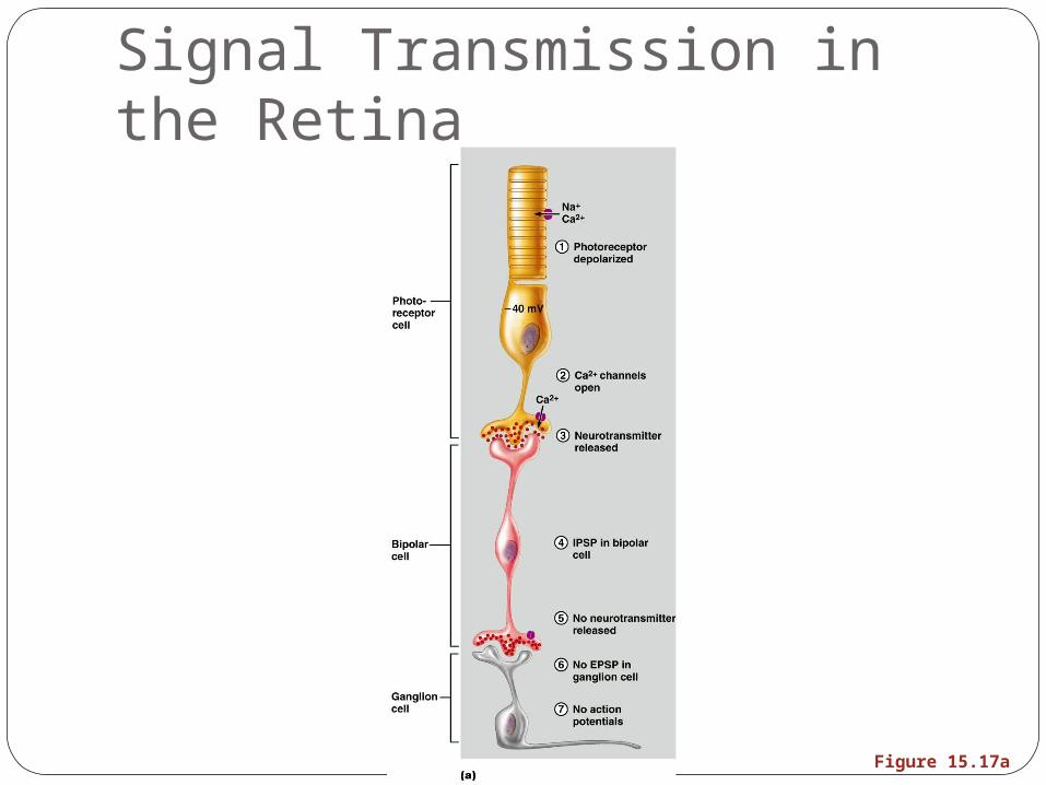
Signal Transmission in the Retina
Figure 15.17a
PhototransductionLight energy splits rhodopsin into all-trans
retinal, releasing activated opsinThe freed opsin activates the G protein
transducinTransducin catalyzes activation of
phosphodiesterase (PDE)PDE hydrolyzes cGMP to GMP and releases
it from sodium channelsWithout bound cGMP, sodium channels
close, the membrane hyperpolarizes, and neurotransmitter cannot be released
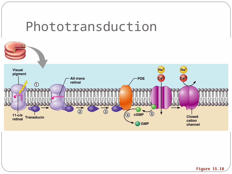
Phototransduction
Figure 15.18
AdaptationAdaptation to bright light (going from dark to
light) involves:Dramatic decreases in retinal sensitivity – rod
function is lostSwitching from the rod to the cone system –
visual acuity is gainedAdaptation to dark is the reverse
Cones stop functioning in low lightRhodopsin accumulates in the dark and retinal
sensitivity is restored
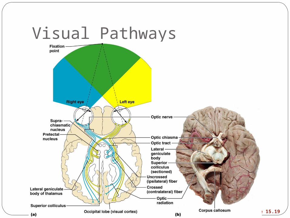
Visual PathwaysAxons of retinal ganglion cells form the optic
nerve Medial fibers of the optic nerve decussate at
the optic chiasmMost fibers of the optic tracts continue to the
lateral geniculate body of the thalamus
Visual PathwaysOther optic tract fibers end in superior
colliculi (initiating visual reflexes) and pretectal nuclei (involved with pupillary reflexes)
Optic radiations travel from the thalamus to the visual cortex
Visual Pathways
Figure 15.19
Visual PathwaysSome nerve fibers send tracts to the
midbrain ending in the superior colliculiA small subset of visual fibers contain
melanopsin (circadian pigment) which:Mediates papillary light reflexesSets daily biorhythms
Depth PerceptionAchieved by both eyes viewing the same
image from slightly different anglesThree-dimensional vision results from
cortical fusion of the slightly different images
If only one eye is used, depth perception is lost and the observer must rely on learned clues to determine depth
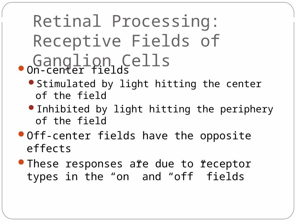
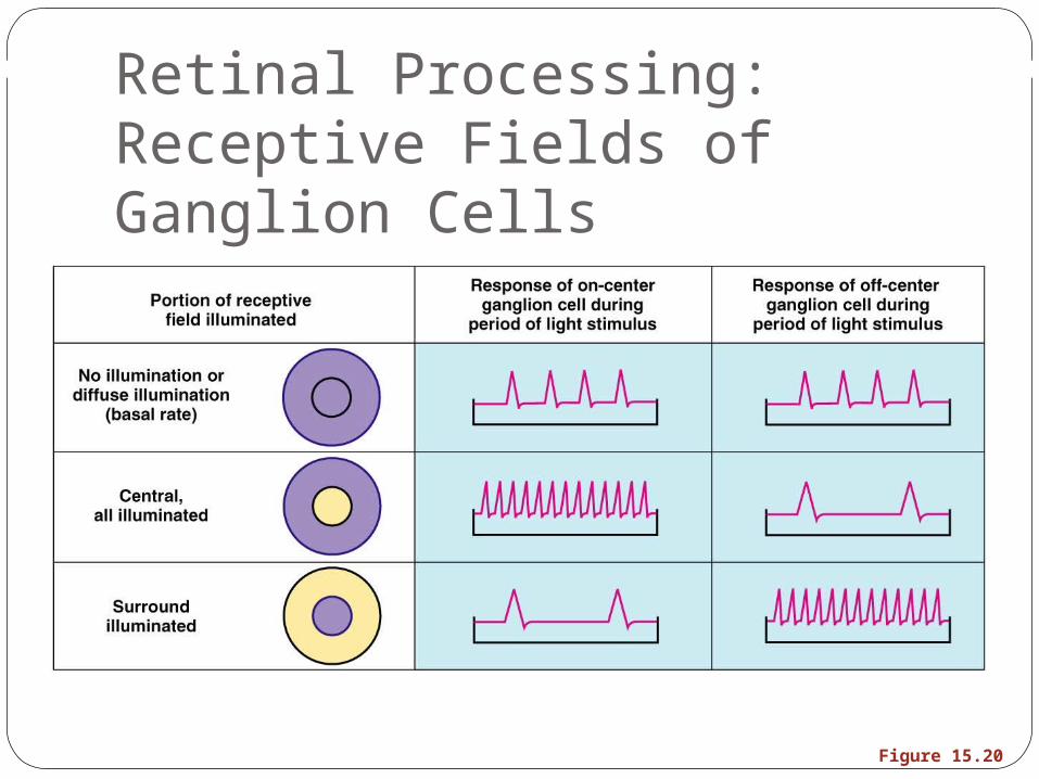
On-center fieldsStimulated by light hitting the center of the fieldInhibited by light hitting the periphery of the
fieldOff-center fields have the opposite effects These responses are due to receptor types in
the “on” and “off” fields
Retinal Processing: Receptive Fields of Ganglion Cells
Retinal Processing: Receptive Fields of Ganglion Cells
Figure 15.20
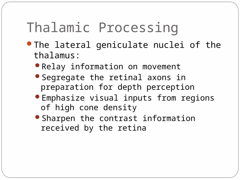
Thalamic ProcessingThe lateral geniculate nuclei of the
thalamus:Relay information on movementSegregate the retinal axons in preparation
for depth perceptionEmphasize visual inputs from regions of high
cone densitySharpen the contrast information received by
the retina
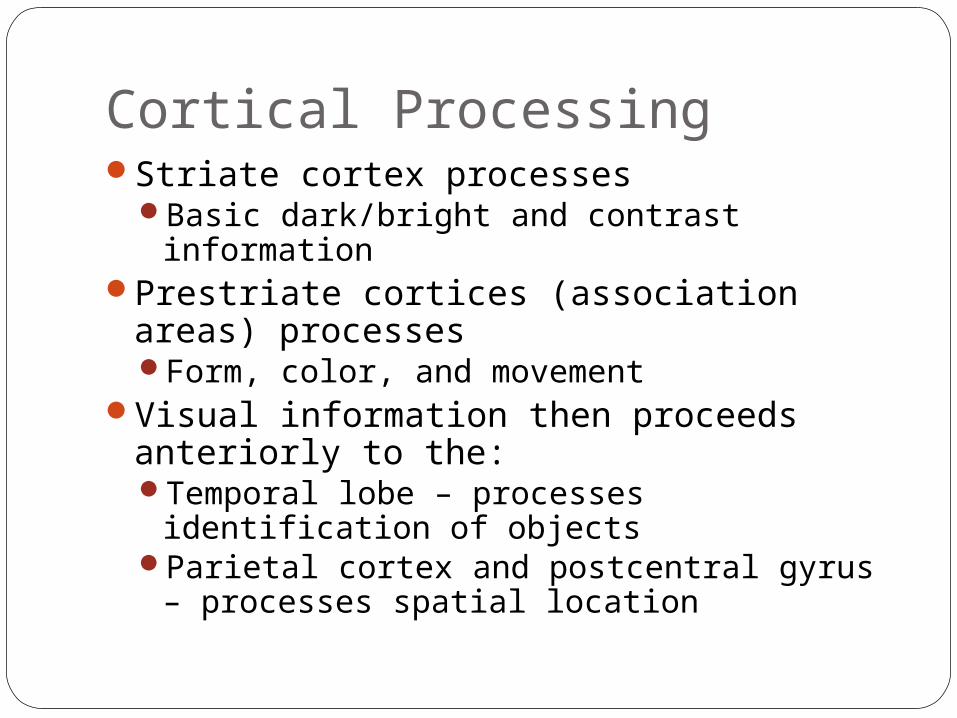
Cortical ProcessingStriate cortex processes
Basic dark/bright and contrast informationPrestriate cortices (association areas)
processesForm, color, and movement
Visual information then proceeds anteriorly to the:Temporal lobe – processes identification of
objectsParietal cortex and postcentral gyrus –
processes spatial location
Chemical SensesChemical senses – gustation (taste) and
olfaction (smell) Their chemoreceptors respond to chemicals
in aqueous solutionTaste – to substances dissolved in salivaSmell – to substances dissolved in fluids of
the nasal membranes
Sense of SmellThe organ of smell is the olfactory
epithelium, which covers the superior nasal concha
Olfactory receptor cells are bipolar neurons with radiating olfactory cilia
Olfactory receptors are surrounded and cushioned by supporting cells
Basal cells lie at the base of the epithelium
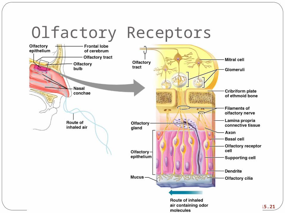
Olfactory Receptors
Figure 15.21
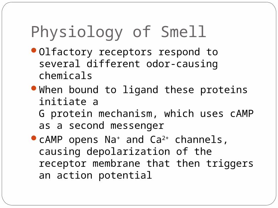
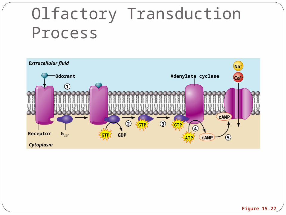
Physiology of SmellOlfactory receptors respond to several
different odor-causing chemicalsWhen bound to ligand these proteins
initiate a G protein mechanism, which uses cAMP as a second messenger
cAMP opens Na+ and Ca2+ channels, causing depolarization of the receptor membrane that then triggers an action potential
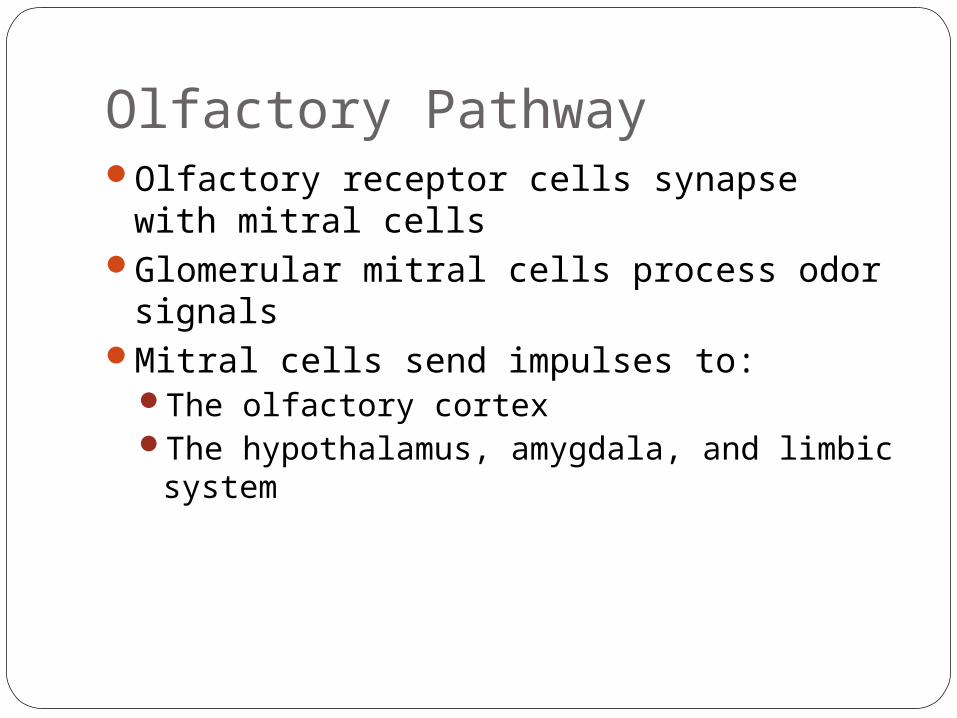
Olfactory PathwayOlfactory receptor cells synapse with mitral
cellsGlomerular mitral cells process odor signalsMitral cells send impulses to:
The olfactory cortex The hypothalamus, amygdala, and limbic
system
GolfReceptor
Extracellular fluid
Cytoplasm
Odorant Adenylate cyclase
Na+
Ca2+
GTP
GTP GTP
GDP cAMP
cAMP
ATP
1
2 34
5
Figure 15.22
Olfactory Transduction Process
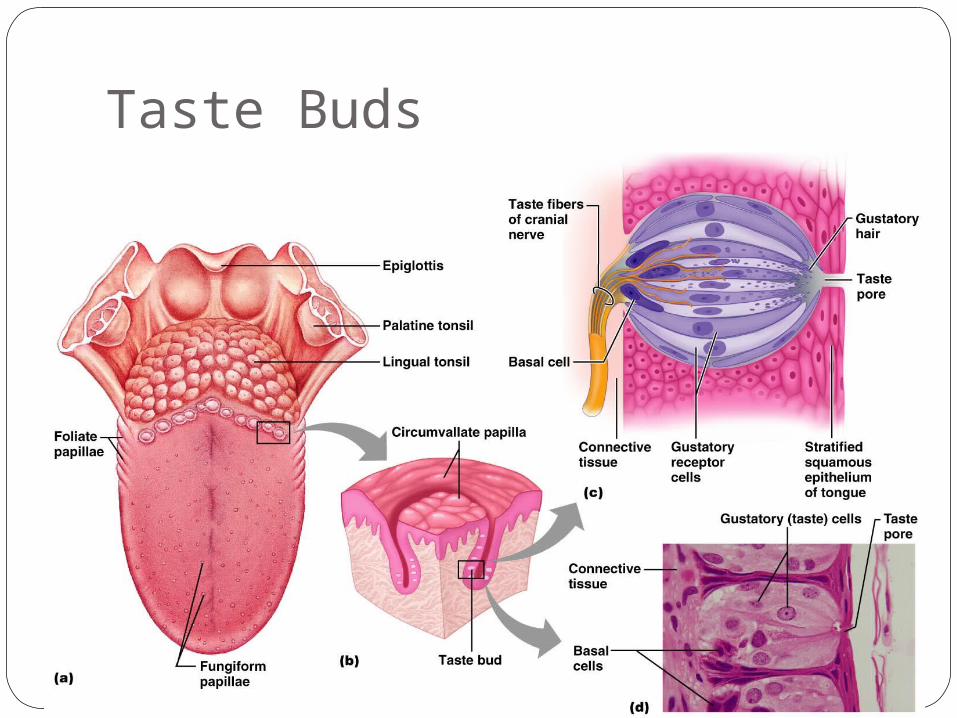
Taste BudsMost of the 10,000 or so taste buds are
found on the tongueTaste buds are found in papillae of the
tongue mucosaPapillae come in three types: filiform,
fungiform, and circumvallateFungiform and circumvallate papillae
contain taste buds
Taste Buds
Figure 15.23
Structure of a Taste BudEach gourd-shaped taste bud consists of
three major cell typesSupporting cells – insulate the receptor Basal cells – dynamic stem cells Gustatory cells – taste cells
Taste SensationsThere are five basic taste sensations
Sweet – sugars, saccharin, alcohol, and some amino acids
Salt – metal ionsSour – hydrogen ionsBitter – alkaloids such as quinine and nicotineUmami – elicited by the amino acid glutamate
Physiology of TasteIn order to be tasted, a chemical:
Must be dissolved in salivaMust contact gustatory hairs
Binding of the food chemical:Depolarizes the taste cell membrane,
releasing neurotransmitterInitiates a generator potential that elicits an
action potential
Taste TransductionThe stimulus energy of taste is converted
into a nerve impulse by:Na+ influx in salty tastesH+ in sour tastes (by directly entering the
cell, by opening cation channels, or by blockade of K+ channels)
Gustducin in sweet and bitter tastes
Gustatory PathwayCranial Nerves VII and IX carry impulses
from taste buds to the solitary nucleus of the medulla
These impulses then travel to the thalamus, and from there fibers branch to the:Gustatory cortex (taste)Hypothalamus and limbic system
(appreciation of taste)
Influence of Other Sensations on TasteTaste is 80% smellThermoreceptors, mechanoreceptors,
nociceptors also influence tastesTemperature and texture enhance or
detract from taste
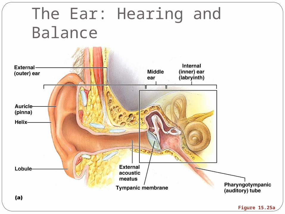
The Ear: Hearing and BalanceThe three parts of the ear are the inner,
outer, and middle earThe outer and middle ear are involved with
hearingThe inner ear functions in both hearing and
equilibriumReceptors for hearing and balance:
Respond to separate stimuliAre activated independently
The Ear: Hearing and Balance
Figure 15.25a
Outer EarThe auricle (pinna) is composed of:
The helix (rim)The lobule (earlobe)
External auditory canalShort, curved tube filled with ceruminous
glands
Outer EarTympanic membrane (eardrum)
Thin connective tissue membrane that vibrates in response to sound
Transfers sound energy to the middle ear ossicles
Boundary between outer and middle ears
Middle Ear (Tympanic Cavity)A small, air-filled, mucosa-lined cavity
Flanked laterally by the eardrumFlanked medially by the oval and round
windowsEpitympanic recess – superior portion of
the middle earPharyngotympanic tube – connects the
middle ear to the nasopharynxEqualizes pressure in the middle ear cavity
with the external air pressure
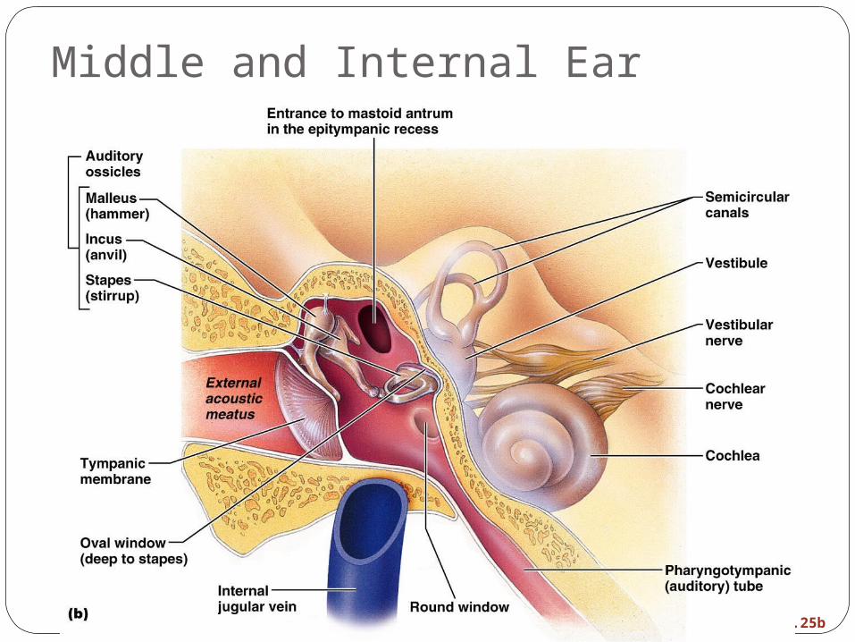
Middle and Internal Ear
Figure 15.25b
Ear OssiclesThe tympanic cavity contains three small
bones: the malleus, incus, and stapesTransmit vibratory motion of the eardrum to
the oval windowDampened by the tensor tympani and
stapedius muscles
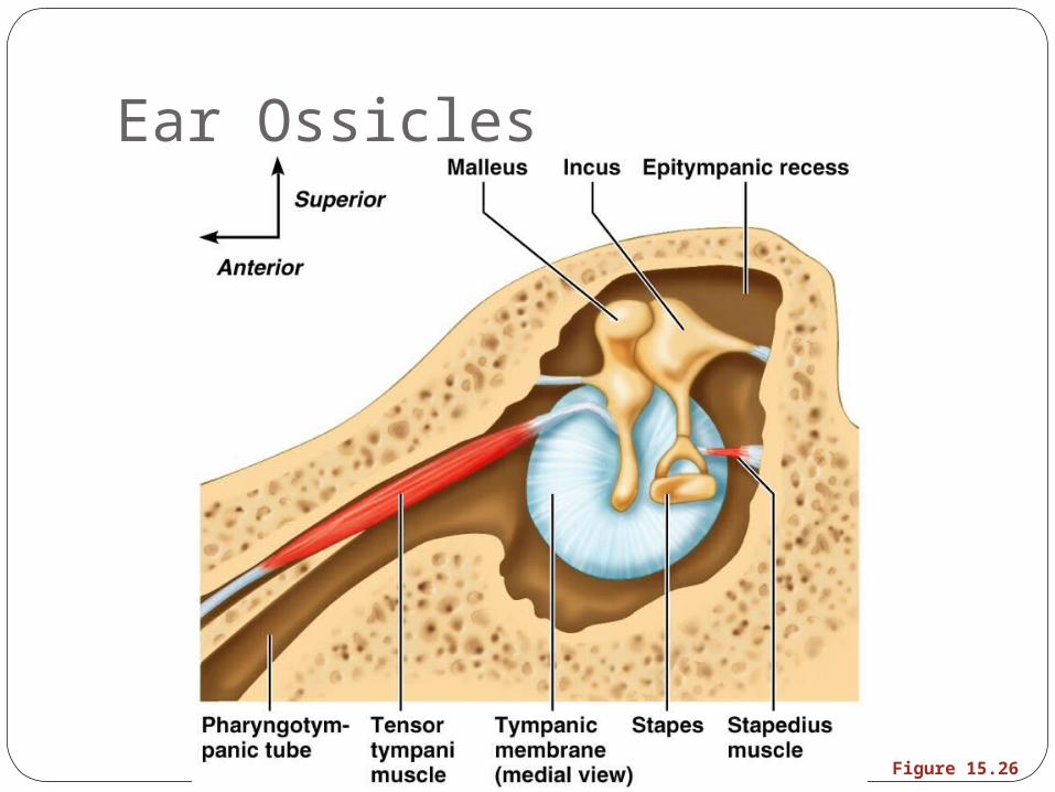
Ear Ossicles
Figure 15.26
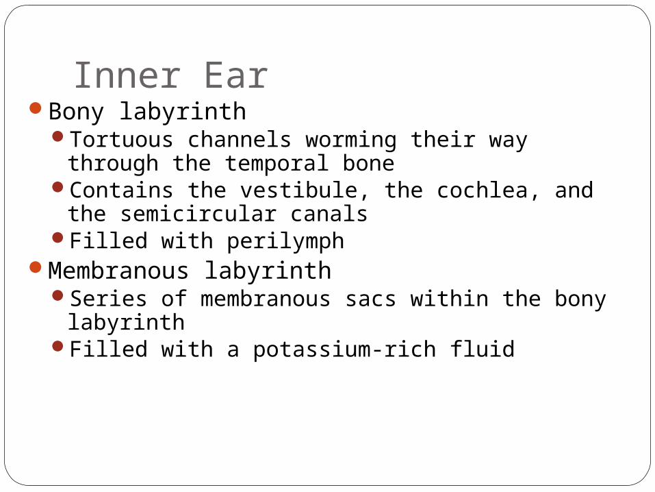
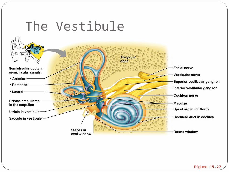
Inner EarBony labyrinth
Tortuous channels worming their way through the temporal bone
Contains the vestibule, the cochlea, and the semicircular canals
Filled with perilymphMembranous labyrinth
Series of membranous sacs within the bony labyrinth
Filled with a potassium-rich fluid
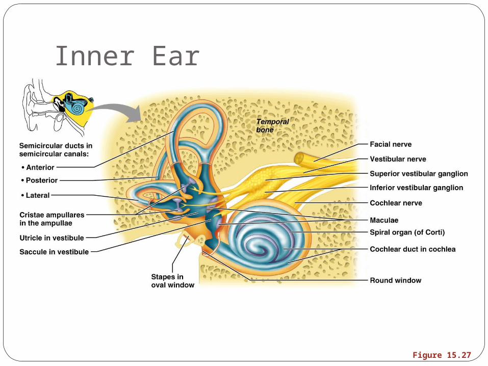
Inner Ear
Figure 15.27
The VestibuleThe central egg-shaped cavity of the bony
labyrinthSuspended in its perilymph are two sacs:
the saccule and utricleThe saccule extends into the cochlea
The VestibuleThe utricle extends into the semicircular
canalsThese sacs:
House equilibrium receptors called maculaeRespond to gravity and changes in the
position of the head
The Vestibule
Figure 15.27
The Semicircular CanalsThree canals that each define two-thirds of
a circle and lie in the three planes of space Membranous semicircular ducts line each
canal and communicate with the utricleThe ampulla is the swollen end of each
canal and it houses equilibrium receptors in a region called the crista ampullaris
These receptors respond to angular movements of the head
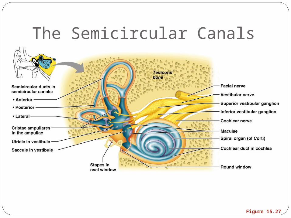
The Semicircular Canals
Figure 15.27
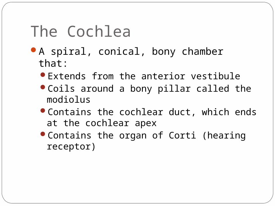
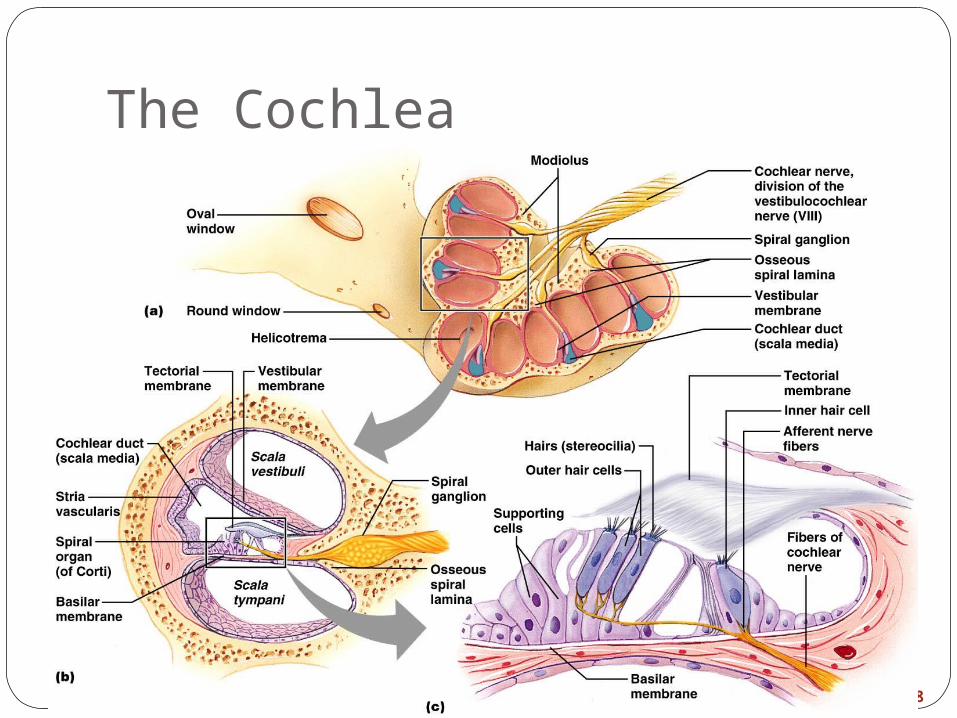
The CochleaA spiral, conical, bony chamber that:
Extends from the anterior vestibuleCoils around a bony pillar called the modiolusContains the cochlear duct, which ends at
the cochlear apexContains the organ of Corti (hearing
receptor)
The CochleaThe cochlea is divided into three chambers:
Scala vestibuliScala mediaScala tympani
The CochleaThe scala tympani terminates at the round
windowThe scalas tympani and vestibuli:
Are filled with perilymphAre continuous with each other via the
helicotremaThe scala media is filled with endolymph
The CochleaThe “floor” of the cochlear duct is
composed of:The bony spiral laminaThe basilar membrane, which supports the
organ of CortiThe cochlear branch of nerve VIII runs from
the organ of Corti to the brain
The Cochlea
Figure 15.28
Sound and Mechanisms of HearingSound vibrations beat against the eardrumThe eardrum pushes against the ossicles,
which presses fluid in the inner ear against the oval and round windowsThis movement sets up shearing forces that
pull on hair cellsMoving hair cells stimulates the cochlear
nerve that sends impulses to the brain
Properties of SoundSound is:
A pressure disturbance (alternating areas of high and low pressure) originating from a vibrating object
Composed of areas of rarefaction and compression
Represented by a sine wave in wavelength, frequency, and amplitude
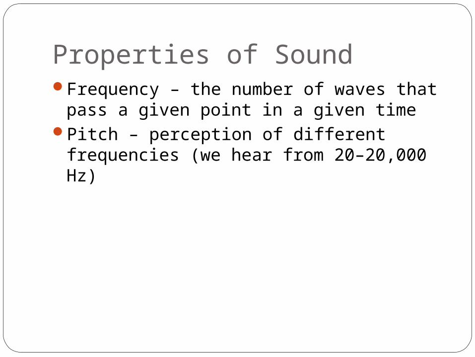
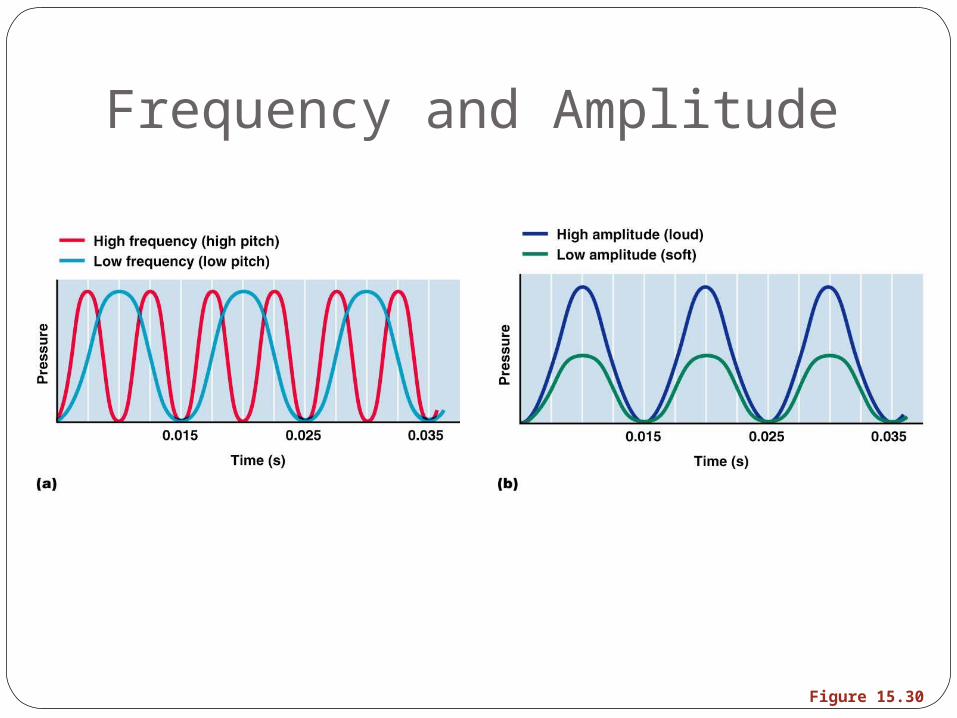
Properties of SoundFrequency – the number of waves that pass
a given point in a given timePitch – perception of different frequencies
(we hear from 20–20,000 Hz)
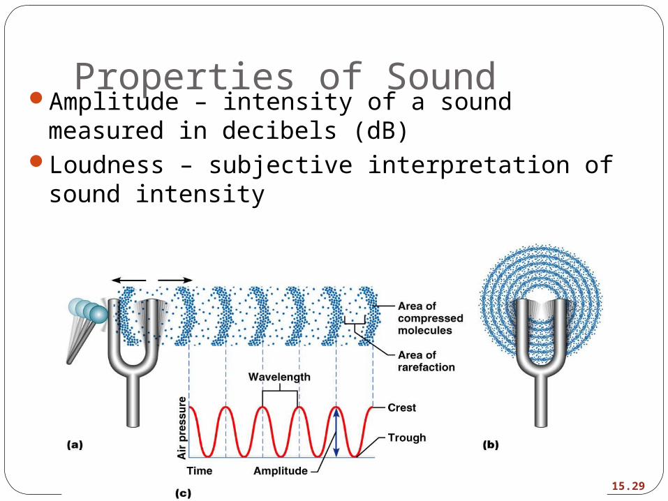
Properties of SoundAmplitude – intensity of a sound measured in
decibels (dB)Loudness – subjective interpretation of sound
intensity
Figure 15.29
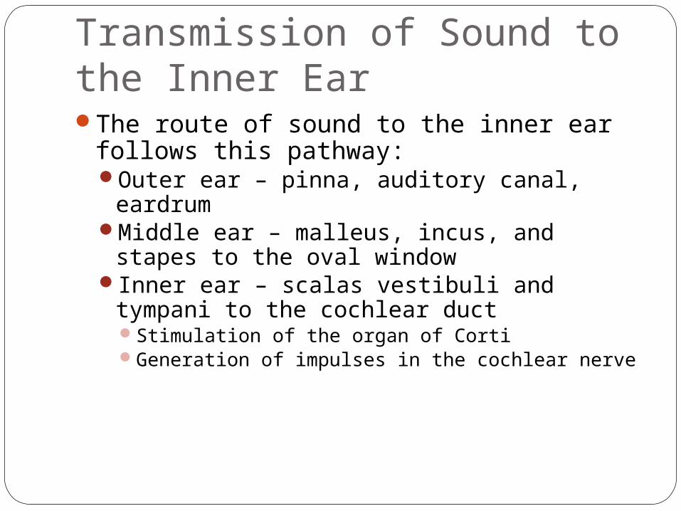
Transmission of Sound to the Inner EarThe route of sound to the inner ear follows
this pathway:Outer ear – pinna, auditory canal, eardrumMiddle ear – malleus, incus, and stapes to the
oval windowInner ear – scalas vestibuli and tympani to
the cochlear duct Stimulation of the organ of CortiGeneration of impulses in the cochlear nerve
Frequency and Amplitude
Figure 15.30
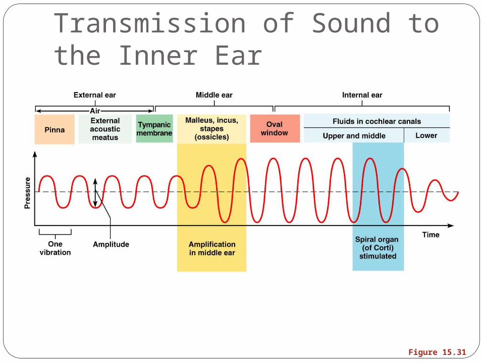
Transmission of Sound to the Inner Ear
Figure 15.31
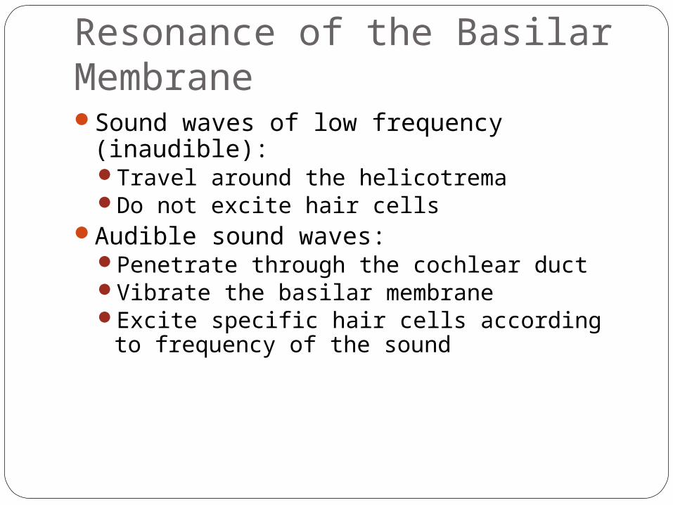
Resonance of the Basilar MembraneSound waves of low frequency (inaudible):
Travel around the helicotrema Do not excite hair cells
Audible sound waves:Penetrate through the cochlear ductVibrate the basilar membraneExcite specific hair cells according to
frequency of the sound
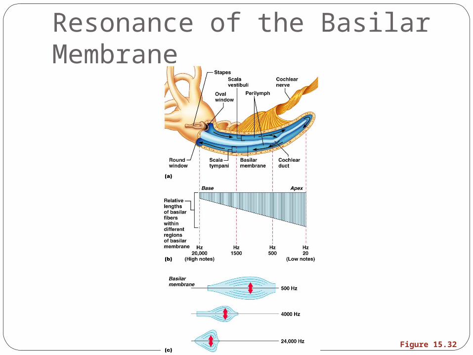
Resonance of the Basilar Membrane
Figure 15.32

The Organ of CortiIs composed of supporting cells and outer
and inner hair cellsAfferent fibers of the cochlear nerve attach
to the base of hair cellsThe stereocilia (hairs):
Protrude into the endolymphTouch the tectorial membrane
Excitation of Hair Cells in the Organ of CortiBending cilia:
Opens mechanically gated ion channelsCauses a graded potential and the release of
a neurotransmitter (probably glutamate)The neurotransmitter causes cochlear
fibers to transmit impulses to the brain, where sound is perceived
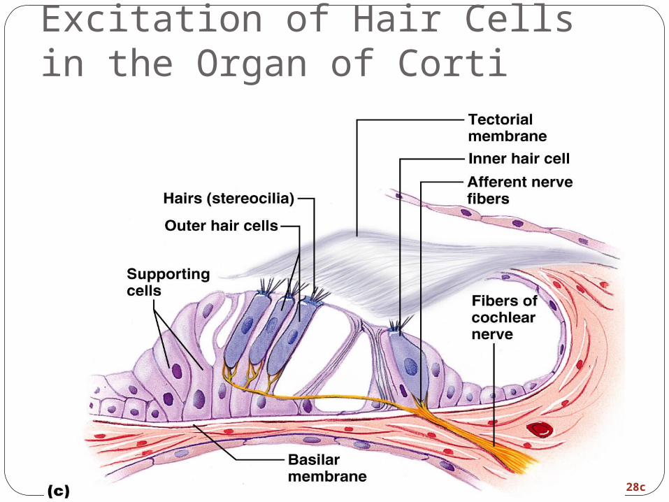
Excitation of Hair Cells in the Organ of Corti
Figure 15.28c
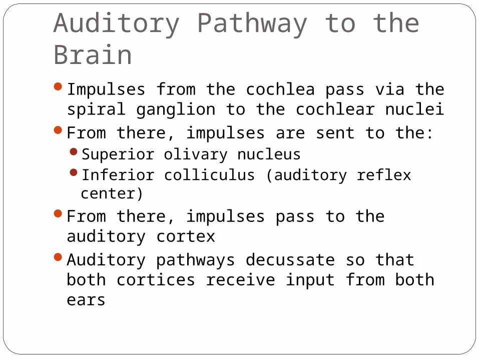
Auditory Pathway to the BrainImpulses from the cochlea pass via the
spiral ganglion to the cochlear nuclei From there, impulses are sent to the:
Superior olivary nucleus Inferior colliculus (auditory reflex center)
From there, impulses pass to the auditory cortex
Auditory pathways decussate so that both cortices receive input from both ears
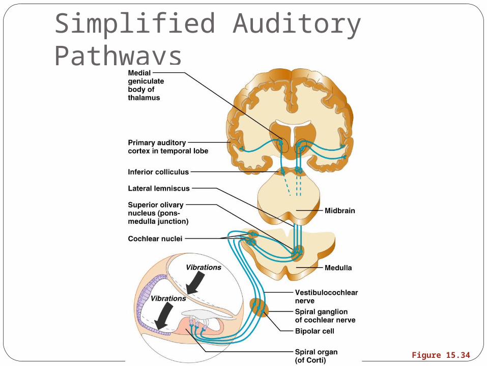
Simplified Auditory Pathways
Figure 15.34
Auditory ProcessingPitch is perceived by:
The primary auditory cortexCochlear nuclei
Loudness is perceived by:Varying thresholds of cochlear cellsThe number of cells stimulated
Localization is perceived by superior olivary nuclei that determine sound
DeafnessConduction deafness – something hampers
sound conduction to the fluids of the inner ear (e.g., impacted earwax, perforated eardrum, osteosclerosis of the ossicles)
Sensorineural deafness – results from damage to the neural structures at any point from the cochlear hair cells to the auditory cortical cells
DeafnessTinnitus – ringing or clicking sound in the
ears in the absence of auditory stimuliMeniere’s syndrome – labyrinth disorder
that affects the cochlea and the semicircular canals, causing vertigo, nausea, and vomiting
Mechanisms of Equilibrium and OrientationVestibular apparatus – equilibrium
receptors in the semicircular canals and vestibuleMaintains our orientation and balance in
spaceVestibular receptors monitor static
equilibriumSemicircular canal receptors monitor
dynamic equilibrium
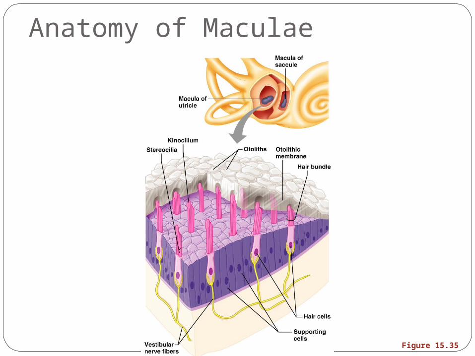
Anatomy of MaculaeMaculae are the sensory receptors for
static equilibriumContain supporting cells and hair cellsEach hair cell has stereocilia and kinocilium
embedded in the otolithic membraneOtolithic membrane – jellylike mass
studded with tiny CaCO3 stones called otoliths
Utricular hairs respond to horizontal movement
Saccular hairs respond to vertical movement
Anatomy of Maculae
Figure 15.35
Effect of Gravity on Utricular Receptor Cells
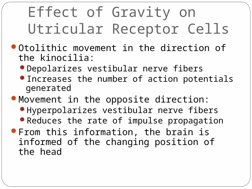
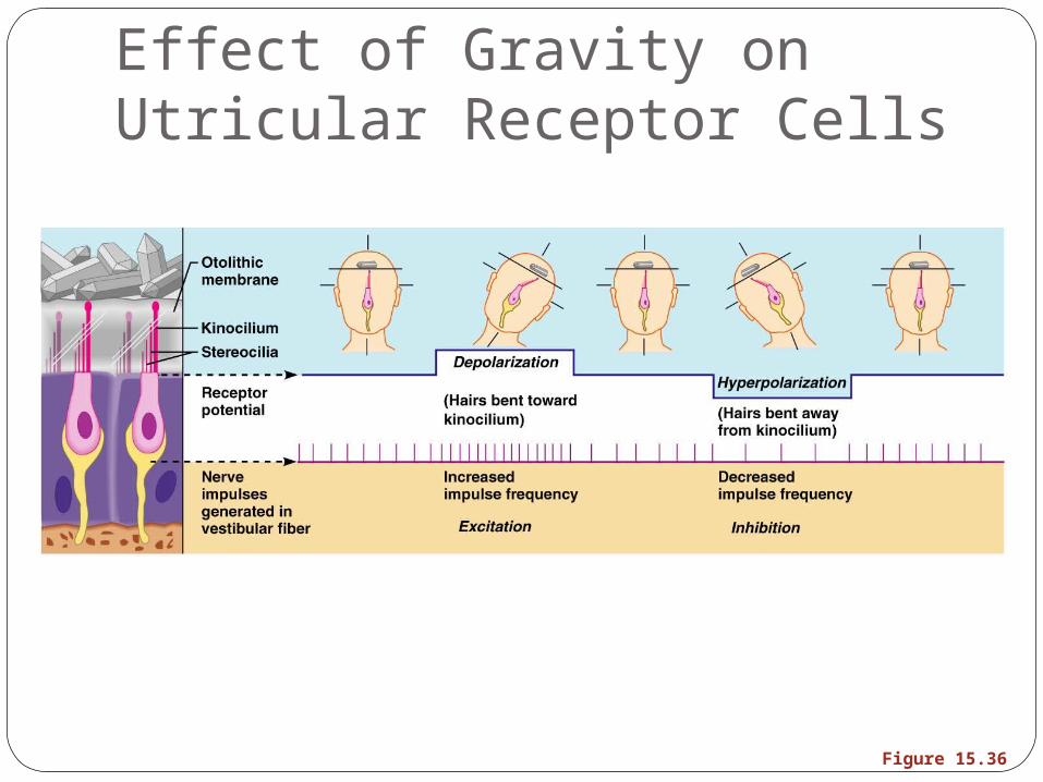
Otolithic movement in the direction of the kinocilia:Depolarizes vestibular nerve fibersIncreases the number of action potentials
generatedMovement in the opposite direction:
Hyperpolarizes vestibular nerve fibersReduces the rate of impulse propagation
From this information, the brain is informed of the changing position of the head
Effect of Gravity on Utricular Receptor Cells
Figure 15.36
Crista Ampullaris and Dynamic EquilibriumThe crista ampullaris (or crista):
Is the receptor for dynamic equilibriumIs located in the ampulla of each semicircular
canalResponds to angular movements
Each crista has support cells and hair cells that extend into a gel-like mass called the cupula
Dendrites of vestibular nerve fibers encircle the base of the hair cells
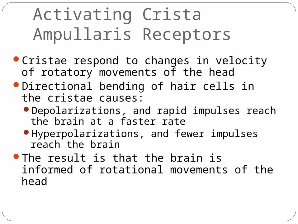
Activating Crista Ampullaris Receptors
Cristae respond to changes in velocity of rotatory movements of the head
Directional bending of hair cells in the cristae causes:Depolarizations, and rapid impulses reach the
brain at a faster rateHyperpolarizations, and fewer impulses reach
the brainThe result is that the brain is informed of
rotational movements of the head
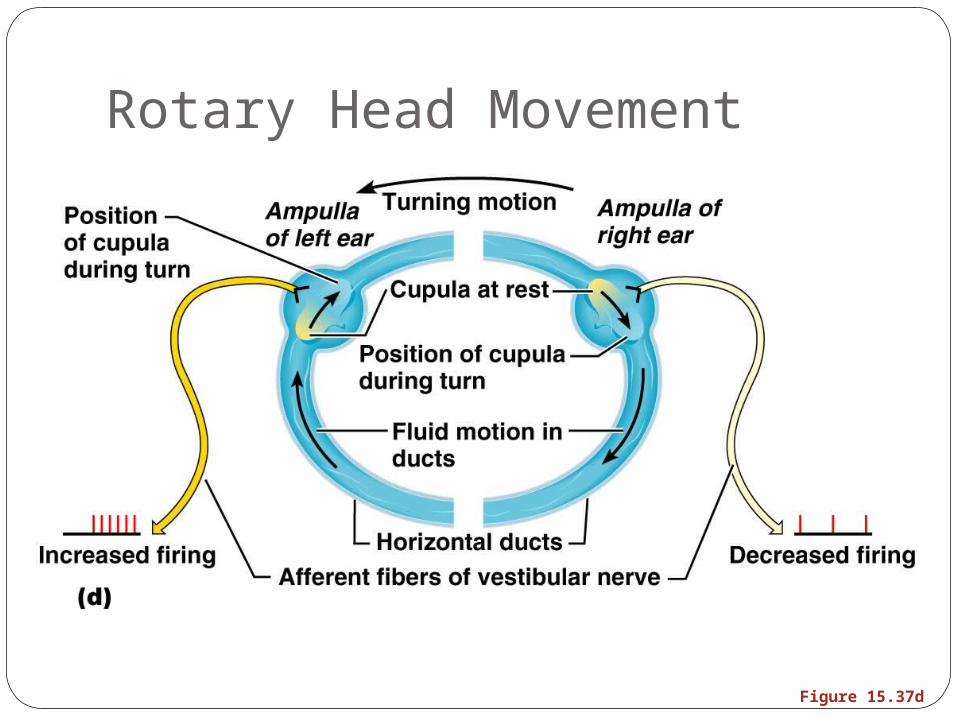
Rotary Head Movement
Figure 15.37d
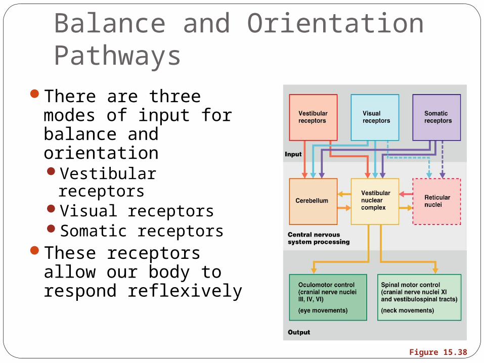
Balance and Orientation Pathways
There are three modes of input for balance and orientationVestibular receptorsVisual receptorsSomatic receptors
These receptors allow our body to respond reflexively
Figure 15.38
Developmental AspectsAll special senses are functional at birthChemical senses – few problems occur until
the fourth decade, when these senses begin to decline
Vision – optic vesicles protrude from the diencephalon during the fourth week of developmentThese vesicles indent to form optic cups and
their stalks form optic nervesLater, the lens forms from ectoderm
Developmental AspectsVision is not fully functional at birthBabies are hyperopic, see only gray tones,
and eye movements are uncoordinatedDepth perception and color vision is well
developed by age five and emmetropic eyes are developed by year six
With age the lens loses clarity, dilator muscles are less efficient, and visual acuity is drastically decreased by age 70
Developmental AspectsEar development begins in the three-week
embryoInner ears develop from otic placodes, which
invaginate into the otic pit and otic vesicleThe otic vesicle becomes the membranous
labyrinth, and the surrounding mesenchyme becomes the bony labyrinth
Middle ear structures develop from the pharyngeal pouches
The branchial groove develops into outer ear structures