Embed Size (px)
Citation preview
Extramural Initiatives:Technology Research and Development
An Evaluative Update of the NCI’s Innovative Molecular Analysis Technologies Program
1
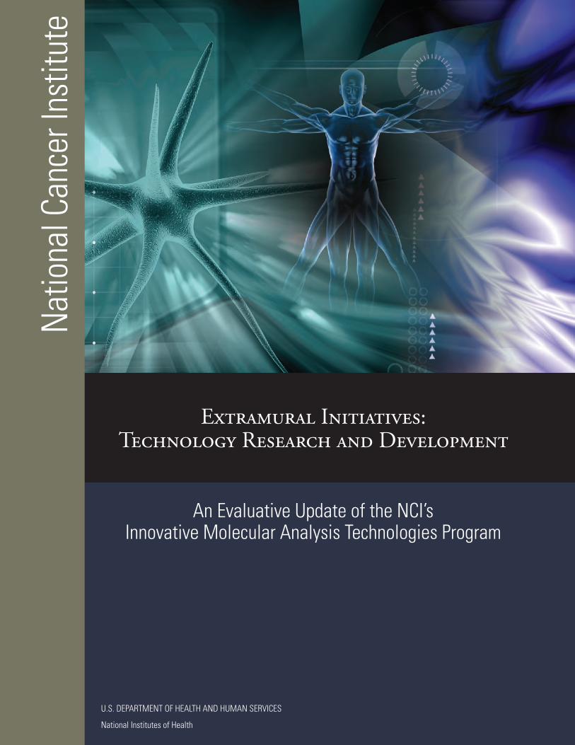
Extramural Initiatives: Technology Research and Development An Evaluative Update of the NCI’s Innovative Molecular Analysis Technologies
Program Independent 3rd Party Source: Science and Technology Policy Institute (STPI), Washington D.C. The Science and Technology Policy Institute (STPI) assists the Executive Branch of the US government as it formulates federal S&T policy by providing objective, high‐quality analytic support to inform policymakers. Chartered by an act of Congress in 1991, STPI provides the highest quality and rigorously objective technical analytical support for the Office of Science and Technology Policy and other government users, under the sponsorship of the National Science Foundation. This report was prepared in conjunction with STPI and the NCI Office of Planning and Assessment.
Purpose, Mandate, and Mission In 1998 the National Cancer Institute (NCI) established the groundwork for a highly successful program focused on early-stage technology development to meet the specific needs of cancer researchers and clinicians by stimulating the next wave of technologies capable of being applied toward the field of cancer research. Unlike other initiatives of the time, the Innovative Molecular Analysis Technologies (IMAT) program solicited only the most cutting-edge ideas, thus restricting its application pool to those projects with the potential to be truly transformative. By doing so, the program filled a void that no other program at the NCI or the National Institutes of Health (NIH) filled. Taking risks on early-stage transformative technologies, IMAT has contributed to many of the blockbuster technologies that are now on the market and in almost ubiquitous use across the cancer research and clinical communities. Successfully commercialized products such as RNALater, Affymetrix gene chips, Illumina bead platforms, and quantum dot labeling technology were all considered high-risk ideas at the time of their inception and initial funding through the IMAT program. Yet, their current widespread use and applicability to multiple clinical and basic sciences research settings are a testament to the high payoff and impact that such transformative technologies have provided to the field of cancer research. By soliciting and supporting these otherwise risky technologies through the IMAT program, the NCI has not only supported the development of these new transformative technologies in and of themselves but also supported them in a manner consistent with providing researchers rapid access to such platforms through appropriate commercialization and dissemination. The NCI has thus taken risks to substantiate the ultimate value and utility of such technologies even in cases where venture capital firms may have been reluctant to do so because of the inherent risks associated with innovative technology development. Currently, there are new challenges facing cancer researchers and clinicians, and as such, the need for a sustained technology development pipeline encompassing inception and initiation (i.e., the “bright idea” stage) through dissemination and commercialization has never been greater. Challenges represented by the need to rapidly assess all of the epigenetic changes in single cells, directly measure microenvironment impact on cancer metastasis, and collect rare cells from the blood of patients with recurrent disease require creative thinking and risk-taking to enable research in a manner similar to the way that gene expression profiling is currently enabled. IMAT seeks to fill this void by:
(1) Empowering individual investigators and small commercial entities such as small business concerns to think creatively;
(2) Stimulating biologists and clinicians to partner with technologists, engineers, physical
scientists, and individuals from other sectors who face similar technical challenges; and
2
(3) Taking the risk needed to break through common technical barriers that currently impede biomedical research and effective clinical decision-making.
By accomplishing the above goals, the IMAT program seeks to stimulate progress in the field of cancer research at a pace that is revolutionary rather than evolutionary or incremental, and to ensure the adequate and equal dissemination of knowledge that stems from such an approach. In 2006 the NCI Executive Committee commissioned an external evaluation of the IMAT program in order to assess the program’s progress toward meeting its stated objectives. The Office of Biorepositories and Biospecimen Research presented the results of this third-party outcomes evaluation to members of the NCI Executive Committee during the first quarter of 2008. This evaluative update provides an assessment of the program’s progress over a 2 year and 5 month period (i.e. since publication and presentation of the 2007 program evaluation) and lays the groundwork for IMAT’s continued future success in keeping the forefront and collaborative nature of cancer research moving forward. Unique Aspects of IMAT Among the strengths of the IMAT Program is the diversity of disciplines from which potentially transformative technologies are solicited. IMAT supports technology development from a variety of cross-cutting, research-enabling scientific and technical disciplines in an attempt to provide directionality to the flow of technology development while not inhibiting the creativity of the individual investigator in tackling the most challenging barriers to cancer research. Highly innovative technologies carrying a certain degree of technical risk but that also have the capability or potential to uncover new directions and paradigms in cancer research are desired and supported through a variety of unique and fully cooperative program characteristics:
Multidisciplinary – technologies are solicited and drawn from a variety of disciplines and fields
Investigator Initiated – utilizes investigator-initiated research project grant mechanisms with the intent of developing better tools and technologies capable of being put back into the hands of the R01 research community
Trans-Divisional – comprised of representatives from all NCI divisions having a common
purpose of ensuring the development of cutting-edge, high-impact technologies and platforms
Innovation Focused – solicits and supports the development, maturation, and dissemination of technically innovative, high risk, high impact and potentially high payoff technologies
Technology Focused – does not support traditional hypothesis driven research but rather supports
the development, transfer, and commercialization of tools and platforms to enable more effective and comprehensive R01 research
Commitment to New or First-Time Investigators – approximately 1/3 of the program’s
portfolio is comprised of new or first-time investigators
Commitment to Diversity – in collaboration with the Center to Reduce Cancer Health Disparities (CRCHD), provides novel education and training programs for historically disadvantaged individuals from high school to post-doctoral levels in areas of emerging technology
Unique Review Structure and Process – utilizes and commissions special emphasis panels
(SEPs) rather than standing Center for Scientific Review (CSR)-appointed study sections in order to ensure that each review panel is focused on the development of innovative technologies and reflects the breadth and depth of the constantly evolving science being reviewed
3
In addition to the above, IMAT has distinguished itself among NIH/NCI programs through a series of “firsts”:
First fully functionally cooperative and trans-divisional program administered by and through the NCI Office of the Director
First program to establish and implement a phased investigator-initiated funding mechanism to accelerate innovation (R21/33 Phased Innovation Award)
First program within the Office of the NCI Director to utilize 100% investigator-initiated
research project grant funding with no infrastructure
First program to support technology-centric, high-risk, high-impact/payoff research and development projects at the NCI and NIH
First program to establish multiple review and receipt cycles per FOA and to utilize ‘ad-
hoc’ Special Emphasis Panels (SEPs) to review submitted applications based on nature, number, and type of applications received per round of receipt
First program to utilize individual SEPs composed of reviewers from traditionally
disparate fields (mix of biologists, technologists, clinicians, engineers and statisticians/mathematicians from both academia and industry)
First program to implement responsiveness and suitability assessments and
requirements in order to ensure alignment and compliance with programmatic objectives and responsible stewardship of public funds
First program to require incorporation of quantitative project milestones in all proposed
proof-of-principle establishment projects and to use such milestones as an evaluation criterion during peer review
First program to delineate a particular scope and to create a list of exclusions of scope in
order to prevent duplication with other NCI programs and initiatives (e.g. Cancer Imaging Program, Biomedical Information Sciences and Technologies Initiative, Cancer Therapeutics Evaluation Program)
Program Integrity, Transparency, and Accountability: The IMAT Program has always attempted to ensure a certain degree of transparency and accountability throughout the technology development process. These actions not only increase the probability of success for the individual investigator but also ensure the responsible use and stewardship of public funds. Varying degrees of accountability are built into the IMAT application, management, and award processes as described below: Application and Review Phase: Innovation, Novelty, and Significance Statement In order to ensure concordance with the stated objectives of the IMAT Program in recruiting only the most innovative and highly transformative technologies in multiple disciplines, all applicants to the program are required to address the following questions in their submitted applications:
How is the [proposed] project potentially transformative and why may it be expected to produce an unusually high impact on biomedical research and technologies?
What are the pioneering approaches for which the potential for groundbreaking or paradigm-shifting results compensates and justifies any associated risks?
4
What concrete evidence can be provided to substantiate the claim of innovativeness?
Benchmarks and Demonstration of Progress All exploratory/pilot-phase applications submitted to the IMAT Program are required to contain quantitative milestones as benchmarks for progress. Other than being a requirement for submission, such milestones are also utilized in the evaluation of the technical and scientific merits associated with the application by peer review. Inclusion of milestones serves two purposes: 1) it increases the probability of the accomplishment of the application’s specific aims, and 2) serves as a benchmark of progress for transition to the next stage of technical development (e.g. transition from proof-of-principle to feasibility or developmental phase). Milestones must be well described, quantitative, and scientifically justified. Specific aims are not regarded as milestones (unless they include quantitative end points). The specific aims describe the goals and intended path of the research while quantitative milestones are a way of determining whether an applicant has successfully reached the specified goals. Milestones are required to be clearly stated and presented in a quantitative manner, such as numerical specifications of sensitivity and specificity or a count of some desired or newly discovered molecule, etc. An application lacking quantitative milestones as determined by the program staff may be returned to the applicant without review. Responsiveness of Applications Commensurate with the IMAT Program’s continual programmatic evolution, all applications submitted to the program since 2003 are initially screened by program staff for responsiveness. Such an examination ensures that all applications submitted to, reviewed by, and potentially funded under the IMAT Program are in-line with objectives for which the program was founded and the larger technology development objectives of the National Cancer Institute. Review of responsiveness by program staff in no way represents an attempt to evaluate the science proposed in a given application. Rather, such a review is made to ensure consistency with program objectives, observance of NIH policies pertaining to research project grants, and the responsible use of public funds. The process also serves to notify non-responsive applicants as early in the application process as possible so that they may submit their applications to other, more appropriate solicitations with minimal loss of time and resources. Post-Application and Award Phase: Communication of Relevant Results Communication of relevant results related to IMAT technology-development grants have been communicated via various publications in peer-reviewed academic journals, acquisition of patents or patent applications, and/or various technology transfer activities. In addition to such activities, an annual meeting of all IMAT-funded Principal Investigators is held in the Fall of each year as a means of providing an open forum by which to hold fruitful discussions, stimulate collaborations, and discuss significant progress and results. All IMAT-funded principal investigators are asked to attend the meeting and to communicate the relevant results from their work in the form of either an oral presentation or poster session. Keynote speakers representing established and well-known experts in the field of biomedical and/or industrial technology development are also invited to attend. All applicants to the program are asked to appropriately budget for the cost of attendance in their research project grant applications. The meeting has traditionally been held on the East and West coasts of the United States on an alternating, yearly basis. Reporting Requirement(s) All IMAT awardees are required to submit a Non-Competing Continuation Grant Progress Report (PHS 2590) annually and financial statements as required in the NIH Grants Policy Statement. A final progress report, invention statement, and Financial Status Report are required when an award is relinquished when a recipient changes institutions or when an award is terminated.
5
Reporting requirements are meant to promote transparency and communication between IMAT awardees and program staff. Program-Level Accountability: Program-Wide Third Party Evaluation In 2006, the IMAT Program, with authorization from the NCI Executive Committee, commissioned an external program-wide outcomes evaluation in order to assess the program’s progress toward meeting its stated objectives, ensure transparency, and assess areas in need of improvement. The evaluation was preceded by a comprehensive feasibility study performed by a third-party (ORC Macro / Macro International) in order to more effectively inform the development of an appropriate and meaningful evaluation structure and construct. The outcome evaluation itself was conducted by a second external entity (SAIC). Results of both the feasibility study and outcome evaluation were presented to members of the NCI Executive Committee as well as the National Cancer Institute’s external Board of Scientific Advisors (BSA). The evaluation provided an assessment of the program’s progress and laid the groundwork for its continued future successes in keeping the forefront of cancer research moving forward. Some of the program’s achievements that were highlighted in the evaluation can be found by viewing the Outputs, Achievements, and Accountability section of the program’s website. The more recent outputs and achievements are described below. Prospective Evaluative Updates and Evaluations In addition to the program-wide evaluation described above, the IMAT program undergoes periodic evaluative updates as part of a prospective (rather than retrospective) approach to ensuring program-wide integrity, transparency, and accountability. In all cases, such updates are always conducted by an independent, third-party entity with minimal input from program staff. This report encompasses the 2010 evaluative update, as conducted by the Science and Technology Policy Institute (STPI), Washington D.C.
6
Continuing Program Successes (Specific Outputs Between 2008 - 2010): Since the completion of its program-wide evaluation and the subsequent presentation of its results to the NCI Executive Committee during the first quarter of 2008, the IMAT program has continued to produce high-quality outputs, consistent with its mission to empower cancer research and treatment through technological innovation. The following represent a snap-shot of the program’s most recent successes between 2008 and 2010 and are a continuance of previously documented program successes:
Individual Technologies and Platforms
COLD PCR® Technology: Commercialized by TransGenomic Corporation (2010)
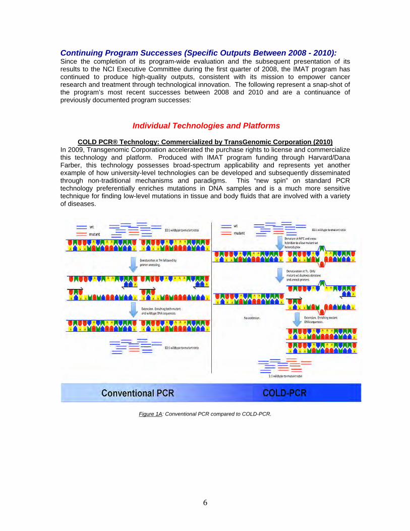
In 2009, Transgenomic Corporation accelerated the purchase rights to license and commercialize this technology and platform. Produced with IMAT program funding through Harvard/Dana Farber, this technology possesses broad-spectrum applicability and represents yet another example of how university-level technologies can be developed and subsequently disseminated through non-traditional mechanisms and paradigms. This “new spin” on standard PCR technology preferentially enriches mutations in DNA samples and is a much more sensitive technique for finding low-level mutations in tissue and body fluids that are involved with a variety of diseases.
Figure 1A: Conventional PCR compared to COLD-PCR.
7
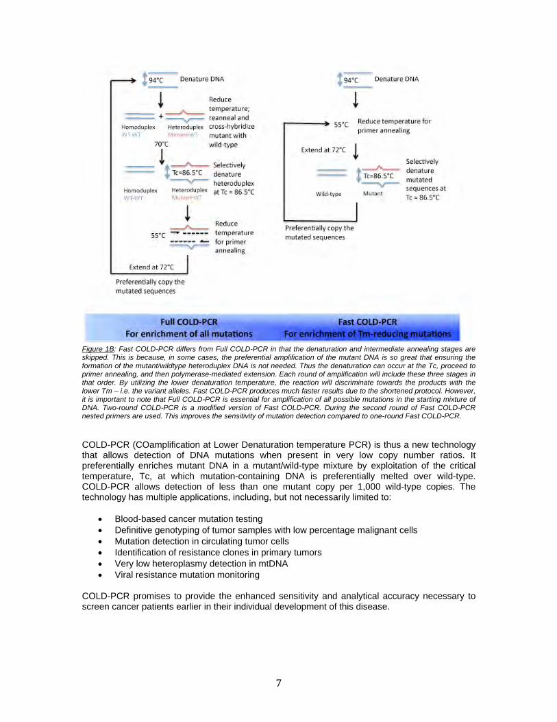
Figure 1B: Fast COLD-PCR differs from Full COLD-PCR in that the denaturation and intermediate annealing stages are skipped. This is because, in some cases, the preferential amplification of the mutant DNA is so great that ensuring the formation of the mutant/wildtype heteroduplex DNA is not needed. Thus the denaturation can occur at the Tc, proceed to primer annealing, and then polymerase-mediated extension. Each round of amplification will include these three stages in that order. By utilizing the lower denaturation temperature, the reaction will discriminate towards the products with the lower Tm – i.e. the variant alleles. Fast COLD-PCR produces much faster results due to the shortened protocol. However, it is important to note that Full COLD-PCR is essential for amplification of all possible mutations in the starting mixture of DNA. Two-round COLD-PCR is a modified version of Fast COLD-PCR. During the second round of Fast COLD-PCR nested primers are used. This improves the sensitivity of mutation detection compared to one-round Fast COLD-PCR.
COLD-PCR (COamplification at Lower Denaturation temperature PCR) is thus a new technology that allows detection of DNA mutations when present in very low copy number ratios. It preferentially enriches mutant DNA in a mutant/wild-type mixture by exploitation of the critical temperature, Tc, at which mutation-containing DNA is preferentially melted over wild-type. COLD-PCR allows detection of less than one mutant copy per 1,000 wild-type copies. The technology has multiple applications, including, but not necessarily limited to:
Blood-based cancer mutation testing Definitive genotyping of tumor samples with low percentage malignant cells Mutation detection in circulating tumor cells Identification of resistance clones in primary tumors Very low heteroplasmy detection in mtDNA Viral resistance mutation monitoring
COLD-PCR promises to provide the enhanced sensitivity and analytical accuracy necessary to screen cancer patients earlier in their individual development of this disease.
8
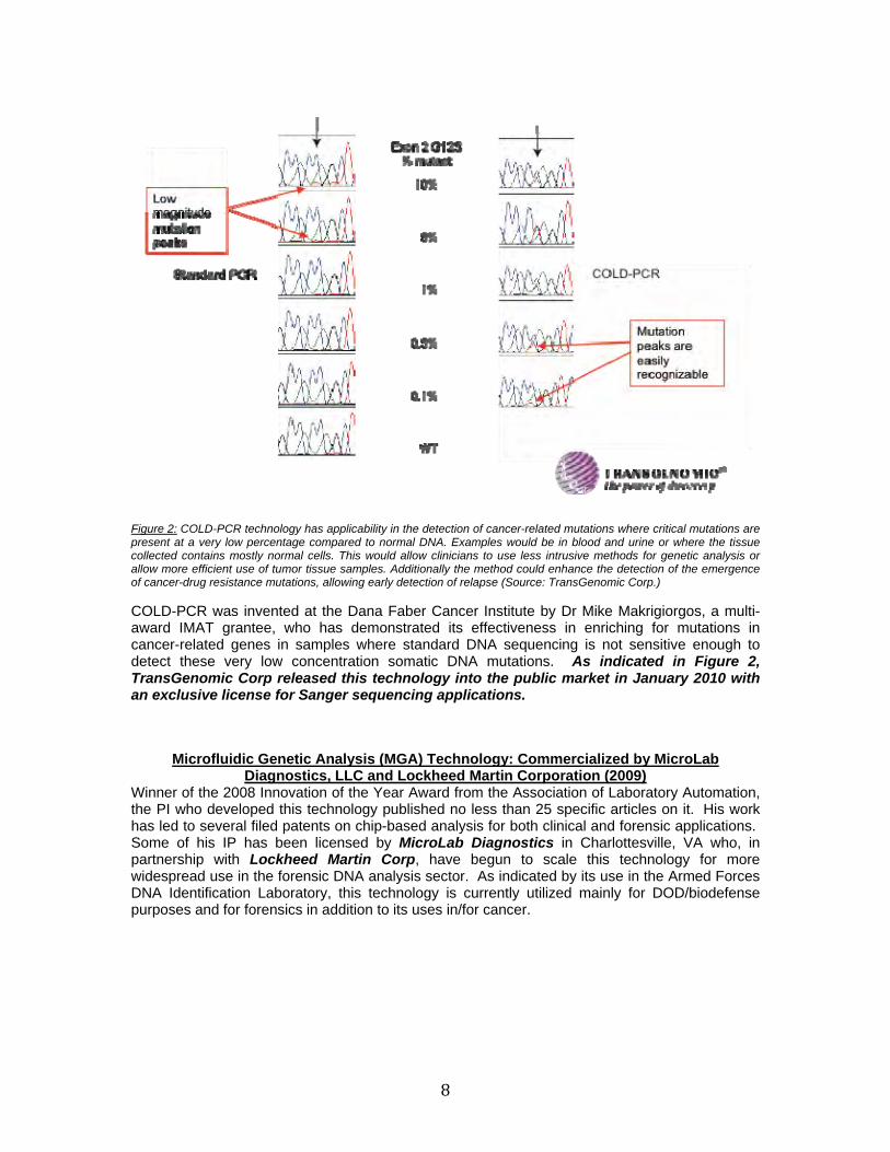
Figure 2: COLD-PCR technology has applicability in the detection of cancer-related mutations where critical mutations are present at a very low percentage compared to normal DNA. Examples would be in blood and urine or where the tissue collected contains mostly normal cells. This would allow clinicians to use less intrusive methods for genetic analysis or allow more efficient use of tumor tissue samples. Additionally the method could enhance the detection of the emergence of cancer-drug resistance mutations, allowing early detection of relapse (Source: TransGenomic Corp.)
COLD-PCR was invented at the Dana Faber Cancer Institute by Dr Mike Makrigiorgos, a multi-award IMAT grantee, who has demonstrated its effectiveness in enriching for mutations in cancer-related genes in samples where standard DNA sequencing is not sensitive enough to detect these very low concentration somatic DNA mutations. As indicated in Figure 2, TransGenomic Corp released this technology into the public market in January 2010 with an exclusive license for Sanger sequencing applications.
Microfluidic Genetic Analysis (MGA) Technology: Commercialized by MicroLab Diagnostics, LLC and Lockheed Martin Corporation (2009)
Winner of the 2008 Innovation of the Year Award from the Association of Laboratory Automation, the PI who developed this technology published no less than 25 specific articles on it. His work has led to several filed patents on chip-based analysis for both clinical and forensic applications. Some of his IP has been licensed by MicroLab Diagnostics in Charlottesville, VA who, in partnership with Lockheed Martin Corp, have begun to scale this technology for more widespread use in the forensic DNA analysis sector. As indicated by its use in the Armed Forces DNA Identification Laboratory, this technology is currently utilized mainly for DOD/biodefense purposes and for forensics in addition to its uses in/for cancer.

9
Figure 3: Images of the MGA device. (a) Dyes are placed in the channels for visualization (Scale bar, 10 mm.). Domains for DNA extraction (yellow), PCR amplification (red), injection (green), and separation (blue) are connected through a network of channels and vias. SPE reservoirs are labeled for sample inlet (SI), sidearm (SA), and extraction waste (EW). Injection reservoirs are labeled for PCR reservoir (PR), marker reservoir (MR), and sample waste (SW). Electrophoresis reservoirs are labeled for buffer reservoir (BR) and buffer waste (BW). Additional domains patterned onto the device include the temperature reference (TR) chamber and fluorescence alignment (FA) channel. The flow control region is outlined by a dashed box. Device dimensions are 30.0 × 63.5 mm, with a total solution volume <10 μl. (Scale bar, 10 mm.) (b) Schematic of flow control region. Valves are shown as open rectangles. V1 separates the SPE and PCR domains. V2 and V5 are inlet valves for the pumping injection, V3 is the diaphragm valve, and V4 is an outlet valve. (c) Device loaded into the manifold. (d) Intersection between SI and SA inlet channels, with the EW channel tapering to increase flow resistance. (Scale bar, 1 mm.) (e) Image of PCR chamber with exit channel tapering before intersecting with the MR inlet channel. (Scale bar, 1 mm.) (f) Image of cross-tee intersection. (Scale bar, 1 mm.) The relative sizes of the BR, SW, and BW channels create the difference in volume displacement during the pumping injection and affect how the resistance is dropped under an applied separation voltage.
The MGA platform is a unique device and resembles a common microscope slide, but it houses the analytical tools of an entire laboratory. Vastly complex and distinct procedures take place within millimeters of one another in tiny troughs that are etched into the chip. Minute tissue or blood samples are placed into the chip and electric charge is applied to the samples—for electrophoresis—to separate out particular sections of DNA based on what type of diagnosis is needed. Once the DNA is separated, it is replicated on one portion of the chip and then pushed to yet another area to be screened for irregularities.
10
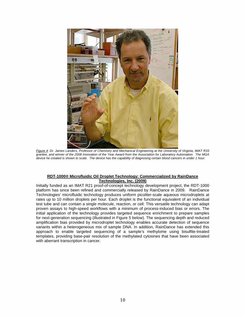
Figure 4: Dr. James Landers, Professor of Chemistry and Mechanical Engineering at the University of Virginia, IMAT R33 grantee, and winner of the 2008 Innovation of the Year Award from the Association for Laboratory Automation. The MGA device he created is shown to scale. The device has the capability of diagnosing certain blood cancers in under 1 hour.
RDT-1000® Microfluidic Oil Droplet Technology: Commercialized by RainDance Technologies, Inc. (2009)
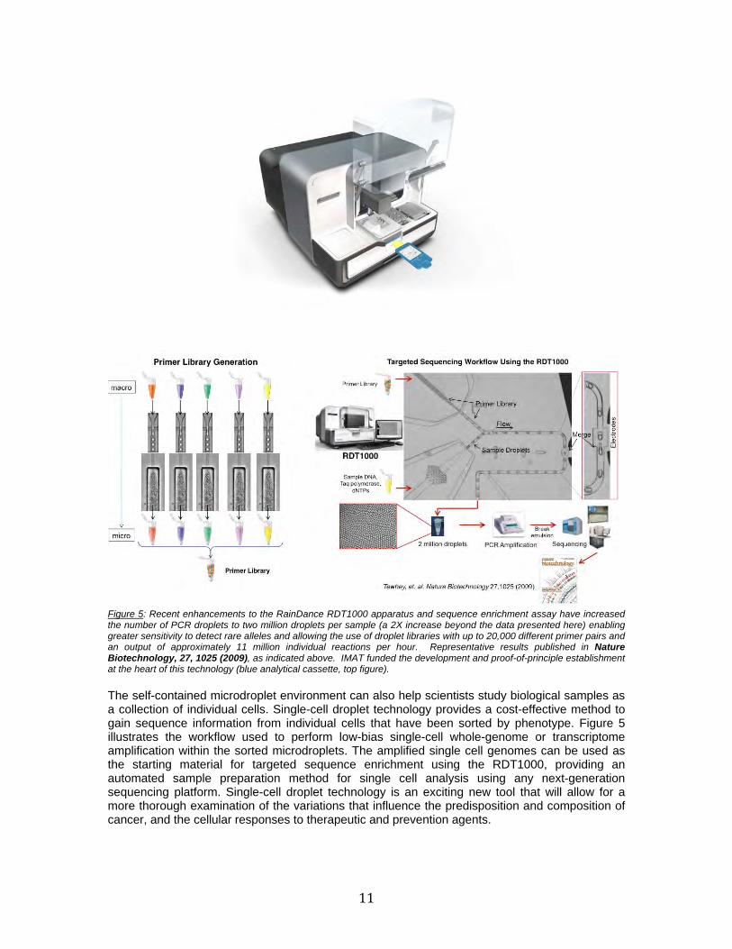
Initially funded as an IMAT R21 proof-of-concept technology development project, the RDT-1000 platform has since been refined and commercially released by RainDance in 2009. RainDance Technologies’ microfluidic technology produces uniform picoliter-scale aqueous microdroplets at rates up to 10 million droplets per hour. Each droplet is the functional equivalent of an individual test tube and can contain a single molecule, reaction, or cell. This versatile technology can adapt proven assays to high-speed workflows with a minimum of process-induced bias or errors. The initial application of the technology provides targeted sequence enrichment to prepare samples for next-generation sequencing (illustrated in Figure 5 below). The sequencing depth and reduced amplification bias provided by microdroplet technology enables accurate detection of sequence variants within a heterogeneous mix of sample DNA. In addition, RainDance has extended this approach to enable targeted sequencing of a sample’s methylome using bisulfite-treated templates, providing base-pair resolution of the methylated cytosines that have been associated with aberrant transcription in cancer.
11
Figure 5: Recent enhancements to the RainDance RDT1000 apparatus and sequence enrichment assay have increased the number of PCR droplets to two million droplets per sample (a 2X increase beyond the data presented here) enabling greater sensitivity to detect rare alleles and allowing the use of droplet libraries with up to 20,000 different primer pairs and an output of approximately 11 million individual reactions per hour. Representative results published in Nature Biotechnology, 27, 1025 (2009), as indicated above. IMAT funded the development and proof-of-principle establishment at the heart of this technology (blue analytical cassette, top figure). The self-contained microdroplet environment can also help scientists study biological samples as a collection of individual cells. Single-cell droplet technology provides a cost-effective method to gain sequence information from individual cells that have been sorted by phenotype. Figure 5 illustrates the workflow used to perform low-bias single-cell whole-genome or transcriptome amplification within the sorted microdroplets. The amplified single cell genomes can be used as the starting material for targeted sequence enrichment using the RDT1000, providing an automated sample preparation method for single cell analysis using any next-generation sequencing platform. Single-cell droplet technology is an exciting new tool that will allow for a more thorough examination of the variations that influence the predisposition and composition of cancer, and the cellular responses to therapeutic and prevention agents.
12
NanoTrap® Biomarker Discovery and Protein Enrichment Platform: Commercialized by Shimadzu Scientific, Inc. (2010)
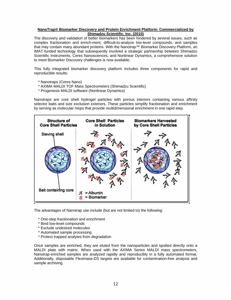
The discovery and validation of better biomarkers has been hindered by several issues, such as complex fractionation and enrich-ment, difficult-to-analyze low-level compounds, and samples that may contain many abundant proteins. With the Nanotrap™ Biomarker Discovery Platform, an IMAT-funded technology that subsequently involved a strategic partnership between Shimadzu Scientific Instruments, Ceres Nanosciences, and Nonlinear Dynamics, a comprehensive solution to meet Biomarker Discovery challenges is now available. This fully integrated biomarker discovery platform includes three components for rapid and reproducible results: * Nanotraps (Ceres Nano) * AXIMA MALDI TOF Mass Spectrometers (Shimadzu Scientific) * Progenesis MALDI software (Nonlinear Dynamics) Nanotraps are core shell hydrogel particles with porous interiors containing various affinity selector baits and size exclusion exteriors. These particles simplify fractionation and enrichment by serving as molecular mops that provide multidimensional enrichment in one rapid step.
The advantages of Nanotrap use include (but are not limited to) the following: * One-step fractionation and enrichment * Bind low-level compounds * Exclude undesired molecules * Automated sample processing * Protect trapped analytes from degradation Once samples are enriched, they are eluted from the nanoparticles and spotted directly onto a MALDI plate with matrix. When used with the AXIMA Series MALDI mass spectrometers, Nanotrap-enriched samples are analyzed rapidly and reproducibly in a fully automated format. Additionally, disposable Fleximass-DS targets are available for contamination-free analysis and sample archiving.
13
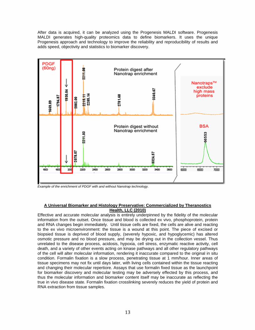
After data is acquired, it can be analyzed using the Progenesis MALDI software. Progenesis MALDI generates high-quality proteomics data to define biomarkers. It uses the unique Progenesis approach and technology to improve the reliability and reproducibility of results and adds speed, objectivity and statistics to biomarker discovery.
Example of the enrichment of PDGF with and without Nanotrap technology.
A Universal Biomarker and Histology Preservative: Commercialized by Theranostics Health, LLC (2010)
Effective and accurate molecular analysis is entirely underpinned by the fidelity of the molecular information from the outset. Once tissue and blood is collected ex vivo, phosphoprotein, protein and RNA changes begin immediately. Until tissue cells are fixed, the cells are alive and reacting to the ex vivo microenvironment: the tissue is a wound at this point. The piece of excised or biopsied tissue is deprived of blood supply, (severely hypoxic, and hypoglycemic) has altered osmotic pressure and no blood pressure, and may be drying out in the collection vessel. Thus unrelated to the disease process, acidosis, hypoxia, cell stress, enzymatic reactive activity, cell death, and a variety of other events acting on kinase pathways and all other regulatory pathways of the cell will alter molecular information, rendering it inaccurate compared to the original in situ condition. Formalin fixation is a slow process, penetrating tissue at 1 mm/hour. Inner areas of tissue specimens may not fix until days later, with living cells contained within the tissue reacting and changing their molecular repertiore. Assays that use formalin fixed tissue as the launchpoint for biomarker discovery and molecular testing may be adversely effected by this process, and thus the molecular information and biomarker content itself may be inaccurate as reflecting the true in vivo disease state. Formalin fixation crosslinking severely reduces the yield of protein and RNA extraction from tissue samples.
14
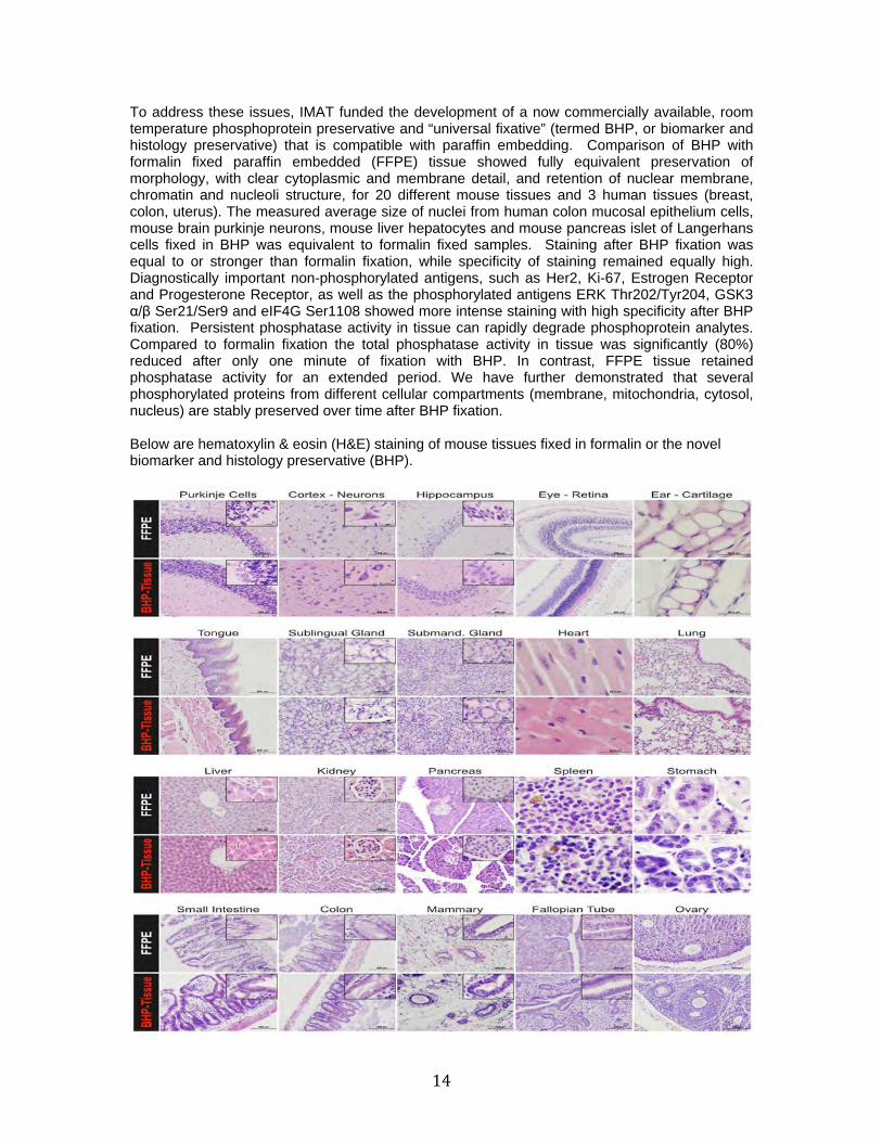
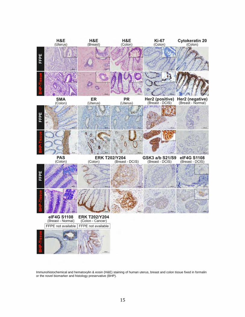
To address these issues, IMAT funded the development of a now commercially available, room temperature phosphoprotein preservative and “universal fixative” (termed BHP, or biomarker and histology preservative) that is compatible with paraffin embedding. Comparison of BHP with formalin fixed paraffin embedded (FFPE) tissue showed fully equivalent preservation of morphology, with clear cytoplasmic and membrane detail, and retention of nuclear membrane, chromatin and nucleoli structure, for 20 different mouse tissues and 3 human tissues (breast, colon, uterus). The measured average size of nuclei from human colon mucosal epithelium cells, mouse brain purkinje neurons, mouse liver hepatocytes and mouse pancreas islet of Langerhans cells fixed in BHP was equivalent to formalin fixed samples. Staining after BHP fixation was equal to or stronger than formalin fixation, while specificity of staining remained equally high. Diagnostically important non-phosphorylated antigens, such as Her2, Ki-67, Estrogen Receptor and Progesterone Receptor, as well as the phosphorylated antigens ERK Thr202/Tyr204, GSK3 α/β Ser21/Ser9 and eIF4G Ser1108 showed more intense staining with high specificity after BHP fixation. Persistent phosphatase activity in tissue can rapidly degrade phosphoprotein analytes. Compared to formalin fixation the total phosphatase activity in tissue was significantly (80%) reduced after only one minute of fixation with BHP. In contrast, FFPE tissue retained phosphatase activity for an extended period. We have further demonstrated that several phosphorylated proteins from different cellular compartments (membrane, mitochondria, cytosol, nucleus) are stably preserved over time after BHP fixation. Below are hematoxylin & eosin (H&E) staining of mouse tissues fixed in formalin or the novel biomarker and histology preservative (BHP).
15
Immunohistochemical and hematoxylin & eosin (H&E) staining of human uterus, breast and colon tissue fixed in formalin or the novel biomarker and histology preservative (BHP).
16
BHP “Universal Fixative” in Clinical Trials The BHP fixative has been used in a series of sponsored clinical research trials involving both national and international clinical and academic centers. Two trials have been ongoing for at least 2 years. US Oncology 05-074/GSK LPT109096 Phase I/II Breast Cancer Trial – Breast tissue core needle biopsies for 100 patients, before and after molecular targeted therapy, were immersed in the novel fixative, subjected to laser capture microdissection, and analyzed for signal pathway activation. NSABP Protocol FB-7– Breast tissue will be immersed in our BHP fixative and analyzed for signal pathway activation in a Phase II randomized clinical trial evaluating neoadjuvant therapy regimens. Multiple Myeloma ex vivo treatment research trial (USA) – Bone marrow aspirates of 45 patients have been treated ex vivo with a series of molecular targeted inhibitors followed by fixation with our novel fixative and analysis for signal pathway activation. The fixative stabilizes and preserves the bone marrow cellular constituents which allows the red blood cell lysis to be performed post fixation. Multiple Myeloma ex vivo treatment research trial (Italy) – Bone marrow aspirates of 30 patients have been treated as described above for the USA trial. In addition peripheral blood and bone marrow aspirates preserved in the novel fixative have been analyzed by flow cytometry.
International Clinical Pathology Evaluation (Italy): 5 clinical research centers throughout Italy are participating in the clinical evaluation of the fixative. Matched tissue samples are being collected in formalin and BHP. Samples are being processed following the standard methods used in each individual pathology lab. Objectives: a) assess ease of use of the fixative in a clinical setting, b) assess diagnostic quality of samples preserved in BHP compared to formalin, and c) evaluate effects of various processing methods/instrument parameters on tissue morphology and immunohistochemistry. Ireland: the fixative is being evaluated at St. James’s Hospital, Dublin, a community hospital, for protein and RNA stability and yield in comparison to FFPE. IuVo® MicroConduit Array Platform and Product Line: Commercialized by BellBrook Labs,
Inc. (2010) Iuvo Microconduit Arrays enable the use of highly miniaturized, advanced cell models and functional assays with automated liquid handling and HCA platforms, with the goal of more accurate replication of in vivo processes in drug discovery. Instead of the "buckets" of various sizes used for cell culture in multiwell plates, iuvo plates have cell culture compartments with geometries designed specifically to support the biology and/or functions of interest. Iuvo plates comply with SBS standards, and the liquid in the microchannels can be displaced with passive pumping as often as necessary using standard automated liquid dispensing equipment. No specialized instrumentation is required to carry out assays or acquire the data. Three dimensional cell culture becomes easy to carry out, and the entire compartment is accessible to microscope-based HCA instruments as well as plate scanners. The microchannel environment reduces dilution of secreted signaling molecules, and the media can be sampled for analysis of secreted factors.
17
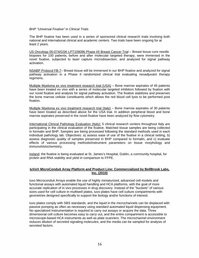
Invasion through laminin-rich extracellular matrix (Matrigel™). A) A CyBi®-Well was used to set up an invasion assay in iuvo Microchannel 5250. B) PC3-M cells invade matrix one day after plating. C) Invasion 5 days after plating. Cells viewed with phase contrast microscopy. D) Overlay image of phalloidin (red) and DAPI (blue). (Source: Bellbrook Labs, Inc.)
TrIP-Chip Technology: Offered Provisional Commercialization License by OceanRidge
Biosciences, LLC (2010) One of the latest up-and-coming technologies funded and developed through the IMAT program involves a new approach to systematically study post-transcriptional regulation in real time in a small number of cells. Actively translating mRNAs are associated with polysomes and the newly synthesized peptide chains are closely associated with molecular chaperones such as hsp70s, which assist in the proper folding of nascent polypeptides into higher ordered structures. These chaperones provide an anchor with which to separate actively translating mRNAs associated with polysomes from free mRNAs. Affinity capture beads were developed to capture hsp70 chaperones associated with the polysome complexes. The isolated actively translating mRNAs were used for high-throughput expression profiling analysis. Feasibility was demonstrated using an in vitro translation system with known translationally regulated mRNA transcript thymidylate synthase (TS). We further developed the approach using HCT-116 colon cancer cells with both TS and p53 as positive controls. The steady-state levels of TS and p53 mRNAs were unaltered after 5-fluorouracil treatment as assessed by real-time qRT-PCR analysis. In contrast, the protein expression and polysome-associated mRNA levels of both genes were increased. These differences in translational rate were revealed with our new approach from 500 cells. This technology has the potential to make investigation of translational control feasible with limited quantities of clinical specimens. OceanRidge Biosciences, LLC has offered an initial commercialization license to the developer of this technology (a university investigator), thus providing an additional example of the IMAT program’s success at the commercializing and disseminating university-developed technologies.
18
Cross-Over Technologies
NIH Transformative R01 Award Acquisitions The Roadmap Transformative R01 Research Projects Program (TR01) was specifically created under the NIH Roadmap for Medical Research to support exceptionally innovative, high risk, original and/or unconventional research projects that have the potential to create or overturn fundamental paradigms. These projects tend to be inherently risky, but if successful can profoundly impact a broad area of biomedical research. As compared to the NIH Director’s Pioneer and New Innovator Programs, the primary emphasis of the Roadmap Transformative Research Projects Program is on creative ideas—projects that have the potential to transform a field of science and to provide adequate support for the work—rather than creative individuals who have proven themselves to be innovative researchers and to provide them with funds to go in a new pioneering direction.
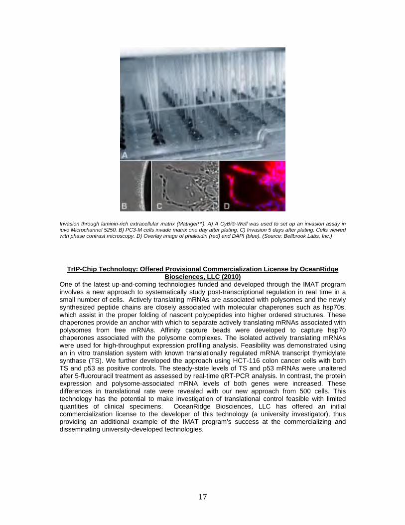
Transformative Research Projects Program
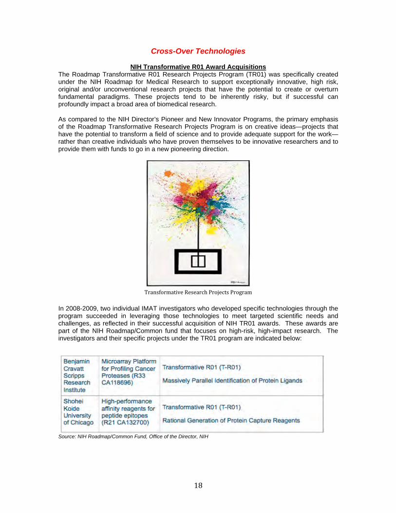
In 2008-2009, two individual IMAT investigators who developed specific technologies through the program succeeded in leveraging those technologies to meet targeted scientific needs and challenges, as reflected in their successful acquisition of NIH TR01 awards. These awards are part of the NIH Roadmap/Common fund that focuses on high-risk, high-impact research. The investigators and their specific projects under the TR01 program are indicated below:
Source: NIH Roadmap/Common Fund, Office of the Director, NIH
19
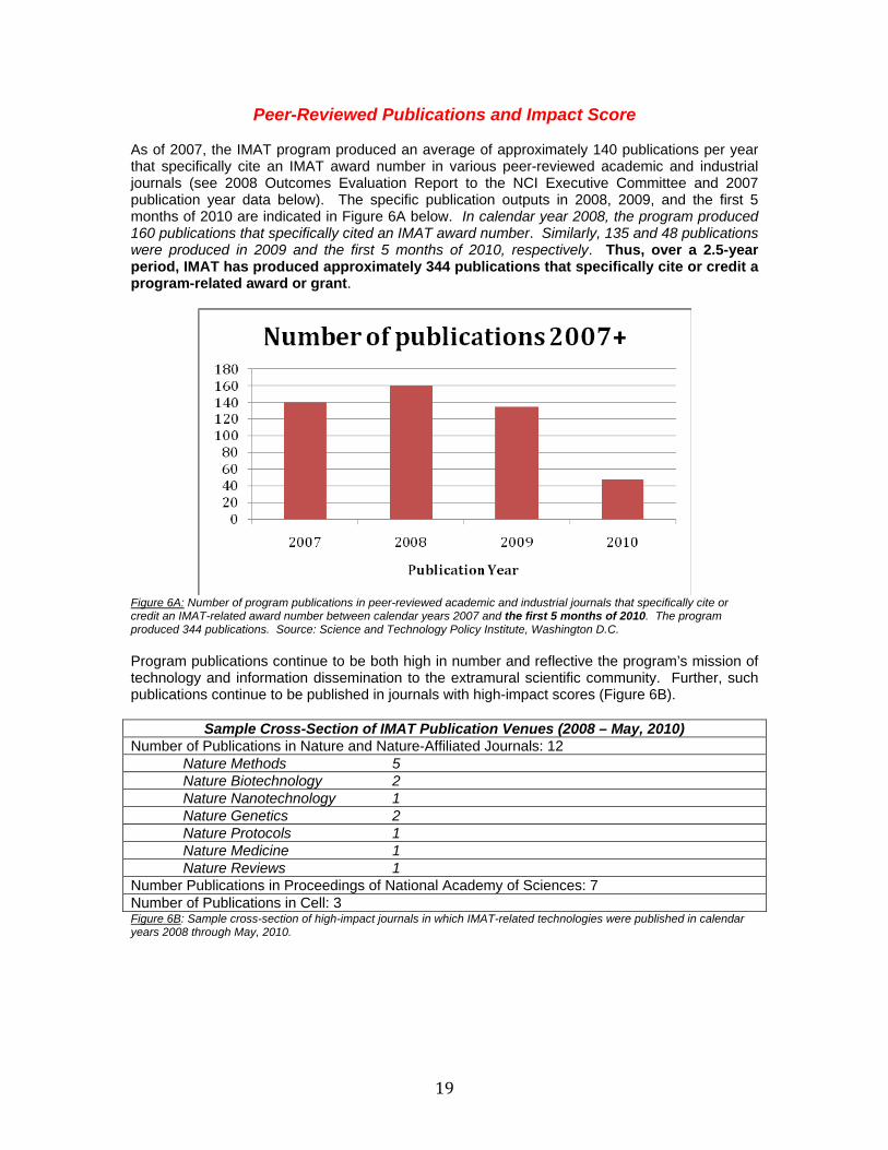
Peer-Reviewed Publications and Impact Score As of 2007, the IMAT program produced an average of approximately 140 publications per year that specifically cite an IMAT award number in various peer-reviewed academic and industrial journals (see 2008 Outcomes Evaluation Report to the NCI Executive Committee and 2007 publication year data below). The specific publication outputs in 2008, 2009, and the first 5 months of 2010 are indicated in Figure 6A below. In calendar year 2008, the program produced 160 publications that specifically cited an IMAT award number. Similarly, 135 and 48 publications were produced in 2009 and the first 5 months of 2010, respectively. Thus, over a 2.5-year period, IMAT has produced approximately 344 publications that specifically cite or credit a program-related award or grant.
Figure 6A: Number of program publications in peer-reviewed academic and industrial journals that specifically cite or credit an IMAT-related award number between calendar years 2007 and the first 5 months of 2010. The program produced 344 publications. Source: Science and Technology Policy Institute, Washington D.C. Program publications continue to be both high in number and reflective the program’s mission of technology and information dissemination to the extramural scientific community. Further, such publications continue to be published in journals with high-impact scores (Figure 6B).
Sample Cross-Section of IMAT Publication Venues (2008 – May, 2010) Number of Publications in Nature and Nature-Affiliated Journals: 12 Nature Methods 5 Nature Biotechnology 2 Nature Nanotechnology 1 Nature Genetics 2 Nature Protocols 1 Nature Medicine 1 Nature Reviews 1 Number Publications in Proceedings of National Academy of Sciences: 7 Number of Publications in Cell: 3 Figure 6B: Sample cross-section of high-impact journals in which IMAT-related technologies were published in calendar years 2008 through May, 2010.
20
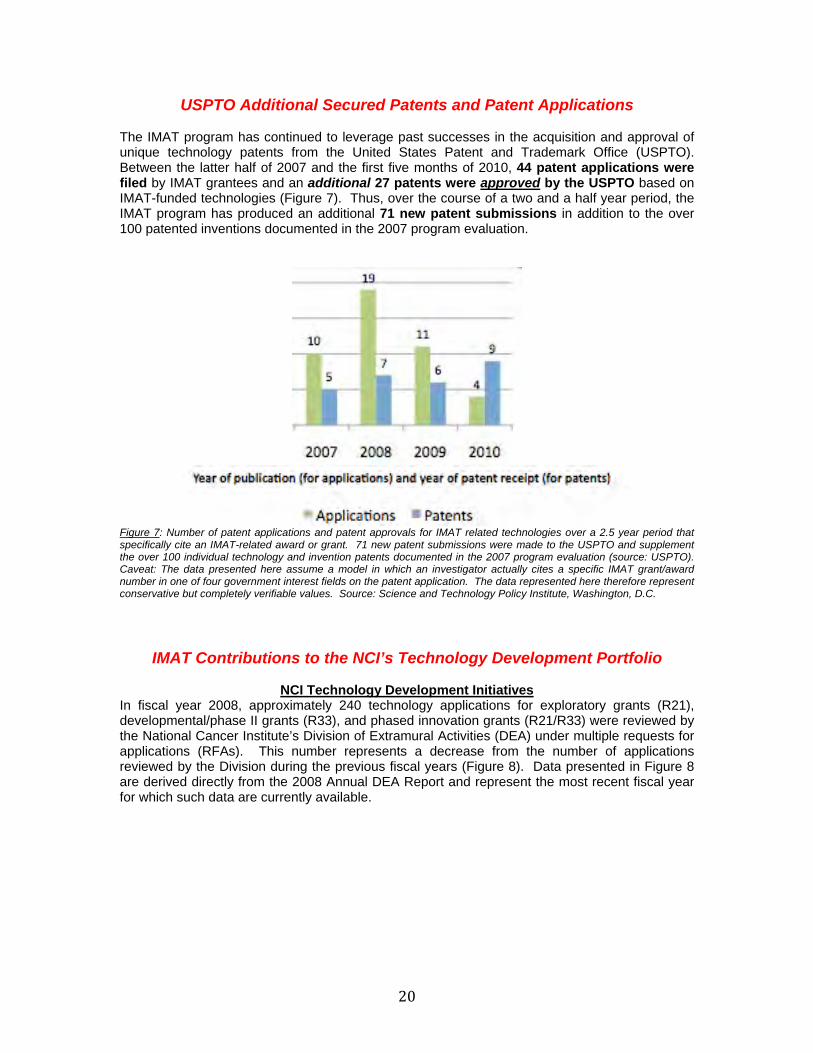
USPTO Additional Secured Patents and Patent Applications The IMAT program has continued to leverage past successes in the acquisition and approval of unique technology patents from the United States Patent and Trademark Office (USPTO). Between the latter half of 2007 and the first five months of 2010, 44 patent applications were filed by IMAT grantees and an additional 27 patents were approved by the USPTO based on IMAT-funded technologies (Figure 7). Thus, over the course of a two and a half year period, the IMAT program has produced an additional 71 new patent submissions in addition to the over 100 patented inventions documented in the 2007 program evaluation.
Figure 7: Number of patent applications and patent approvals for IMAT related technologies over a 2.5 year period that specifically cite an IMAT-related award or grant. 71 new patent submissions were made to the USPTO and supplement the over 100 individual technology and invention patents documented in the 2007 program evaluation (source: USPTO). Caveat: The data presented here assume a model in which an investigator actually cites a specific IMAT grant/award number in one of four government interest fields on the patent application. The data represented here therefore represent conservative but completely verifiable values. Source: Science and Technology Policy Institute, Washington, D.C.
IMAT Contributions to the NCI’s Technology Development Portfolio
NCI Technology Development Initiatives
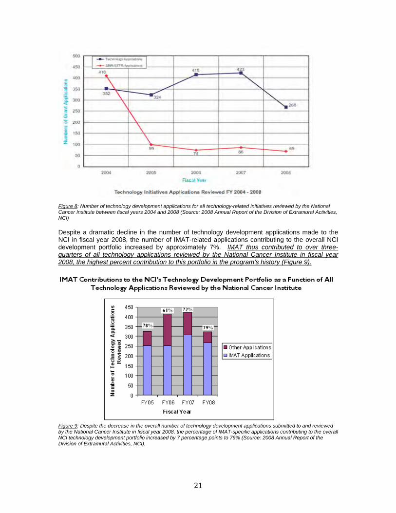
In fiscal year 2008, approximately 240 technology applications for exploratory grants (R21), developmental/phase II grants (R33), and phased innovation grants (R21/R33) were reviewed by the National Cancer Institute’s Division of Extramural Activities (DEA) under multiple requests for applications (RFAs). This number represents a decrease from the number of applications reviewed by the Division during the previous fiscal years (Figure 8). Data presented in Figure 8 are derived directly from the 2008 Annual DEA Report and represent the most recent fiscal year for which such data are currently available.
21
Figure 8: Number of technology development applications for all technology-related initiatives reviewed by the National Cancer Institute between fiscal years 2004 and 2008 (Source: 2008 Annual Report of the Division of Extramural Activities, NCI) Despite a dramatic decline in the number of technology development applications made to the NCI in fiscal year 2008, the number of IMAT-related applications contributing to the overall NCI development portfolio increased by approximately 7%. IMAT thus contributed to over three-quarters of all technology applications reviewed by the National Cancer Institute in fiscal year 2008, the highest percent contribution to this portfolio in the program’s history (Figure 9).
Figure 9: Despite the decrease in the overall number of technology development applications submitted to and reviewed by the National Cancer Institute in fiscal year 2008, the percentage of IMAT-specific applications contributing to the overall NCI technology development portfolio increased by 7 percentage points to 79% (Source: 2008 Annual Report of the Division of Extramural Activities, NCI).
22
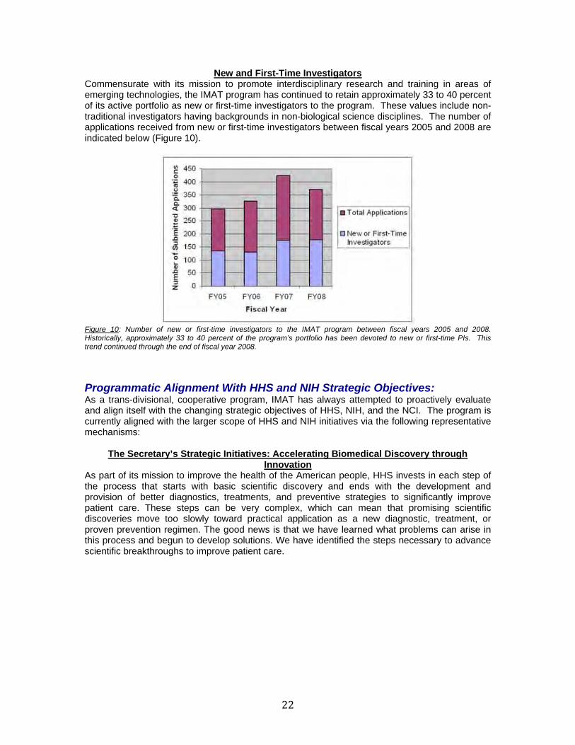
New and First-Time Investigators Commensurate with its mission to promote interdisciplinary research and training in areas of emerging technologies, the IMAT program has continued to retain approximately 33 to 40 percent of its active portfolio as new or first-time investigators to the program. These values include non-traditional investigators having backgrounds in non-biological science disciplines. The number of applications received from new or first-time investigators between fiscal years 2005 and 2008 are indicated below (Figure 10).
Figure 10: Number of new or first-time investigators to the IMAT program between fiscal years 2005 and 2008. Historically, approximately 33 to 40 percent of the program’s portfolio has been devoted to new or first-time PIs. This trend continued through the end of fiscal year 2008.
Programmatic Alignment With HHS and NIH Strategic Objectives: As a trans-divisional, cooperative program, IMAT has always attempted to proactively evaluate and align itself with the changing strategic objectives of HHS, NIH, and the NCI. The program is currently aligned with the larger scope of HHS and NIH initiatives via the following representative mechanisms:
The Secretary’s Strategic Initiatives: Accelerating Biomedical Discovery through Innovation
As part of its mission to improve the health of the American people, HHS invests in each step of the process that starts with basic scientific discovery and ends with the development and provision of better diagnostics, treatments, and preventive strategies to significantly improve patient care. These steps can be very complex, which can mean that promising scientific discoveries move too slowly toward practical application as a new diagnostic, treatment, or proven prevention regimen. The good news is that we have learned what problems can arise in this process and begun to develop solutions. We have identified the steps necessary to advance scientific breakthroughs to improve patient care.
23
A key component of the Secretary’s Strategic Initiatives published in 2010 is for HHS is to accelerate biomedical discovery through innovative methodologies and technology. Under this objective, HHS will continue to support fundamental discoveries that expand the knowledge base in the biomedical and associated sciences. Also of critical importance is ongoing cross-agency development of information systems capable of storing, organizing and sharing vast amounts of data with researchers around the globe, which will accelerate scientific discovery. As a cooperative program allowing international applicants and fostering international collaboration in addition to its focus on the development, production, and subsequent dissemination of more effective cutting-edge tools and platforms to accelerate and empower biomedical research, IMAT is in alignment with this functional area of the Secretary’s Strategic Plan. The record of the program’s productive ability is outlined in its individual and collective achievements to date.
NIH Common Fund
The past two decades have brought tremendous scientific advances that can greatly benefit medical research. While this unprecedented period of progress in the biological, behavioral, clinical, social, physical, chemical, engineering, and mathematical sciences will continue into the foreseeable future, human health and well-being would benefit from accelerating the current pace of discovery. One way to achieve this goal is to support scientists of exceptional creativity who propose highly innovative approaches to major contemporary challenges in biomedical research. By bringing their unique perspectives and abilities to bear on key research questions, these visionary scientists may develop seminal theories or technologies that will propel fields forward and speed the translation of research into improved health. NIH has traditionally supported research projects, not individual investigators. However, complementary means might be necessary to identify scientists with ideas that have the potential for high impact, but that may be too novel, span too diverse a range of disciplines, or be at a stage too early to fare well in the traditional peer review process.
To address this issue, the NIH Roadmap/Common Fund has created three programs with funding opportunities, the NIH Director's Pioneer, New Innovator, and Transformative R01 Awards, to
24
encourage creative, outside-the-box thinkers to pursue exciting and innovative ideas about biomedical research. Given the unique nature of these awards, candidates must undergo a rigorous evaluation process to identify those investigators with the highest likelihood of pursuing a pioneering approach to a significant biomedical problem. Awardees will have the intellectual freedom to pursue their ideas and follow them in expected or even unexpected directions. IMAT has at least 2 individual investigators who have succeeded in acquiring such prestigious awards by leveraging individual technologies they developed through the program. Summary: Between 2008 and the first five months of 2010, the IMAT program has continued to fulfill its mission of bringing innovation to cross-cutting, research-enabling sectors through a cooperative approach to translational technology development. Specific outputs of the program over the most recent 2.5 year period include the following:
COLD-PCR Technology licensed, marketed, and commercialized by TransGenomic Corporation (2010)
Universal Biomarker and Histology Preservative (BHP) licensed and commercialized by Theranostics, LLC (2010)
TrIP-Chip Technology – developed at Stony Brook University Medical Center - offered
initial commercialization license by OceanRidge Biosciences, LLC (confidential and in progress, 2010)
NanoTrap Biomarker Discovery Platform Technology licensed and commercialized by
Shimadzu Scientific, Inc. (2010)
IuVo product line (note: this constitutes multiple products) – developed at University of Wisconsin – licensed and commercialized by BellBrooks Labs, Inc. (2009/2010)
Microfluidic RDT-1000 analytical platform marketed and commercialized by RainDance
Technologies (2009)
Microfluidic Genetic Analysis (MGA) Technology licensed and scaled by Lockheed Martin Corporation and MicroLab Diagnostics; technology utilized by multiple federal departments, including the Department of Defense for forensic purposes, in addition to its uses in cancer diagnostics by the clinical research community (2008)
Acquisition of at least two individual trans-NIH T-R01 awards by IMAT investigators that
utilize technologies developed through IMAT to conduct ground-breaking research in high-impact areas as defined by T-R01 solicitation
344 publications that specifically cite or credit a program-related award in both academic
and industrial peer-reviewed journals; this number includes at least 13 publications in Nature-family journals, 3 publications in Cell, and 7 publications in the Proceedings of the National Academy of Sciences
At least 71 new patent submissions to the USPTO that specifically cite or credit a
program-related award number in one of four government-interest fields; of these, at least 27 patents have already been approved by the USPTO
2 finalists for the Association for Laboratory Automation’s “Innovation of the Year” Award,
including a 2010 finalist and the 2008 winner

25
79% contribution to the NCI’s overall technology development portfolio, as reflected in the 2008 Report of the NCI Division of Extramural Activities, the highest percent contribution in the program’s history
Approximately 40% of the program’s portfolio represented by new, first-time, or non-
traditional principal investigators, the highest in the program’s history