Embed Size (px)
Citation preview

Cell Physiol Biochem 2020;54:88-109DOI: 10.33594/000000207Published online: 29 January 2020 88
Cellular Physiology and Biochemistry
Cellular Physiology and Biochemistry
© 2020 The Author(s). Published by Cell Physiol Biochem Press GmbH&Co. KG
Medeiros et al.: EVs as Biomarkers of Kidney Injury
Extracellular Vesicles: Cell-Derived Biomarkers of Glomerular and Tubular InjuryThalia Medeirosa,b Robert L. Myettea,c Jorge Reis Almeidab
Andrea Alice Silvab Dylan Burgera
aKidney Research Centre, Department of Cellular and Molecular Medicine, The Ottawa Hospital Research Institute, University of Ottawa, Ottawa, ON, Canada, bMultiuser Laboratory for Research in Nephrology and Medical Sciences (LAMAP), Faculty of Medicine, Universidade Federal Fluminense, Rio de Janeiro, Brazil, cDivision of Nephrology, The Children’s Hospital of Eastern Ontario, Ottawa, ON, Canada
Key WordsExtracellular vesicles • Kidney • Urine • Biomarker
AbstractExtracellular vesicles (EVs) are important mediators of intercellular communication. Since EVs are also released during pathological conditions, there has been considerable interest in their potential as sensitive biomarkers of cellular stress and/or injury. In the context of kidney disease, urinary EVs are promising indicators of glomerular and tubular damage. In the present review we discuss the role of urinary EVs in kidney health and disease. Our focus is to explore urinary large EVs (lEVs, often referred to as microparticles or microvesicles) as direct and non-invasive early biomarkers of renal injury. In this regard, studies have been demonstrating altered levels of urinary lEVs, especially podocyte-derived lEVs, preceding the decrease of renal function assessed by classical markers. In addition, we discuss the role of small EVs (sEVs, often referred to as exosomes) and their contents in kidney pathophysiology. Even though results concerning the production of sEVs during diseased conditions are varied, there has been a consensus on the importance of urinary sEV content assessment in kidney disease. These mediators, including EV-released miRNAs and mRNAs, are responsible for EV-mediated signaling in the regulation of renal cellular homeostasis, pathogenesis and regeneration. Finally, steps necessary for the validation of EVs as reliable markers will be discussed.
Introduction
Extracellular vesicles (EVs) are emergent mediators of intercellular communication as well as biomarkers of disease [1–3]. These biologically active membrane-coated vesicles
Review
Accepted: 23 December 2019
This article is licensed under the Creative Commons Attribution-NonCommercial-NoDerivatives 4.0 Interna-tional License (CC BY-NC-ND). Usage and distribution for commercial purposes as well as any distribution of modified material requires written permission.
DOI: 10.33594/000000207Published online: 29 January 2020
© 2020 The Author(s)Published by Cell Physiol Biochem Press GmbH&Co. KG, Duesseldorfwww.cellphysiolbiochem.com
© 2020 The Author(s). Published by Cell Physiol Biochem Press GmbH&Co. KG
Dylan Burger Kidney Research Centre, Department of Cellular and Molecular MedicineThe Ottawa Hospital Research Institute, University of Ottawa, 2513-/451 Smyth Road, Ottawa, ON K1H 8M5 (Canada)E-Mail [email protected]

Cell Physiol Biochem 2020;54:88-109DOI: 10.33594/000000207Published online: 29 January 2020 89
Cellular Physiology and Biochemistry
Cellular Physiology and Biochemistry
© 2020 The Author(s). Published by Cell Physiol Biochem Press GmbH&Co. KG
Medeiros et al.: EVs as Biomarkers of Kidney Injury
are released by cells during physiological conditions in addition to conditions of stress, injury or death. Mechanisms of EV-mediated signaling occur through antigen presentation, receptor-mediated signaling, cell membrane fusion and/or endocytosis [4, 5]. EV content or “cargo” includes functional cytoplasmic proteins, peptides, lipids, nucleic acids (DNA, mRNA, microRNA, lncRNAs) and other signaling molecules that modulate cellular function, promoting autocrine or paracrine responses [6, 7]. On their surface, EVs present characteristic protein markers which can be used to identify their origin [7].
Size and mechanism of biogenesis should be considered when distinguishing different types of EVs: (i) apoptotic bodies are > 1 µm EVs that are formed during the late stages of cellular death by apoptotic pathways; (ii) microparticles (MPs), also termed microvesicles (MVs) are ~100 – 1000 nm fragments released by membrane blebbing and eventual shedding into the extracellular milieu; and, (iii) exosomes are smaller EVs (~20 – 150 nm), formed in a multi-step mechanism, where intracellular vesicles accumulate within multivesicular bodies, which merge with the plasma membrane and release exosomes. Other EV subpopulations (i.e. apoptotic nanovesicles, exomeres, oncosomes, migrasomes) have also been described, often to refer to EV populations in a specific setting (i.e. oncosomes are EVs released by cancer cells; migrasomes are generated during cell migration) [8]. These subpopulations tend to be much less pervasive in literature. A summary of the biogenesis of major EV classes is described in Fig. 1 and readers are directed to Chuo et al. 2018, D’Souza-Schorey & Schorey 2018, Abels & Breakefield, 2016 and Kalra et al. 2016 for more detailed reviews of EV biogenesis [4, 7–9]. Recent guidance from the International Society of Extracellular Vesicles suggests identifying ~100-1000 nm vesicles as “large EVs” (lEVs) and ~40-100 nm vesicles as “small EVs” (sEVs) when size is the primary descriptor and their origin is unclear. This represents a convenient distinction as many separation techniques effectively segregate based on size, however it is important to note that there is significant overlap of sizes between exosomes and MVs such that one cannot simply conclude that large EVs ≠ MV and small EVs ≠ exosomes. Regardless, these terms sEV and lEV will be used as appropriate throughout the present discussion, regardless of the terminology used in the original manuscript. When the size of the EVs is not clear from a manuscript we will employ the term “EV”.
Several studies have reported circulating lEVs as biomarkers of vascular disease, reflecting early vascular damage during prothrombotic and proinflammatory states [6]. In plasma, lEVs derived from endothelial cells, platelets and leukocytes have been associated with the development/progression of kidney diseases, in acute kidney injury (AKI), chronic
Fig. 1. Biogenesis of major extracellular vesicles classes. Apoptotic bodies are larger EVs (> 1 µm) formed during cellular death by apoptotic pathways. Microparticles (MPs) are sized ~100 – 1000 nm and released by membrane blebbing and shedding into the extracellular space. Exosomes are smaller (~40 – 100 nm), formed in a multi-step mechanism, with formation of multivesicular bodies (accumulation of intracellular vesicles) and posterior merge with the plasma membrane.
Cell Physiol Biochem 2020;54:88-109DOI: 10.33594/000000207Published online: 29 January 2020 90
Cellular Physiology and Biochemistry
Cellular Physiology and Biochemistry
© 2020 The Author(s). Published by Cell Physiol Biochem Press GmbH&Co. KG
Medeiros et al.: EVs as Biomarkers of Kidney Injury
kidney disease (CKD), diabetic nephropathy (DN), lupus nephritis and nephrotic syndrome, among others [2, 3,10, 11]. However, urine is also a rich source of EVs [12]. In the present review we discuss the role of urinary EVs in kidney disease. In this context, circulating EVs are not likely able to cross the glomerular filtration barrier in significant numbers due to their size, thus, EVs in urine are believed to arise mainly from epithelial and parenchymal cells in direct contact with the urine throughout the nephron and bladder [12, 13]. This has led to the assessment of urinary EVs as putative non-invasive markers of renal injury [12–14].
Urinary lEVs as biomarkers of kidney injury
Podocyte-derived vesicles were first described as membrane-bound vesicles enriched in phospholipids and cholesterol, found in the urine of patients with minimal change disease, membranous nephropathy, focal sclerosis, and DN [15]. These vesicles were approximately 100-200nm in size, consistent with the definition of lEVs. In 2010, Hara et al. demonstrated that podocyte-lEVs were derived from the tip vesiculation of podocyte’s apical membrane, more specifically by microvilli transformation and shedding into Bowman’s space. These urinary lEVs from nephritic and nephrotic patients had a mean size of 200nm and absence of “exosomal” markers, such as CD24 and CD63 [16]. In addition, podocalyxin-associated lEVs were isolated from the urine of patients with idiopathic membranous nephropathy and focal segmental glomerulosclerosis and then characterized as released by podocytes in glomerular diseases [17]. Based on these observations, podocyte-lEVs gained consideration as direct, non-invasive markers of podocyte injury.
Our group has been investigating the role of podocyte-lEVs in diabetic kidney disease. We previously demonstrated that podocytes release lEVs in vitro when exposed to high glucose for 24h [18]. In addition, we were amongst the first to use nanoscale flow cytometry for assessment of urinary lEVs. Using this approach, we reported that urinary levels of podocyte-lEVs (podocalyxin+ or podoplanin+) were increased in diabetic mice (streptozotocin-treated, db/db, OVE26 and Akita mice) [18]. Levels of urinary podocyte lEVs were strongly correlated with albuminuria in these mice; however, elevated levels of podocyte lEVs could be identified before the development of albuminuria. Recently, we reported that significant increases in podocyte-lEVs may be detected in type 1 diabetes patients also in the absence of albuminuria, nephrinuria or glomerular filtration rate (GFR) decline [19]. In the same study, we observed that podocyte-lEVs (podoplanin+) were significantly higher during clamped hyperglycemia, suggesting glucose-mediated induction of lEVs formation in accordance with previous in vitro experiments [19]. In type 2 diabetes, urinary lEVs were reported to be associated with DN progression. De and colleagues (2017) observed a progressive increase in urinary total EVs and ≥130nm lEVs (podocalyxin+) in diabetic patients with increases in albuminuria [20]. In addition, Kamińska et al. (2016) identified ~100nm sized urinary EVs inversely correlated with GFR in diabetic patients; however, no differences were observed in patients with advanced kidney disease [21]. Collectively, these findings indicate that in diabetic kidney disease, total and podocyte-derived EVs can be altered when classical markers of kidney function are still unaltered, which suggests that they are reflective of early glomerular damage. Of note, higher levels of urinary lEVs during hyperglycemic states suggest that more podocyte damage could occur in diabetic patients with poor glycemic control. However, whether these events are associated with a premature development of kidney disease in type 1 or 2 diabetes, is a matter of further investigation.
Other studies have examined EVs derived from podocytes, in various kidney disorders. For example, Kwon et al. (2017) reported higher levels of podocyte-EVs (podocalyxin+/nephrin+) in patients with renovascular hypertension when compared with hypertensive patients with similar blood pressure values [22]. They concluded that this reflects podocyte injury associated with the decrease of renal blood flow and perfusion. It is worth noting that the authors did not provide any information regarding the size of the vesicles studied and
Cell Physiol Biochem 2020;54:88-109DOI: 10.33594/000000207Published online: 29 January 2020 91
Cellular Physiology and Biochemistry
Cellular Physiology and Biochemistry
© 2020 The Author(s). Published by Cell Physiol Biochem Press GmbH&Co. KG
Medeiros et al.: EVs as Biomarkers of Kidney Injury
only flow cytometry was used to assess EVs in this study. Accordingly, it may be that only a fraction of urinary EVs were analyzed. In contrast with these observations, podocyte-EVs were not altered in renovascular hypertension in the study of Santelli et al. (2019). Despite that, the authors reported an increase in tubule-derived p16+ (marker of cellular senescence) EVs in patients with renovascular hypertension and essential hypertension, which were directly associated with circulant proinflammatory biomarkers and negatively associated with GFR [23]. Recent work by Zhang et al. (2019) reported podocyte-lEVs (podocalyxin+/nephrin+) to be significantly increased in obese patients as well as in an experimental porcine model of metabolic syndrome. In this study, the authors reported that urinary lEVs were directly associated with podocyte damage (foot process effacement, podocyte size and number, nephrin and podocalyxin tissue expression) and with renal dysfunction [24]. Higher levels of podocyte-derived lEVs were associated with podocyte injury in lupus nephritis and with increased lupus disease activity [25]. In patients with idiopathic membranous nephropathy, the same group reported that the decrease in urinary podocyte-lEVs were associated with disease remission after immunosuppressive therapy [26]. In addition, urinary lEVs of podocyte origin (podocin+/nephrin+) were significantly correlated with albuminuria in preeclampsia [27]. Taken together, these studies highlight the potential of urinary podocyte EVs as early biomarkers of podocyte damage in a wide range of clinical and metabolic disorders.
Similar results have been obtained when looking at urinary EVs from other cell types. For example, in kidney transplant recipients, urinary CD133+ EVs (derived from progenitor cells) were demonstrated to be released by the donor’s glomeruli (CD2AP+) and proximal tubule cells (megalin+). The presence of CD133+ vesicles was associated with graft function, since patients with poor graft function presented lower urinary levels of these progenitor EVs. This suggests a potential protective role of progenitor cell-derived EVs during the reestablishment of kidney function. Consistent with this, CD133+ EVs were not detected in the urine of patients with end-stage kidney disease [28]. We recently studied proximal tubule-derived lEVs (megalin+) in a mouse model of adenine-CKD treated with PBI-4050. PBI-4050 is an agonist of the G protein-coupled receptor 40 that has been consistently shown to reduce kidney fibrosis and inflammation in preclinical models [29, 30]. We observed that PBI-4050 treatment was associated with lower levels of tubular lEVs parallel to improvement in tubular injury in adenine-fed mice [30]. Thus tubular lEVs appeared to be indicative of response to therapy in this animal model. Turco et al. (2016) identified that urinary EV levels decrease in parallel with the decline of kidney function associated with aging. Significant reductions in urinary EVs were also associated with nephron hypertrophy and global glomerulosclerosis in the same study [31]. The authors suggested that a significant decrease in EV production could be correlated with the renal function decline, but at which stage of kidney disease progression the “drop” in EV levels happens and if this is directly associated with extensive cellular death and nephron loss remains to be clarified. Of note in this study is the fact that the authors identified EVs by flow cytometry and that certain antibodies used targeted intracellular antigens (i.e. cytokeratins). Thus it is possible that non-EV cell fragments were enumerated by this approach.
Although there is some evidence suggesting that tubular lEVs are altered in kidney diseases, further studies are necessary to clarify the role of tubule-derived lEVs during acute and chronic renal damage. In this regard, it is interesting to note that De and colleagues have suggested that tubular markers such as megalin are more likely to be associated with sEVs, while podocyte markers are more frequently observed in urinary lEVs [20]. Notably, urinary lEVs from extrarenal cell sources could also provide insights about cellular activation in renal pathophysiology, as recently demonstrated by Burbano et al. (2019), who observed higher levels of urinary lEVs expressing specific markers of monocyte activation in lupus nephritis [32]. The authors speculated that this could reflect on the presence of inflammatory infiltrates in renal parenchyma. Urinary lEVs containing other biomarkers, such as monocyte chemoattractant protein-1 (MCP-1) and neutrophil gelatinase-associated lipocalin (NGAL), were also investigated in kidney stone disease [33]. Therefore, while it is believed that kidney
Cell Physiol Biochem 2020;54:88-109DOI: 10.33594/000000207Published online: 29 January 2020 92
Cellular Physiology and Biochemistry
Cellular Physiology and Biochemistry
© 2020 The Author(s). Published by Cell Physiol Biochem Press GmbH&Co. KG
Medeiros et al.: EVs as Biomarkers of Kidney Injury
cells are the major source of lEVs in urine, these findings suggest that the assessment of renal endothelial and inflammatory responses might be possible when investigating urinary lEVs.
In summary, urinary lEVs are promising biomarkers of kidney injury, with significant increases observed in early stages of kidney diseases. Podocyte-lEVs, in particular, have been shown to associate with early glomerular damage in metabolic disease, renovascular hypertension, pre-eclampsia and lupus. An important consideration here is the fact that urinary flow rate can be highly variable between and even within individuals. This can have a profound impact on the concentration of urinary EVs. As such it is advisable to normalize in some fashion, although this has not always been done. We, and others have normalized to urinary creatinine levels in an effort to address this issue [18, 19, 33]. Approaches such as measuring levels over a 24 hour time-period or normalizing to urine osmolality may also be considered however at the moment there is no consensus on the best approach to optimization and we await guidance on this.
Urinary sEVs as biomarkers of kidney diseases
Urinary sEVs can be derived from renal cells in all nephron segments, as well as the bladder and prostate [34]. Conflicting results concerning the production of sEVs by kidney cells in the setting of clinical diseases can be found in the literature. On one hand, some authors reported unaltered urinary levels of sEVs in kidney disease, [28, 35–39] while others have demonstrated significant increases in urinary sEV levels, especially during AKI [40–43]. Evidence supporting the latter hypothesis includes the production 60-80% more sEVs by proximal tubular epithelial cells subjected to inflammatory or hypoxic conditions in vitro [44]. In addition, a recent study reported significant increases in sEV production in IgA nephropathy, which was correlated with tubular injury, histologic activity and proteinuria [41]. Similarly, Yu et al. (2018) observed higher numbers of urinary sEVs in CKD patients [45]. Further studies are needed to confirm whether direct increases in sEV release by kidney cells is reflected in urine as increased urinary sEVs.
There has been considerable focus on urinary sEV content in kidney disease. Of note, sEV content is protected from degradation by proteases and nucleases in urine by the vesicle’s bilayered lipid membrane. This may facilitate analysis of protein expression inside sEVs as a form of “liquid biopsy” to identify molecular determinants of renal pathology. Early studies examining sEVs in human urine focused on proteomic analysis of the sEV urinary fraction after ultracentrifugation [46]. Pisitikun et al. (2004) initially identified ~300 proteins including many proteins well-established to play a critical role in kidney function. More recent studies report over 2000 different proteins expressed within urinary sEVs [47, 48]. Next, we discuss the evaluation of sEV content in kidney disease with a specific focus on transcription factors, sodium (Na+) and water transporters and RNA species.
Ion and water transportersEmerging evidences suggest that levels of Na+ transporters within sEVs are consistent
with their expression in kidney tissue in experimental models [49, 50]. Du Cheyron et al. (2003) reported the presence of Na+/H+ exchanger 3 in “urine membrane fractions” of patients with prerenal azotemia and acute tubular necrosis [51]. Of note, as sEVs were not explicitly demonstrated in these urine samples, the authors used the term “membrane fractions” to describe urine pellets obtained after ultracentrifugation (200,000g for 120 minutes). Further studies demonstrated urinary sEV excretion of other Na+ transporters as sensitive markers of Na+ handling and blood pressure regulation in kidney diseases: Na-Cl-K co-transporter 2 (NKCC2) is significantly elevated in urine samples of patients with pre-eclampsia [52] and DN [53]; increased activation of the epithelial sodium channel (ENaC) was also reported in diabetic patients [54]; alterations in sEV sodium-chloride cotransporter (NCC) were associated with mineralocorticoid administration [55] and calcineurin-induced hypertension in kidney transplant recipients [38]. Significant decreases in sEV-NCC and
Cell Physiol Biochem 2020;54:88-109DOI: 10.33594/000000207Published online: 29 January 2020 93
Cellular Physiology and Biochemistry
Cellular Physiology and Biochemistry
© 2020 The Author(s). Published by Cell Physiol Biochem Press GmbH&Co. KG
Medeiros et al.: EVs as Biomarkers of Kidney Injury
NKCC2 has been used to phenotypically differentiate hereditary salt-losing tubulopathies [56]. Moreover, the detection of the B1 subunit of the distal tubule V-ATPase ion exchanger in urinary sEVs during induced metabolic acidosis suggests that these EVs could be involved in the regulation of acid-base homeostasis [57].
Aquaporins (AQPs) were also reported in urinary EVs [58]. Ikeda’s group demonstrated significant reductions in urinary sEV APQ-1 and -2 in early stages of I/R-AKI [35, 40]. In gentamicin-treated rats, they observed significant early increases in urinary sEV excretion of AQP-2 followed by significant reductions six days after, which was accompanied by urinary concentration defects [42]. The dynamics of AQP-1 and -2 release in EVs after cisplatin treatment were also studied; while urinary sEV AQP-1 increases at early stages and significantly decreases in late AKI, dramatic reductions in AQP-2 can be found even at 24h after AKI induction, which suggests that sEV APQ-2 in urine can be used as an early biomarker of cisplatin-induced AKI [59]. Recently, the group observed that reductions of sEV AQP-2 are significantly associated with lower urine osmolality in kidney transplant recipients. This alteration may reflect concentration defects in transplant recipients, however it is difficult to determine whether the reduction in AQP-2 precedes the drop in urine osmolality or vice-versa [60]. In addition, the role of sEV APQs has been also investigated in DN. In fact, one of the first studies that demonstrated urinary EVs, focusing on the increase of AQP-2 expression in response to vasopressin, was performed in diabetic patients [61]. More recently, Rossi et al. (2017) reported significant increases in urinary excretion of AQP-1, -2 and -5 in diabetic patients with albuminuria, and AQP-2 and -5 were significantly associated with the progression of DN [53]. These results can elucidate mechanisms of urinary concentration defects in some disorders.
Transcription factorsThe presence of transcription factors in urine was specifically demonstrated in
sEV fractions [62, 63]. The presence of various transcription factors in urinary sEVs has been reported in both AKI and CKD [62]. Significant increases in urinary sEV activating transcription factor 3 (ATF3) was shown in early sepsis-induced and ischemic AKI [63, 64]. ATF3 may, in fact, play a causal role in AKI as Chen et al. (2014) have shown that ATF3 inhibits MCP1-expression in vitro and ATF3-knockout mice showed a higher I/R-induced inflammation [63].
Urinary sEV Wilm’s tumor protein 1 (WT-1) was demonstrated to be significantly increased in patients with type 1 diabetes and associated with the decline of kidney function in diabetic kidney disease [65]. Recently, the work of Abe et al. (2018) reported that hyperglycemia can stimulate the mobilization of WT-1 from the podocyte cytoplasm to be released within sEVs. This may explain the association between sEV WT-1 and DN progression, since they also observed that higher basal levels of urinary WT-1 were significantly associated with rapid decline of kidney function in type 2 diabetes [66]. In summary, these studies demonstrated that the alterations in urinary sEV levels of transcription factors could reflect the regulation of gene expression during kidney injury.
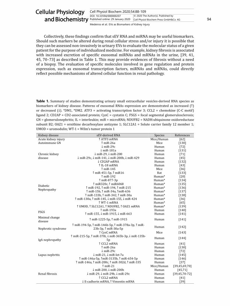
mRNAs and miRNAsNucleic acid content in urine sEV has also emerged as a possible avenue for biomarker
discovery. As sEVs can be released by kidney cells during stress conditions (i.e. hypoxia, hyperglycemia, oxidative stress, inflammation, acidosis), [67] several studies have examined the role of mRNAs and miRNAs delivered by sEVs during kidney injury and their effects on the recipient cell. In this regard, the transference/exchange of mRNAs and miRNAs between cells from different segments of the nephron (as discussed in the next topic) is emerging as a novel regulator of kidney function [14]. Independent of this, sEVs secreted by renal cells in contact with the urinary space may be assessed for their RNA content. In fact, sEV RNAs are more stable in urine samples than “free” RNA [68, 69]. In Table 1, studies investigating urinary sEV RNAs in kidney disease are summarized.
Cell Physiol Biochem 2020;54:88-109DOI: 10.33594/000000207Published online: 29 January 2020 94
Cellular Physiology and Biochemistry
Cellular Physiology and Biochemistry
© 2020 The Author(s). Published by Cell Physiol Biochem Press GmbH&Co. KG
Medeiros et al.: EVs as Biomarkers of Kidney Injury
Collectively, these findings confirm that sEV RNA and miRNA may be useful biomarkers. Should such markers be altered during renal cellular stress and/or injury it is possible that they can be assessed non-invasively in urinary EVs to evaluate the molecular status of a given patient for the purpose of individualized medicine. For example, kidney fibrosis is associated with increased excretion of specific exosomal miRNAs and mRNAs in the urine, [39, 41, 45, 70–73] as described in Table 1. This may provide evidences of fibrosis without a need of a biopsy. The evaluation of specific molecules involved in gene regulation and protein expression, such as exosomal transcription factors, miRNAs and mRNAs, could directly reflect possible mechanisms of altered cellular function in renal pathology.
Table 1. Summary of studies demonstrating urinary small extracellular vesicles-derived RNA species as biomarkers of kidney disease. Patterns of exosomal RNAs expression are demonstrated as increased (↑) or decreased (↓). #DM1, *DM2. ATF3 = activating transcription factor 3; CCL2 = chemokine (C-C motif) ligand 2; CD2AP = CD2-associated protein; CysC = cystatin C; FSGS = focal segmental glomerulosclerosis; GN = glomerulonephritis; IL = interleukin; miR = microRNA; NDUFB2 = NADH:ubiquinone oxidoreductase subunit B2; OAZ1 = ornithine decarboxylase antizyme 1; SLC12A1 = Solute carrier family 12 member 1; UMOD = uromodulin; WT-1 = Wilm’s tumor protein 1
↑ ATF3 mRNA↑↓
↓ miR↓ miR 29, ↓ miR
↓ miR 29c, ↓ miR 141, ↓ miR 200b, ↓ miR↓ CD2AP mRNA↑ IL
↑ miR↑ miR 5p, ↑ miR16
↑ miR↑ miR
↑ miR320c, ↑ ↑ miR 192, ↑ miR 194, ↑ miR↑ miR 15b, ↑ miR 34a, ↑miR
↑ miR 133b, ↑ miR 342, ↑ miR↑ miR 130a, ↑ miR 145, ↓ miR 155, ↓ miR
↑ WT↑ UMOD, ↑ SLC12A1, ↑ NDUFB2, ↑ OAZ1 mRNA
↑ miR↑ miR 155, ↓ miR 1915, ↓ miR
↑ miR 5p, ↑ miR↑ miR 5p, ↑ miR 5p, ↑ miR 3p, ↑
3p, ↑ miR↑ CysC mRNA
↑ miR 5p, ↑ miR 378i, ↓ miR 3p, ↓ miR
↑ CCL2 mRNA↑ miR↓
↓ ↓ miR↑ miR 5p, ↑miR 3135b, ↑ miR
↑ miR 146a, ↑ miR 200c, ↑ miR 302d, ↑ miR↑ miR
↓ miR 200, ↓ miR↓ miR 29, ↓ miR 29b, ↓ miR
↑ CCL2 mRNA↓ E cadherin mRNA, ↑ Vimentin mRNA
Cell Physiol Biochem 2020;54:88-109DOI: 10.33594/000000207Published online: 29 January 2020 95
Cellular Physiology and Biochemistry
Cellular Physiology and Biochemistry
© 2020 The Author(s). Published by Cell Physiol Biochem Press GmbH&Co. KG
Medeiros et al.: EVs as Biomarkers of Kidney Injury
EV signaling across the nephron
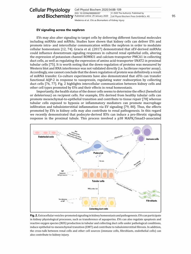
EVs may also alter signaling to target cells by delivering different functional molecules including miRNAs and mRNAs. Studies have shown that kidney cells can deliver EVs and promote intra- and intercellular communication within the nephron in order to modulate cellular homeostasis [12, 74]. Gracia et al. (2017) demonstrated that sEV-derived miRNAs could influence downstream signaling responses in cultured renal epithelial cells, altering the expression of potassium channel ROMKI1 and calcium-transporter PMCA1 in collecting duct cells, as well as regulating the expression of amino acid-transporter SNAT2 in proximal tubular cells [75]. It is worth noting that the down-regulation of proteins was measured by Western blot and RNA interference was not validated directly (i.e. luciferase reporter assay). Accordingly, one cannot conclude that the down regulation of protein was definitively a result of miRNA transfer. Co-culture experiments have also demonstrated that sEVs can transfer functional AQP-2 in response to vasopressin, regulating water reabsorption by collecting duct cells [76, 77]. Fig. 2 highlights intercellular communication between kidney cells and other cell types promoted by EVs and their effects in renal homeostasis.
Importantly, the health status of the donor cells seems to determine the effect (beneficial or deleterious) on recipient cells. For example, EVs derived from healthy tubular cells can promote mesenchymal-to-epithelial transition and contribute to tissue repair [78] whereas tubular cells exposed to hypoxia or inflammatory mediators can promote macrophage infiltration and tubulointerstitial inflammation via EV signaling [79, 80]. Thus, the effects promoted by EVs in kidney cells may also contribute to renal pathogenesis. In this regard we recently demonstrated that podocyte-derived lEVs can induce a pro-fibrotic signaling response in the proximal tubule. This process involved a p38 MAPK/Smad3-associated
Fig. 2. Extracellular vesicles-promoted signaling in kidney homeostasis and pathogenesis. EVs can participate in kidney physiological processes, such as transference of aquaporins. EVs can also regulate apoptosis and reactive oxygen species (ROS) production in tubular and collecting duct cells under pathological conditions, induce epithelial-to-mesenchymal transition (EMT) and contribute to tubulointerstitial fibrosis. In addition, the cross-talk between renal cells and other cell sources (immune cells, fibroblasts, endothelial cells) can also contribute to kidney injury.

Cell Physiol Biochem 2020;54:88-109DOI: 10.33594/000000207Published online: 29 January 2020 96
Cellular Physiology and Biochemistry
Cellular Physiology and Biochemistry
© 2020 The Author(s). Published by Cell Physiol Biochem Press GmbH&Co. KG
Medeiros et al.: EVs as Biomarkers of Kidney Injury
TGF-β activation and extracellular matrix production [81]. Consistent with this, sEVs enriched with TGF-β released by cells exposed to hypoxia or high glucose are implicated in the development of renal fibrosis [82–84]. In addition, other studies have also reported EV signaling contributing to other pathological processes such as ROS production [85] and epithelial-to-mesenchymal transition [70, 84]. Lastly, the cross-talk between kidney and immune cells promoted by EV signaling has also been recognized as an important factor associated with renal tubulointerstitial inflammation. In vitro studies demonstrated that lEVs and sEVs released by immune cells stimulate the production of inflammatory mediators by podocytes and epithelial tubule cells, respectively [86, 87]. The work of Lv et al. (2018) demonstrated that tubule-derived sEVs are packed with cytokine associated mRNAs in AKI and CKD experimental models. In addition, the group observed an increase in CCL2 expression within sEVs from cells exposed to albumin-induced inflammation, which then induced an up-regulation of inflammatory makers by macrophages [80]. Consistent with this, urinary exosomal CCL2 mRNA is significantly associated with proteinuria in IgA nephropathy patients [41].
EVs and kidney repairIt is important to emphasize that intercellular communication promoted by EVs are not
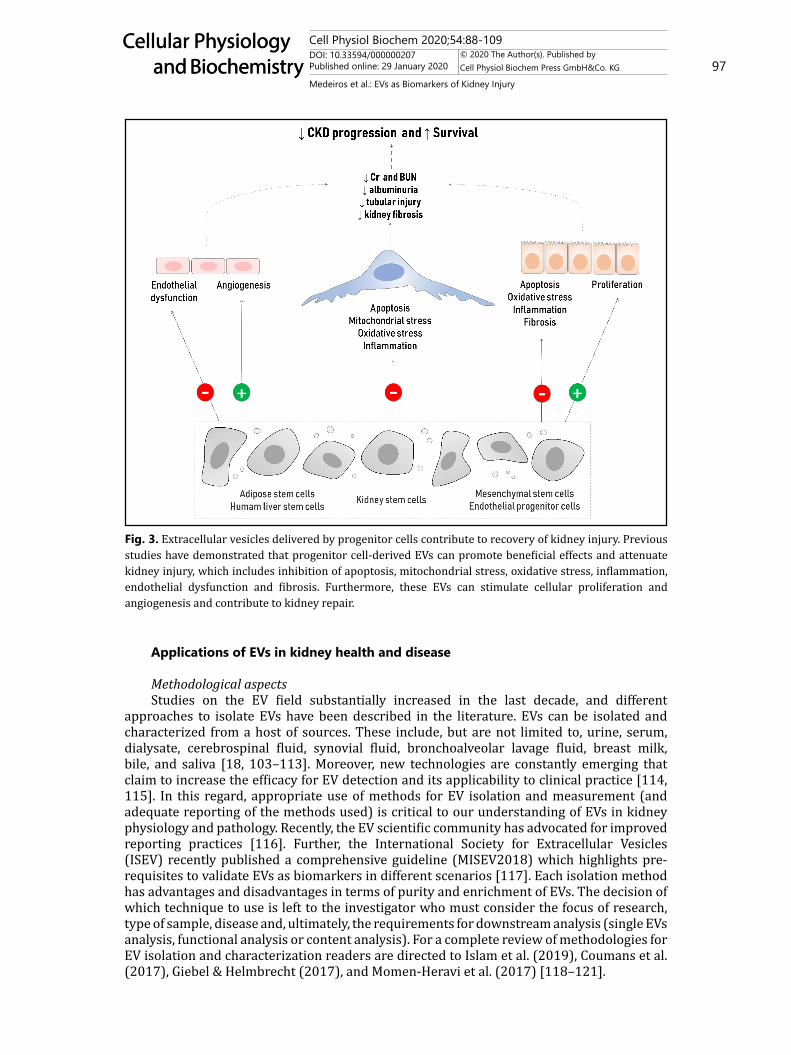
always deleterious. In this regard, pioneering studies investigating the therapeutic potential of stem cells in kidney disease consistently observed that treatment with different types of progenitor cells (or their conditioned media) could ameliorate kidney function, however, the mechanisms involved in the recovery were not well understood [88–90]. As EVs became increasingly recognized as bioactive mediators of horizontal communication between cells, the role of progenitor cell-derived EVs in kidney regeneration was investigated. Several studies have reported that EVs released by different types of progenitor cells (e.g. mesenchymal stromal cells, endothelial cells, liver and kidney stem cells, adipose cells) were involved in tissue repair, cellular recovery and reduction of apoptosis [12, 91–94]. In Fig. 3, the protective role of EVs delivered by progenitor cells during kidney injury is illustrated.
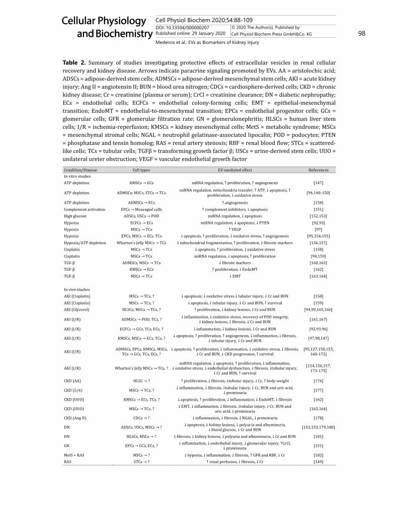
The beneficial effects promoted by EVs in kidney disease have been reported in experimental models of AKI, CKD and DN as summarized in Table 2. Biodistribution experiments from two studies demonstrated that mesenchymal stem cell (MSC)- and endothelial progenitor cell-derived EVs are significantly increased in kidney tissue during AKI, accumulating within endothelial and injured tubular cells at 2h after I/R [95, 96]. In control animals, MSC-derived EVs did not traffic specifically to the kidney [95, 96]. Similarly, Viňas et al. (2018) recently reported that sEVs can target to kidney cells at 30min and 4h after I/R and transfer miR-486-5p to glomerular, tubular and endothelial cells. CXCR4/SDF-1α was observed to be involved in sEV uptake, microRNA transfer and improvement of kidney function during ischemic conditions [97]. Despite increasing evidence, additional in vivo studies are needed to clarify mechanisms of action and relative efficacy of sEVs from various origins in promoting kidney repair.
In summary, EVs are not only promising biomarkers of kidney disease, but active contributors to both pathogenesis and recovery. Importantly, as reviewed in this section, the health status of the donor cell directly impacts EV signaling. The cellular effects can be either beneficial, or deleterious, depending on whether the donor cells are injured or not. However, how and when the switch of the EV content during disease state happens, remains unclear. Moreover, progenitor cell-derived sEVs are associated with cellular recovery but lEVs have been reported to promote beneficial [98, 99] and deleterious effects [93, 100] during disease conditions. Thus, this it may not simply be a case of “small EV= good” and “large EV = bad”? Further study will be necessary to gain a greater appreciation for the role of EVs in intercellular communication in the kidney and the impact of disease on this. Finally, EV size categorization studies have shown that different molecular signatures can be observed according to sEV size [101, 102]. Even EVs within the same “category” (i.e. sEVs or lEVs) can be sub-categorized according to size, and this can directly impact the effect promoted by EVs in recipient cells [101, 102]. This challenges the current classification of major EV subtypes.
Cell Physiol Biochem 2020;54:88-109DOI: 10.33594/000000207Published online: 29 January 2020 97
Cellular Physiology and Biochemistry
Cellular Physiology and Biochemistry
© 2020 The Author(s). Published by Cell Physiol Biochem Press GmbH&Co. KG
Medeiros et al.: EVs as Biomarkers of Kidney Injury
Applications of EVs in kidney health and disease
Methodological aspectsStudies on the EV field substantially increased in the last decade, and different
approaches to isolate EVs have been described in the literature. EVs can be isolated and characterized from a host of sources. These include, but are not limited to, urine, serum, dialysate, cerebrospinal fluid, synovial fluid, bronchoalveolar lavage fluid, breast milk, bile, and saliva [18, 103–113]. Moreover, new technologies are constantly emerging that claim to increase the efficacy for EV detection and its applicability to clinical practice [114, 115]. In this regard, appropriate use of methods for EV isolation and measurement (and adequate reporting of the methods used) is critical to our understanding of EVs in kidney physiology and pathology. Recently, the EV scientific community has advocated for improved reporting practices [116]. Further, the International Society for Extracellular Vesicles (ISEV) recently published a comprehensive guideline (MISEV2018) which highlights pre-requisites to validate EVs as biomarkers in different scenarios [117]. Each isolation method has advantages and disadvantages in terms of purity and enrichment of EVs. The decision of which technique to use is left to the investigator who must consider the focus of research, type of sample, disease and, ultimately, the requirements for downstream analysis (single EVs analysis, functional analysis or content analysis). For a complete review of methodologies for EV isolation and characterization readers are directed to Islam et al. (2019), Coumans et al. (2017), Giebel & Helmbrecht (2017), and Momen-Heravi et al. (2017) [118–121].
Fig. 3. Extracellular vesicles delivered by progenitor cells contribute to recovery of kidney injury. Previous studies have demonstrated that progenitor cell-derived EVs can promote beneficial effects and attenuate kidney injury, which includes inhibition of apoptosis, mitochondrial stress, oxidative stress, inflammation, endothelial dysfunction and fibrosis. Furthermore, these EVs can stimulate cellular proliferation and angiogenesis and contribute to kidney repair.
Cell Physiol Biochem 2020;54:88-109DOI: 10.33594/000000207Published online: 29 January 2020 98
Cellular Physiology and Biochemistry
Cellular Physiology and Biochemistry
© 2020 The Author(s). Published by Cell Physiol Biochem Press GmbH&Co. KG
Medeiros et al.: EVs as Biomarkers of Kidney Injury
Table 2. Summary of studies investigating protective effects of extracellular vesicles in renal cellular recovery and kidney disease. Arrows indicate paracrine signaling promoted by EVs. AA = aristolochic acid; ADSCs = adipose-derived stem cells; ADMSCs = adipose-derived mesenchymal stem cells; AKI = acute kidney injury; Ang II = angiotensin II; BUN = blood urea nitrogen; CDCs = cardiosphere-derived cells; CKD = chronic kidney disease; Cr = creatinine (plasma or serum); CrCl = creatinine clearance; DN = diabetic nephropathy; ECs = endothelial cells; ECFCs = endothelial colony-forming cells; EMT = epithelial-mesenchymal transition; EndoMT = endothelial-to-mesenchymal transition; EPCs = endothelial progenitor cells; GCs = glomerular cells; GFR = glomerular filtration rate; GN = glomerulonephritis; HLSCs = human liver stem cells; I/R = ischemia-reperfusion; KMSCs = kidney mesenchymal cells; MetS = metabolic syndrome; MSCs = mesenchymal stromal cells; NGAL = neutrophil gelatinase-associated lipocalin; POD = podocytes; PTEN = phosphatase and tensin homolog; RAS = renal artery stenosis; RBF = renal blood flow; STCs = scattered-like cells; TCs = tubular cells; TGFβ = transforming growth factor β; USCs = urine-derived stem cells; UUO = unilateral ureter obstruction; VEGF = vascular endothelial growth factor
KMSCs → ECs mRNA regulation, ↑ proliferation, ↑ angiogenesis
ADMSCs; MSCs, STCs → TCs miRNA regulation, mitochondria transfer, ↑ ATP, ↓ apoptosis, ↑ proliferation, ↓ oxidative stress
ADMSCs → ECs ↑ angiogenesisEPCs → Mesangial cells ↑ complement inhibitors, ↓ apoptosis
ADSCs, USCs → POD miRNA regulation, ↓ apoptosisECFCs → ECs miRNA regulation, ↓ apoptosis, ↓ PTENMSCs → TCs ↑ VEGF
EPCs, MSCs → ECs, TCs ↓ apoptosis, ↑ proliferation, ↓ oxidative stress, ↑ angiogenesisWharton’s Jelly MSCs → TCs ↓ mitochondrial fragmentation, ↑ proliferation, ↓ fibrotic markers
MSCs → TCs ↓ apoptosis, ↑ proliferation, ↓ oxidative stress MSCs → TCs miRNA regulation, ↓ apoptosis, ↑ proliferation
β ADMSCs, MSCs → TCs ↓ fibrotic markersβ KMSCs → ECs ↑ proliferation, ↓ EndoMTβ → TCs ↓ EMT
MSCs → TCs, ↓ apoptosis; ↓ oxidative stress ↓ tubular injury, ↓ Cr and BUNMSCs → TCs, ↓ apoptosis, ↓ tubular injury, ↓ Cr and BUN, ↑ s
→ TCs, ? ↑ proliferation, ↓ kidney lesions, ↓ Cr and BUN
ADMSCs → POD, TCs, ↓ inflammation, ↓ oxidative stress, recovery of POD integrity, ↓ kidney lesions, ↓ fibrosis, ↓ Cr and BUN
ECFCs → GCs, TCs, ECs, ↓ inflammation, ↓ kidney lesions, ↓ Cr and BUN
→ ECs, TCs, ? ↓ apoptosis, ↑ proliferation, ↑ angiogenesis, ↓ inflammation, ↓ fibrosis,↓ tubular injury, ↓ Cr and BUN
TCs → GCs, TCs, ECs, ?↓ apoptosis, ↑ proliferation, ↓ inflammation, ↓ oxidative stress, ↓ fibrosis,
↓ Cr and BUN ↓ CKD progression, ↑ s
Wharton’s Jelly MSCs → TCs,↓ apoptosis, ↑ proliferation, ↓ inflammation,
↓ oxidative stress, ↓ endothelial dysfunction, ↓ fibrosis, ↓tubular injury,↓ Cr and BUN, ↑ s
HLSC → ? ↑ proliferation, ↓ fibrosis, ↓tubular injury, ↓ Cr, ↑ body weight
MSCs → TCs, ? ↓ inflammation, ↓ fibrosis, ↓tubular injury, ↓ Cr, BUN and uric acid,↓ proteinuria
→ ECs, TCs, ? ↓ apoptosis, ↑ proliferation, ↓ inflammation, ↓ EndoMT, ↓ fibrosis
MSCs → TCs, ? ↓ EMT, ↓ inflammation, ↓ fibrosis, ↓tubular injury, ↓ Cr, BUN anduric acid, ↓ proteinuria
CDCs → ? ↓ inflammation, ↓ fibrosis, ↓ NGAL, ↓ proteinuria
ADSCs, USCs, MSCs → ↓ apoptosis, ↓ kidney lesions, ↓ ↓ blood glucose, ↓ Cr and BUN
HLSCs, MSCs → ↓ fibrosis, ↓ kidney lesions, ↓ polyuria and albuminuria, ↓ Cr and BUN
EPCs → GCs, ECs, ? ↓ inflammation, ↓ endothelial injury, ↓ glomerular injury, ↑CrCl,↓
MSCs → ? ↓ hypoxia, ↓ inflammation, ↓ fibrosis, ↑ GFR and RBF, ↓ CrSTCs → ↑ renal perfusion, ↓ fibrosis, ↓ Cr

Cell Physiol Biochem 2020;54:88-109DOI: 10.33594/000000207Published online: 29 January 2020 99
Cellular Physiology and Biochemistry
Cellular Physiology and Biochemistry
© 2020 The Author(s). Published by Cell Physiol Biochem Press GmbH&Co. KG
Medeiros et al.: EVs as Biomarkers of Kidney Injury
Clinical perspectivesGiven that EVs are measurable, specific and becoming well-studied, it is certainly
feasible that they will ultimately be developed as new biomarkers of renal disease in clinical practice. Furthermore, their potential use as biological delivery systems are also an area of great interest. As mentioned above, in order for EVs to be reliable markers in clinical practice, they need to be easily obtained, quantified and characterized. This also needs to occur in a reasonably fast and affordable manner. As such, there are several groups studying the use of EVs in pre-clinical and clinical studies as they pertain to medical conditions including cancer, [122, 123] rheumatologic disease, [124] cardiovascular disease [125] and renal disease [2, 13].
As biologically active biomarkers, EVs present a new and exciting opportunity. Regarding urinary EVs in particular, it is thought they can function as both endogenous communication links between renal cells and perhaps exogenous tools to protect against injury or promote recovery [2, 14, 91, 100]. Because it is easy and non-invasive to collect urine, using urinary EVs as markers is very clinically appealing. Fresh urine is ideal; [126] however, samples can be centrifuged and stored frozen, making batched analysis a viable option that would be clinically feasible. Nevertheless, numerous challenges remain and further information is needed with respect to normalization, optimal collection parameters, and an appreciation of the influence of protein aggregates, urine viscosity, pH and osmolality. As such one must be careful not to over-conclude based on our current knowledge. While the development of individual patient urinary EV proteomes or transcriptomes to diagnose renal conditions, or monitor disease progression is appealing, much is still not known about possible confounding variables. In this context, further “omic”-based studies should continue to elucidate specific changes in the expression EV-derived factors in kidney disease (i.e. exosomal derived-mRNAs and miRNAs) but also remain conscious of the need to ensure reproducibility.
Using EVs as clinical tools for delivering “cargo” was recently discussed in an ISEV-led white paper [127]. An example of this would be the ability of EVs isolated from human MSCs to protect against I/R injury following both acute and chronic kidney injury [91, 128]. As a singular example, one can see the importance of translating this finding to clinical practice. AKI remains a leading cause of morbidity and mortality among adult and pediatric patients, alike [129, 130]. This paper highlights several additional examples of clinically applicable uses for EVs in diagnosis of disease ranging from genetic disorders (i.e. exosomal NKCC2), water homeostasis defects (i.e. urinary EV-AQP2), and glomerular injury secondary to podocytopathies (i.e. podocyte-derived lEVs). Importantly, these conditions normally require serum testing and long wait times. Could urinary EVs facilitate a faster diagnosis and be employed as markers of disease severity or remission? At present, it is impossible to answer this, but it would appear this is a clinically important question to ask.
Our approach for this review was to highlight the potential of EVs as mediators of intercellular communication and biomarkers of kidney disease. The number of studies on the EV field is increasing substantially in the past few years, however there are still several unanswered questions. For example, to identify lEV’s cellular origin, a range of surface markers have been studied individually or at most in combination of two; however, it is unclear if the EV phenotype (density of specific markers) can change according the diseased condition. Therefore, a panel of markers for the same cellular origin in different scenarios should be performed. In the context of glomerulopathies, for example, the expression of podocyte markers in lEVs such as podocin, podocalyxin, nephrin and podoplanin; could vary according to the etiology and phenotype of glomerular disease.
We have also discussed EV-mediated interactions involved in both kidney pathophysiology and repair, however, the possible routes for EV signaling and how donor cells target different cells across the nephron require further study. Recent studies suggest that cells can communicate by EV delivery throughout urinary space (e.g. podocytes → proximal tubular cells), but we speculate that it may also be possible for mesangial and endothelial cells to transfer EVs in the vasa recta and promote “interstitial signaling”. Future
Cell Physiol Biochem 2020;54:88-109DOI: 10.33594/000000207Published online: 29 January 2020 100
Cellular Physiology and Biochemistry
Cellular Physiology and Biochemistry
© 2020 The Author(s). Published by Cell Physiol Biochem Press GmbH&Co. KG
Medeiros et al.: EVs as Biomarkers of Kidney Injury
experimental studies performing renal punctures and isolation of mesangial-derived EVs could test this hypothesis.
Conclusion
In conclusion, this review summarizes some of the many applications for urinary EVs in clinical medicine. While assessment of EV levels and their content shows promise it is clear that we are only scratching the surface of our what is possible. Future studies should focus on standardization of methodology to facilitate adoption to routine clinical practice and further expanding our knowledge of EVs in kidney health and disease.
Acknowledgements
FundingTM is supported by the Coordenação de Aperfeiçoamento de Pessoal de Nível Superior
(CAPES, Brazil). DB is supported by grants from the Canadian Institutes of Health Research, an Ontario Early Researcher Award, the Canada Foundation for Innovation, and The Ottawa Hospital Department of Medicine.
Author’s ContributionsTM, RM, JRA, AAS and DB contributed to the manuscript and approved the final version.
Disclosure Statement
The authors declare no conflict of interest.
References
1 Burger D, Touyz RM: Cellular biomarkers of endothelial health: microparticles, endothelial progenitor cells, and circulating endothelial cells. J Am Soc Hypertens 2012;6:85–99.
2 Erdbrügger U, Le TH: Extracellular Vesicles in Renal Diseases: More than Novel Biomarkers? J Am Soc Nephrol 2016;27:12–26.
3 Helmke A, von Vietinghoff S: Extracellular vesicles as mediators of vascular inflammation in kidney disease. World J Nephrol 2016;5:125–138.
4 Abels ER, Breakefield XO: Introduction to Extracellular Vesicles: Biogenesis, RNA Cargo Selection, Content, Release, and Uptake. Cell Mol Neurobiol 2016;36:301–312.
5 Mulcahy LA, Pink RC, Carter DRF: Routes and mechanisms of extracellular vesicle uptake. J Extracell Vesicles 2014;3:2461.
6 Burger D, Schock S, Thompson CS, Montezano AC, Hakim AM, Touyz RM: Microparticles: biomarkers and beyond. Clin Sci 2013;124:423–441.
7 Kalra H, Drummen GPC, Mathivanan S: Focus on Extracellular Vesicles: Introducing the Next Small Big Thing. Int J Mol Sci 2016;17:170.
8 Chuo STY, Chien JCY, Lai CPK: Imaging extracellular vesicles: current and emerging methods. J Biomed Sci 2018;25:91.
9 D’Souza-Schorey C, Schorey JS: Regulation and mechanisms of extracellular vesicle biogenesis and secretion. Essays Biochem 2018;62:125–133.
10 Nielsen CT, Rasmussen NS, Heegaard NHH, Jacobsen S: “Kill” the messenger: Targeting of cell-derived microparticles in lupus nephritis. Autoimmun Rev 2016;15:719–725.
11 Dursun I, Yel S, Unsur E: Dynamics of circulating microparticles in chronic kidney disease and transplantation: Is it really reliable marker? World J Transplant 2015;5:267–275.
Cell Physiol Biochem 2020;54:88-109DOI: 10.33594/000000207Published online: 29 January 2020 101
Cellular Physiology and Biochemistry
Cellular Physiology and Biochemistry
© 2020 The Author(s). Published by Cell Physiol Biochem Press GmbH&Co. KG
Medeiros et al.: EVs as Biomarkers of Kidney Injury
12 Morrison EE, Bailey MA, Dear JW: Renal extracellular vesicles: from physiology to clinical application. J Physiol (Lond) 2016;594:5735–5748.
13 Zhang W, Zhou X, Zhang H, Yao Q, Liu Y, Dong Z: Extracellular vesicles in diagnosis and therapy of kidney diseases. Am J Physiol Renal Physiol 2016;311:F844–F851.
14 Abbasian N, Herbert KE, Pawluczyk I, Burton JO, Bevington A: Vesicles bearing gifts: the functional importance of micro-RNA transfer in extracellular vesicles in chronic kidney disease. Am J Physiol Renal Physiol 2018;315:F1430–F1443.
15 Pascual M, Steiger G, Sadallah S, Paccaud JP, Carpentier JL, James R, Schifferli JA: Identification of membrane-bound CR1 (CD35) in human urine: evidence for its release by glomerular podocytes. J Exp Med 1994;179:889–899.
16 Hara M, Yanagihara T, Hirayama Y, Ogasawara S, Kurosawa H, Sekine S, Kihara I: Podocyte membrane vesicles in urine originate from tip vesiculation of podocyte microvilli. Hum Pathol 2010;41:1265–1275.
17 Rood IM, Deegens JKJ, Merchant ML, Tamboer WPM, Wilkey DW, Wetzels JFM, Klein JB: Comparison of three methods for isolation of urinary microvesicles to identify biomarkers of nephrotic syndrome. Kidney Int 2010;78:810–816.
18 Burger D, Thibodeau J-F, Holterman CE, Burns KD, Touyz RM, Kennedy CRJ: Urinary podocyte microparticles identify prealbuminuric diabetic glomerular injury. J Am Soc Nephrol 2014;25:1401–1407.
19 Lytvyn Y, Xiao F, Kennedy CRJ, Perkins BA, Reich HN, Scholey JW, Cherney DZ, Burger D: Assessment of urinary microparticles in normotensive patients with type 1 diabetes. Diabetologia 2017;60:581–584.
20 De S, Kuwahara S, Hosojima M, Ishikawa T, Kaseda R, Sarkar P, Yoshioka Y, Kabasawa H, Iida T, Goto S, Toba K, Higuchi Y, Suzuki Y, Hara M, Kurosawa H, Narita I, Hirayama Y, Ochiya T, Saito A: Exocytosis-Mediated Urinary Full-Length Megalin Excretion Is Linked With the Pathogenesis of Diabetic Nephropathy. Diabetes 2017;66:1391–1404.
21 Kamińska A, Platt M, Kasprzyk J, Kuśnierz-Cabala B, Gala-Błądzińska A, Woźnicka O, Jany BR, Krok F, Piekoszewski W, Kuźniewski M, Stępień EŁ: Urinary Extracellular Vesicles: Potential Biomarkers of Renal Function in Diabetic Patients. J Diabetes Res 2016;2016:5741518.
22 Kwon SH, Woollard JR, Saad A, Garovic VD, Zand L, Jordan KL, Textor SC, Lerman LO: Elevated urinary podocyte-derived extracellular microvesicles in renovascular hypertensive patients. Nephrol Dial Transplant 2017;32:800–807.
23 Santelli A, Sun IO, Eirin A, Abumoawad AM, Woollard JR, Lerman A, Textor SC, Puranik AS, Lerman LO: Senescent Kidney Cells in Hypertensive Patients Release Urinary Extracellular Vesicles. J Am Heart Assoc 2019;8:e012584.
24 Zhang LH, Zhu XY, Eirin A, Nargesi AA, Woollard JR, Santelli A, Sun IO, Textor SC, Lerman LO: Early podocyte injury and elevated levels of urinary podocyte-derived extracellular vesicles in swine with metabolic syndrome: role of podocyte mitochondria. Am J Physiol Renal Physiol 2019;317:F12–F22.
25 Lu J, Hu ZB, Chen PP, Lu CC, Zhang JX, Li XQ, Yuan BY, Huang SJ, Ma KL: Urinary podocyte microparticles are associated with disease activity and renal injury in systemic lupus erythematosus. BMC Nephrology 2019;20:303.
26 Lu J, Hu ZB, Chen PP, Lu CC, Zhang JX, Li XQ, Yuan BY, Huang SJ, Ma KL: Urinary levels of podocyte-derived microparticles are associated with the progression of chronic kidney disease. Ann Transl Med 2019;7:445.
27 Gilani SI, Anderson UD, Jayachandran M, Weissgerber TL, Zand L, White WM, Milic N, Suarez MLG, Vallapureddy RR, Nääv Å, Erlandsson L, Lieske JC, Grande JP, Nath KA, Hansson SR, Garovic VD: Urinary Extracellular Vesicles of Podocyte Origin and Renal Injury in Preeclampsia. J Am Soc Nephrol 2017;28:3363–3372.
28 Dimuccio V, Ranghino A, Praticò Barbato L, Fop F, Biancone L, Camussi G, Bussolati B: Urinary CD133+ extracellular vesicles are decreased in kidney transplanted patients with slow graft function and vascular damage. PLoS One 2014;9:e104490.
29 Gagnon L, Leduc M, Thibodeau JF, Zhang MZ, Grouix B, Sarra-Bournet F, Gagnon W, Hince K, Tremblay M, Geerts L, Kennedy CRJ, Hébert RL, Gutsol A, Holterman CE, Kamto E, Gervais L, Ouboudinar J, Richard J, Felton A, Laverdure A, et al.: A Newly Discovered Antifibrotic Pathway Regulated by Two Fatty Acid Receptors: GPR40 and GPR84. Am J Pathol 2018;188:1132–1148.
30 Thibodeau JF, Simard JC, Holterman CE, Blais A, Cloutier MP, Medeiros T, Leduc M, Grouix B, Leblond FA, Burger D, Hébert RL, Kennedy CRJ, Gagnon L: PBI-4050 via GPR40 activation improves adenine-induced kidney injury in mice. Clin Sci 2019;133:1587–1602.
Cell Physiol Biochem 2020;54:88-109DOI: 10.33594/000000207Published online: 29 January 2020 102
Cellular Physiology and Biochemistry
Cellular Physiology and Biochemistry
© 2020 The Author(s). Published by Cell Physiol Biochem Press GmbH&Co. KG
Medeiros et al.: EVs as Biomarkers of Kidney Injury
31 Turco AE, Lam W, Rule AD, Denic A, Lieske JC, Miller VM, Larson JJ, Kremers WK, Jayachandran M: Specific renal parenchymal-derived urinary extracellular vesicles identify age-associated structural changes in living donor kidneys. J Extracell Vesicles 2016;5:29642.
32 Burbano C, Gómez-Puerta JA, Muñoz-Vahos C, Vanegas-García A, Rojas M, Vásquez G, Castaño D: HMGB1+ microparticles present in urine are hallmarks of nephritis in patients with systemic lupus erythematosus. Eur J Immunol 2019;49:323–335.
33 Chirackal RS, Jayachandran M, Wang X, Edeh S, Haskic Z, Perinpam M, Halling TM, Mehta R, Rivera ME, Lieske JC: Urinary extracellular vesicles associated MCP-1 and NGAL derived from specific nephron segments differ between calcium oxalate stone formers and controls. Am J Physiol Renal Physiol 2019;317:F1475-F1482.
34 Miranda KC, Bond DT, Levin JZ, Adiconis X, Sivachenko A, Russ C, Brown D, Nusbaum C, Russo LM: Massively parallel sequencing of human urinary exosome/microvesicle RNA reveals a predominance of non-coding RNA. PLoS One 2014;9:e96094.
35 Sonoda H, Yokota-Ikeda N, Oshikawa S, Kanno Y, Yoshinaga K, Uchida K, Ueda Y, Kimiya K, Uezono S, Ueda A, Ito K, Ikeda M: Decreased abundance of urinary exosomal aquaporin-1 in renal ischemia-reperfusion injury. Am J Physiol Renal Physiol 2009;297:F1006-F1016.
36 Barutta F, Tricarico M, Corbelli A, Annaratone L, Pinach S, Grimaldi S, Bruno G, Cimino D, Taverna D, Deregibus MC, Rastaldi MP, Perin PC, Gruden G: Urinary exosomal microRNAs in incipient diabetic nephropathy. PLoS One 2013;8:e73798.
37 Perez-Hernandez J, Forner MJ, Pinto C, Chaves FJ, Cortes R, Redon J: Increased Urinary Exosomal MicroRNAs in Patients with Systemic Lupus Erythematosus. PLoS One 2015;10:e0138618.
38 Tutakhel OAZ, Moes AD, Valdez-Flores MA, Kortenoeven MLA, Vrie MVD, Jeleń S, Fenton RA, Zietse R, Hoenderop JGJ, Hoorn EJ, Hilbrands L, Bindels RJM: NaCl cotransporter abundance in urinary vesicles is increased by calcineurin inhibitors and predicts thiazide sensitivity. PLoS One 2017;12:e0176220.
39 Chun-Yan L, Zi-Yi Z, Tian-Lin Y, Yi-Li W, Bao L, Jiao L, Wei-Jun D: Liquid biopsy biomarkers of renal interstitial fibrosis based on urinary exosome. Exp Mol Pathol 2018;105:223–228.
40 Asvapromtada S, Sonoda H, Kinouchi M, Oshikawa S, Takahashi S, Hoshino Y, Sinlapadeelerdkul T, Yokota-Ikeda N, Matsuzaki T, Ikeda M: Characterization of urinary exosomal release of aquaporin-1 and -2 after renal ischemia-reperfusion in rats. Am J Physiol Renal Physiol 2018;314:F584–F601.
41 Feng Y, Lv LL, Wu WJ, Li ZL, Chen J, Ni HF, Zhou LT, Tang TT, Wang FM, Wang B, Chen PS, Crowley SD, Liu BC: Urinary Exosomes and Exosomal CCL2 mRNA as Biomarkers of Active Histologic Injury in IgA Nephropathy. Am J Pathol 2018;188:2542–2552.
42 Abdeen A, Sonoda H, El-Shawarby R, Takahashi S, Ikeda M: Urinary excretion pattern of exosomal aquaporin-2 in rats that received gentamicin. Am J Physiol Renal Physiol 2014;307:F1227-F1237.
43 Peake PW, Pianta TJ, Succar L, Fernando M, Pugh DJ, McNamara K, Endre ZH: A comparison of the ability of levels of urinary biomarker proteins and exosomal mRNA to predict outcomes after renal transplantation. PLoS One 2014;9:e98644.
44 Wang X, Wilkinson R, Kildey K, Potriquet J, Mulvenna J, Lobb RJ, Möller A, Cloonan N, Mukhopadhyay P, Kassianos AJ, Healy H: Unique molecular profile of exosomes derived from primary human proximal tubular epithelial cells under diseased conditions. J Extracell Vesicles 2017;6:1314073.
45 Yu Y, Bai F, Qin N, Liu W, Sun Q, Zhou Y, Yang J: Non-Proximal Renal Tubule-Derived Urinary Exosomal miR-200b as a Biomarker of Renal Fibrosis. Nephron 2018;139:269–282.
46 Pisitkun T, Shen R-F, Knepper MA: Identification and proteomic profiling of exosomes in human urine. Proc Natl Acad Sci USA 2004;101:13368–13373.
47 Vesiclepedia [Internet] [cited 2019 Dec 12]. URL: microvesicles.org.48 Fujita K, Kume H, Matsuzaki K, Kawashima A, Ujike T, Nagahara A, Uemura M, Miyagawa Y, Tomonaga T,
Nonomura N: Proteomic analysis of urinary extracellular vesicles from high Gleason score prostate cancer. Sci Rep 2017;7:42961.
49 Esteva-Font C, Wang X, Ars E, Guillén-Gómez E, Sans L, González Saavedra I, Torres F, Torra R, Masilamani S, Ballarín JA, Fernández-Llama P: Are sodium transporters in urinary exosomes reliable markers of tubular sodium reabsorption in hypertensive patients? Nephron Physiol 2010;114:p25-p34.
50 Salih M, Fenton RA, Zietse R, Hoorn EJ: Urinary extracellular vesicles as markers to assess kidney sodium transport. Curr Opin Nephrol Hypertens 2016;25:67–72.
Cell Physiol Biochem 2020;54:88-109DOI: 10.33594/000000207Published online: 29 January 2020 103
Cellular Physiology and Biochemistry
Cellular Physiology and Biochemistry
© 2020 The Author(s). Published by Cell Physiol Biochem Press GmbH&Co. KG
Medeiros et al.: EVs as Biomarkers of Kidney Injury
51 du Cheyron D, Daubin C, Poggioli J, Ramakers M, Houillier P, Charbonneau P, Paillard M: Urinary measurement of Na+/H+ exchanger isoform 3 (NHE3) protein as new marker of tubule injury in critically ill patients with ARF. Am J Kidney Dis 2003;42:497–506.
52 Hu C-C, Katerelos M, Choy S-W, Crossthwaite A, Walker SP, Pell G, Lee M, Cook N, Mount PF, Paizis K, Power DA: Pre-eclampsia is associated with altered expression of the renal sodium transporters NKCC2, NCC and ENaC in urinary extracellular vesicles. PLoS One 2018;13:e0204514.
53 Rossi L, Nicoletti MC, Carmosino M, Mastrofrancesco L, Di Franco A, Indrio F, Lella R, Laviola L, Giorgino F, Svelto M, Gesualdo L, Procino G: Urinary Excretion of Kidney Aquaporins as Possible Diagnostic Biomarker of Diabetic Nephropathy. J Diabetes Res 2017;2017:4360357.
54 Andersen H, Friis UG, Hansen PBL, Svenningsen P, Henriksen JE, Jensen BL: Diabetic nephropathy is associated with increased urine excretion of proteases plasmin, prostasin and urokinase and activation of amiloride-sensitive current in collecting duct cells. Nephrol Dial Transplant 2015;30:781–789.
55 Wolley MJ, Wu A, Xu S, Gordon RD, Fenton RA, Stowasser M: In Primary Aldosteronism, Mineralocorticoids Influence Exosomal Sodium-Chloride Cotransporter Abundance. J Am Soc Nephrol 2017;28:56–63.
56 Corbetta S, Raimondo F, Tedeschi S, Syrèn M-L, Rebora P, Savoia A, Baldi L, Bettinelli A, Pitto M: Urinary exosomes in the diagnosis of Gitelman and Bartter syndromes. Nephrol Dial Transplant 2015;30:621–630.
57 Pathare G, Dhayat NA, Mohebbi N, Wagner CA, Bobulescu IA, Moe OW, Fuster DG: Changes in V-ATPase subunits of human urinary exosomes reflect the renal response to acute acid/alkali loading and the defects in distal renal tubular acidosis. Kidney Int 2018;93:871–880.
58 Oshikawa S, Sonoda H, Ikeda M: Aquaporins in Urinary Extracellular Vesicles (Exosomes). Int J Mol Sci 2016;17:957.
59 Sonoda H, Oshikawa-Hori S, Ikeda M: An Early Decrease in Release of Aquaporin-2 in Urinary Extracellular Vesicles After Cisplatin Treatment in Rats. Cells 2019;8:139.
60 Oshikawa-Hori S, Yokota-Ikeda N, Sonoda H, Ikeda M: Urinary extracellular vesicular release of aquaporins in patients with renal transplantation. BMC Nephrol 2019;20:216.
61 Kanno K, Sasaki S, Hirata Y, Ishikawa S, Fushimi K, Nakanishi S, Bichet DG, Marumo F: Urinary excretion of aquaporin-2 in patients with diabetes insipidus. N Engl J Med 1995;332:1540–1545.
62 Zhou H, Cheruvanky A, Hu X, Matsumoto T, Hiramatsu N, Cho ME, Berger A, Leelahavanichkul A, Doi K, Chawla LS, Illei GG, Kopp JB, Balow JE, Austin HA, Yuen PS, Star RA: Urinary exosomal transcription factors, a new class of biomarkers for renal disease. Kidney Int 2008;74:613–621.
63 Chen HH, Lai PF, Lan YF, Cheng CF, Zhong WB, Lin YF, Chen TW, Lin H: Exosomal ATF3 RNA attenuates pro-inflammatory gene MCP-1 transcription in renal ischemia-reperfusion. J Cell Physiol 2014;229:1202–1211.
64 Panich T, Chancharoenthana W, Somparn P, Issara-Amphorn J, Hirankarn N, Leelahavanichkul A: Urinary exosomal activating transcriptional factor 3 as the early diagnostic biomarker for sepsis-induced acute kidney injury. BMC Nephrol 2017;18:10.
65 Kalani A, Mohan A, Godbole MM, Bhatia E, Gupta A, Sharma RK, Tiwari S: Wilm’s tumor-1 protein levels in urinary exosomes from diabetic patients with or without proteinuria. PLoS One 2013;8:e60177.
66 Abe H, Sakurai A, Ono H, Hayashi S, Yoshimoto S, Ochi A, Ueda S, Nishimura K, Shibata E, Tamaki M, Kishi F, Kishi S, Murakami T, Nagai K, Doi T: Urinary Exosomal mRNA of WT1 as Diagnostic and Prognostic Biomarker for Diabetic Nephropathy. J Med Invest 2018;65:208–215.
67 Fang DY, King HW, Li JY, Gleadle JM: Exosomes and the kidney: blaming the messenger. Nephrology (Carlton) 2013;18:1–10.
68 Miranda KC, Bond DT, McKee M, Skog J, Păunescu TG, Da Silva N, Brown D, Russo LM: Nucleic acids within urinary exosomes/microvesicles are potential biomarkers for renal disease. Kidney Int 2010;78:191–199.
69 Cheng L, Sun X, Scicluna BJ, Coleman BM, Hill AF: Characterization and deep sequencing analysis of exosomal and non-exosomal miRNA in human urine. Kidney Int 2014;86:433–444.
70 Zhou Y, Xiong M, Fang L, Jiang L, Wen P, Dai C, Zhang CY, Yang J: miR-21-containing microvesicles from injured tubular epithelial cells promote tubular phenotype transition by targeting PTEN protein. Am J Pathol 2013;183:1183–1196.
71 Lv CY, Ding WJ, Wang YL, Zhao ZY, Li JH, Chen Y, Lv J: A PEG-based method for the isolation of urinary exosomes and its application in renal fibrosis diagnostics using cargo miR-29c and miR-21 analysis. Int Urol Nephrol 2018;50:973–982.
72 Lv LL, Cao YH, Ni HF, Xu M, Liu D, Liu H, Chen PS, Liu BC: MicroRNA-29c in urinary exosome/microvesicle as a biomarker of renal fibrosis. Am J Physiol Renal Physiol 2013;305:F1220–F1227.
Cell Physiol Biochem 2020;54:88-109DOI: 10.33594/000000207Published online: 29 January 2020 104
Cellular Physiology and Biochemistry
Cellular Physiology and Biochemistry
© 2020 The Author(s). Published by Cell Physiol Biochem Press GmbH&Co. KG
Medeiros et al.: EVs as Biomarkers of Kidney Injury
73 Solé C, Cortés-Hernández J, Felip ML, Vidal M, Ordi-Ros J: miR-29c in urinary exosomes as predictor of early renal fibrosis in lupus nephritis. Nephrol Dial Transplant 2015;30:1488–1496.
74 Ranghino A, Dimuccio V, Papadimitriou E, Bussolati B: Extracellular vesicles in the urine: markers and mediators of tissue damage and regeneration. Clin Kidney J 2015;8:23–30.
75 Gracia T, Wang X, Su Y, Norgett EE, Williams TL, Moreno P, Micklem G, Karet Frankl FE: Urinary Exosomes Contain MicroRNAs Capable of Paracrine Modulation of Tubular Transporters in Kidney. Sci Rep 2017;7:40601.
76 Street JM, Birkhoff W, Menzies RI, Webb DJ, Bailey MA, Dear JW: Exosomal transmission of functional aquaporin 2 in kidney cortical collecting duct cells. J Physiol (Lond) 2011;589:6119–6127.
77 Oosthuyzen W, Scullion KM, Ivy JR, Morrison EE, Hunter RW, Starkey Lewis PJ, O’Duibhir E, Street JM, Caporali A, Gregory CD, Forbes SJ, Webb DJ, Bailey MA, Dear JW: Vasopressin Regulates Extracellular Vesicle Uptake by Kidney Collecting Duct Cells. J Am Soc Nephrol 2016;27:3345–3355.
78 Chiabotto G, Bruno S, Collino F, Camussi G: Mesenchymal Stromal Cells Epithelial Transition Induced by Renal Tubular Cells-Derived Extracellular Vesicles. PLoS One 2016;11:e0159163.
79 Li ZL, Lv LL, Tang TT, Wang B, Feng Y, Zhou LT, Cao JY, Tang RN, Wu M, Liu H, Crowley SD, Liu BC: HIF-1α inducing exosomal microRNA-23a expression mediates the cross-talk between tubular epithelial cells and macrophages in tubulointerstitial inflammation. Kidney Int 2019;95:388–404.
80 Lv LL, Feng Y, Wen Y, Wu WJ, Ni HF, Li ZL, Zhou LT, Wang B, Zhang JD, Crowley SD, Liu BC: Exosomal CCL2 from Tubular Epithelial Cells Is Critical for Albumin-Induced Tubulointerstitial Inflammation. J Am Soc Nephrol 2018;29:919–935.
81 Munkonda MN, Akbari S, Landry C, Sun S, Xiao F, Turner M, Holterman CE, Nasrallah R, Hébert RL, Kennedy CRJ, Burger D: Podocyte-derived microparticles promote proximal tubule fibrotic signaling via p38 MAPK and CD36. J Extracell Vesicles 2018;7:1432206.
82 Borges FT, Melo SA, Özdemir BC, Kato N, Revuelta I, Miller CA, Gattone VH, LeBleu VS, Kalluri R: TGF-β1-containing exosomes from injured epithelial cells activate fibroblasts to initiate tissue regenerative responses and fibrosis. J Am Soc Nephrol 2013;24:385–392.
83 Wu XM, Gao YB, Cui FQ, Zhang N: Exosomes from high glucose-treated glomerular endothelial cells activate mesangial cells to promote renal fibrosis. Biol Open 2016;5:484–491.
84 Wu X, Gao Y, Xu L, Dang W, Yan H, Zou D, Zhu Z, Luo L, Tian N, Wang X, Tong Y, Han Z: Exosomes from high glucose-treated glomerular endothelial cells trigger the epithelial-mesenchymal transition and dysfunction of podocytes. Sci Rep 2017;7:9371.
85 Gildea JJ, Seaton JE, Victor KG, Reyes CM, Bigler Wang D, Pettigrew AC, Courtner CE, Shah N, Tran HT, Van Sciver RE, Carlson JM, Felder RA: Exosomal transfer from human renal proximal tubule cells to distal tubule and collecting duct cells. Clin Biochem 2014;47:89–94.
86 Eyre J, Burton JO, Saleem MA, Mathieson PW, Topham PS, Brunskill NJ: Monocyte- and endothelial-derived microparticles induce an inflammatory phenotype in human podocytes. Nephron Exp Nephrol 2011;119:e58-66.
87 Singhto N, Thongboonkerd V: Exosomes derived from calcium oxalate-exposed macrophages enhance IL-8 production from renal cells, neutrophil migration and crystal invasion through extracellular matrix. J Proteomics 2018;185:64–76.
88 Krause D, Cantley LG: Bone marrow plasticity revisited: protection or differentiation in the kidney tubule? J Clin Invest 2005;115:1705–1708.
89 Tögel F, Hu Z, Weiss K, Isaac J, Lange C, Westenfelder C: Administered mesenchymal stem cells protect against ischemic acute renal failure through differentiation-independent mechanisms. Am J Physiol Renal Physiol 2005;289:F31-F42.
90 Bi B, Schmitt R, Israilova M, Nishio H, Cantley LG: Stromal cells protect against acute tubular injury via an endocrine effect. J Am Soc Nephrol 2007;18:2486–2496.
91 Allan DS, Tieu A, Lalu M, Burger D: Concise Review: Mesenchymal Stromal Cell-Derived Extracellular Vesicles for Regenerative Therapy and Immune Modulation: Progress and Challenges Toward Clinical Application. Stem Cells Transl Med 2020;9:39-46.
92 Grange C, Iampietro C, Bussolati B: Stem cell extracellular vesicles and kidney injury. Stem Cell Investig 2017;4:90.

Cell Physiol Biochem 2020;54:88-109DOI: 10.33594/000000207Published online: 29 January 2020 105
Cellular Physiology and Biochemistry
Cellular Physiology and Biochemistry
© 2020 The Author(s). Published by Cell Physiol Biochem Press GmbH&Co. KG
Medeiros et al.: EVs as Biomarkers of Kidney Injury
93 Burger D, Viñas JL, Akbari S, Dehak H, Knoll W, Gutsol A, Carter A, Touyz RM, Allan DS, Burns KD: Human endothelial colony-forming cells protect against acute kidney injury: role of exosomes. Am J Pathol 2015;185:2309–2323.
94 Viñas JL, Burger D, Zimpelmann J, Haneef R, Knoll W, Campbell P, Gutsol A, Carter A, Allan DS, Burns KD: Transfer of microRNA-486-5p from human endothelial colony forming cell-derived exosomes reduces ischemic kidney injury. Kidney Int 2016;90:1238–1250.
95 Bruno S, Grange C, Deregibus MC, Calogero RA, Saviozzi S, Collino F, Morando L, Busca A, Falda M, Bussolati B, Tetta C, Camussi G: Mesenchymal stem cell-derived microvesicles protect against acute tubular injury. J Am Soc Nephrol 2009;20:1053–1067.
96 Cantaluppi V, Gatti S, Medica D, Figliolini F, Bruno S, Deregibus MC, Sordi A, Biancone L, Tetta C, Camussi G: Microvesicles derived from endothelial progenitor cells protect the kidney from ischemia-reperfusion injury by microRNA-dependent reprogramming of resident renal cells. Kidney Int 2012;82:412–427.
97 Viñas JL, Spence M, Gutsol A, Knoll W, Burger D, Zimpelmann J, Allan DS, Burns KD: Receptor-Ligand Interaction Mediates Targeting of Endothelial Colony Forming Cell-derived Exosomes to the Kidney after Ischemic Injury. Sci Rep 2018;8:16320.
98 Zou X, Gu D, Xing X, Cheng Z, Gong D, Zhang G, Zhu Y: Human mesenchymal stromal cell-derived extracellular vesicles alleviate renal ischemic reperfusion injury and enhance angiogenesis in rats. Am J Transl Res 2016;8:4289–4299.
99 Zou X, Gu D, Zhang G, Zhong L, Cheng Z, Liu G, Zhu Y: NK Cell Regulatory Property is Involved in the Protective Role of MSC-Derived Extracellular Vesicles in Renal Ischemic Reperfusion Injury. Hum Gene Ther 2016;27:926–35.
100 Bruno S, Tapparo M, Collino F, Chiabotto G, Deregibus MC, Soares Lindoso R, Neri F, Kholia S, Giunti S, Wen S, Quesenberry P, Camussi G: Renal Regenerative Potential of Different Extracellular Vesicle Populations Derived from Bone Marrow Mesenchymal Stromal Cells. Tissue Eng Part A 2017;23:1262–1273.
101 Guan S, Yu H, Yan G, Gao M, Sun W, Zhang X: Size-dependent sub-proteome analysis of urinary exosomes. Anal Bioanal Chem 2019;411:4141–4149.
102 Collino F, Pomatto M, Bruno S, Lindoso RS, Tapparo M, Sicheng W, Quesenberry P, Camussi G: Exosome and Microvesicle-Enriched Fractions Isolated from Mesenchymal Stem Cells by Gradient Separation Showed Different Molecular Signatures and Functions on Renal Tubular Epithelial Cells. Stem Cell Rev Rep 2017;13:226–243.
103 Mobarrez F, Vikerfors A, Gustafsson JT, Gunnarsson I, Zickert A, Larsson A, Pisetsky DS, Wallén H, Svenungsson E: Microparticles in the blood of patients with systemic lupus erythematosus (SLE): phenotypic characterization and clinical associations. Sci Rep 2016;6:36025.
104 Welton JL, Loveless S, Stone T, von Ruhland C, Robertson NP, Clayton A: Cerebrospinal fluid extracellular vesicle enrichment for protein biomarker discovery in neurological disease; multiple sclerosis. J Extracell Vesicles 2017;6:1369805.
105 Foers AD, Chatfield S, Dagley LF, Scicluna BJ, Webb AI, Cheng L, Hill AF, Wicks IP, Pang KC: Enrichment of extracellular vesicles from human synovial fluid using size exclusion chromatography. J Extracell Vesicles 2018;7:1490145.
106 Shin TS, Kim JH, Kim YS, Jeon SG, Zhu Z, Gho YS, Kim YK: Extracellular vesicles are key intercellular mediators in the development of immune dysfunction to allergens in the airways. Allergy 2010;65:1256–1265.
107 Lee H, Zhang D, Laskin DL, Jin Y: Functional Evidence of Pulmonary Extracellular Vesicles in Infectious and Noninfectious Lung Inflammation. J Immunol 2018;201:1500–1509.
108 Wang X: Isolation of Extracellular Vesicles from Breast Milk. Methods Mol Biol 2017;1660:351–353.109 Li L, Masica D, Ishida M, Tomuleasa C, Umegaki S, Kalloo AN, Georgiades C, Singh VK, Khashab M, Amateau
S, Li Z, Okolo P, Lennon AM, Saxena P, Geschwind JF, Schlachter T, Hong K, Pawlik TM, Canto M, Law J, et al.: Human bile contains microRNA-laden extracellular vesicles that can be used for cholangiocarcinoma diagnosis. Hepatology 2014;60:896–907.
110 Niu Z, Pang RTK, Liu W, Li Q, Cheng R, Yeung WSB: Polymer-based precipitation preserves biological activities of extracellular vesicles from an endometrial cell line. PLoS One 2017;12:e0186534.
111 Ng YH, Rome S, Jalabert A, Forterre A, Singh H, Hincks CL, Salamonsen LA: Endometrial exosomes/microvesicles in the uterine microenvironment: a new paradigm for embryo-endometrial cross talk at implantation. PLoS One 2013;8:e58502.

Cell Physiol Biochem 2020;54:88-109DOI: 10.33594/000000207Published online: 29 January 2020 106
Cellular Physiology and Biochemistry
Cellular Physiology and Biochemistry
© 2020 The Author(s). Published by Cell Physiol Biochem Press GmbH&Co. KG
Medeiros et al.: EVs as Biomarkers of Kidney Injury
112 Sun Y, Xia Z, Shang Z, Sun K, Niu X, Qian L, Fan LY, Cao CX, Xiao H: Facile preparation of salivary extracellular vesicles for cancer proteomics. Sci Rep 2016;6:24669.
113 Chance TC, Rathbone CR, Kamucheka RM, Peltier GC, Cap AP, Bynum JA: The effects of cell type and culture condition on the procoagulant activity of human mesenchymal stromal cell-derived extracellular vesicles. J Trauma Acute Care Surg 2019;87:S74–S82.
114 Dong D, Zhu L, Hu J, Pang D-W, Zhang Z-L: Simple and rapid extracellular vesicles quantification via membrane biotinylation strategy coupled with fluorescent nanospheres-based lateral flow assay. Talanta 2019;200:408–414.
115 Kalimuthu K, Kwon WY, Park KS: A simple approach for rapid and cost-effective quantification of extracellular vesicles using a fluorescence polarization technique. J Biol Eng 2019;13:31.
116 EV-TRACK Consortium, Van Deun J, Mestdagh P, Agostinis P, Akay Ö, Anand S, Anckaert J, Martinez ZA, Baetens T, Beghein E, Bertier L, Berx G, Boere J, Boukouris S, Bremer M, Buschmann D, Byrd JB, Casert C, Cheng L, Cmoch A, et al.: EV-TRACK: transparent reporting and centralizing knowledge in extracellular vesicle research. Nat Methods 2017;14:228–232.
117 Théry C, Witwer KW, Aikawa E, Alcaraz MJ, Anderson JD, Andriantsitohaina R, Antoniou A, Arab T, Archer F, Atkin-Smith GK, Ayre DC, Bach JM, Bachurski D, Baharvand H, Balaj L, Baldacchino S, Bauer NN, Baxter AA, Bebawy M, Beckham C, et al.: Minimal information for studies of extracellular vesicles 2018 (MISEV2018): a position statement of the International Society for Extracellular Vesicles and update of the MISEV2014 guidelines. J Extracell Vesicles 2018;7:1535750.
118 Giebel B, Helmbrecht C: Methods to Analyze EVs. Methods Mol Biol 2017;1545:1–20.119 Coumans FAW, Brisson AR, Buzas EI, Dignat-George F, Drees EEE, El-Andaloussi S, Emanueli C, Gasecka A,
Hendrix A, Hill AF, Lacroix R, Lee Y, van Leeuwen TG, Mackman N, Mäger I, Nolan JP, van der Pol E, Pegtel DM, Sahoo S, Siljander PRM, et al.: Methodological Guidelines to Study Extracellular Vesicles. Circ Res 2017;120:1632–1648.
120 Islam MK, Syed P, Lehtinen L, Leivo J, Gidwani K, Wittfooth S, Pettersson K, Lamminmäki U: A Nanoparticle-Based Approach for the Detection of Extracellular Vesicles. Sci Rep 2019;9:10038.
121 Momen-Heravi F, Balaj L, Alian S, Mantel PY, Halleck AE, Trachtenberg AJ, Soria CE, Oquin S, Bonebreak CM, Saracoglu E, Skog J, Kuo WP: Current methods for the isolation of extracellular vesicles. Biol Chem 2013;394:1253–1262.
122 Rak J: Extracellular vesicles - biomarkers and effectors of the cellular interactome in cancer. Front Pharmacol 2013;4:21.
123 Nawaz M, Camussi G, Valadi H, Nazarenko I, Ekström K, Wang X, Principe S, Shah N, Ashraf NM, Fatima F, Neder L, Kislinger T: The emerging role of extracellular vesicles as biomarkers for urogenital cancers. Nat Rev Urol 2014;11:688–701.
124 Ragni E, Perucca Orfei C, De Luca P, Colombini A, Viganò M, Lugano G, Bollati V, de Girolamo L: Identification of miRNA Reference Genes in Extracellular Vesicles from Adipose Derived Mesenchymal Stem Cells for Studying Osteoarthritis. Int J Mol Sci 2019;20:pii:E1108.
125 Chong SY, Lee CK, Huang C, Ou YH, Charles CJ, Richards AM, Neupane YR, Pavon MV, Zharkova O, Pastorin G, Wang JW: Extracellular Vesicles in Cardiovascular Diseases: Alternative Biomarker Sources, Therapeutic Agents, and Drug Delivery Carriers. Int J Mol Sci 2019;20:pii:E3272.
126 Salih M, Zietse R, Hoorn EJ: Urinary extracellular vesicles and the kidney: biomarkers and beyond. Am J Physiol Renal Physiol 2014;306:F1251-F1259.
127 Lener T, Gimona M, Aigner L, Börger V, Buzas E, Camussi G, Chaput N, Chatterjee D, Court FA, Del Portillo HA, O’Driscoll L, Fais S, Falcon-Perez JM, Felderhoff-Mueser U, Fraile L, Gho YS, Görgens A, Gupta RC, Hendrix A, Hermann DM, et al.: Applying extracellular vesicles based therapeutics in clinical trials - an ISEV position paper. J Extracell Vesicles 2015;4:30087.
128 Gatti S, Bruno S, Deregibus MC, Sordi A, Cantaluppi V, Tetta C, Camussi G: Microvesicles derived from human adult mesenchymal stem cells protect against ischaemia-reperfusion-induced acute and chronic kidney injury. Nephrol Dial Transplant 2011;26:1474–1483.
129 Susantitaphong P, Cruz DN, Cerda J, Abulfaraj M, Alqahtani F, Koulouridis I, Jaber BL, Acute Kidney Injury Advisory Group of the American Society of Nephrology: World incidence of AKI: a meta-analysis. Clin J Am Soc Nephrol 2013;8:1482–1493.
130 Silver SA, Cardinal H, Colwell K, Burger D, Dickhout JG: Acute kidney injury: preclinical innovations, challenges, and opportunities for translation. Can J Kidney Health Dis 2015;2:30.
Cell Physiol Biochem 2020;54:88-109DOI: 10.33594/000000207Published online: 29 January 2020 107
Cellular Physiology and Biochemistry
Cellular Physiology and Biochemistry
© 2020 The Author(s). Published by Cell Physiol Biochem Press GmbH&Co. KG
Medeiros et al.: EVs as Biomarkers of Kidney Injury
131 Ichii O, Otsuka-Kanazawa S, Horino T, Kimura J, Nakamura T, Matsumoto M, Toi M, Kon Y: Decreased miR-26a expression correlates with the progression of podocyte injury in autoimmune glomerulonephritis. PLoS One 2014;9:e110383.
132 Khurana R, Ranches G, Schafferer S, Lukasser M, Rudnicki M, Mayer G, Hüttenhofer A: Identification of urinary exosomal noncoding RNAs as novel biomarkers in chronic kidney disease. RNA 2017;23:142–152.
133 Lv LL, Cao YH, Pan MM, Liu H, Tang RN, Ma KL, Chen PS, Liu BC: CD2AP mRNA in urinary exosome as biomarker of kidney disease. Clin Chim Acta 2014;428:26–31.
134 Mohan A, Singh RS, Kumari M, Garg D, Upadhyay A, Ecelbarger CM, Tripathy S, Tiwari S: Urinary Exosomal microRNA-451-5p Is a Potential Early Biomarker of Diabetic Nephropathy in Rats. PLoS One 2016;11:e0154055.
135 Xie Y, Jia Y, Cuihua X, Hu F, Xue M, Xue Y: Urinary Exosomal MicroRNA Profiling in Incipient Type 2 Diabetic Kidney Disease. J Diabetes Res 2017;2017:6978984.
136 Delić D, Eisele C, Schmid R, Baum P, Wiech F, Gerl M, Zimdahl H, Pullen SS, Urquhart R: Urinary Exosomal miRNA Signature in Type II Diabetic Nephropathy Patients. PLoS One 2016;11:e0150154.
137 Jia Y, Guan M, Zheng Z, Zhang Q, Tang C, Xu W, Xiao Z, Wang L, Xue Y: miRNAs in Urine Extracellular Vesicles as Predictors of Early-Stage Diabetic Nephropathy. J Diabetes Res 2016;2016:7932765.
138 Eissa S, Matboli M, Aboushahba R, Bekhet MM, Soliman Y: Urinary exosomal microRNA panel unravels novel biomarkers for diagnosis of type 2 diabetic kidney disease. J Diabetes Complicat 2016;30:1585–1592.
139 Eissa S, Matboli M, Bekhet MM: Clinical verification of a novel urinary microRNA panal: 133b, -342 and -30 as biomarkers for diabetic nephropathy identified by bioinformatics analysis. Biomed Pharmacother 2016;83:92–99.
140 Yamamoto CM, Murakami T, Oakes ML, Mitsuhashi M, Kelly C, Henry RR, Sharma K: Uromodulin mRNA from Urinary Extracellular Vesicles Correlate to Kidney Function Decline in Type 2 Diabetes Mellitus. Am J Nephrol 2018;47:283–291.
141 Huang Z, Zhang Y, Zhou J, Zhang Y: Urinary Exosomal miR-193a Can Be a Potential Biomarker for the Diagnosis of Primary Focal Segmental Glomerulosclerosis in Children. Biomed Res Int 2017;2017:7298160.
142 Ramezani A, Devaney JM, Cohen S, Wing MR, Scott R, Knoblach S, Singhal R, Howard L, Kopp JB, Raj DS: Circulating and urinary microRNA profile in focal segmental glomerulosclerosis: a pilot study. Eur J Clin Invest 2015;45:394–404.
143 Chen T, Wang C, Yu H, Ding M, Zhang C, Lu X, Zhang CY, Zhang C: Increased urinary exosomal microRNAs in children with idiopathic nephrotic syndrome. EBioMedicine 2019;39:552–561.
144 Spanu S, van Roeyen CRC, Denecke B, Floege J, Mühlfeld AS: Urinary exosomes: a novel means to non-invasively assess changes in renal gene and protein expression. PLoS One 2014;9:e109631.
145 Min QH, Chen XM, Zou YQ, Zhang J, Li J, Wang Y, Li SQ, Gao QF, Sun F, Liu J, Xu YM, Lin J, Huang LF, Huang B, Wang XZ: Differential expression of urinary exosomal microRNAs in IgA nephropathy. J Clin Lab Anal 2018; DOI:10.1002/jcla.22226.
146 Tangtanatakul P, Klinchanhom S, Sodsai P, Sutichet T, Promjeen C, Avihingsanon Y, Hirankarn N: Down-regulation of let-7a and miR-21 in urine exosomes from lupus nephritis patients during disease flare. Asian Pac J Allergy Immunol 2018;37:189-197.
147 Li Y, Xu X, Tang X, Bian X, Shen B, Zhao H, Luo S, Chen Z, Zhang K: MicroRNA expression profile of urinary exosomes in Type IV lupus nephritis complicated by cellular crescent. J Biol Res (Thessalon) 2018;25:16.
148 Choi HY, Moon SJ, Ratliff BB, Ahn SH, Jung A, Lee M, Lee S, Lim BJ, Kim BS, Plotkin MD, Ha SK, Park HC: Microparticles from kidney-derived mesenchymal stem cells act as carriers of proangiogenic signals and contribute to recovery from acute kidney injury. PLoS One 2014;9:e87853.
149 Lindoso RS, Collino F, Bruno S, Araujo DS, Sant’Anna JF, Tetta C, Provero P, Quesenberry PJ, Vieyra A, Einicker-Lamas M, Camussi G: Extracellular vesicles released from mesenchymal stromal cells modulate miRNA in renal tubular cells and inhibit ATP depletion injury. Stem Cells Dev 2014;23:1809–1819.
150 Zou X, Kwon SH, Jiang K, Ferguson CM, Puranik AS, Zhu X, Lerman LO: Renal scattered tubular-like cells confer protective effects in the stenotic murine kidney mediated by release of extracellular vesicles. Sci Rep 2018;8:1263.
151 Collino F, Lopes JA, Corrêa S, Abdelhay E, Takiya CM, Wendt CHC, de Miranda KR, Vieyra A, Lindoso RS: Adipose-Derived Mesenchymal Stromal Cells Under Hypoxia: Changes in Extracellular Vesicles Secretion and Improvement of Renal Recovery after Ischemic Injury. Cell Physiol Biochem 2019;52:1463–1483.
Cell Physiol Biochem 2020;54:88-109DOI: 10.33594/000000207Published online: 29 January 2020 108
Cellular Physiology and Biochemistry
Cellular Physiology and Biochemistry
© 2020 The Author(s). Published by Cell Physiol Biochem Press GmbH&Co. KG
Medeiros et al.: EVs as Biomarkers of Kidney Injury
152 Cantaluppi V, Medica D, Mannari C, Stiaccini G, Figliolini F, Dellepiane S, Quercia AD, Migliori M, Panichi V, Giovannini L, Bruno S, Tetta C, Biancone L, Camussi G: Endothelial progenitor cell-derived extracellular vesicles protect from complement-mediated mesangial injury in experimental anti-Thy1.1 glomerulonephritis. Nephrol Dial Transplant 2015;30:410–422.
153 Jiang Z, Liu Y, Niu X, Yin J, Hu B, Guo S, Fan Y, Wang Y, Wang NS: Exosomes secreted by human urine-derived stem cells could prevent kidney complications from type I diabetes in rats. Stem Cell Res Ther 2016;7:24.
154 Jin J, Shi Y, Gong J, Zhao L, Li Y, He Q, Huang H: Exosome secreted from adipose-derived stem cells attenuates diabetic nephropathy by promoting autophagy flux and inhibiting apoptosis in podocyte. Stem Cell Res Ther 2019;10:95.
155 Zhang G, Zou X, Huang Y, Wang F, Miao S, Liu G, Chen M, Zhu Y: Mesenchymal Stromal Cell-Derived Extracellular Vesicles Protect Against Acute Kidney Injury Through Anti-Oxidation by Enhancing Nrf2/ARE Activation in Rats. Kidney Blood Press Res 2016;41:119–128.
156 Yuan X, Li D, Chen X, Han C, Xu L, Huang T, Dong Z, Zhang M: Extracellular vesicles from human-induced pluripotent stem cell-derived mesenchymal stromal cells (hiPSC-MSCs) protect against renal ischemia/reperfusion injury via delivering specificity protein (SP1) and transcriptional activating of sphingosine kinase 1 and inhibiting necroptosis. Cell Death Dis 2017;8:3200.
157 Gu D, Zou X, Ju G, Zhang G, Bao E, Zhu Y: Mesenchymal Stromal Cells Derived Extracellular Vesicles Ameliorate Acute Renal Ischemia Reperfusion Injury by Inhibition of Mitochondrial Fission through miR-30. Stem Cells Int 2016;2016:2093940.
158 Chen W, Yan Y, Song C, Ding Y, Du T: Microvesicles derived from human Wharton’s Jelly mesenchymal stem cells ameliorate ischemia-reperfusion-induced renal fibrosis by releasing from G2/M cell cycle arrest. Biochem J 2017;474:4207–4218.
159 Zhou Y, Xu H, Xu W, Wang B, Wu H, Tao Y, Zhang B, Wang M, Mao F, Yan Y, Gao S, Gu H, Zhu W, Qian H: Exosomes released by human umbilical cord mesenchymal stem cells protect against cisplatin-induced renal oxidative stress and apoptosis in vivo and in vitro. Stem Cell Res Ther 2013;4:34.
160 Bruno S, Grange C, Collino F, Deregibus MC, Cantaluppi V, Biancone L, Tetta C, Camussi G: Microvesicles derived from mesenchymal stem cells enhance survival in a lethal model of acute kidney injury. PLoS One 2012;7:e33115.
161 Wang B, Yao K, Huuskes BM, Shen H-H, Zhuang J, Godson C, Brennan EP, Wilkinson-Berka JL, Wise AF, Ricardo SD: Mesenchymal Stem Cells Deliver Exogenous MicroRNA-let7c via Exosomes to Attenuate Renal Fibrosis. Mol Ther 2016;24:1290–1301.
162 Zhu F, Chong Lee Shin OLS, Pei G, Hu Z, Yang J, Zhu H, Gao H, Yao W, Luo X, Liao W, Xu G, Zeng R, Yao Y: Adipose-derived mesenchymal stem cells employed exosomes to attenuate AKI-CKD transition through tubular epithelial cell dependent Sox9 activation. Oncotarget 2017;8:70707–70726.
163 Choi HY, Lee HG, Kim BS, Ahn SH, Jung A, Lee M, Lee JE, Kim HJ, Ha SK, Park HC: Mesenchymal stem cell-derived microparticles ameliorate peritubular capillary rarefaction via inhibition of endothelial-mesenchymal transition and decrease tubulointerstitial fibrosis in unilateral ureteral obstruction. Stem Cell Res Ther 2015;6:18.
164 He J, Wang Y, Lu X, Zhu B, Pei X, Wu J, Zhao W: Micro-vesicles derived from bone marrow stem cells protect the kidney both in vivo and in vitro by microRNA-dependent repairing. Nephrology (Carlton) 2015;20:591–600.
165 Wang Y, Lu X, He J, Zhao W: Influence of erythropoietin on microvesicles derived from mesenchymal stem cells protecting renal function of chronic kidney disease. Stem Cell Res Ther 2015;6:100.
166 Herrera Sanchez MB, Bruno S, Grange C, Tapparo M, Cantaluppi V, Tetta C, Camussi G: Human liver stem cells and derived extracellular vesicles improve recovery in a murine model of acute kidney injury. Stem Cell Res Ther 2014;5:124.
167 Collino F, Bruno S, Incarnato D, Dettori D, Neri F, Provero P, Pomatto M, Oliviero S, Tetta C, Quesenberry PJ, Camussi G: AKI Recovery Induced by Mesenchymal Stromal Cell-Derived Extracellular Vesicles Carrying MicroRNAs. J Am Soc Nephrol 2015;26:2349–2360.
168 Lin KC, Yip HK, Shao PL, Wu SC, Chen KH, Chen YT, Yang CC, Sun CK, Kao GS, Chen SY, Chai HT, Chang CL, Chen CH, Lee MS: Combination of adipose-derived mesenchymal stem cells (ADMSC) and ADMSC-derived exosomes for protecting kidney from acute ischemia-reperfusion injury. Int J Cardiol 2016;216:173–185.

Cell Physiol Biochem 2020;54:88-109DOI: 10.33594/000000207Published online: 29 January 2020 109
Cellular Physiology and Biochemistry
Cellular Physiology and Biochemistry
© 2020 The Author(s). Published by Cell Physiol Biochem Press GmbH&Co. KG
Medeiros et al.: EVs as Biomarkers of Kidney Injury
169 Ju G, Cheng J, Zhong L, Wu S, Zou X, Zhang G, Gu D, Miao S, Zhu YJ, Sun J, Du T: Microvesicles derived from human umbilical cord mesenchymal stem cells facilitate tubular epithelial cell dedifferentiation and growth via hepatocyte growth factor induction. PLoS One 2015;10:e0121534.
170 Ranghino A, Bruno S, Bussolati B, Moggio A, Dimuccio V, Tapparo M, Biancone L, Gontero P, Frea B, Camussi G: The effects of glomerular and tubular renal progenitors and derived extracellular vesicles on recovery from acute kidney injury. Stem Cell Res Ther 2017;8:24.
171 Dominguez JH, Liu Y, Gao H, Dominguez JM, Xie D, Kelly KJ: Renal Tubular Cell-Derived Extracellular Vesicles Accelerate the Recovery of Established Renal Ischemia Reperfusion Injury. J Am Soc Nephrol 2017;28:3533–3544.
172 Dominguez JM, Dominguez JH, Xie D, Kelly KJ: Human extracellular microvesicles from renal tubules reverse kidney ischemia-reperfusion injury in rats. PLoS One 2018;13:e0202550.
173 Alzahrani FA: Melatonin improves therapeutic potential of mesenchymal stem cells-derived exosomes against renal ischemia-reperfusion injury in rats. Am J Transl Res 2019;11:2887–2907.
174 Zhang G, Zou X, Miao S, Chen J, Du T, Zhong L, Ju G, Liu G, Zhu Y: The anti-oxidative role of micro-vesicles derived from human Wharton-Jelly mesenchymal stromal cells through NOX2/gp91(phox) suppression in alleviating renal ischemia-reperfusion injury in rats. PLoS One 2014;9:e92129.
175 Zou X, Zhang G, Cheng Z, Yin D, Du T, Ju G, Miao S, Liu G, Lu M, Zhu Y: Microvesicles derived from human Wharton’s Jelly mesenchymal stromal cells ameliorate renal ischemia-reperfusion injury in rats by suppressing CX3CL1. Stem Cell Res Ther 2014;5:40.
176 Wu X, Yan T, Wang Z, Wu X, Cao G, Zhang C, Tian X, Wang J: Micro-vesicles derived from human Wharton’s Jelly mesenchymal stromal cells mitigate renal ischemia-reperfusion injury in rats after cardiac death renal transplantation. J Cell Biochem 2018;119:1879–1888.
177 Kholia S, Herrera Sanchez MB, Cedrino M, Papadimitriou E, Tapparo M, Deregibus MC, Brizzi MF, Tetta C, Camussi G: Human Liver Stem Cell-Derived Extracellular Vesicles Prevent Aristolochic Acid-Induced Kidney Fibrosis. Front Immunol 2018;9:1639.
178 He J, Wang Y, Sun S, Yu M, Wang C, Pei X, Zhu B, Wu J, Zhao W: Bone marrow stem cells-derived microvesicles protect against renal injury in the mouse remnant kidney model. Nephrology (Carlton) 2012;17:493–500.
179 Cambier L, Giani JF, Liu W, Ijichi T, Echavez AK, Valle J, Marbán E: Angiotensin II-Induced End-Organ Damage in Mice Is Attenuated by Human Exosomes and by an Exosomal Y RNA Fragment. Hypertension 2018;72:370–380.
180 Nagaishi K, Mizue Y, Chikenji T, Otani M, Nakano M, Konari N, Fujimiya M: Mesenchymal stem cell therapy ameliorates diabetic nephropathy via the paracrine effect of renal trophic factors including exosomes. Sci Rep 2016;6:34842.
181 Ebrahim N, Ahmed IA, Hussien NI, Dessouky AA, Farid AS, Elshazly AM, Mostafa O, Gazzar WBE, Sorour SM, Seleem Y, Hussein AM, Sabry D: Mesenchymal Stem Cell-Derived Exosomes Ameliorated Diabetic Nephropathy by Autophagy Induction through the mTOR Signaling Pathway. Cells 2018;7:pii:E226.
182 Grange C, Tritta S, Tapparo M, Cedrino M, Tetta C, Camussi G, Brizzi MF: Stem cell-derived extracellular vesicles inhibit and revert fibrosis progression in a mouse model of diabetic nephropathy. Sci Rep 2019;9:4468.
183 Eirin A, Zhu XY, Puranik AS, Tang H, McGurren KA, van Wijnen AJ, Lerman A, Lerman LO: Mesenchymal stem cell-derived extracellular vesicles attenuate kidney inflammation. Kidney Int 2017;92:114–124.