Embed Size (px)
Citation preview
Expression of Transcriptional Factor Genes (Oct-4, Nanog,and Sox-2) and Embryonic Stem Cell-Like Charactersin Placental Membrane of Buffalo (Bubalus bubalis)
Kapil Dev • Shiv Kumar Giri • Anil Kumar •
Anita Yadav • Birbal Singh • Sanjeev Kumar Gautam
Received: 11 November 2011 / Accepted: 1 April 2012 / Published online: 22 April 2012
� Springer Science+Business Media, LLC 2012
Abstract The aim of the study was to assess the
expression of transcriptional factor genes and embryonic
stem cell-like characters in the placental membrane of
buffalo (Bubalus bubalis). Along with the placenta, amni-
otic fluid, maternal peripheral blood, and umbilical cord
blood samples were taken for the future study. The isola-
tion and culture of cells from the placental membrane was
followed by the determination of RT-PCR-based markers
(Oct-4, Nanog, Sox-2, alkaline phosphatase, stem cell
factor, and Nestin) of these cells. Placental membrane cells
also positively expressed alkaline phosphatase staining. We
isolated adherent cells from trypsin–EDTA-digested pla-
centas and examined these cells for morphology, surface
markers, and differentiation potential and found that they
expressed several stem cell markers. They also showed
neurogenic and adipogenic differentiation potentials under
appropriate guided conditions. We suggest that placenta-
derived cells have multilineage differentiation potential
similar to mesenchymal stem cells in terms of morphology
and cell-surface antigen expression. The placenta may
prove to be a useful source of mesenchymal stem cells.
Keywords Amniotic membrane � Differentiation �Placenta � Stem cells
Introduction
Regenerative medicine based on cell therapy and tissue
engineering methodologies is a newly emerging, multi-
disciplinary field involving biology, medicine, and genetic
manipulation (Parolini and Soncini 2006). Modern
improvement in the study of stem cells has unlocked new
perspective for their application in cell treatment. The
present resources of stem cells are embryonic stem cells
and adult-type stem cells; however, their use poses both
ethical and technical problems. The finding of other stem
cell sources that do not raise ethical problems, that are
easily accessible, and that are sufficiently numerous to be
used for therapeutic purposes has been attempted (Mihu
et al. 2009). A new source that meets all these requirements
is placenta.
In this study, we focused on the presence of stem cell
characters in the placental membrane because these are less
studied in the bubaline species and are speculated to have
good differentiation related to a high number of cell vari-
eties—significantly greater than that of adult-type stem
cells. Because they are located at the maternal–fetal border,
they appear to have high immunological acceptance, which
makes them simple to apply in the transplantation process.
The aim of this study was to isolate placental membrane
cells and to assess the evidence of their stem cell like
properties.
The water buffalo is an important livestock species con-
tributing significantly to dairy, agriculture, leather and meat
production in several countries (Singh et al. 2009), and
research is going on to enhance its production efficiency and
conservation of elite and native genotypes (Gautam et al.
2008; Singh et al. 2009). Establishing stem cells for assisted
reproduction and health application has been empha-
sized in this species (Dev et al. 2011a, b; Singh et al. 2011;
K. Dev � S. K. Giri � A. Kumar � A. Yadav � S. K. Gautam (&)
Department of Biotechnology, Kurukshetra University,
Kurukshetra 136119, Haryana, India
e-mail: [email protected]
K. Dev
e-mail: [email protected]
B. Singh
Regional Station, Indian Veterinary Research Institute,
Palampur 176061, Himachal Pradesh, India
123
J Membrane Biol (2012) 245:177–183
DOI 10.1007/s00232-012-9427-5
Yadav et al. 2012). In this study, we report characterization
of pluripotency molecular markers and embryonic stem cells
like characters in placental membrane from bubaline.
Ability of prolonged cultured cells to differentiate in vitro
into various cell lineages is investigated.
Materials and Methods
Chemicals and Media
All chemicals, reagents, culture media, and antibiotics used
in this study were of cell culture grade and were obtained
from Sigma Chemical Company (USA) unless otherwise
indicated. Fetal bovine serum (FBS) was from Hyclone
(Thermo Scientific, USA), and Trizol was from Invitrogen
(USA). Disposable 35 9 10 mm cell culture petri dishes,
six-well tissue culture plates, and centrifuge tubes were
procured from Tarsons Products (India). Membrane filters
were from Advanced Micro-Devices (India). The primers
were synthesized by GenxBio (India). The culture media
was filter sterilized (0.22 lm) before use.
Sampling and Transportation
Of all the placentas gathered, six were from natural
deliveries and one was from a dead fetus. They were
transported in cold Dulbecco phosphate-buffered saline
(DPBS) solution in a thermally insulated container on ice.
Along with the placentas, different samples from amniotic
fluid and umbilical cord blood were taken. All the samples
were directly processed following laboratory-standardized
protocols. Some of samples were separated by centrifuga-
tion with a Ficoll density gradient and amassed in liquid
nitrogen awaiting its later use. The placentas were assessed
macroscopically and microscopically. Stained histologic
preparations for placenta were completed.
Isolation and Culturing of Placental Membrane Cells
The placental cells were separated by chopping followed
by centrifugation (5000g, 10 min), then washed three times
with DPBS. The cells were seeded at a density of 103 cells/
cm2 in six-well culture plates containing cell culture
medium (Dulbecco modified Eagle medium supplemented
with 16 % FBS, 1 % penicillin/streptomycin, and 1 %
vitamin solution) and incubated in a humidified CO2
incubator (Lark, China) at 38.5 �C in the presence of 5 %
CO2 (Dev et al. 2010).
The placental cells were allowed to grow and were
subcultured by passaging after achieving [80 % conflu-
ence. Viability of the cells was monitored by standard
protocols of exclusion of trypan blue dye, and the cells
were counted with a hemocytometer (Rohem, India).
Morphologic features of the cells and their anchorage to
culture plates were monitored and recorded regularly.
Characterization of Stem Cells
Alkaline Phosphatase (AP) Expression
The cultured placental cells were screened for embryonic
stem cell-like cells and AP expression with an AP staining
kit (Sigma Chemical Company, catalog no. 86C). For this,
cell culture medium was removed and the cells were fixed
using the fixative 157 ll citrate solution, 50 ll formalde-
hyde, and 406 ll acetone for 30 s. After fixation, the cells
were washed three times with DPBS for 60 s, and 60 ll
alkaline dye (10 ll sodium nitrate, 10 ll fast blue base
alkaline, 10 ll naphthalene, and 470 ll water) was added.
The cells were left at room temperature for 15 min. The
treated cells were washed 8–10 times with DPBS. Natural
red dye was added and removed after 30 s. The cells were
observed under an inverted microscope (Radical Instru-
ments, India).
Oct-4, Sox-2, Nanog, AP, and Nestin Expression
with RT-PCR
The method proposed by Hummon et al. (2007), with
minor modifications, was used for extracting total cellular
RNA. RNA was extracted from approximately 0.6 9 107
cells with Trizol (Invitrogen, USA) reagent. The Trizol
extract (with the cell pellet) was transferred to 2 ml cen-
trifuge tubes and mixed with 200 ll chloroform and iso-
amyl alcohol (24:1). Aqueous and organic phases were
mixed by gentle shaking followed by centrifugation at
12,0009g for 15 min at room temperature. The supernatant
was collected, and 500 ll of isopropyl alcohol was added
to 1 ml of Trizol extract. The contents were remixed gently
and centrifuged at 95009g for 15 min. The RNA pellet
was washed with 500 ll of 70 % chilled ethanol, then dried
at room temperature. The dried RNA pellet was dissolved
in 190 ll of diethylpyrocarbonate-treated water and treated
with RNase-free DNase for removing DNA, if any. The
RNA concentration was measured with a spectrophotom-
eter (ND-1000; NanoDrop Technologies, USA).
The cDNA was synthesized by reverse transcription of
mRNA purified from the cultured placental cells. The
reaction mixture was composed of total cellular 5 ng RNA,
0.2 lg/ll random hexamer, 7 lg/ll cDNA direct RT,
10 lM/ ll AMV reverse transcriptase, and 40 U/ll RNase
inhibitor in a total volume of 20 ll. RT-PCR was carried
out at 42 �C for 60 min followed by denaturation at 95 �C
for 8 min. The cDNA taken was generally 5–10 ng/lL, and
178 K. Dev et al.: Expression of Transcriptional Factor Genes
123
ultrapure water instead of standard DNA was taken as
negative control. The final volume of the PCR reaction
consisted of 60 ng cDNA, 20 pmol of each primer
(GenxBio, India), 10 mM dNTPs mixture, 25 mM of
MgCl2, and 3 U of Taq polymerase (all from Bangalore
GeNei, India). The primer sequences used were for Oct-4,
Sox-2, Nanog, AP, stem cell factor, and Nestin (Table 1).
The PCR conditions were the same except for the anneal-
ing temperature (Table 1): 94 �C for 5 min for initial
denaturation, denaturation at 94 �C for 30 s, elongation at
72 �C for 1 min (35 cycles), and final extension at 72 �C
for 10 min. The amplified DNA fragments were resolved
on a 2 % agarose gel containing 10 mg/ml ethidium
bromide.
Karyotyping
Standard protocols were used to investigate the chromo-
somal profiles of the fibroblastic cells at different passages.
The actively growing cells were incubated with colchicine
(0.1 lg/ml) for 4 h at 37 �C. The treated cultures were
washed twice with DPBS and trypsinized (as above). The
cells were suspended and incubated in a hypotonic solution
(68 mM KCl) for 20 min at 37 �C. The cells were collected
by centrifugation and fixed in a chilled fixative (methanol
and glacial acetic acid, 3:1) for 10 min. The cell pellet was
obtained and suspended in 5 ml of chilled fixative for
another 10 min. The metaphase spreads were prepared by
dropping the cell suspension onto prechilled glass slides.
The air-dried cell spreads were stained with Giemsa stain
and observed under oil immersion. Additionally, the cells
were also examined for appearance of micronuclei as
indicators of genetic abnormalities during culturing (Tho-
mas et al. 2009).
Results
Isolation and Culturing of Placental Membrane Cells
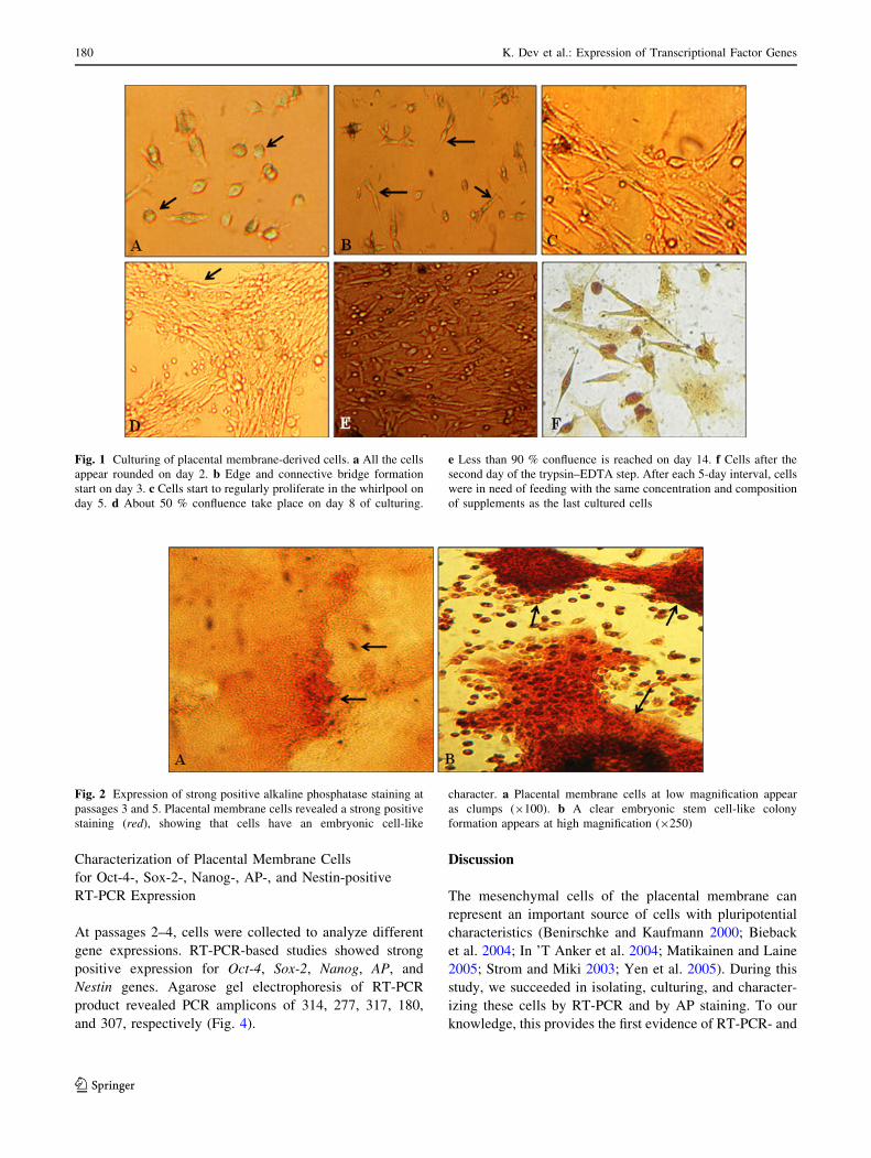
After collection, all the cells were in network form and
were similar in size and shape (Fig. 1). Some of the cells
were rounded and some were edge-pointed after 5 days of
culturing. All the cells displayed anchorage after 9 days of
culturing. After day 14, there was \80 % confluence with
morphologically similar cells (fibroblastic cells).
Instead of forming uniform cell monolayers, cell clumps
were also observed. Initially the cells reached 70–80 %
confluence after 2 weeks. However, the passaged cells
exhibited a higher growth rate, reaching 90–100 % con-
fluence after day 16 of culturing.
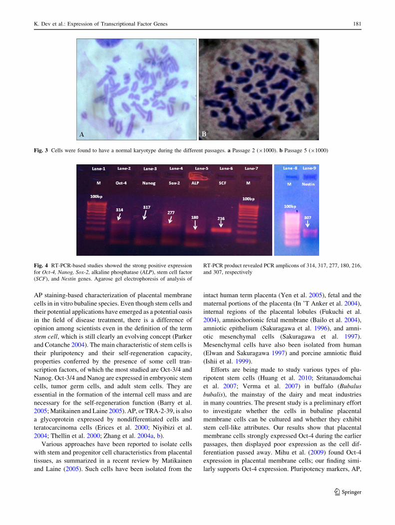
Characterization of Placental Membrane Cells by AP
Staining
The placental cells were found to expand extensively in
fibroblast appearance. The cells were found to stain posi-
tive for AP. Cells showed embryonic stem cell-like cells
properties by AP staining. Whereas the other cell (Pinna)-
cultured fibroblasts did not acquire stain, the cultured cells
stained red and were considered positive for AP expression
(Fig. 2).
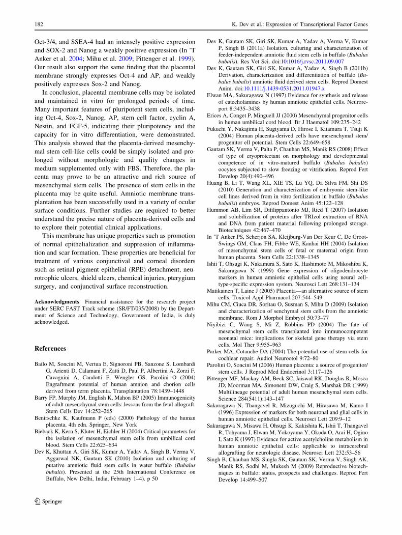
Karyotyping
The cells had a standard karyotype at different passages.
No noticeable genomic aberrations (e.g., appearance of
micronuclei, chromosomal fragmentation) were observed
(Fig. 3).
Table 1 Primer sequences, size of amplification products, and annealing temperatures
Gene Primer sequence Product
size (bp)
Accession
no.
Annealing
temperature (�C)
Octamer binding-4 (Oct-4) CTTCAATCGCATATTCTTTAACCA
GGAGGAAGCTGACAACAACG
314 FI907061 58.0
Nanog GCCCCTTAGTAAGCTGCTTTT
GGGGTGGTGGAAATCAGTAA
317 DQ487022 58.0
Sox-2 AACCAAGACGCTCATGAAGAA
GTACTGCAGGGCGCTCAC
277 EU627692 61.0
Alkaline phosphatase (AP) ACCAATGGCAACCTGCTGTA
CTCCTCCAGGATCTTGGCTA
180 X93604 60.0
Stem cell factor (SCF) TCCCTGCTACCATCCCTATG
GCTTCCCAAATCTGGATCAT
216 AY667192 59.5
Nestin ACC TGC TGT ACA TCG GCT TT
GAGGATGGTGAAGACGGAGA
307 X93604 60.0
K. Dev et al.: Expression of Transcriptional Factor Genes 179
123
Characterization of Placental Membrane Cells
for Oct-4-, Sox-2-, Nanog-, AP-, and Nestin-positive
RT-PCR Expression
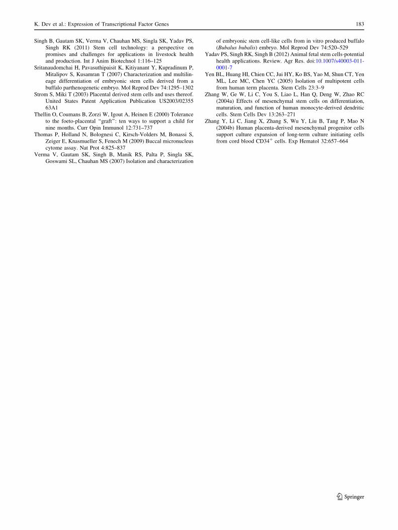
At passages 2–4, cells were collected to analyze different
gene expressions. RT-PCR-based studies showed strong
positive expression for Oct-4, Sox-2, Nanog, AP, and
Nestin genes. Agarose gel electrophoresis of RT-PCR
product revealed PCR amplicons of 314, 277, 317, 180,
and 307, respectively (Fig. 4).
Discussion
The mesenchymal cells of the placental membrane can
represent an important source of cells with pluripotential
characteristics (Benirschke and Kaufmann 2000; Bieback
et al. 2004; In ’T Anker et al. 2004; Matikainen and Laine
2005; Strom and Miki 2003; Yen et al. 2005). During this
study, we succeeded in isolating, culturing, and character-
izing these cells by RT-PCR and by AP staining. To our
knowledge, this provides the first evidence of RT-PCR- and
Fig. 1 Culturing of placental membrane-derived cells. a All the cells
appear rounded on day 2. b Edge and connective bridge formation
start on day 3. c Cells start to regularly proliferate in the whirlpool on
day 5. d About 50 % confluence take place on day 8 of culturing.
e Less than 90 % confluence is reached on day 14. f Cells after the
second day of the trypsin–EDTA step. After each 5-day interval, cells
were in need of feeding with the same concentration and composition
of supplements as the last cultured cells
Fig. 2 Expression of strong positive alkaline phosphatase staining at
passages 3 and 5. Placental membrane cells revealed a strong positive
staining (red), showing that cells have an embryonic cell-like
character. a Placental membrane cells at low magnification appear
as clumps (9100). b A clear embryonic stem cell-like colony
formation appears at high magnification (9250)
180 K. Dev et al.: Expression of Transcriptional Factor Genes
123
AP staining-based characterization of placental membrane
cells in in vitro bubaline species. Even though stem cells and
their potential applications have emerged as a potential oasis
in the field of disease treatment, there is a difference of
opinion among scientists even in the definition of the term
stem cell, which is still clearly an evolving concept (Parker
and Cotanche 2004). The main characteristic of stem cells is
their pluripotency and their self-regeneration capacity,
properties conferred by the presence of some cell tran-
scription factors, of which the most studied are Oct-3/4 and
Nanog. Oct-3/4 and Nanog are expressed in embryonic stem
cells, tumor germ cells, and adult stem cells. They are
essential in the formation of the internal cell mass and are
necessary for the self-regeneration function (Barry et al.
2005; Matikainen and Laine 2005). AP, or TRA-2-39, is also
a glycoprotein expressed by nondifferentiated cells and
teratocarcinoma cells (Erices et al. 2000; Niyibizi et al.
2004; Thellin et al. 2000; Zhang et al. 2004a, b).
Various approaches have been reported to isolate cells
with stem and progenitor cell characteristics from placental
tissues, as summarized in a recent review by Matikainen
and Laine (2005). Such cells have been isolated from the
intact human term placenta (Yen et al. 2005), fetal and the
maternal portions of the placenta (In ’T Anker et al. 2004),
internal regions of the placental lobules (Fukuchi et al.
2004), amniochorionic fetal membrane (Bailo et al. 2004),
amniotic epithelium (Sakuragawa et al. 1996), and amni-
otic mesenchymal cells (Sakuragawa et al. 1997).
Mesenchymal cells have also been isolated from human
(Elwan and Sakuragawa 1997) and porcine amniotic fluid
(Ishii et al. 1999).
Efforts are being made to study various types of plu-
ripotent stem cells (Huang et al. 2010; Sritanaudomchai
et al. 2007; Verma et al. 2007) in buffalo (Bubalus
bubalis), the mainstay of the dairy and meat industries
in many countries. The present study is a preliminary effort
to investigate whether the cells in bubaline placental
membrane cells can be cultured and whether they exhibit
stem cell-like attributes. Our results show that placental
membrane cells strongly expressed Oct-4 during the earlier
passages, then displayed poor expression as the cell dif-
ferentiation passed away. Mihu et al. (2009) found Oct-4
expression in placental membrane cells; our finding simi-
larly supports Oct-4 expression. Pluripotency markers, AP,
Fig. 3 Cells were found to have a normal karyotype during the different passages. a Passage 2 (91000). b Passage 5 (91000)
Fig. 4 RT-PCR-based studies showed the strong positive expression
for Oct-4, Nanog, Sox-2, alkaline phosphatase (ALP), stem cell factor
(SCF), and Nestin genes. Agarose gel electrophoresis of analysis of
RT-PCR product revealed PCR amplicons of 314, 317, 277, 180, 216,
and 307, respectively
K. Dev et al.: Expression of Transcriptional Factor Genes 181
123
Oct-3/4, and SSEA-4 had an intensely positive expression
and SOX-2 and Nanog a weakly positive expression (In ’T
Anker et al. 2004; Mihu et al. 2009; Pittenger et al. 1999).
Our result also support the same finding that the placental
membrane strongly expresses Oct-4 and AP, and weakly
positively expresses Sox-2 and Nanog.
In conclusion, placental membrane cells may be isolated
and maintained in vitro for prolonged periods of time.
Many important features of pluripotent stem cells, includ-
ing Oct-4, Sox-2, Nanog, AP, stem cell factor, cyclin A,
Nestin, and FGF-5, indicating their pluripotency and the
capacity for in vitro differentiation, were demonstrated.
This analysis showed that the placenta-derived mesenchy-
mal stem cell-like cells could be simply isolated and pro-
longed without morphologic and quality changes in
medium supplemented only with FBS. Therefore, the pla-
centa may prove to be an attractive and rich source of
mesenchymal stem cells. The presence of stem cells in the
placenta may be quite useful. Amniotic membrane trans-
plantation has been successfully used in a variety of ocular
surface conditions. Further studies are required to better
understand the precise nature of placenta-derived cells and
to explore their potential clinical applications.
This membrane has unique properties such as promotion
of normal epithelialization and suppression of inflamma-
tion and scar formation. These properties are beneficial for
treatment of various conjunctival and corneal disorders
such as retinal pigment epithelial (RPE) detachment, neu-
rotrophic ulcers, shield ulcers, chemical injuries, pterygium
surgery, and conjunctival surface reconstruction.
Acknowledgments Financial assistance for the research project
under SERC FAST Track scheme (SR/FT/035/2008) by the Depart-
ment of Science and Technology, Government of India, is duly
acknowledged.
References
Bailo M, Soncini M, Vertua E, Signoroni PB, Sanzone S, Lombardi
G, Arienti D, Calamani F, Zatti D, Paul P, Albertini A, Zorzi F,
Cavagnini A, Candotti F, Wengler GS, Parolini O (2004)
Engraftment potential of human amnion and chorion cells
derived from term placenta. Transplantation 78:1439–1448
Barry FP, Murphy JM, English K, Mahon BP (2005) Immunogenicity
of adult mesenchymal stem cells: lessons from the fetal allograft.
Stem Cells Dev 14:252–265
Benirschke K, Kaufmann P (eds) (2000) Pathology of the human
placenta, 4th edn. Springer, New York
Bieback K, Kern S, Kluter H, Eichler H (2004) Critical parameters for
the isolation of mesenchymal stem cells from umbilical cord
blood. Stem Cells 22:625–634
Dev K, Khuttan A, Giri SK, Kumar A, Yadav A, Singh B, Verma V,
Aggarwal NK, Gautam SK (2010) Isolation and culturing of
putative amniotic fluid stem cells in water buffalo (Bubalusbubalis). Presented at the 25th International Conference on
Buffalo, New Delhi, India, February 1–4). p 50
Dev K, Gautam SK, Giri SK, Kumar A, Yadav A, Verma V, Kumar
P, Singh B (2011a) Isolation, culturing and characterization of
feeder-independent amniotic fluid stem cells in buffalo (Bubalusbubalis). Res Vet Sci. doi:10:1016/j.rvsc.2011.09.007
Dev K, Gautam SK, Giri SK, Kumar A, Yadav A, Singh B (2011b)
Derivation, characterization and differentiation of buffalo (Bu-balus bubalis) amniotic fluid derived stem cells. Reprod Domest
Anim. doi:10.1111/j.1439-0531.2011.01947.x
Elwan MA, Sakuragawa N (1997) Evidence for synthesis and release
of catecholamines by human amniotic epithelial cells. Neurore-
port 8:3435–3438
Erices A, Conget P, Minguell JJ (2000) Mesenchymal progenitor cells
in human umbilical cord blood. Br J Haematol 109:235–242
Fukuchi Y, Nakajima H, Sugiyama D, Hirose I, Kitamura T, Tsuji K
(2004) Human placenta-derived cells have mesenchymal stem/
progenitor ell potential. Stem Cells 22:649–658
Gautam SK, Verma V, Palta P, Chauhan MS, Manik RS (2008) Effect
of type of cryoprotectant on morphology and developmental
competence of in vitro-matured buffalo (Bubalus bubalis)
oocytes subjected to slow freezing or vitrification. Reprod Fert
Develop 20(4):490–496
Huang B, Li T, Wang XL, XIE TS, Lu YQ, Da Silva FM, Shi DS
(2010) Generation and characterization of embryonic stem-like
cell lines derived from in vitro fertilization in buffalo (Bubalusbubalis) embryos. Reprod Domest Anim 45:122–128
Hummon AB, Lim SR, Difilippantonio MJ, Ried T (2007) Isolation
and solubilization of proteins after TRIzol extraction of RNA
and DNA from patient material following prolonged storage.
Biotechniques 42:467–470
In ’T Anker PS, Scherjon SA, Kleijburg-Van Der Keur C, De Groot-
Swings GM, Claas FH, Fibbe WE, Kanhai HH (2004) Isolation
of mesenchymal stem cells of fetal or maternal origin from
human placenta. Stem Cells 22:1338–1345
Ishii T, Ohsugi K, Nakamura S, Sato K, Hashimoto M, Mikoshiba K,
Sakuragawa N (1999) Gene expression of oligodendrocyte
markers in human amniotic epithelial cells using neural cell-
type-specific expression system. Neurosci Lett 268:131–134
Matikainen T, Laine J (2005) Placenta—an alternative source of stem
cells. Toxicol Appl Pharmacol 207:544–549
Mihu CM, Ciuca DR, Soritau O, Susman S, Mihu D (2009) Isolation
and characterization of senchymal stem cells from the amniotic
membrane. Rom J Morphol Embryol 50:73–77
Niyibizi C, Wang S, Mi Z, Robbins PD (2004) The fate of
mesenchymal stem cells transplanted into immunocompetent
neonatal mice: implications for skeletal gene therapy via stem
cells. Mol Ther 9:955–963
Parker MA, Cotanche DA (2004) The potential use of stem cells for
cochlear repair. Audiol Neurootol 9:72–80
Parolini O, Soncini M (2006) Human placenta: a source of progenitor/
stem cells. J Reprod Med Endocrinol 3:117–126
Pittenger MF, Mackay AM, Beck SC, Jaiswal RK, Douglas R, Mosca
JD, Moorman MA, Simonetti DW, Craig S, Marshak DR (1999)
Multilineage potential of adult human mesenchymal stem cells.
Science 284(5411):143–147
Sakuragawa N, Thangavel R, Mizuguchi M, Hirasawa M, Kamo I
(1996) Expression of markers for both neuronal and glial cells in
human amniotic epithelial cells. Neurosci Lett 209:9–12
Sakuragawa N, Misawa H, Ohsugi K, Kakishita K, Ishii T, Thangavel
R, Tohyama J, Elwan M, Yokoyama Y, Okuda O, Arai H, Ogino
I, Sato K (1997) Evidence for active acetylcholine metabolism in
human amniotic epithelial cells: applicable to intracerebral
allografting for neurologic disease. Neurosci Lett 232:53–56
Singh B, Chauhan MS, Singla SK, Gautam SK, Verma V, Singh AK,
Manik RS, Sodhi M, Mukesh M (2009) Reproductive biotech-
niques in buffalo: status, prospects and challenges. Reprod Fert
Develop 14:499–507
182 K. Dev et al.: Expression of Transcriptional Factor Genes
123
Singh B, Gautam SK, Verma V, Chauhan MS, Singla SK, Yadav PS,
Singh RK (2011) Stem cell technology: a perspective on
promises and challenges for applications in livestock health
and production. Int J Anim Biotechnol 1:116–125
Sritanaudomchai H, Pavasuthipaisit K, Kitiyanant Y, Kupradinum P,
Mitalipov S, Kusamran T (2007) Characterization and multilin-
eage differentiation of embryonic stem cells derived from a
buffalo parthenogenetic embryo. Mol Reprod Dev 74:1295–1302
Strom S, Miki T (2003) Placental derived stem cells and uses thereof.
United States Patent Application Publication US2003/02355
63A1
Thellin O, Coumans B, Zorzi W, Igout A, Heinen E (2000) Tolerance
to the foeto-placental ‘‘graft’’: ten ways to support a child for
nine months. Curr Opin Immunol 12:731–737
Thomas P, Holland N, Bolognesi C, Kirsch-Volders M, Bonassi S,
Zeiger E, Knasmueller S, Fenech M (2009) Buccal micronucleus
cytome assay. Nat Prot 4:825–837
Verma V, Gautam SK, Singh B, Manik RS, Palta P, Singla SK,
Goswami SL, Chauhan MS (2007) Isolation and characterization
of embryonic stem cell-like cells from in vitro produced buffalo
(Bubalus bubalis) embryo. Mol Reprod Dev 74:520–529
Yadav PS, Singh RK, Singh B (2012) Animal fetal stem cells-potential
health applications. Review. Agr Res. doi:10.1007/s40003-011-
0001-7
Yen BL, Huang HI, Chien CC, Jui HY, Ko BS, Yao M, Shun CT, Yen
ML, Lee MC, Chen YC (2005) Isolation of multipotent cells
from human term placenta. Stem Cells 23:3–9
Zhang W, Ge W, Li C, You S, Liao L, Han Q, Deng W, Zhao RC
(2004a) Effects of mesenchymal stem cells on differentiation,
maturation, and function of human monocyte-derived dendritic
cells. Stem Cells Dev 13:263–271
Zhang Y, Li C, Jiang X, Zhang S, Wu Y, Liu B, Tang P, Mao N
(2004b) Human placenta-derived mesenchymal progenitor cells
support culture expansion of long-term culture initiating cells
from cord blood CD34? cells. Exp Hematol 32:657–664
K. Dev et al.: Expression of Transcriptional Factor Genes 183
123
![Hepatobiliary diseases in buffalo ( Bubalus bubalis ... · diseases of internal organs, including hepatic diseases in buffalo under field conditions [3,5]. A complete ultrasonographic](https://img.pdfslide.us/doc/110x75/5eb4d20a1ae6da71cd66ea30/hepatobiliary-diseases-in-buffalo-bubalus-bubalis-diseases-of-internal-organs.jpg)