Embed Size (px)
Citation preview

J. Pathol. 189: 66–71 (1999)
EXPRESSION AND LOCALIZATION OF PLACENTAGROWTH FACTOR AND PlGF RECEPTORS IN HUMAN
MENINGIOMAS
1, . 2, . 2 . 1*1Department of Gene Regulation and Differentiation, National Research Center for Biotechnology (GBF),
38124 Braunschweig, Germany2Department of Neuropathology, Neurocenter, Freiburg University Medical School, Breisacherstrasse 64, 79106 Freiburg, Germany
SUMMARY
It has previously been suggested that in human brain tumours, endothelial cell proliferation during angiogenesis is regulated by aparacrine mechanism involving vascular endothelial growth factor (VEGF) and its receptors (VEGF receptor 1 and VEGF receptor 2).The mechanism of growth factor up-regulation is based on hypoxic activation of mRNA expression and mRNA stabilization and geneticevents, leading to an increase of growth factor gene expression. The role of the other newly discovered VEGF family members with ahigh specificity for endothelial cells in the pathogenesis of glial neoplasms is unknown. To investigate which other members of the VEGFfamily are overexpressed in human brain tumours, the mRNA levels of placenta growth factor (PlGF), VEGF-A, and VEGF-B geneswere determined by northern blot analysis in surgically obtained human meningiomas. In the 16 meningiomas examined, the mRNA forPlGF was highly expressed in four tumours and VEGF-A mRNA was highly abundant in three tumour samples. There was no closecorrelation between PlGF mRNA levels and VEGF-A expression levels. VEGF-B mRNA was abundantly expressed in all tumoursamples at uniform levels. In a PlGF-positive tumour sample, immunoreactive VEGFR-1 and VEGFR-2 were detected in endothelialcells of the blood vessels. PlGF protein was detectable in most but not all capillaries of the tumour. PlGF is thus highly up-regulated ina subset of human meningiomas and may therefore have functions, in some tumour vessels, connected to endothelial cell maturation andtube formation. These findings suggest that PlGF, in addition to VEGF-A, may be another positive factor in tumour angiogenesis inhuman meningiomas. Copyright ? 1999 John Wiley & Sons, Ltd.
KEY WORDS—placenta growth factor (PlGF); vascular endothelial growth factor (VEGF); receptors; meningiomas; angiogenesis; braintumours
*Correspondence to: Herbert A. Weich, Dept. RD1F/GBF,Mascheroder Weg 1b, D-38124 Braunschweig, Germany.E-mail: [email protected]
Contract/grant sponsor: EU Biomed II Program; Contract/grantnumber: PL 950669.
Contract/grant sponsor: Deutsche Krebshilfe; Contract/grant
INTRODUCTION
The progression and growth of solid tumours aredependent on the formation of new blood vessels, aprocess called tumour angiogenesis, which is regulatedby growth factors that are secreted by tumour cells andoften act specifically on vascular endothelial cells.1 Wehave previously reported that vascular endothelialgrowth factor (VEGF-A) is an angiogenesis factor inbrain tumours and mediates tumour vascularizationin vivo.2 VEGF and its high affinity receptors (VEGFR-1and VEGFR-2) are expressed in normal brain at lowlevels, but are up-regulated up to 50-fold in tumourtissues.3,4 These observations have strongly supportedthe concept that vascularization in brain tumours isregulated by paracrine mechanisms, VEGF-A being akey molecule for this process.
Another member of the VEGF growth factor family isplacenta growth factor (PlGF), a dimeric glycoprotein
number: 10-1302-Ri3.
CCC 0022–3417/99/100066–06$17.50Copyright ? 1999 John Wiley & Sons, Ltd.
with 53 per cent homology to VEGF-A.5,6 PlGF bindsto only one of the two receptors, namely VEGFR-1.7PlGF is chemotactic for monocytes and therefore activein signal transduction, but it is not angiogenic in thechicken CAM assay.8,9 However, it was very recentlyreported that PlGF-1, a non-heparin-binding splice formof PlGF, is also angiogenic in vivo in the rabbit corneapocket assay.10 Recently, three new members of theVEGF family were described as VEGF-B, VEGF-C,and VEGF-D (for a review see ref. 11). Whereas the roleof VEGF-A in tumour development has been welldocumented, few data have been reported for the role ofPlGF or of VEGF-B and the other new members intumour-associated angiogenesis.
Besides our description of PlGF overexpression insome brain tumours,4 PlGF expression has beenreported in hypervascular renal cell carcinomas and insome thyroid and germ-cell tumours.12 PlGF is alsoup-regulated in fetal growth retardation.13 In contrast toVEGF-A, PlGF and VEGF-B are not regulated byhypoxia and their physiological roles are largelyunknown.11 PlGF is highly expressed in placenta14 andits expression may be sensitive to steroid hormones.Because meningioma growth has been reported to besteroid-dependent,15 we investigated in this studywhether PlGF and other members of the VEGF familyare up-regulated in human meningiomas.
Received 22 July 1998Revised 1 December 1998
Accepted 14 April 1999
67PlGF EXPRESSION IN MENINGIOMAS
MATERIALS AND METHODS
Tissue specimens
Sixteen cases of intracerebral meningioma classifiedaccording to the WHO classification (1993) wereincluded in this study. Tumour specimens were receiveddirectly from the neurosurgical theatre. Part of thespecimen was fixed in 4 per cent buffered formalin,embedded in paraffin, and processed for routine histo-logical diagnosis. An adjacent part of the tissue wassnap-frozen in liquid nitrogen and stored at "70)Cprior to use.
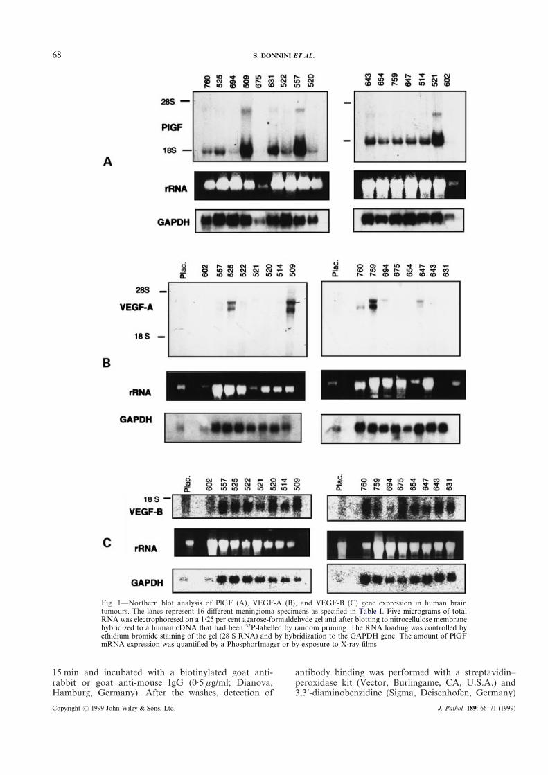
Probes, RNA isolation, and northern blotting
The human VEGF-A probe was a fragment of 0·7 kb,generated as previously described.4 Human VEGF-BcDNA was a gift from Drs Kari Alitalo and UlfEriksson.16 PlGF cDNA was cloned from humanplacenta as described earlier.6 Total RNA isolationand northern blotting were performed as before.4 Theblots were analysed by a PhosphorImager (MolecularDynamics, Sunnyvale, CA, U.S.A.) and/or exposed toKodak XAR films with an intensifying screen at "70)Cover 2–7 days. Ribosomal RNA bands were indicated assize markers. For control of RNA loading, the blotswere stripped and rehybridized with a 32P-labelledGAPDH cDNA probe.
Antibodies
Affinity-purified PlGF antibodies were obtained fromimmune rabbit serum and from protein-A purified total
Copyright ? 1999 John Wiley & Sons, Ltd.
IgG. The antibodies were raised against a peptide con-jugated to KLH, containing the first 20 NH3-terminalamino acids of human PlGF protein. The affinity puri-fication of the PlGF antibodies was very similar to thatdescribed for the VEGF antibodies.2 The generation ofmouse monoclonal antibodies against VEGFR-1 andVEGFR-2 has been described in detail.17 The mouseclones KDR-1 and FLT-19 were used in this study. Themonoclonal antibodies did not cross-react with eachother or with the related soluble extracellular FLT-4protein (a gift from Dr Kari Alitalo), nor with solublePDGF-âR proteins (a gift from Dr Michael Pech). Forimmunostaining, ascites fluid diluted 1:50 was used.
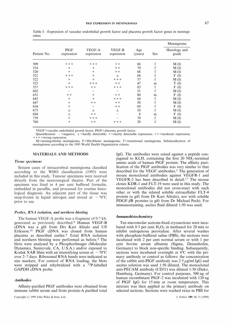
Table I—Expression of vascular endothelial growth factor and placenta growth factor genes in meningi-omas
Patient No.PlGF
expressionVEGF-Aexpression
VEGF-Bexpression
Age(years) Sex
Meningioma
Histology andgrade
509 + + + + + + + + 66 f M (I)514 + + + + 70 f M (I)520 + + + + 68 f M (I)521 + + + + & 68 f F (I)522 + + + + + 57 f M (I)525 + + + + + + 47 m T (I)557 + + + + + + + + 83 f F (I)602 " " " 51 f M (I)631 + + + + + 80 m F (I)643 + + + + + 64 f M (I)647 + + + + + 58 f M (I)654 + " + + 69 f F (I)675 & & & 10 f M (I)694 & + + " 8 m F (I)759 + + + + " 70 f M (I)760 + + + + + + 29 f M (I)
VEGF=vascular endothelial growth factor; PlGF=placenta growth factor.Quantification: " =negative; & =hardly detectable; + =clearly detectable expression; + + =moderate expression;
+ + + =strong expression.M=meningothelial meningioma; F=fibroblastic meningioma; T=transitional meningioma. Subclassification of
meningiomas according to the 1993 World Health Organization criteria.
Immunohistochemistry
Ten micromolar acetone-fixed cryosections were incu-bated with 0·3 per cent H2O2 in methanol for 20 min toinhibit endogenous peroxidase. After several washeswith phosphate-buffered saline (PBS), the sections wereincubated with 2 per cent normal serum or with 1 percent bovine serum albumin (Sigma, Deisenhofen,Germany) to block non-specific binding. Subsequently,sections were incubated overnight at 4)C with the pri-mary antibody or control as follows: the concentrationof the rabbit anti-PlGF antibody was 2·5 ìg/ml IgG andascites solution was used 1:50 diluted. The monoclonalanti-PECAM antibody (CD31) was diluted 1:30 (Dako,Hamburg, Germany). For control purposes, 500 ng ofhuman recombinant PlGF-2 was incubated with 120 ngof PlGF IgG for 15 min at room temperature. Thismixture was then applied as the primary antibody onselected sections. Sections were washed twice in PBS for
J. Pathol. 189: 66–71 (1999)
68 S. DONNINI ET AL.
Fig. 1—Northern blot analysis of PlGF (A), VEGF-A (B), and VEGF-B (C) gene expression in human braintumours. The lanes represent 16 different meningioma specimens as specified in Table I. Five micrograms of totalRNA was electrophoresed on a 1·25 per cent agarose-formaldehyde gel and after blotting to nitrocellulose membranehybridized to a human cDNA that had been 32P-labelled by random priming. The RNA loading was controlled byethidium bromide staining of the gel (28 S RNA) and by hybridization to the GAPDH gene. The amount of PlGFmRNA expression was quantified by a PhosphorImager or by exposure to X-ray films
15 min and incubated with a biotinylated goat anti-rabbit or goat anti-mouse IgG (0·5 ìg/ml; Dianova,Hamburg, Germany). After the washes, detection of
Copyright ? 1999 John Wiley & Sons, Ltd.
antibody binding was performed with a streptavidin–peroxidase kit (Vector, Burlingame, CA, U.S.A.) and3,3*-diaminobenzidine (Sigma, Deisenhofen, Germany)
J. Pathol. 189: 66–71 (1999)
69PlGF EXPRESSION IN MENINGIOMAS
Copyright ? 1999 John Wiley & Sons, Ltd.
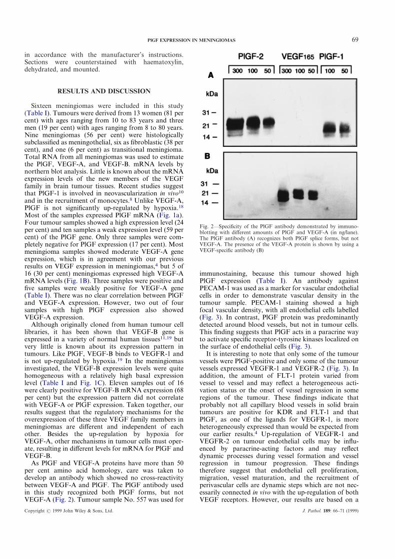
Fig. 2—Specificity of the PlGF antibody demonstrated by immuno-blotting with different amounts of PlGF and VEGF-A (in ng/lane).The PlGF antibody (A) recognizes both PlGF splice forms, but notVEGF-A. The presence of the VEGF-A protein is shown by using aVEGF-specific antibody (B)
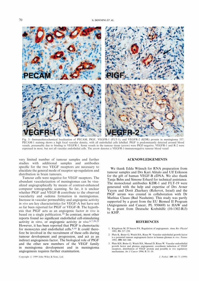
immunostaining, because this tumour showed highPlGF expression (Table I). An antibody againstPECAM-1 was used as a marker for vascular endothelialcells in order to demonstrate vascular density in thetumour sample. PECAM-1 staining showed a highfocal vascular density, with all endothelial cells labelled(Fig. 3). In contrast, PlGF protein was predominantlydetected around blood vessels, but not in tumour cells.This finding suggests that PlGF acts in a paracrine wayto activate specific receptor-tyrosine kinases localized onthe surface of endothelial cells (Fig. 3).
It is interesting to note that only some of the tumourvessels were PlGF-positive and only some of the tumourvessels expressed VEGFR-1 and VEGFR-2 (Fig. 3). Inaddition, the amount of FLT-1 protein varied fromvessel to vessel and may reflect a heterogeneous acti-vation status or the onset of vessel regression in someregions of the tumour. These findings indicate thatprobably not all capillary blood vessels in solid braintumours are positive for KDR and FLT-1 and thatPlGF, as one of the ligands for VEGFR-1, is moreheterogeneously expressed than would be expected fromour earlier results.4 Up-regulation of VEGFR-1 andVEGFR-2 on tumour endothelial cells may be influ-enced by paracrine-acting factors and may reflectdynamic processes during vessel formation and vesselregression in tumour progression. These findingstherefore suggest that endothelial cell proliferation,migration, vessel maturation, and the recruitment ofperivascular cells are dynamic steps which are not nec-essarily connected in vivo with the up-regulation of bothVEGF receptors. However, our results are based on a
in accordance with the manufacturer’s instructions.Sections were counterstained with haematoxylin,dehydrated, and mounted.
RESULTS AND DISCUSSION
Sixteen meningiomas were included in this study(Table I). Tumours were derived from 13 women (81 percent) with ages ranging from 10 to 83 years and threemen (19 per cent) with ages ranging from 8 to 80 years.Nine meningiomas (56 per cent) were histologicallysubclassified as meningothelial, six as fibroblastic (38 percent), and one (6 per cent) as transitional meningioma.Total RNA from all meningiomas was used to estimatethe PlGF, VEGF-A, and VEGF-B. mRNA levels bynorthern blot analysis. Little is known about the mRNAexpression levels of the new members of the VEGFfamily in brain tumour tissues. Recent studies suggestthat PlGF-1 is involved in neovascularization in vivo10
and in the recruitment of monocytes.8 Unlike VEGF-A,PlGF is not significantly up-regulated by hypoxia.18
Most of the samples expressed PlGF mRNA (Fig. 1a).Four tumour samples showed a high expression level (24per cent) and ten samples a weak expression level (59 percent) of the PlGF gene. Only three samples were com-pletely negative for PlGF expression (17 per cent). Mostmeningioma samples showed moderate VEGF-A geneexpression, which is in agreement with our previousresults on VEGF expression in meningiomas,4 but 5 of16 (30 per cent) meningiomas expressed high VEGF-AmRNA levels (Fig. 1B). Three samples were positive andfive samples were weakly positive for VEGF-A gene(Table I). There was no clear correlation between PlGFand VEGF-A expression. However, two out of foursamples with high PlGF expression also showedVEGF-A expression.
Although originally cloned from human tumour celllibraries, it has been shown that VEGF-B gene isexpressed in a variety of normal human tissues11,19 butvery little is known about its expression pattern intumours. Like PlGF, VEGF-B binds to VEGFR-1 andis not up-regulated by hypoxia.19 In the meningiomasinvestigated, the VEGF-B expression levels were quitehomogeneous with a relatively high basal expressionlevel (Table I and Fig. 1C). Eleven samples out of 16were clearly positive for VEGF-B mRNA expression (68per cent) but the expression pattern did not correlatewith VEGF-A or PlGF expression. Taken together, ourresults suggest that the regulatory mechanisms for theoverexpression of these three VEGF family members inmeningiomas are different and independent of eachother. Besides the up-regulation by hypoxia forVEGF-A, other mechanisms in tumour cells must oper-ate, resulting in different levels for mRNA for PlGF andVEGF-B.
As PlGF and VEGF-A proteins have more than 50per cent amino acid homology, care was taken todevelop an antibody which showed no cross-reactivitybetween VEGF-A and PlGF. The PlGF antibody usedin this study recognized both PlGF forms, but notVEGF-A (Fig. 2). Tumour sample No. 557 was used for
J. Pathol. 189: 66–71 (1999)
70 S. DONNINI ET AL.
Copyright ? 1999 John Wiley & Sons, Ltd.
ACKNOWLEDGEMENTS
We thank Edda Wunsch for RNA preparation fromtumour samples and Drs Kari Alitalo and Ulf Erikssonfor the gift of human VEGF-B cDNA. We also thankTanja Behn and Simone Erhard for technical assistance.The monoclonal antibodies KDR-1 and FLT-19 weregenerated with the help and expertise of Drs AvnerYayon and Dorit Zharhary (Rehovot, Israel) and thePlGF serum was created in collaboration with DrMathias Clauss (Bad Nauheim). This study was partlysupported by a grant from the EU Biomed II Program(Angiogenesis and Cancer, PL 950669) to HAW andby a grant from Deutsche Krebshilfe (10-1302-Ri3)to KHP.
Fig. 3—Immunohistochemical localization of PECAM, PlGF, VEGFR-1 (FLT-1), and VEGFR-2 (KDR) protein in meningioma 557.PECAM-1 staining shows a high focal vascular density, with all endothelial cells labelled. PlGF is predominantly detected around bloodvessels, presumably due to binding to VEGFR-1. Some vessels in the tumour tissue (arrow) were PlGF-negative. VEGFR-1 and R-2 wereexpressed in most, but not all vascular endothelial cells. The arrow denotes a VEGFR-1-immunonegative tumour blood vessel
REFERENCES
1. Klagsbrun M, D’Amore PA. Regulation of angiogenesis. Annu Rev Physiol1991; 53: 217–239.
2. Plate K, Breier G, Weich HA, Risau W. Vascular endothelial growth factoris a potential tumour angiogenesis factor in human gliomas in vivo. Nature1992; 359: 845–848.
3. Plate KH, Breier G, Weich HA, Mennel D, Risau W. Vascular endoethelialgrowth factor and gliomas angiogenesis: coordinate induction of VEGFreceptors, distribution of VEGF protein and possible in vivo regulatorymechanisms. Int J Cancer 1994; 5: 51–10.
very limited number of tumour samples and furtherstudies with additional samples and antibodiesspecific for the two VEGF receptors are necessary toelucidate the general mode of receptor up-regulation anddistribution in brain tumours.
Tumour cells were negative for VEGF receptors. Theabundant vascularization of meningiomas can be visu-alized angiographically by means of contrast-enhancedcomputer tomographic scanning. So far, it is unclearwhether PlGF and VEGF-B contribute to the observedvascularity and oedema formation in meningiomas.Increase in vascular permeability and angiogenic activityin vivo are key characteristics for VEGF-A but have notso far been reported for PlGF or VEGF-B. The hypoth-esis that PlGF acts as an angiogenic factor in vivo isbased on a single publication.10 In contrast, most otherreports found no significant endothelial cell-stimulatingactivity in vitro, or angiogenic activity in vivo.6,7,20,21
However, it has been reported that PlGF is chemotacticfor monocytes and endothelial cells.8,10 It could there-fore be involved in the recruitment of these cells duringtumour development and progression, and act as anindirect angiogenesis factor. The biological role of PlGFand the other new members of the VEGF familyin meningioma development and in meningiomaangiogenesis requires further examination.
J. Pathol. 189: 66–71 (1999)
71PlGF EXPRESSION IN MENINGIOMAS
4. Weindel K, Moringlane JR, Marme D, Weich HA. Detection and quanti-fication of vascular endothelial growth factor/vascular permeability factorin brain tumour tissue and cyst fluid: the key to angiogenesis? Neurosurgery1994; 35: 439–449.
5. Maglione D, Guerriero V, Viglietto G, Delli-Bovi P, Persico MG. Isolationof a human placenta cDNA coding for a protein related to the vascularpermeability factor. Proc Natl Acad Sci U S A 1991; 88: 9267–9271.
6. Hauser S, Weich HA. A heparin-binding from of placenta growth factor(PlGF-2) is expressed in human umbilical vein endothelial cells and inplacenta. Growth Factors 1993; 9: 259–268.
7. Park JEC, Ferrara N. Placenta growth factor. J Biol Chem 1994; 269:25 646–25 654.
8. Clauss M, Weich HA, Breier G, et al. The vascular endothelial growthfactor receptor flt-1 mediates biological activities. J Biol Chem 1996; 271:17 629–17 634.
9. Birkenhager R, Schneppe B, Rockl W, et al. Synthesis and physiologicalactivity of heterodimers comprising different splice-forms of vascularendothelial growth factor and placenta growth factor. Biochem J 1996;316: 703–707.
10. Ziche M, Maglione D, Ribatti D, et al. Placenta growth factor-1 ischemotactic, mitogenic, and angiogenic. Lab Invest 1997; 76: 517–531.
11. Joukov V, Kaipainen A, Jeltsch M, et al. Vascular endothelial growthfactors VEGF-B and VEGF-C. J Cell Physiol 1997; 173: 211–215.
12. Takahashi A, Sasaki H, Tobisu K, et al. Markly increased amount ofmessenger RNAs for vascular endothelial growth factor and placentagrowth factor in renal cell carcinoma associated with angiogenesis. CancerRes 1994; 54: 4233–4237.
Copyright ? 1999 John Wiley & Sons, Ltd.
13. Khaliq A, Li XF, Shams M, et al. Localisation of placenta growth factor(PlGF) in human term placenta. Growth Factors 1996; 13: 243–250.
14. Khaliq A, Dunk C, Jiang J, et al. Hypoxia down-regulates placenta growthfactor (PlGF) while fetal growth restriction upregulates PlGF expression:molecular evidence for ‘placental hyperoxia’ in IURG. Lab Invest 1999; 79:151–170.
15. Speirs V, Boyle-Walsh E, Fraser WD. Constitutive co-expression of estro-gen and progesterone receptor mRNA in human meningiomas by RT-PCRand response of in vitro cell cultures to steroid hormones. Int J Cancer 1997;72: 714–719.
16. Olofsson B, Pajusola K, Kaipanen A, et al. Vascular endothelial growthfactor B, a novel growth factor for endothelial cells. Proc Natl Acad SciU S A 1996; 93: 2576–2581.
17. Simon M, Rockl W, Hornig C, et al. Receptors of vascular endothelialgrowth factor, vascular permeability factor (VEGF/VPF) in renal ontogen-esis: localization and activity. J Am Soc Nephrol 1998; 9: 1032–1044.
18. Gleadle JM, Ebert BL, Firth JD, Rathcliffe PJ. Regulation of angiogenicgrowth factor expression by hypoxia, transition metals, and chelatingagents. Am J Physiol 1995; 268: C1362–C1368.
19. Enholm B, Paavonen K, Ristimake A, et al. Comparison of VEGF,VEGF-B, VEGF-C and Ang-1 mRNA regulation by serum, growth factors,oncoproteins and hypoxia. Oncogene 1997; 14: 2475–2483.
20. Oh SJ, Jeltsch MM, Birkenhaeger R, et al. VEGF and VEGF-C: specificinduction of angiogenesis and lymphangiogenesis in the differentiated avianchorioallantoic membrane. Dev Biol 1997; 188: 96–109.
21. Cao Y, Linden P, Shima D, Browne F, Folkman J. In vivo angiogenicactivity and hypoxia induction of heterodimers of placenta growth factor/vascular endothelial growth factor. J Clin Invest 1996; 98: 2507–2511.
J. Pathol. 189: 66–71 (1999)


![Amino acid transporter LAT1 in tumor-associated vascular ...patients with bevacizumab, an anti-VEGF antibody, increased the PlGF in plasma [3]. FGF-2 and PlGF were increased in glioblastoma](https://img.pdfslide.us/doc/110x75/609eaca54cba15523b358952/amino-acid-transporter-lat1-in-tumor-associated-vascular-patients-with-bevacizumab.jpg)