Embed Size (px)
Citation preview
406
Biochimica et Biophysica Acta, 504 (1978) 406--412 © Elsevier/North-Holland Biomedical Press
BBA 47611
EXCITATION ENERGY TRANSFER BETWEEN PIGMENT SYSTEM II UNITS IN BLUE-GREEN ALGAE
MAMORU MIMURO and YOSHIHIKO FUJITA
Ocean Research Institute, University of Tokyo, Nakano, Tokyo 164 (Japan)
(Received November 29th, 1977) (Revised manuscript received May 2nd, 1978)
Summary
Efficiency in excitation energy transfer from closed to open reaction center II in blue-green and red algae was estimated by the method developed by Joliot and Joliot (C.R. Acad. Sci. (1964) 258, 4622--4625) after slight modification; the number of open reaction centers II was counted from the mean 02 yield of repetitive short flashes.
The efficiency in energy transfer in Chlorella pyrenoidosa was the same in our measurement as that reported by Joliot and Joliot (0.55 + 0.02). However, the values obtained with four blue-green algae and one red alga were very small, in a range of 0.00--0.07. The low efficiency was always obtained independently of the size of the apparent photosynthetic unit which was varied by growth conditions. Results indicated that pigment system II forms a unit in which only one reaction center II is operative.
Introduction
Photosynthetic pigment system of blue-green and red algae consists of chlorophyll a, phycobilins and carotenoids. Phycobilins act as a major compo- nent of pigment system II. However, their location in the photosynthetic orga- nelles is different from that of chlorophyll a and carotenoids. Phycobilins are bound to hydrophilic proteins. Electron microscopic analyses have revealed that phycobiliproteins in a highly associated form are located on the surface of thylakoids; each unit of the protein associates takes a granular structure (phycobilisome) and is arranged at a regular distance (40--50 nm) on the thyl- akoid surface [ 1 ].
We found that the size of one phycobilisome is almost equal to the phyco-
Abbreviation: DCMU, 3-(3,4-dichlorophenyl)-l,l-dimethylurea.
4 0 7
bilin size per one reaction center II [ 2 ]. This suggests that together with chloro- phyll a in thylakoids, each phycobilisome forms a unit of pigment system II. In blue-green and red algae, the amount of pigment system II chlorophyll a is very small (approx. 10% of total chlorophyll a in Anabaena variabilis [2] 16% of Anacystis nidulans [3] and approx. 5% of Porphyridium cruentum [4]). Our estimate indicated that in A. variabilis, only 20 molecules of chlorophyll a function in one pigment system II unit [2]. Pigment system II chlorophyll a is probably located only at the thylakoid locus where phycobilisome resides. These features suggest that in blue-green and red algae, pigment system II units are isolated in the excitation energy transfer between them. To examine this possibility, we tried to estimate the efficiency in excitation energy transfer between pigment system II units in blue-green and red algae. Results reported here indicate that pigment system II units in these algae show a typical separate package nature in the energy transfer between them.
Materials and Methods
Algal culture. Algal strains used in the present experiments were originally supplied from the Algal Collection at the Institute of Applied Microbiology, University of Tokyo [5]. Cells were grown autotrophically under continuous illumination of incandescent light at 26°C. Air containing 0.5% CO2 was con- tinuously supplied. Algal strains, growth conditions and specific growth rates are summarized in Table I. Anabaena cylindrica was grown under N2-fixing conditions with modified Detmer's medium [5] free from KNO3, and P. cruentum was grown in artificial seawater medium of Provasoli [6] enriched with 2-fold NaNO3 and 5-fold K2HPO4. Cells at the late log-growth phase were used in all experiments.
Polarographic measurements. Cells washed with a fresh culture medium were suspended in the fresh medium at a concentration equivalent to 3 ug chloro- phyll a/ml. In our experimental procedure described below, this algal concen-
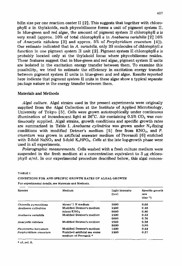
T A B L E I
C O N D I T I O N S F O R A N D SPECIFIC G R O W T H R A T E S OF A L G A L G R O W T H
For e x p e r i m e n t a l detai ls , see Materials and M e t h o d s .
Spec ie s M e d i u m Light in t ens i ty ( lux)
Spec i f i c g r o w t h rate (day -~)
Chlorella p y r e n o i d o s a Myers ' 1 N m e d i u m 3 0 0 0 Anabaena cy l indr ica M o d i f i e d D e t m e r ' s m e d i u m 1500
m i n u s K N O 3 6 0 0 0 Anabaena variabilis M o d i f i e d D e t m e r ' s m e d i u m 1500
6 0 0 0 A n a c y s t i s nidulans Modi f i ed D e t m e r ' s m e d i u m 1500
6 0 0 0 P l e c t o n e m a boryanurn M o d i f i e d D e t m e r ' s m e d i u m 1500 Porphyr id iurn c r u e n t u m Enriched artif icial sea w a t e r 1500
m e d i u m o f Provasol i *
0 .66 0 . 3 8 0 .80 0 .43 0 .76 0 . 5 8 0 .90 0 .54 0 .37
* cf . ref . 6.
408
tration gave 40--50% (in most cases, 42--45%) light absorption at the wave- length for phycobilin peak. Until measurements were taken cell suspensions were kept under the same light conditions as the algal culture.
O2 evolution was measured by a Teflon-covered oxygen electrode (YSI 4004) in a closed cylindrical vessel (20 mm height and 12 mm diameter; total volume, 2 ml) with two light sources at 26°C. The one was xenofi short flash. Flash source was a Sugawara Storoboscope MS-230. The flash duration was 4.4 us at 1/2. An ultraviolet-cut filter (Toshiba UV-39) was inserted between the electrode vessel and the flash source. Heat irradiance was cut by a lucite block (10 mm thickness). The other was an I2-W lamp light (Ushio 5V 150W E.H.). Heat irradiance was removed by an infrared cut filter and a lucite block (10 mm thickness). The light intensity was varied by a neutral density filter (Hoya Glass ND series). The flash source gave an intensity of at least 3.8 • 103 erg/cm 2 per flash at the surface of the electrode vessel; an actual intensity must be higher than the above value because we set a concave mirror on the opposite side of the flash source so as to give focusing of the reflecting light at the electrode vessel. This flash intensity gave an estimation that far more than 95% of targets (reaction centers I and II) can be hit by each flash, even when we count only the light absorbed by phycobilins which are present in vivo as a phycobil isome unit (30 nm in diameter, cf. ref. 1). The estimate was experi- mentally proved by the relationship between 02 flash yield and flash intensity (Kawamura, M., Mimuro, M. and Fujita, Y., in preparation).
Maximum 02 flash yield was obtained for Chlorella pyrenoidosa at a repeti- tion rate as high as 20 Hz in our measuring system. However, we found that the maximum 02 flash yield was obtained only at a repetition rate as low as 10 Hz in all blue-green and red algae, and thus, we applied repetitive flashes at 5 Hz to all measurements. Details of condit ions for our repetitive flash experiments will be reported elsewhere (Kawamura, M., Mimuro, M. and Fujita, Y., in prepara- tion).
Cells placed in the vessel were kept in the dark for 7 min for the dark stan- dardization and the temperature equilibration. After that, repetitive flashes for 3 min and cont inuous light for 3 min were alternately given with a 5 min dark interval.
The efficiency (p) in excitation energy transfer from closed to open reaction center II can be obtained from Joliot 's equation between a steady-state and light-limiting photosynthesis (V) and a fraction of open reaction center II (e), V/Vmax = e/1 - - p ( 1 - - e ) where Vma x is the rate of steady state photosynthesis under maximum number of open reaction centers II [7]. Joliot, Jol iot and Kok [8] reported that the dark reaction between two reaction centers in spinach chloroplasts and Chlorella cells has a low apparent equilibrium constant {K). Thus, the 02 flash yield does not necessarily count the total number of reaction centers II. In the experiments of Jol iot and Jol iot [7] and Jol iot [9], the frac- tion of open reaction centers II was estimated from the 02 yield of a single short flash superimposed on a weak continuous light, and thus, the number of open reaction centers II under cont inuous illumination can be equal to that under the flash illumination. Our method consists of alternate measurements of 02 evolution under cont inuous and repetitive flash illumination. Thus, the fraction of open reaction centers II is not necessarily equal under the two,
409
steady-state and quasi-steady state, condi t ions . The white light used for the cont inuous illumination had the color temperature of 3400 K; the energy distribution in a visible wavelength region indicates that photons absorbed by phycobilins and transferred to pigment system II chlorophyll a are a little larger (1.1--1.4 times) than those absorbed by pigment system I chlorophyll a. The light quanta must be equally distributed to pigment systems I and II, so that the fraction of open reaction centers II under the cont inuous illumination can approximate that under the repetitive short flashes.
Chlorophyll content was determined with acetone extracts with use of the absorption coefficient of Mackinney [10].
Results and Discussion
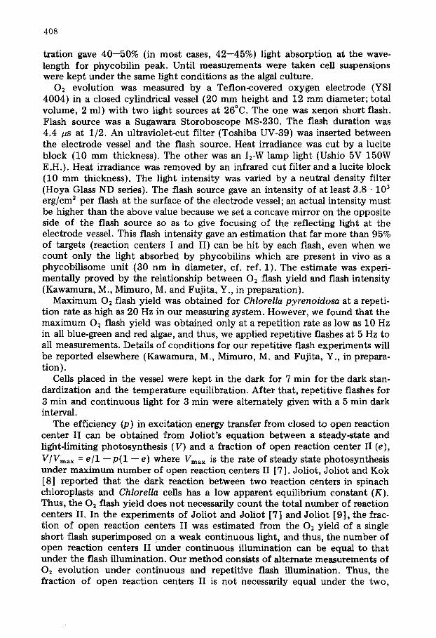
Fig. 1 shows relative rates of steady-state photosynthesis in C. pyrenoidosa as a function of relative number of open reaction centers II. A varied propor- tion of open reaction centers II was set by addition of 3-(3,4<lichlorophenyl)- 1,1~limethylurea (DCMU) at various concentrations, and expressed as a value relative to that wi thout DCMU addition. The pattern is identical to that reported by Jol iot and Joliot [7] and Joliot [9]. The p calculated by the least- square method is 0.55 + 0.02, in good agreement with the value reported by Joliot and Joliot [7]. Our method is valid for the experiment for green algal system.
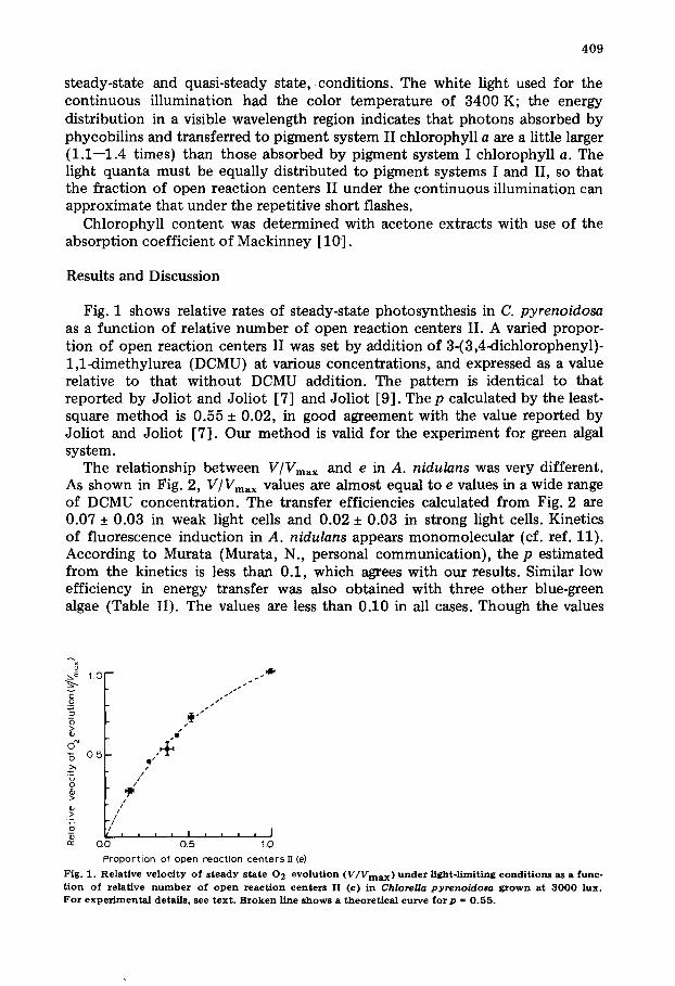
The relationship between V/Vmax and e in A. nidulans was very different. As shown in Fig. 2, V/Vmax values are almost equal to e values in a wide range of DCMU concentration. The transfer efficiencies calculated from Fig. 2 are 0.07 + 0.03 in weak light cells and 0.02 + 0.03 in strong light cells. Kinetics of fluorescence induction in A. nidulans appears monomolecular (cf. ref. 11). According to Murata (Murata, N., personal communicat ion), the p estimated from the kinetics is less than 0.1, which agrees with our results. Similar low efficiency in energy transfer was also obtained with three other blue-green algae (Table II). The values are less than 0.10 in all cases. Though the values
~.E 1 0
E _o 5 ~>
"5 o.5
_o
#i I
eO
.+ O I ¢
, J
f
I ,+
0.0 0.5 1.0
Propor t ion of open react ion cen te rs fT (e)
Fig . I . Relat ive v e l o c i t y o f s t e a d y s tate 0 2 e v o l u t i o n (V/Vmax) under Hght- l imit ing c o n d i t i o n s as a f u n c -
t i o n o f re lat ive n u m b e r o f o p e n reac t ion c e n te r s II ( e ) in Chlorella pyrenoidosa g r o w n at 3 0 0 0 l u x . For e x p e r i m e n t a l detai ls , see t e x t . B r o k e n l ine s h o w s a th e ore t i ca l curve f o r p = 0 . 5 5 .
410
.o
o" "6
>~ >~ o
r~
1 . 0 - -
0 . ~
(~)
/
: r = ' /
/ /
• z . ' , z •
/ /
/ /
J ' , I I , I
0.0 0.5
/ / + ,
/
( b )
/ /
/ /
10!-
0 ~ /
,+ /
/ /
/ 1
. . . . I / . . . . J , , , , I 1 . 0 0 . 0 0 . 5 1 . 0
P r o p o r t i o n o f o p e n r e e c t i o n c e n t e r s IT ( e )
Fig. 2. Relat ive ve loc i ty o f s teady state 0 2 evo lu t ion ( V / V m a x ) under l ight-l imiting cond i t ions as a func t ion o f relative number o f o p e n react ion centers II (e) in A n a c y s t i s n i d u l a n s grown at 1 5 0 0 (a) and 6 0 0 0 lux (b). For exper imenta l details , see text . Broken l ines s h o w e d theoret ica l curves f o r p = 0 .07 (a) and 0 . 0 2 (b) , the m o s t probable p values es t imated by the least-square m e t h o d .
were somewhat variable, they clearly indicate that energy transfer from closed to open reaction centers II hardly occurs in blue-green algae. A low efficiency was again obtained with the red alga P. cruentum (Table II) suggesting that this feature is not limited to the system in blue-green algae but a general character of pigment system II in blue-green and red algae.
Apparent photosynthetic unit (phycobilins plus chlorophyll a/02) in blue- green algae is markedly variable depending on the light conditions for algal growth (Kawamura, M., Mimuro, M. and Fujita, Y., in preparation); it is smaller in cells grown under strong light (cf. Table II) similarly to the case of C. pyrenoidosa reported by Myers and Graham [12]. This variation may be
T A B L E II
T R A N S F E R E F F I C I E N C Y (P) OF E X C I T A T I O N E N E R G Y B E T W E E N PIGMENT S Y S T E M II U N I T S
Values are calculated from the equat ion of V / V m a x = e/1--p(1---e) . In a, are values for grown under w e a k l ight ( 1 5 0 0 lux) e x c e p t for C. p y r e n o i d o s a ( 3 0 0 0 lux) . In b, are values for cells grown under strong light ( 6 0 0 0 lux) . Values were obta ined from 6 measurements . For exper imenta l details , see text .
Species Transfer e f f ic iency Apparent p h o t o s y n t h e t i c unit ( p ) (phycobi l lns plus ch lorophy l l a 1 0 2 per flash)
(a) Chlore l la p y r e n o i d o s a 0 . 5 5 ± 0 . 0 2 - -
A n a c y s t i 8 n i d u l a n s 0 . 0 7 -+ 0 .03 2240 ± 300 A n a b a e n a c y l i n d r f c a 0 . 0 7 ± 0 . 0 2 2 2 0 0 ± 2 1 0
A n a b a e n a variabi l is 0 . 0 0 ± 0 .01 1910 ± 190 P l e c t o n e m a b o r y a n u m 0 . 0 0 ± 0 .03 2420 ± 160 P o r p h y r i d i u m c r u e n t u m 0.~)1 ± 0 . 0 4 1930 ± 230
Co) A n a c y s t h r niduJans 0 . 0 2 ± 0 . 0 3 1 1 8 0 ± 1 2 0
A n o b a e n a c y U n d r f c a 0 . 0 0 ± 0 .03 1 2 2 0 ± 8 0
A n a b a e n a varfabili~ 0 . 0 2 ± 0 . 0 1 1 1 0 0 ± 1 2 0
411
accompanied with a modification in thylakoid architecture including pigment system II array in the thylakoids. As shown in Fig. 2b and Table II, however, no significant difference in p values was observed between cells grown under weak and strong light. Modification of pigment system II array in the thyl- akoids, if any, may be not so extensive as to cause a variation in energy trans- fer efficiency between reaction centers II.
The p values less than 0.1 probably mean that (1) each pigment system II unit associates with one reaction center II and (2) the energy transfer between pigment system II units does not occur with a weak coupling mechanism but it depends only on the reabsorption of fluorescence emitted by pigment system II chlorophyll a. An occurrence of uphill energy transfer from pigment system II chlorophyll a to phycobilins [13,14] suggests that phycobilin emission also somewhat contributes to the energy transfer. This separate package nature of pigment system II unit strongly supports our previous view that each reaction center II has a pigment system which consists of one phycobil isome and a small amount of chlorophyll a [2].
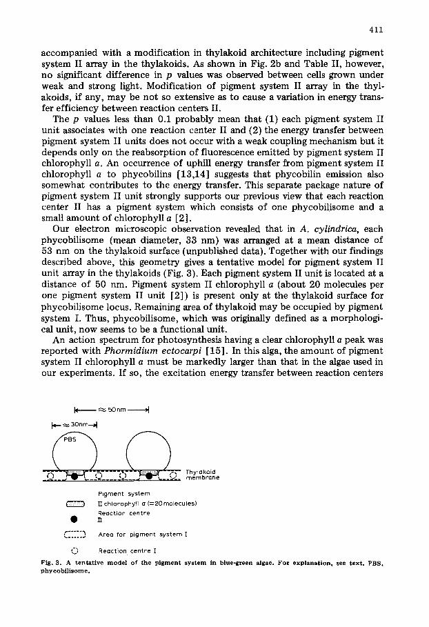
Our electron microscopic observation revealed that in A. cylindrica, each phycobil isome (mean diameter, 33 nm) was arranged at a mean distance of 53 nm on the thylakoid surface (unpublished data). Together with our findings described above, this geometry gives a tentative model for pigment system II unit array in the thylakoids (Fig. 3). Each pigment system II unit is located at a distance of 50 nm. Pigment system II chlorophyll a (about 20 molecules per one pigment system II unit [2]) is present only at the thylakoid surface for phycobil isome locus. Remaining area of thylakoid may be occupied by pigment system I. Thus, phycobil isome, which was originally defined as a morphologi- cal unit, now seems to be a functional unit.
An action spectrum for photosynthesis having a clear chlorophyll a peak was reported with Phormidium ectocarpi [15]. In this alga, the amount of pigment system II chlorophyll a must be markedly larger than that in the algae used in our experiments. If so, the excitation energy transfer between reaction centers
k ~ 5Onto ~1
I~- ~ 3onm-, I
_~_- ~ w ',__.= . . . . . . = . . . . ',. _ _ =-__ m e m b r a n e
P i g m e n t s y s t e m
] ] c h l o r o p h y l l a ( = 2 O m o l e c u l e s )
R e a c t i o n c e n t r e
C . . . . . . . . . . ~' A r e a f o r p i g m e n t s y s t e m ]
~ R e a c t i o n c e n t r e I
Fig. 3. A tentat ive m o d e l o f the p igment sys tem in blue-green algae. For explanat ion , see text . PBS, p h y c o b i ] i s o m e .
412
II may occur with a weak coupling at chlorophyll a level. We note that our model for pigment system II in blue-green and red algae is applicable to the system in which only a small fraction of chlorophyll a functions in pigment system II.
Acknowledgements
We wish to express our thanks to Dr. S. Murakami, College of General Education, University of Tokyo for his kind help in electron microscopic observation. This work was partly supported by grants for scientific research from the Ministry of Education, Japan.
References
1 Edwards, M.R. and Gantt. E. (1971) J. Cell Biol. 50, 896--900 2 Mimuro, M. and Fujita, Y. (1977) Biochim. Biophys. Acta 459,376--389 3 Wang, T.W., Stevens, C.L.R. and Myers, J. (1977) Photochem. Pbotobiol. 25. 103--108 4 Ley, A°C. and Butler, W.L. (1977) in Photosynthetic Organelles0 special issue Plant Cell Physiol.
No. 3, (Miyachi, S., Katoh, S., Fujita, Y. and Shibata, K., eds.), PP. 33--46, Japanese Society of Plant Physiologist, Kyoto and Center for Academic Publications. Japan, Tokyo
5 Watanabe. A. (1960) J. Gen. Appl. Microbiol. 6, 283--292 6 Provasoli, L., McLanghHn, J.J. and Droop, M.R. (1957) Arch. Mikrobiol. 25, 392--428 7 Joliot, A. and Joliot, P. (1964) C.R. Acad. Sci. 258, 4622--4625 8 Joliot, P., Joliot. A. and Kok, B. (1968) Biochim. Biophys. Acta 153, 635--652 9 Joliot, A. (1965) Physiol. Veg. 3,329--344
10 Mackinney, G. (1941) J. Biol. Chem. 140,315--322 11 Murata, N. and Takamiya, A. (1967) Plant Cell Physiol. 8, 683--694 12 Myers, J. and Graham, J. (1971) Plant Physiol. 48, 282--286 13 Wang, R.T. and Myers, J. (1977) in Photosynthetic Organelles, special issue Plant Cell Physiol. No. 3,
(Miyachi, S., Katoh, S.. Fujita. Y. and Shibata, K., eds.), pp. 3--7, Japanese Society of Plant Physiolo- gist, Kyoto and Center for Academic Publications, Japan, Tokyo
14 Mumta, N. (1977) in Photosynthetic Organelles, special issue Plant Cell Physiol. No. 3, (Miyachi. S., Katoh, S.. Fujita, Y. and Shibata, K., eds.), pp. 9--13, Japanese Society of Plant Physiologist, Kyoto and Center of Academic Publications, Japan, Tokyo
15 Haxo, F.T. (1960) in Comparative Biochemistry of Photoreactivc Systems (Alien, M.B., ed.), PP. 339--360, Academic Press, New York

![Review Article Coupling of Algal Biofuel Production …downloads.hindawi.com/journals/tswj/2014/210504.pdfphotosynthetic pigment [ ]. Algae can be divided into two main categories,](https://img.pdfslide.us/doc/110x75/5e676521238c2166da70fe9a/review-article-coupling-of-algal-biofuel-production-photosynthetic-pigment-.jpg)