Embed Size (px)
Citation preview
In organisms from viruses to man, the fidelity withwhich genetic information is replicated depends on theability of polymerases to select correct nucleotides—rather than incorrect and/or damaged nucleotides—forincorporation without adding or deleting nucleotides.Polymerase selectivity is the prime determinant offidelity both at the replication fork and during synthesisto repair DNA damage generated by endogenous cellularmetabolism or exposure to the environment (Fried berg etal. 2006). In many organisms, fidelity can be in creasedby exonucleolytic proofreading of mismatches duringreplication and by DNA mismatch repair (MMR) (forreview, see Kunkel and Erie 2005; Iyer et al. 2006; Hsiehand Yamane 2008). Certain proteins involved in MMRalso can also signal for DNA-damage responses, preventhomologous recombination, promote meiotic re combina- tion, modulate somatic hyper mu tation of im muno glob u-lin genes, or even stabilize certain misaligned repetitiveDNA sequences. When DNA damage is not removedbefore replication, helix-distorting lesions can impedereplication fork progression. In such circumstances, cellsurvival can be enhanced by specialized DNA transac-tions, some of which can be mutagenic via translesionDNA synthesis (Jansen et al. 2007; Yang and Woodgate2007; Chang and Cimprich 2009). Con sidered here is theamazing diversity of evolutionarily conserved DNApolymerases involved in these transactions, many ofwhich have been discovered relatively recently. Empha -sis is on their fidelity and on the contributions of proof-reading and MMR to replication fidelity, which can varyover a much wider range than was appreciated even adecade ago.
MULTIPLE POLYMERASES WITH MULTIPLEOVERLAPPING FUNCTIONS
DNA polymerases were first discovered using assaysfor polymerization activity (Bessman et al. 1956). Thisapproach revealed that bacteria and eukaryotes harbormultiple polymerases (Kornberg and Baker 1992). How -ever, just how many only came to light more re centlywhen sequence alignments and recombinant DNA tech-nology were used to find low-activity low-abundancepolymerases. Sequence alignments now permit classifi-cation of DNA polymerases into several different fami-lies, with most organisms encoding more than one(Shcherbakova et al. 2003a; Bebenek and Kunkel 2004;Loeb and Monnat 2008). For example, Escherichia coliencodes five polymerases (Friedberg et al. 2005), oneeach from families A, B, and C and two from differentsubfamilies of family Y, each with important but some-what different functions. The human genome encodeseven more (Table 1) from families A (three polymerases),B (four polymerases), X (four poly merases), Y (four poly-merases), and reverse transcriptase (RT) (telomerase).Because polymerases can have multiple functions (Table1) and can sometimes com pensate one for another, it is acontinuing challenge to understand exactly where andwhen each polymerase operates in vivo.Despite differences in primary sequence, DNA poly-
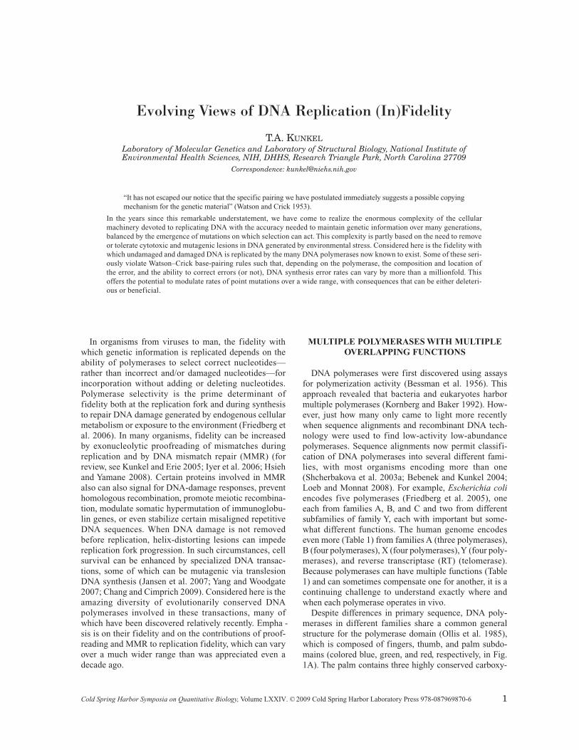
merases in different families share a common generalstructure for the polymerase domain (Ollis et al. 1985),which is composed of fingers, thumb, and palm subdo-mains (colored blue, green, and red, respectively, in Fig.1A). The palm contains three highly conserved carboxy-
Evolving Views of DNA Replication (In)Fidelity
T.A. KUNKELLaboratory of Molecular Genetics and Laboratory of Structural Biology, National Institute of Environmental Health Sciences, NIH, DHHS, Research Triangle Park, North Carolina 27709
Correspondence: [email protected]
“It has not escaped our notice that the specific pairing we have postulated immediately suggests a possible copyingmechanism for the genetic material” (Watson and Crick 1953).
In the years since this remarkable understatement, we have come to realize the enormous complexity of the cellularmachinery devoted to replicating DNA with the accuracy needed to maintain genetic information over many generations,balanced by the emergence of mutations on which selection can act. This complexity is partly based on the need to removeor tolerate cytotoxic and mutagenic lesions in DNA generated by environmental stress. Considered here is the fidelity withwhich undamaged and damaged DNA is replicated by the many DNA polymerases now known to exist. Some of these seri-ously violate Watson–Crick base-pairing rules such that, depending on the polymerase, the composition and location ofthe error, and the ability to correct errors (or not), DNA synthesis error rates can vary by more than a millionfold. Thisoffers the potential to modulate rates of point mutations over a wide range, with consequences that can be either deleteri-ous or beneficial.
Cold Spring Harbor Symposia on Quantitative Biology,Volume LXXIV. ©2009 Cold Spring Harbor Laboratory Press 978-087969870-6 1
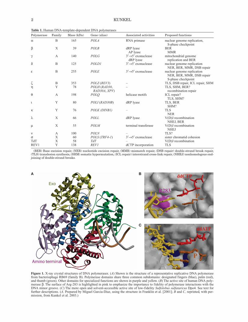
Figure 1. X-ray crystal structures of DNA polymerases. (A) Shown is the structure of a representative replicative DNA polymerasefrom bacteriophage RB69 (family B). Polymerase domains share three common subdomains: designated fingers (blue), palm (red),and thumb (green). Other domains for specialized functions are shown in purple and yellow. (B) The active site of human DNA poly-merase β. The surface of Arg-283 is highlighted in pink to emphasize the importance to fidelity of polymerase interactions with theDNA minor groove. (C) The more open and solvent-accessible active site of low-fidelity Sulfolobus sulfataricus Dpo4. See text forfurther descriptions. (A, Prepared by Miguel Garcia-Diaz, using the structure in Franklin et al. [2001]; B and C, reprinted, with per-mission, from Kunkel et al. 2003.)
2 KUNKEL
A B
C
Table 1. Human DNA-template-dependent DNA polymerases
Polymerase Family Mass (kDa) Gene (alias) Associated activities Proposed functions
α B 165 POLA RNA primase nuclear genome replication,S-phase checkpoint
β X 39 POLB dRP lyase BERAP lyase MMR
γ A 140 POLG 3′→5′ exonuclease mitochondrial genomedRP lyase replication and BER
δ B 125 POLD1 3′→5′ exonuclease nuclear genome replicationNER, BER, MMR, DSB repair
ε B 255 POLE 3′→5′ exonuclease nuclear genome replicationNER, BER, MMR, DSB repairS-phase checkpoint
ζ B 353 POLZ (REV3) – TLS, DSB repair, ICL repair, SHMη Y 78 POLH (RAD30, – TLS, SHM, BER?
RAD30A, XPV) recombination repairθ A 198 POLQ helicase motifs ICL repair?
TLS, SHM?ι Y 80 POLI (RAD30B) dRP lyase TLS, BER
SHM?κ Y 76 POLK (DINB1) – TLS
NERλ X 66 POLL dRP lyase V(D)J recombination
NHEJ, BERµ X 55 POLM terminal transferase V(D)J recombination
NHEJν A 100 POLN – TLS?σ X 60 POLS (TRF4-1) 3′→5′ exonuclease sister chromatid cohesionTdT X 58 TdT – V(D)J recombinationREV1 Y 138 REV1 dCTP incorporation TLS
(BER) Base excision repair, (NER) nucleotide excision repair, (MMR) mismatch repair, (DSB repair) double-strand break repair,(TLS) translesion synthesis, (SHM) somatic hypermutation, (ICL repair) interstrand cross-link repair, (NHEJ) nonhomologous end-joining of double-strand breaks.
lates that bind two divalent metal ions required for cataly-sis via an in-line nucleophilic attack of the 3′-OH on the α-phosphate of the incoming dNTP. This mechanism isthought to be common to all DNA polymerases (Steitz1993), yet it appears to have resulted from convergent evo-lution, because some polymerase families have the activesite carboxylates in a “right-handed” configuration andothers (families X and C) have it in a “left-handed” con-figuration (see, e.g., Wing et al. 2008 and referencestherein).The polymerase domains are usually attached to other
domains needed for the variable functions of these pro-teins. For example, polymerases that perform the bulk ofgenome replication often have a domain harboring 3′exonuclease activity that proofreads replication errors(Fig. 1A). Nonetheless, most DNA polymerases lack anintrinsic 3′ exonuclease activity (Table 1), which is inter-esting given the importance of proofreading to genomestability (see below). Other specialized domains includea “little finger” domain (Yang and Woodgate 2007)unique to family-Y members involved in translesionDNA synthesis, and an 8-kDa domain unique to family-X polymerases (Moon et al. 2007) that assists in fillingsmall gaps during DNA repair and that, in polymerases βand λ, harbors a dRP (deoxyribose phosphate) lyaseactivity needed for repair (Table 1). Still other domainsinclude the BRCT (BRCA1 carboxy terminal) domainsof family-X polymerases involved in nonhomologousend-joining of double-stranded DNA breaks and amino-or carboxy-terminal regions of polymerase catalytic sub-units that are involved in cellular responses to DNA dam-age, including via partnerships with other proteins. Infact, DNA polymerases typically operate in DNA trans-actions that require coordinated interactions with manyother proteins (e.g., noncatalytic accessory subunits, pro-cessivity clamps, and single-stranded DNA-binding pro-teins), whose properties and functions are subjects ofcontinuing interest (see, e.g., Shcherbakova et al. 2003a;Bebenek and Kunkel 2004; Friedberg et al. 2005; Jansenet al. 2007; Loeb and Monnat 2008; Burgers 2009;Chang and Cimprich 2009 and references therein).
THE FIDELITY OF DNA SYNTHESIS
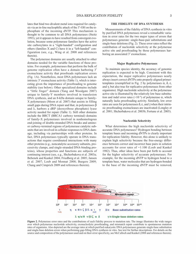
Measurements of the fidelity of DNA synthesis in vitroby purified DNA polymerases reveal a remarkable varia-tion in error rates for the two major types of errors thatpolymerases generate: single-base-pair substitutions andsingle-base deletions (Fig. 2). These error rates reflect thecontribution of nucleotide selectivity at the polymeraseactive site and proofreading by those polymerases har-boring an associated 3′ exonuclease.
Major Replicative Polymerases
To maintain species identity, the accuracy of genomicreplication is expected to be high. Consistent with thisexpectation, the major replicative polymerases nearlyalways insert correct dNTPs onto properly aligned primer-templates (exemplified in Fig. 2 by polymerases α, δ, ε,and γ, but also true for replicative polymerases from otherorganisms). High nucleotide selectivity at the polymeraseactive site is illustrated by the relatively low base substitu-tion and indel error rates (~10–4) of polymerase α, whichnaturally lacks proofreading activity. Sim i lar ly, low errorrates are seen for polymerases δ, ε, and γwhen their intrin-sic proofreading exonucleases are inactivated (Longley etal. 2001; Shcherbakova et al. 2003b; Fortune et al. 2005).
Nucleotide Selectivity
What determines the high nucleotide selectivity ofaccurate DNA polymerases? Hydrogen bonding betweentemplate bases and incoming dNTPs is clearly importantfor replication fidelity. However, this alone is unlikely toexplain high selectivity because the free-energy differ-ence between correct and incorrect base pairs in solutionaccounts for error rates of ~1:100 (Loeb and Kunkel1982). Thus, other ideas have been put forth to accountfor the higher selectivity of accurate polymerases. Forexample, for the incoming dNTP to hydrogen bond to atemplate base, water molecules that are hydrogen-bondedto the base of the incoming dNTP must be removed,
DNA REPLICATION FIDELITY 3
Figure 2. Polymerase error rates and the contributions of each fidelity process to mutation rate. The image illustrates the wide rangesover which polymerase nucleotide selectivity, exonucleolytic proofreading, and mismatch repair contribute to spontaneous mutationrates of organisms. Also depicted are the average rates at which purified eukaryotic DNA polymerases generate single-base substitutionand single-base deletion errors when performing gap-filling DNA synthesis in vitro. See text for further descriptions. For details on thesource and composition of the polymerases used and on their error specificity, see McCulloch and Kunkel (2008 and references therein).
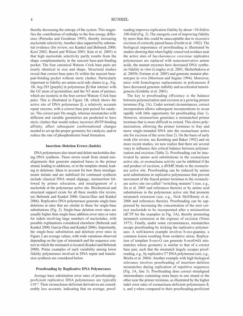
thereby decreasing the entropy of the system. This magni-fies the contribution of enthalpy to the free-energy differ-ence (Petruska and Goodman 1995), thereby increas ingnucleotide selectivity. Another idea supported by substan-tial evidence (for review, see Kunkel and Bebenek 2000;Kool 2002; Beard and Wilson 2003; Kim et al. 2005) isthat high nucleotide selectivity partly results from theshape complementarity in the nascent base-pair-bindingpocket. The four canonical Watson–Crick base pairs arenearly identical in size and shape. Structural studiesreveal that correct base pairs fit within the nascent base-pair-binding pocket without steric clashes. Particularlyimportant to fidelity are amino acid side chains (e.g., Fig.1B, Arg-283 [purple] in polymerase β) that interact withthe O2 atom of pyrimidines and the N3 atom of purines,which are isosteric in the four correct Watson–Crick basepairs. This is illustrated in Figure 1B, which shows theactive site of DNA polymerase β, a relatively accuraterepair enzyme, with a correct base pair poised for cataly-sis. The correct pair fits snugly, whereas mismatches withdifferent and variable geometries are predicted to havesteric clashes that would reduce incorrect dNTP-bindingaffinity, affect subsequent conformational changesneeded to set up the proper geometry for catalysis, and/orreduce the rate of phosphodiester bond formation.
Insertion–Deletion Errors (Indels)
DNA polymerases also insert and delete nucleotides dur-ing DNA synthesis. These errors result from strand mis-alignments that generate unpaired bases in the primerstrand, leading to additions, or in the template strand, lead-ing to deletions. Ideas to account for how these misalign-ments initiate and are stabilized for continued synthesisinclude classical DNA strand slippage, mis insertion fol-lowed by primer relocation, and misalignment of anucleotide at the polymerase active site. Biochemical andstructural support exists for all three models (for review,see Bebenek and Kunkel 2000; Garcia-Diaz and Kunkel2006). Replicative DNA polymerases generate single-basedeletions at rates that are similar to those for single-basesubstitutions (Fig. 2). Single-base deletion error rates areusually higher than single-base addition error rates or ratesfor indels involving large numbers of nucleotides, withpossible explanations considered elsewhere (Bebenek andKunkel 2000; Garcia-Diaz and Kunkel 2006). Importantly,the single-base substitution and deletion error rates inFigure 2 are average values, with wide variations observeddepending on the type of mismatch and the sequence con-text in which the mismatch is located (Kunkel and Bebenek2000). Prime examples of such variability among lowerfidelity polymerases involved in DNA repair and transle-sion synthesis are considered below.
Proofreading by Replicative DNA Polymerases
Average base substitution error rates of proofreading-proficient replicative DNA polymerases are typically≥10–6. Their exonuclease-deficient derivatives are consid-erably less accurate, indicating that on average, proof-
reading improves replication fidelity by about ~10-fold to100-fold (Fig. 2). The energetic cost of improving fidelityby more than this could be unacceptable due to excessiveexcision of correctly paired bases (Fersht et al. 1982). Thebiological importance of proofreading is illustrated bystudies showing that when highly conserved residues nearthe active sites of Saccharomyces cerevisiae replicativepolymerases are replaced with nonconservative aminoacids, the mutant enzymes have decreased DNA synthe-sis fidelity in vitro (Longley et al. 2001; Shcherbakova etal. 2003b; Fortune et al. 2005) and generate mutator phe-notypes in vivo (Morrison and Sugino 1994). Moreover,mice with homologous replacements in polymerase δhave decreased genomic stability and accelerated tumori-genesis (Goldsby et al. 2001).The key to proofreading efficiency is the balance
between polymerization and excision at a growing primerterminus (Fig. 3A). Under normal circumstances, correctincorporation allows subsequent incorporations to occurrapidly with little opportunity for proofreading (line 1).However, misinsertion generates a mismatched primerterminus that is more difficult to extend. This slows poly-merization, allowing the primer terminus to fray andmove single-stranded DNA into the exonuclease activesite for excision of the error (line 2). On the basis of earlywork (for review, see Kornberg and Baker 1992) and onmore recent studies, we now realize that there are severalways to influence this critical balance between polymer-ization and excision (Table 2). Proofreading can be inac-tivated by amino acid substitutions in the exonucleaseactive site, or exonuclease activity can be inhibited if theend product of excision, a dNMP, binds to the exonucle-ase active site. Proofreading can be reduced by aminoacid substitutions in replicative polymerases that preventmovement of the frayed primer terminus to the exonucle-ase active site (so-called “switching mutants”) (see, e.g.,Jin et al. 2005 and references therein) or by amino acidsubstitutions in the polymerase active site that promotemismatch extension (see, e.g., Nick McElhinny et al.2008 and references therein). Proofreading can be sup-pressed by increasing the concentration of the next cor-rect nucleotide to be incorporated after a misinsertion(dCTP for the examples in Fig. 3A), thereby promotingmismatch extension at the expense of excision (Ninio1975). Finally, under some circumstances, mismatchesescape proofreading by tricking the replicative polymer-ases. A well-known example involves 8-oxo-guanine, acommon lesion resulting from oxidative stress. Repli ca -tion of template 8-oxo-G can generate 8-oxoG•dA mis-matches whose geometry is similar to that of a correctbase pair, such that the mismatch largely escapes proof-reading, e.g., by replicative T7 DNA polymerase (see, e.g.,Brieba et al. 2004). Another example with high biologicalrelevance involves proofreading of insertion–deletionmismatches during replication of repetitive se quences(Fig. 3A, line 3). Proofreading does correct misalignedintermediates containing extra bases in one strand or theother near the primer terminus, as illustrated by the higherindel error rates of exonuclease-deficient polymerases δ,ε, and γ when compared to their proofreading-proficient
4 KUNKEL
DNA REPLICATION FIDELITY 5
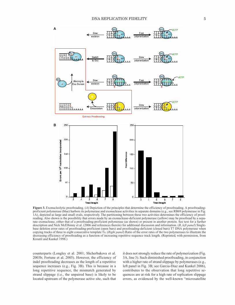
Figure 3. Exonucleolytic proofreading. (A) Depiction of the principles that determine the efficiency of proofreading. A proofreading-proficient polymerase (blue) harbors its polymerase and exonuclease activities in separate domains (e.g., see RB69 polymerase in Fig.1A), depicted as large and small ovals, respectively. The partitioning between these two activities determines the efficiency of proof-reading. Also shown is the possibility that errors made by an exonuclease-deficient polymerase (yellow) may be proofread by a sepa-rate exonuclease, either that of a proofreading-proficient polymerase (as shown) or present in another protein. See text for a furtherdescription and Nick McElhinny et al. (2006 and references therein) for additional discussion and information. (B, left panel) Single-base deletion error rates of proofreading-proficient (open bars) and proofreading-deficient (closed bars) T7 DNA polymerase whencopying tracks of three to eight consecutive template Ts. (Right panel) Ratio of the error rates of the two polymerases to illustrate thedecreasing efficiency of proofreading as a function of increasing repetitive sequence track length. (Reprinted, with permission, fromKroutil and Kunkel 1998.)
A
B
counterparts (Longley et al. 2001; Shcherbakova et al.2003b; Fortune et al. 2005). However, the efficiency ofindel proofreading decreases as the length of a repetitivesequence increases (e.g., Fig. 3B). This is because in along repetitive sequence, the mismatch generated bystrand slippage (i.e., the unpaired base) is likely to belocated upstream of the polymerase active site, such that
it does not strongly reduce the rate of polymerization (Fig.3A, line 3). Such diminished proofreading, in conjunctionwith a higher rate of strand slippage by polymerases (e.g.,left panel in Fig. 3B; see Garcia-Diaz and Kunkel 2006),contributes to the observation that long repetitive se -quences are at risk for a high rate of replication slippageerrors, as evidenced by the well-known “microsatellite
instability” phenotype of eukaryotic cells defective inDNA mismatch (see below). On the basis of the logic inFigure 3 and the parameters in Table 2, it is now very clearthat just as for nucleotide selectivity, the contribution ofproofreading to replication fidelity can vary over a widerange (Fig. 2), from almost none (8-oxoG•dA mis-matches) to several hundredfold (e.g., for bacteriophageT7 replication; see, e.g., Donlin et al. 1991).
“Extrinsic” Proofreading May Also Contribute to Genome Stability
Interestingly, among many mammalian DNA polymer-ases, only those responsible for the bulk of chain elonga-tion during replication (δ, ε, and γ) contain intrinsic 3′exonucleolytic proofreading activity. Nonetheless, theexonuclease-deficient polymerases have very importantroles in maintaining genome stability (Table 1). Areerrors made by exonuclease-deficient polymerases sub-ject to “extrinsic” proofreading by a separate exonucle-ase? The idea (Fig. 3A, line 4) is that, after making amismatch, the polymerase would dissociate, allowing theexonuclease activity of another protein to excise the mis-match. Indeed, the major E. coli replicative polymerase,DNA polymerase III, harbors its polymerase and exonu-clease activities in two different subunits (the α and ε sub-units, respectively), and these two proteins work inconcert to achieve high replication fidelity. Proofreadingby a separate protein may also occur in eukaryotes. Forexample, yeast DNA polymerase α lacks its own proof-reading activity yet synthesizes perhaps 10% of eachOkazaki fragment on the lagging strand, i.e., ~5% of thehuman genome. Given a base substitution error rate of~10–4 (Fig. 2), this amount of replication would generate
30,000 mismatches during each replication cycle. Canpolymerase α errors be proofread by a separate exonucle-ase? This possibility was recently ex amined in a geneticstudy of yeast polymerase αwith a Leu-868Met (L868M)substitution at the polymerase active site (Pavlov et al.2006). L868M polymerase α copies DNA in vitro withnormal activity and processivity but with reduced fidelity.In vivo, the pol1-L868M allele confers a mutator pheno-type, which is strongly increased upon inactivation of the3′ exonuclease of polymerase δ but not that of ε. Amongseveral possible (nonexclusive) explanations, the resultssupport the hypothesis that the 3′ exonuclease of poly-merase δ proofreads errors generated by polymerase αduring initiation of Okazaki fragments. Given the ex ist -ence of many other specialized, naturally proof reading-deficient DNA polymerases with even lower fidelity thanpolymerase α, intrinsic proofreading could be relevant toother DNA transactions that control genome stability (forreview, see Nick McElhinny et al. 2006), such as baseexcision repair and possibly translesion synthesis bypolymerase η (see below).
REPLICATION ASYMMETRY AND FIDELITY
The two strands of duplex DNA are oriented antiparal-lel to each other, and DNA polymerases copy DNA in onlythe 5′ to 3′ direction. Thus, replication of duplex DNA isintrinsically asymmetric. This asymmetry is illustrated bythe simple model of a eukaryotic replication fork shown inFigure 4A, left. Recent evidence in budding yeast suggeststhat the leading strand is primarily replicated by polymer-ase ε (Pursell et al. 2007), whereas Okazaki fragments onthe lagging strand are initiated by polymerase α-primaseand then primarily completed by polymerase δ (NickMcElhinny et al. 2008). These enzymes differ from eachother in primary sequence, subunit composition, interac-tions with other proteins, and several biochemical proper-ties, including processivity, proofreading capacity,fidelity, and error specificity. It is therefore possible thatthe fidelity of leading- and lagging-strand replication maydiffer, perhaps even more so under nonstandard replica-tion conditions arising under stress (Fig. 4A, right), eitherenvironmental (DNA lesions) or genetic (mutations in keygenes). Evidence for differences in leading- and lagging-strand replication fidelity have been reported in E. coli(Fijalkowska et al. 1998), where both strands are replicatedby the same polymerase acting as a multisubunit dimer,DNA polymerase III holoenzyme.
FIDELITY OF DNA REPAIR POLYMERASES
Efficient and accurate replication requires clean sub-strates, such that many organisms devote great attentionand energy to repairing DNA lesions that can result fromendogenous metabolic processes and from exposure tophysical and chemical agents in the external environment.Many different repair processes exist and can be distin-guished by lesion specificity, the enzymes involved, andwhen they operate (for review, see Friedberg et al. 2006).For many of these repair pathways, e.g., BER, NER, NHEJ,
6 KUNKEL
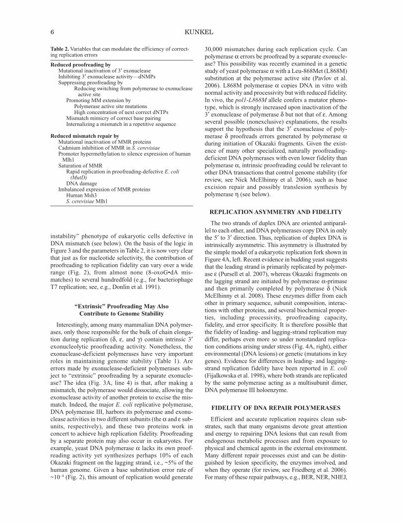
Table 2.Variables that can modulate the efficiency of correct-ing replication errors
Reduced proofreading byMutational inactivation of 3′ exonucleaseInhibiting 3′ exonuclease activity—dNMPsSuppressing proofreading by
Reducing switching from polymerase to exonucleaseactive site
Promoting MM extension byPolymerase active site mutationsHigh concentration of next correct dNTPs
Mismatch mimicry of correct base pairingInternalizing a mismatch in a repetitive sequence
Reduced mismatch repair byMutational inactivation of MMR proteinsCadmium inhibition of MMR in S. cerevisiaePromoter hypermethylation to silence expression of humanMlh1Saturation of MMRRapid replication in proofreading-defective E. coli(MutD)DNA damage
Imbalanced expression of MMR proteinsHuman Msh3S. cerevisiaeMlh1
MMR, and ICL repair, excision of a lesion is followed bygap-filling DNA synthesis and ligation (Fried berg et al.2006). The gap filling is conducted by poly merases that arehighly accurate (e.g., polymerase δ for filling long gapsduring NER and MMR) or moderately accurate (polymer-ase β for filling short gaps during BER). However, certaingap-filling transactions in cells may involve inaccurateDNA polymerases, e.g., κ in NER, ζ/θ in ICL repair, and µin NHEJ, such that DNA synthesis errors occurring duringrepair may contribute to mutagenesis. Perhaps the bestexamples are for polymerases ζ and η, both of which areimplicated in somatic hypermutation of immunoglobulingenes, a process that involves processing uracil in DNAgenerated by cytosine deamination catalyzed by activation-induced cytosine deaminase (Diaz and Lawrence 2005).
Fidelity of Translesion Synthesis Polymerases
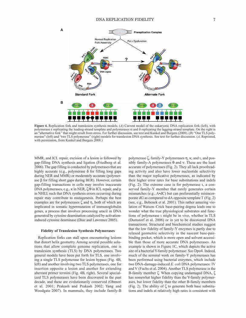
Replication forks can stall upon encountering lesionsthat distort helix geometry. Among several possible solu-tions that allow complete genome replication, one istranslesion synthesis (TLS) by DNA polymerases. Twogeneral models have been put forth for TLS, one involv-ing a single TLS polymerase for lesion bypass (Fig. 4B,left) and another involving two TLS polymerases, one forinsertion opposite a lesion and another for extendingaberrant primer termini (Fig. 4B, right). Several special-ized TLS polymerases have been discovered in the pastdecade, and these are evolutionarily conserved (Ohmoriet al. 2001; Prakash and Prakash 2002; Yang andWoodgate 2007). In mammals, they include family-B
polymerase ζ, family-Y polymerases η, κ, and ι, and pos-sibly family-A polymerases θ and ν. These are the leastaccurate of polymerases (Fig. 2). They all lack proofread-ing activity and also have lower nucleotide selectivitythan the major replicative polymerases, as indicated bytheir higher error rates for base substitutions and indels(Fig. 2). The extreme case is for polymerase ι, a con-served family-Y member that rarely generates certainmismatches (e.g., A•dC) but can preferentially misincor-porate dG as compared to dA opposite template T (Fig. 2)(see, e.g., Bebenek et al. 2001). This rather amazing vio-lation of Watson–Crick base-pairing dogma leads one towonder what the true physiological substrates and func-tions of polymerase ι might be in vivo, whether in TLS(Dumstorf et al. 2006) or in yet to be discovered DNAtransactions. Structural and biochemical studies suggestthat the low fidelity of family-Y enzymes is partly due torelaxed geometric selectivity in the nascent base-pair-binding pocket, which is more open and solvent accessi-ble than those of more accurate DNA polymerases. Anexample is shown in Figure 1C, which depicts the activesite of a bacterial Y-family polymerase: SsoDpo4. Indeed,much of the seminal work on family-Y polymerases hasbeen performed using bacterial enzymes, which includetwo DNA-damage-induced E. coli DNA polymerases: IVand V (Fuchs et al. 2004). Another TLS polymerase is theB-family member ζ. When copying undamaged DNA, ζhas somewhat higher fidelity than the Y-family polymer-ases, but lower fidelity than the other B-family members(Fig. 2). The ability of ζ to generate both base substitu-tions and indels at relatively high rates is consistent with
DNA REPLICATION FIDELITY 7
Figure 4. Replication fork and translesion synthesis models. (A) Current model of the eukaryotic DNA replication fork (left), withpolymerase ε replicating the leading-strand template and polymerases α and δ replicating the lagging-strand template. On the right isan “alternative fork” that might result from stress. For further discussion, see text and Kunkel and Burgers (2008). (B) “One TLS poly-merase” (left) and “two TLS polymerase” (right) models for translesion DNA synthesis. See text for further discussion. (A, Reprinted,with permission, from Kunkel and Burgers 2008.)
A
B
its known participation in a large majority of spontaneousmutations, as well as in mutagenesis induced by a varietyof DNA-damaging agents (Lawrence 2002). Polymeraseζ’s high base substitution error rate clearly demonstratesthat it has relatively low nucleotide selectivity, consistentwith a possible direct role in mutagenic misinsertion ofdNTPs in vivo. Polymerase ζ also efficiently extends ter-minal mismatches when copying undamaged DNA, aswell as efficiently extending damaged termini, the latterbeing consistent with a role for ζ in the extension step ofTLS in the two-polymerase model (Fig. 4B). A similarrole has also been proposed for polymerase κ, which, likeζ, is promiscuous for mismatch extension (for review, seePrakash and Prakash 2002). During DNA synthesis invitro, ζ also generates “complex” mutations that containmultiple substitutions and indels within a short tract ofDNA (Sakamoto et al. 2007; Stone et al. 2009). Con sist -ent with this property, ζ also generates complex errors invivo, which could be significant from an evolutionaryperspective. The biological relevance of TLS is perhapsbest illustrated by the role of polymerase V in the muta-genic SOS response in E. coli, and the fact that loss ofpolymerase η function in humans and in mice results insensitivity to sunlight, predisposition to skin cancer, andaltered specificity of somatic hypermutation of im mu no -globulin genes. The topics and the TLS ability and fi del -ity of various polymerases when encountering a widerange of structurally diverse lesions have been describedin great detail elsewhere (see, e.g., Prakash and Prakash2002; Fuchs et al. 2004; Diaz and Lawrence 2005;Friedberg et al. 2005; Yang and Woodgate 2007).
DNA Mismatch Repair
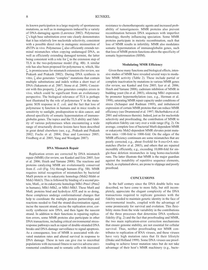
Replication errors are corrected by DNA mismatchrepair (MMR) (for review, see Kunkel and Erie 2005; Iyeret al. 2006; Hsieh and Yamane 2008). The reactions andproteins catalyzing MMR are evolutionarily conservedfrom E. coli (Fig. 5A) through humans (Fig. 5B). MMRrequires initial recognition of mismatches by bacterialMutS protein or its eukaryotic homologs (Msh2-Msh6 orMsh2-Msh3). This is followed by binding of a second pro-tein, MutL, or its eukaryotic homologs Mlh1-Pms1 (Pms2in humans), Mlh1-Mlh2, or Mlh1-Mlh3. These MutS andMutL proteins bind and hydrolyze ATP, and in so doing,these complexes undergo conformational changes thathelp to coordinate the multiple protein partnerships andreactions needed to find the strand-discrimination signal,incise the nascent strand, excise the replication error, cor-rectly synthesize new DNA, and then ligate the nascentstrand. In addition to their functions in repairing replica-tion errors, some MMR proteins also participate in otherDNA transactions, including critical environmental stress-response pathways such as repair of double-stranded DNAbreaks and DNA-damage surveillance to signal apoptosis.As a consequence, loss of MMR is associated with ele-vated mutation rates and altered survival in response toDNA damage. These in turn can give rise to microbialpopulations with increased fitness to survive adverse envi-ronmental conditions and to somatic cells with increased
resistance to chemotherapeutic agents and increased prob-ability of tumorigenesis. MMR proteins also preventrecombination between DNA sequences with imperfecthomology, thereby influencing speciation. Some MMRproteins participate in meiotic recombination, such thatloss of MMR results in infertility. MMR also modulatessomatic hypermutation of immunoglobulin genes, suchthat loss of MMR protein functions alters the specificity ofsomatic hypermutation (SHM).
Modulating MMR Efficiency
Given these many functions and biological effects, inten-sive studies of MMR have revealed several ways to modu-late MMR activity (Table 2). These include partial orcomplete inactivation by mutations in various MMR genes(for review, see Kunkel and Erie 2005; Iyer et al. 2006;Hsieh and Yamane 2008), cadmium inhibition of MMR inbudding yeast (Jin et al. 2003), silencing Mlh1 expressionby promoter hypermethylation (see, e.g., Herman et al.1998), saturating MMR repair capacity under conditions ofstress (Schaaper and Radman 1989), and imbalancedexpression of certain MMR proteins that can reduce MMRefficiency (see Drummond et al. 1997; Shcherbakova et al.2001 and references therein). Indeed, just as for nucleotideselectivity and proofreading, the contribution of MMR toreplication fidelity can vary over a wide range (Fig. 2). Onaverage, complete loss of bacterial MutS-dependent MMRor eukaryotic Msh2-dependent MMR elevates point muta-tion rates ~100-fold to 1000-fold. On the edges of theMMR efficiency continuum are some mismatches that arepoorly corrected, e.g., about fivefold for 8-oxo-G•A mis-matches (Pavlov et al. 2003), and others that are repairedincredibly efficiently, e.g., exceeding 10,000-fold for sin-gle-base deletion mismatches in long homo-nu cle o tideruns. The latter illustrate that MMR is the major guard ianagainst the instability of repetitive se quence elements,which, as explained above, are prone to slippage and poorlyproofread.
CONCLUSIONS
In the half century since the DNA double helix wasdescribed, we have come to more fully, but still incom-pletely, appreciate the elegant complexity of the DNAtransactions required to replicate genomes with thefidelity needed to maintain genetic identity in the face ofenvironmental insults, coupled with the advantage ofsome promiscuity for survival and evolution. This flexi-bility stems from the wide variability in the contributionsof the three processes that determine DNA synthesisfidelity (Fig. 2) and the fact that proofreading and MMR,the two main replication-error correction mechanismsthat ensure genome stability, are not essential for cellularsurvival. Thus, neither proofreading nor MMR con-tributes to replication of RNA viruses, and these viruseshave very high mutation rates that fit their lifestyle(Drake and Holland 1999). Some DNA viruses use proof-reading to achieve lower mutation rates but do not takeadvantage of their host’s MMR machinery (e.g., bacte-
8 KUNKEL
DNA REPLICATION FIDELITY 9
Figure 5. Models for DNA mismatch repair in E. coli (A) and eukaryotes (B). See text for description. (Reprinted, with permission,from Iyer et al. 2006.)
A
B
riophage T4) (Santos and Drake 1994). As a consequence,they have mutation rates per base pair that are higher thanorganisms that do use MMR. Interestingly, given the dif-ferences in genome size, the mutation rate per genome isrelatively constant among DNA-based organisms, at0.003 (Drakes rule; Drake 1991, 1999). Also of interest isthe fact that the genomes of certain bacteria do not encodeobvious homologs of the major MMR genes. This leadsone to wonder whether they forego MMR altogether orcorrect replication errors in a manner yet undiscovered.
ACKNOWLEDGMENTS
I thank Alan Clark and Mercedes Arana for thoughtfulcomments on the manuscript. The research conducted inthe author’s laboratory is supported by the IntramuralResearch Program of the National Institutes of Health,National Institute of Environmental Health Sciences(Projects Z01 ES065070 and Z01 ES065089).
REFERENCES
Beard WA, Wilson SH. 2003. Structural insights into the originsof DNA polymerase fidelity. Structure 11: 489–496.
Bebenek K, Kunkel TA. 2000. Streisinger revisited: DNA syn-thesis errors mediated by substrate misalignments. ColdSpring Harbor Symp Quant Biol 65: 81–91.
Bebenek K, Kunkel TA. 2004. Functions of DNA. Adv ProteinChem 69: 137–165.
Bebenek K, Tissier A, Frank EG, McDonald JP, Prasad R,Wilson SH, Woodgate R, Kunkel TA. 2001. 5′-Deoxyribosephosphate lyase activity of human DNA polymerase ι in vitro.Science 291: 2156–2159.
Bessman MJ, Kornberg A, Lehman IR, Simms ES. 1956. En -zymic synthesis of deoxyribonucleic acid. Biochim BiophysActa 21: 197–198.
Brieba LG, Eichman BF, Kokoska RJ, Doubliâe S, Kunkel TA,Ellenberger T. 2004. Structural basis for the dual codingpotential of 8-oxoguanosine by a high-fidelity DNA polymer-ase. EMBO J 23: 3452–3461.
Burgers PM. 2009. Polymerase dynamics at the eukaryotic DNAreplication fork. J Biol Chem 284: 4041–4045.
Chang DJ, Cimprich KA. 2009. DNA damage tolerance: Whenit’s OK to make mistakes. Nat Chem Biol 5: 82–90.
Diaz M, Lawrence C. 2005. An update on the role of translesionsynthesis DNA polymerases in Ig hypermutation. TrendsImmunol 26: 215–220.
Donlin MJ, Patel SS, Johnson KA. 1991. Kinetic partitioning be -tween the exonuclease and polymerase sites in DNA error cor-rection. Biochemistry 30: 538–546.
Drake JW. 1991. A constant rate of spontaneous mutation inDNA-based microbes. Proc Natl Acad Sci 88: 7160–7164.
Drake JW. 1999. The distribution of rates of spontaneous muta-tion over viruses, prokaryotes, and eukaryotes. Ann NY AcadSci 870: 100–107.
Drake JW, Holland JJ. 1999. Mutation rates among RNA viruses.Proc Natl Acad Sci 96: 13910–13913.
Drummond JT, Genschel J, Wolf E, Modrich P. 1997. DHFR/MSH3 amplification in methotrexate-resistant cells alters thehMutSα/hMutSβ ratio and reduces the efficiency of base-basemismatch repair. Proc Natl Acad Sci 94: 10144–10149.
Dumstorf CA, Clark AB, Lin Q, Kissling GE, Yuan T, Kucher -lapati R, McGregor WG, Kunkel TA. 2006. Par ti ci pa tion ofmouse DNA polymerase ι in strand-biased mutagenic bypassof UV photoproducts and suppression of skin cancer. ProcNatl Acad Sci 103: 18083–18088.
Fersht AR, Knill-Jones JW, Tsui WC. 1982. Kinetic basis ofspon taneous mutation. Misinsertion frequencies, proofread-ing specificities and cost of proofreading by DNA polymer-ases of Escherichia coli. J Mol Biol 156: 37–51.
Fijalkowska IJ, Jonczyk P, Tkaczyk MM, Bialoskorska M,Schaaper RM. 1998. Unequal fidelity of leading strand andlagging strand DNA replication on the Escherichia coli chro-mosome. Proc Natl Acad Sci 95: 10020–10025.
Fortune JM, Pavlov YI, Welch CM, Johansson E, Burgers PM,Kunkel TA. 2005. Saccharomyces cerevisiae DNA polymer-ase δ: High fidelity for base substitutions but lower fidelityfor single- and multi-base deletions. J Biol Chem 280: 29980–29987.
Franklin MC, Wang J, Steitz TA. 2001. Structure of the replicatingcomplex of a pol α family DNA polymerase. Cell 105: 657–667.
Friedberg EC, Lehmann AR, Fuchs RP. 2005. Trading places:How do DNA polymerases switch during translesion DNAsynthesis? Mol Cell 18: 499–505.
Friedberg EC, Walker GC, Siede W, Wood RD, Schultz RA,Ellenberger T. 2006. DNA repair and mutagenesis, 2nd ed.ASM Press, Washington, D.C.
Fuchs RP, Fujii S, Wagner J. 2004. Properties and functions ofEscherichia coli: Pol IV and Pol V. Adv Protein Chem 69:229–264.
Garcia-Diaz M, Kunkel TA. 2006. Mechanism of a genetic glis-sando: Structural biology of indel mutations. Trends BiochemSci 31: 206–214.
Goldsby RE, Lawrence NA, Hays LE, Olmsted EA, Chen X,Singh M, Preston BD. 2001. Defective DNA polymerase-δproofreading causes cancer susceptibility in mice. Nat Med 7:638–639.
Herman JG, Umar A, Polyak K, Graff JR, Ahuja N, Issa JP, Marko -witz S, Willson JK, Hamilton SR, Kinzler KW, et al. 1998.Incidence and functional consequences of hMLH1 promoterhypermethylation in colorectal carcinoma. Proc Natl Acad Sci95: 6870–6875.
Hsieh P, Yamane K. 2008. DNA mismatch repair: Molecular mech- anism, cancer, and ageing. Mech Ageing Dev 129: 391–407.
Iyer RR, Pluciennik A, Burdett V, Modrich PL. 2006. DNA mis-match repair: Functions and mechanisms. Chem Rev 106:302–323.
Jansen JG, Fousteri MI, de Wind N. 2007. Send in the clamps:Control of DNA translesion synthesis in eukaryotes. Mol Cell28: 522–529.
Jin YH, Clark AB, Slebos RJ, Al-Refai H, Taylor JA, Kunkel TA,Resnick MA, Gordenin DA. 2003. Cadmium is a mutagen thatacts by inhibiting mismatch repair. Nat Genet 34: 326–329.
Jin YH, Garg P, Stith CM, Al-Refai H, Sterling J, Murray LJ,Kunkel TA, Resnick MA, Burgers PM, Gordenin DA. 2005.The multiple biological roles of the 3′→5′ exonuclease ofSaccharomyces cerevisiaeDNA polymerase δ require switch-
ing between the polymerase and exonuclease domains. MolCell Biol 25: 461–471.
Kim TW, Delaney JC, Essigmann JM, Kool ET. 2005. Probingthe active site tightness of DNA polymerase in subangstromincrements. Proc Natl Acad Sci 102: 15803–15808.
Kool ET. 2002. Active site tightness and substrate fit in DNA repli-cation. Annu Rev Biochem 71: 191–219.
Kornberg A, Baker T. 1992. DNA replication, 2nd ed. Freeman,New York.
Kroutil LC, Kunkel TA. 1998. DNA replication errors involvingstrand misalignments. In Genetic instabilities and hereditaryneurological diseases (ed. RD Wells and ST Warren), pp.699–716. Academic, San Diego.
Kunkel TA, Bebenek K. 2000. DNA replication fidelity. AnnuRev Biochem 69: 497–529.
Kunkel TA, Burgers PM. 2008. Dividing the workload at aeukaryotic replication fork. Trends Cell Biol 18: 521–527.
Kunkel TA, Erie DA. 2005. DNA mismatch repair. Annu RevBiochem 74: 681–710.
Kunkel TA, Pavlov YI, Bebenek K. 2003. Functions of humanDNA polymerases η, κ and ι suggested by their properties,including fidelity with undamaged DNA templates. DNARepair 2: 135–149.
Lawrence CW. 2002. Cellular roles of DNA polymerase ζ andRev1 protein. DNA Repair 1: 425–435.
Loeb LA, Kunkel TA. 1982. Fidelity of DNA synthesis. AnnuRev Biochem 51: 429–457.
Loeb LA, Monnat RJ Jr. 2008. DNA polymerases and humandisease. Nat Rev Genet 9: 594–604.
Longley MJ, Nguyen D, Kunkel TA, Copeland WC. 2001. Thefidelity of human DNA polymerase γwith and without exonu-cleolytic proofreading and the p55 accessory subunit. J BiolChem 276: 38555–38562.
McCulloch SD, Kunkel TA. 2008. The fidelity of DNA synthe-sis by eukaryotic replicative and translesion synthesis poly-merases. Cell Res 18: 148–161.
Moon AF, Garcia-Diaz M, Bebenek K, Davis BJ, Zhong X,Ramsden DA, Kunkel TA, Pedersen LC. 2007. Structuralinsight into the substrate specificity of DNA polymerase µ.Nat Struct Mol Biol 14: 45–53.
Morrison A, Sugino A. 1994. The 3′→5′ exonucleases of bothDNA polymerases δ and ε participate in correcting errors ofDNA replication in Saccharomyces cerevisiae. Mol GenGenet 242: 289–296.
Nick McElhinny SA, Pavlov YI, Kunkel TA. 2006. Evidence forextrinsic exonucleolytic proofreading. Cell Cycle 5: 958–962.
Nick McElhinny SA, Gordenin DA, Stith CM, Burgers PM,Kunkel TA. 2008. Division of labor at the eukaryotic replica-tion fork. Mol Cell 30: 137–144.
Ninio J. 1975. Kinetic amplification of enzyme discrimination.Biochimie 57: 587–595.
Ohmori H, Friedberg EC, Fuchs RP, Goodman MF, Hanaoka F,Hinkle D, Kunkel TA, Lawrence CW, Livneh Z, Nohmi T, etal. 2001. The Y-family of DNA polymerases. Mol Cell 8: 7–8.
Ollis DL, Brick P, Hamlin R, Xuong NG, Steitz TA. 1985.Structure of large fragment of Escherichia coli DNA poly-merase I complexed with dTMP. Nature 313: 762–766.
Pavlov YI, Mian IM, Kunkel TA. 2003. Evidence for preferentialmismatch repair of lagging strand DNA replication errors inyeast. Curr Biol 13: 744–748.
Pavlov YI, Frahm C, Nick McElhinny SA, Niimi A, Suzuki M,Kunkel TA. 2006. Evidence that errors made by DNA poly-merase α are corrected by DNA polymerase δ. Curr Biol 16:202–207.
Petruska J, Goodman MF. 1995. Enthalpy-entropy compensationin DNA melting thermodynamics. J Biol Chem 270: 746–750.
Prakash S, Prakash L. 2002. Translesion DNA synthesis in eukar -yotes: A one- or two-polymerase affair. Genes Dev 16: 1872–1883.
Pursell ZF, Isoz I, Lundstrom EB, Johansson E, Kunkel TA.2007. Yeast DNA polymerase ε participates in leading-strandDNA replication. Science 317: 127–130.
Sakamoto AN, Stone JE, Kissling GE, McCulloch SD, Pavlov
10 KUNKEL
YI, Kunkel TA. 2007. Mutator alleles of yeast DNA polymer-ase ζ. DNA Repair 6: 1829–1838.
Santos ME, Drake JW. 1994. Rates of spontaneous mutation inbacteriophage T4 are independent of host fidelity determi-nants. Genetics 138: 553–564.
Schaaper RM, Radman M. 1989. The extreme mutator effect of Es -cherichia colimutD5 results from saturation of mismatch repairby excessive DNA replication errors. EMBO J 8: 3511–3516.
Shcherbakova PV, Hall MC, Lewis MS, Bennett SE, Martin KJ,Bushel PR, Afshari CA, Kunkel TA. 2001. Inactivation of DNAmismatch repair by increased expression of yeast MLH1. MolCell Biol 21: 940–951.
Shcherbakova PV, Bebenek K, Kunkel TA. 2003a. Functions ofeukaryotic DNA polymerases. Sci Aging Knowledge Environ2003: RE3.
Shcherbakova PV, Pavlov YI, Chilkova O, Rogozin IB, Johans son
E, Kunkel TA. 2003b. Unique error signature of the four-sub-unit yeast DNA polymerase ε. J Biol Chem 278: 43770–43780.
Steitz TA. 1993. DNA- and RNA-dependent DNA polymerases.Curr Opin Struct Biol 3: 31–38.
Stone JE, Kissling GE, Lujan SA, Rogozin IB, Stith CM,Burgers PM, Kunkel TA. 2009. Low-fidelity DNA synthesisby the L979F mutator derivative of Saccharomyces cerevisiaeDNA polymerase ζ. Nucleic Acids Res 37: 3774–3787.
Watson JD, Crick FH. 1953. Molecular structure of nucleic acids;a structure for deoxyribose nucleic acid. Nature 171: 737–738.
Wing RA, Bailey S, Steitz TA. 2008. Insights into the replisomefrom the structure of a ternary complex of the DNA polymer-ase III α-subunit. J Mol Biol 382: 859–869.
Yang W, Woodgate R. 2007. What a difference a decade makes:Insights into translesion DNA synthesis. Proc Natl Acad Sci104: 15591–15598.
DNA REPLICATION FIDELITY 11