Embed Size (px)
Citation preview
Proc. Natl. Acad. Sci. USAVol. 81, pp. 6881-6885, November 1984Neurobiology
Evolutionary conservation of key structures and binding functionsof neural cell adhesion molecules
(cell-cell adhesion/adhesive specificity/morphogenesis/cell surface proteins/brain development)
STANLEY HOFFMAN, CHENG-MING CHUONG, AND GERALD M. EDELMANThe Rockefeller University, 1230 York Avenue, New York, NY 10021
Contributed by Gerald M. Edelman, July 10, 1984
ABSTRACT The neural cell adhesion molecule N-CAM isa sialic acid-rich, cell surface glycoprotein that mediates celladhesion by a homophilic mechanism. Its binding function hasbeen implicated in both morphogenesis and histogenesis; dur-ing development it changes in amount at the cell surface andperinatally it undergoes a decrease in sialic acid content (em-bryonic -* adult conversion) with an increase in binding effi-cacy. In the present study, salient aspects of the structure andthe mutual binding specificities of N-CAMs from a variety ofvertebrate species were examined to determine whether (N-CAM)-mediated adhesion mechanisms have been conservedduring evolution. N-CAM immunoreactivity was detected in aseries of polypeptides of characteristic molecular weight ex-tracted from brain tissues of all vertebrate species tested, in-cluding mammals, birds, reptiles, amphibia, and bony andcartilaginous fish. Adhesion mediated by N-CAM occurredacross species lines as indicated by the co-aggregation of chick-en and mouse neural cells. By using a quantitative membranevesicle aggregation assay, the efficacy of cross-species brainmembrane vesicle adhesion in various pairings (chicken-mouse, chicken-frog, mouse-frog) was found to be similar tothe efficacy of intra-species adhesion. Effective cross-speciesaggregation of brain membrane vesicles also occurred in em-bryonic-embryonic, adult-adult, and embryonic-adult pair-ings. In a control experiment, embryonic chicken liver mem-brane vesicles (which do not contain N-CAM) did not co-ag-gregate with embryonic chicken brain membrane vesicles.Cross-species co-aggregation could be inhibited by Fab' frag-ments of antibodies to N-CAM and was most effectively inhib-ited in the presence of mixtures made from the Fab' fragmentsof specific antibodies prepared against the N-CAMs from eachof the animal species constituting a co-aggregating pair. Theseresults suggest that, in accord with the proposed role of N-CAM as a regulator of morphogenesis, both the specificity ofthe binding region of the molecule and its basic chemical struc-ture have been highly conserved during evolution.
The histologic differences and differences in form that dis-tinguish vertebrate species probably result from covariant al-terations (1) during evolution in the relative rates of two ormore of the primary morphogenetic processes of cell adhe-sion, cell migration, cell proliferation, cell differentiation,and cell death. Work on cell adhesion molecules or CAMs(2, 3) provides one opportunity to test the hypothesis (1) thatpattern formation may be mediated in part through the regu-latory effects on other primary processes of a relativelysmall number of CAMs of different specificities, providedthat their adhesive efficacies could be graded over a widerange by various forms of surface modulation (3), includingdifferential expression and chemical modification. This hy-pothesis suggests that major changes in vertebrate form at-tributable to adhesion would most likely have arisen by alter-
ations during evolution of various regulatory genes forCAMs rather than by major changes in the structure andspecificity of the CAMs themselves.
Previous studies have indicated that N-CAM, the neuralcell adhesion molecule, mediates cell-cell adhesion in chick-en (4-6), rodent (7, 8), human (9), and frog (10) neural tis-sues. The importance of N-CAM in morphogenesis is sug-gested by sequential changes in its distribution during earlydevelopment (11, 12). N-CAM undergoes alterations in prev-alence at the cell surface during critical periods of primaryand secondary induction in a fashion that is spatio-temporal-ly coordinated with changes in the prevalence and distribu-tion of another non-neural adhesion molecule, liver CAM (L-CAM) (12). These observations raise the possibility that em-bryonic induction and CAM modulation may be causallylinked (1).N-CAM is also involved in later histogenetic processes
(3), including highly patterned events such as the formationof the retinotectal projection (10). During this developmentalepoch, N-CAM converts from a sialic acid-rich embryonic(E) form having a microheterogeneous electrophoretic distri-bution to several adult (A) forms, which contain approxi-mately one-third as much sialic acid and which migrate onNaDodSO4 gel electrophoresis as discrete components ofMr180,000, 150,000, and 120,000 (13-15). E -+ A conversionoccurs gradually, is exhibited to varying degrees and at vary-ing rates in different brain regions (16), and is delayed in thecerebellum of the neurological mutant staggerer (14). Thepossible functional significance of these changes is suggest-ed by analyses (6) of the rates of vesicle aggregation thatindicate that the efficacy ofN-CAM to N-CAM (homophilic)binding is inversely related to the sialic acid content of theparticipating molecules. The predominance of modulationmechanisms and the distribution patterns of N-CAM raisethe question whether N-CAM specificity is conserved in ver-tebrate evolution.
In the present study, an estimate of the degree of conser-vation of N-CAM structure and function was made across awide range of vertebrate species by employing a series ofimmunological, electrophoretic, and aggregation assays. Thetwo main findings are (i) N-CAM polypeptides of character-istic electrophoretic behavior and immunological cross-reac-tivity can be detected in descendants of early vertebratessuch as the shark and in all higher species tested and (ii)within the sensitivity range of present assays, cross-speciesaggregation of membrane vesicles containing N-CAMs pre-pared from mouse, chicken, and frog brains occurs as readilyas intra-species vesicle aggregation.
MATERIALS AND METHODSAnimals. White Leghorn chickens and BALB/c and NCS
mice were obtained from a variety of sources. Adult frogs
Abbreviations: CAM, cell adhesion molecule; N-CAM, neuralCAM; L-CAM, liver CAM; E and A, embryonic and adult forms ofN-CAM.
6881
The publication costs of this article were defrayed in part by page chargepayment. This article must therefore be hereby marked "advertisement"in accordance with 18 U.S.C. §1734 solely to indicate this fact.
6882 Neurobiology: Hoffman etaP
and stage 55 tadpoles (Xenopus laevis) were obtained fromNasco (Fort Atkinson, WI). Other species were obtainedfrom the JBJ Pet Shop (New York).
Antibodies. Rabbit anti-chicken N-CAM (17), anti-mouseN-CAM (7), and anti-frog N-CAM (10) IgG and Fab' frag-ments (18) were prepared following published procedures.
Cross-Species Cell Adhesion. Embryonic mouse (16 day)and embryonic chicken (9 day) cells were prepared by using0.002% trypsin in the presence of 1 mM EDTA (19). Mousecells, labeled with diacetyl fluorescein (19), and chickencells, labeled with rhodamine isothiocyanate (20), were co-incubated (4 x 10⁷ cells from each species in 1.5 ml of mini-mal essential medium for suspension culture, 30 min, 370C,90 rpm) in the presence of either 1 mg of nonimmune Fab' ora mixture of 500 ,ug each of anti-(mouse N-CAM) and anti-(chicken N-CAM) Fab'.
Quantitative Measurement of Membrane Vesicle Aggrega-tion. To prepare brain membrane vesicles, brains in calcium/magnesium-free medium (17)/Trasylol (Mobay Chemical,New York) (200 units/ml)/deoxyribonuclease I (Worthing-ton) (0.5 mg/ml) were homogenized using a Polytron (Brink-mann) (five 2-sec bursts at setting 5) followed by a tight-fit-ting steel Dounce homogenizer (10 strokes). The homoge-nate was mixed with 2 vol of 70% (wt/vol) sucrose, layeredin the bottom of SW27 ultracentrifuge tubes (Beckman),overlaid with 42%, 26%, and 10% sucrose in phosphate-buff-ered saline, pH 7.4 (Pi/NaCl), and centrifuged 2 hr at 40C, at25,000 rpm. The turbid material at the 26%/42% sucrose in-terface was collected, washed, and resuspended in Pi/NaCl.Liver membrane vesicles were prepared as described (21)and material at the 37%/41% and 41%/45% sucrose inter-faces was pooled.Membrane vesicle aggregation experiments were per-
formed and monitored as described (6) with the followingexceptions: (i) vesicles were passed through 0.6-,um pore-size filters (Nucleopore) before each experiment and (it) theexperiment shown in Fig. 3B was done in L-CAM assay buffer(21) containing Ca2 , which is required for the aggregation ofthe liver membrane vesicles. Rates of membrane vesicle ag-gregation varied <10o between duplicate experiments.
RESULTSIn addition to criteria previously established (7, 9, 17), in thepresent studies we used several means to test for evolution-ary conservation of N-CAM structure and function: (i) im-mune cross-reactivity among a broad range of species de-tected by using polyclonal anti-(N-CAM) antibodies, (ii) sim-ilar electrophoretic migration patterns of the adult forms ofN-CAM from these species, and (iii) (N-CAM)-mediated ad-hesion between cells of different species followed by quanti-tative assessment of the relative efficacy of cross-speciesand intra-species aggregation of membrane vesicles.
Conservation of Characteristic N-CAM Antigenic Determi-nants and Electrophoretic Patterns Among Vertebrates. N-CAM was originally isolated from chicken neural tissue andseveral monoclonal antibodies were prepared that recog-nized the molecule. One of the antibodies, 15G8, was used toimmunoaffinity purify N-CAM from human (9), rodent (7),and frog (10) brains. The following members of subphylumvertebrata were surveyed for the existence of N-CAM im-munoreactive material using a mixture of rabbit antibodies tomouse, chicken, and frog N-CAM to maximize potentialcross-reactivity: human, mouse, rat (class Mammalia);chicken, canary (class Aves); turtle, snake, lizard (classReptilia); frog, salamander, newt (class Amphibia); goldfish,blue fish (class Osteichthyes); and shark (class Chondrich-thyes). Immunoblot analysis of these adult brain extracts in-dicated that some or all of the characteristic (13-15) adult-form N-CAM polypeptides (Mr 180,000, 150,000, and
Mr tO C -abGo@\to+6Ofit220<i -n,"16-*
58
43-6-
Dye-
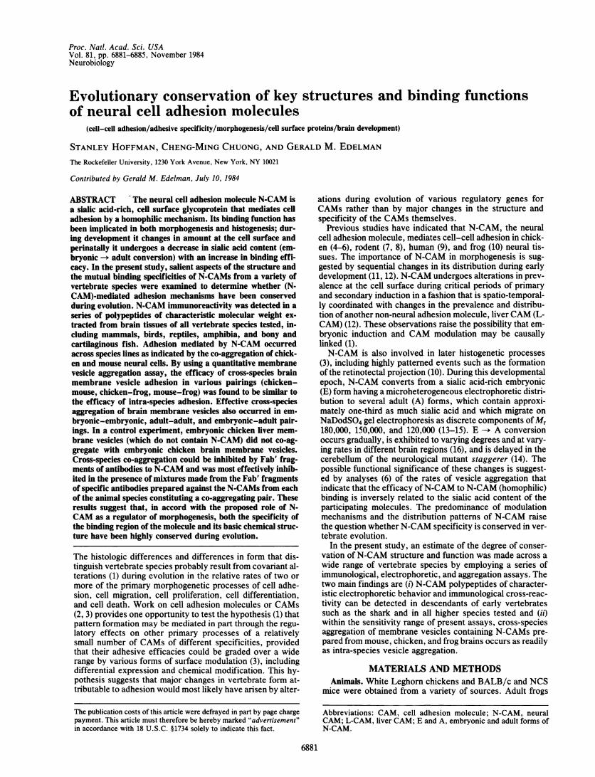
FIG. 1. Polypeptides of characteristic molecular weight from avariety of species react with anti-(N-CAM) antibodies. Nonidet P-40extracts (100 ,ug protein) of crude brain membrane vesicles wereprepared from adults of the indicated species and analyzed by im-munoblotting using a mixture of rabbit anti-mouse, chicken, andfrog N-CAM antibodies (50 ,ug each), and the bound antibodies weredetected as described (16). The migration of reference proteins isindicated by their Mr x lo-3. The leftmost lane shows that a typicalbrain extract (in this case, salamander) is composed of many pro-teins detectable by amido black staining. The right lanes are autora-diographs showing the polypeptides that react with anti-(N-CAM)antibodies. Note that the only polypeptides recognized by the anti-bodies corresponded in molecular weight to the characteristic adult-form N-CAM polypeptides (Mr 180,000, 150,000, and 120,000).
120,000) were present in all of the species tested. Resultsfrom several representative species are shown in Fig. 1. Inevery case, the Mr 180,000 polypeptide was predominant.These polypeptides were also detected by immunoblot anal-ysis using monoclonal antibody 15G8 (7, 14, 16), althoughthe relative intensities of the components differed from thoseobtained with rabbit anti-(N-CAM) antibodies (data notshown).N-CAM Mediates Cross-Species Neural Cell Adhesion. To
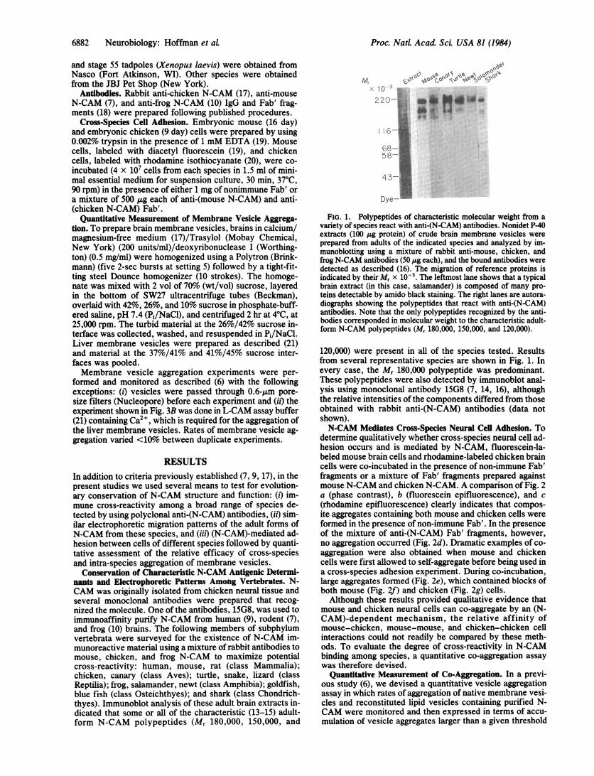
determine qualitatively whether cross-species neural cell ad-hesion occurs and is mediated by N-CAM, fluorescein-la-beled mouse brain cells and rhodamine-labeled chicken braincells were co-incubated in the presence of non-immune Fab'fragments or a mixture of Fab' fragments prepared againstmouse N-CAM and chicken N-CAM. A comparison of Fig. 2a (phase contrast), b (fluorescein epifluorescence), and c(rhodamine epifluorescence) clearly indicates that compos-ite aggregates containing both mouse and chicken cells wereformed in the presence of non-immune Fab'. In the presenceof the mixture of anti-(N-CAM) Fab' fragments, however,no aggregation occurred (Fig. 2d). Dramatic examples of co-aggregation were also obtained when mouse and chickencells were first allowed to self-aggregate before being used ina cross-species adhesion experiment. During co-incubation,large aggregates formed (Fig. 2e), which contained blocks ofboth mouse (Fig. 2f) and chicken (Fig. 2g) cells.Although these results provided qualitative evidence that
mouse and chicken neural cells can co-aggregate by an (N-CAM)-dependent mechanism, the relative affinity ofmouse-chicken, mouse-mouse, and chicken-chicken cellinteractions could not readily be compared by these meth-ods. To evaluate the degree of cross-reactivity in N-CAMbinding among species, a quantitative co-aggregation assaywas therefore devised.
Quantitative Measurement of Co-Aggregation. In a previ-ous study (6), we devised a quantitative vesicle aggregationassay in which rates of aggregation of native membrane vesi-cles and reconstituted lipid vesicles containing purified N-CAM were monitored and then expressed in terms of accu-mulation of vesicle aggregates larger than a given threshold
Proc. NatL Acad Sci. USA 81 (1984)
Proc. NatL Acad. Sci USA 81 (1984) 6883
U_FIG. 2. Co-aggregation between mouse and chicken brain cells.
Co-aggregates formed in the presence of non-immune Fab' (a and e,phase-contrast microscopy) and were composed of both mouse cells(b and f, fluorescein optics) and chicken cells (c and g, rhodamineoptics). In the presence of anti-(N-CAM) Fab', no aggregation oc-curred (d, phase-contrast microscopy). The dashed white lines indi-cate the contours of co-aggregates determined by comparison ofthese fields with a and e. (x80.)
(1.5 Am3). Analyses of plots of the superthreshold aggregateaccumulation revealed two key properties of these vesiclepreparations: (i) there was an apparent lag before the com-mencement of aggregation probably because the earliest ag-gregation events do not generate superthreshold particlesand (it) following this lag, the accumulation of superthresh-old particles proceeded at a rate proportional to the secondpower of the initial vesicle concentration.The quantitative co-aggregation assay devised for the pre-
sent work takes advantage of the apparent second-order de-pendence on vesicle concentration of the rate of super-threshold aggregate formation, using it to discriminate be-tween pairs of vesicle preparations that co-aggregate andthose that do not. For example, if two vesicle preparationsthat do not co-aggregate are mixed, the rate of aggregation ofthe mixture will simply be the sum of the rates of aggregationof each component as if those rates had been estimated inseparate tubes at similar dilutions. On the other hand, mixingof two vesicle preparations from different species that fullyco-aggregate should yield results that are no different thanthose obtained after mixing two aliquots of a vesicle prepara-tion from one species-i.e., doubling the vesicle concentra-tion by adding two equal aliquots of vesicles will quadruplethe initial rate of vesicle aggregation.
Prior to a co-aggregation experiment, dilutions of eachvesicle preparation were tested to determine the concentra-tion that yielded a plateau level of aggregation of about 200nl/ml of superthreshold particles. To assess the degree of co-aggregation, the aggregation of five samples was compared:the predetermined dilution of each preparation as describedabove, one-half that concentration for each preparation,and,- finally, a mixture of the lower concentrations of eachpreparation. This assay readily distinguishes between pairsof membrane vesicle preparations that do co-aggregate (e.g.,embryonic and adult chicken brains; Fig. 3A) and those thatdo not (e.g., embryonic chicken brain and liver; Fig. 3B).
Cross-Species Co-Aggregation of Brain Membrane Vesicles.To evaluate whether the efficacy of the cross-species bind-
-9
C 200
0Mas
-
c Ioo
aLlcn.10 30 50 10 30 50
Time (min)
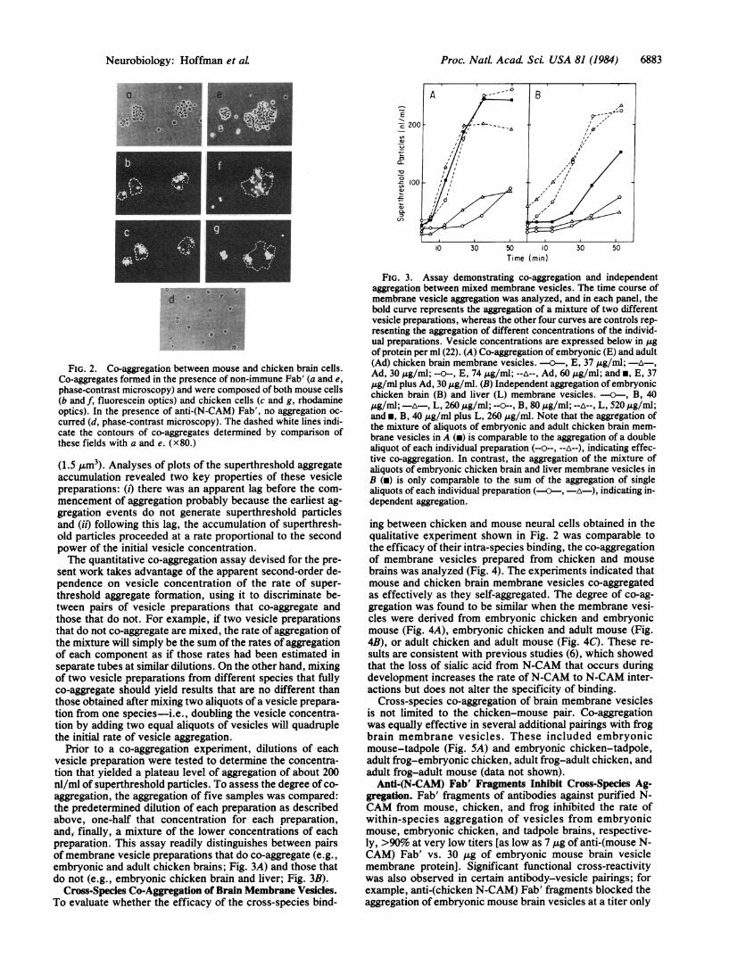
FIG. 3. Assay demonstrating co-aggregation and independentaggregation between mixed membrane vesicles. The time course ofmembrane vesicle aggregation was analyzed, and in each panel, thebold curve represents the aggregation of a mixture of two differentvesicle preparations, whereas the other four curves are controls rep-resenting the aggregation of different concentrations of the individ-ual preparations. Vesicle concentrations are expressed below in ,4gof protein per ml (22). (A) Co-aggregation of embryonic (E) and adult(Ad) chicken brain membrane vesicles. - , E, 37 ,ug/ml; -A ,Ad, 30 ,4g/ml; --o--, E, 74 pg/ml; --A--, Ad, 60 A&g/ml; and *, E, 37gg/ml plus Ad, 30 ug/ml. (B) Independent aggregation of embryonicchicken brain (B) and liver (L) membrane vesicles. - , B, 40,ug/ml;-A-, L, 260ug/mil; ----B, 80,4g/ml;--A--, L, 520Ag/ml;and *, B, 40 ,ug/ml plus L, 260 ,ug/ml. Note that the aggregation ofthe mixture of aliquots of embryonic and adult chicken brain mem-brane vesicles in A (m) is comparable to the aggregation of a doublealiquot of each individual preparation (--0--, --A--), indicating effec-tive co-aggregation. In contrast, the aggregation of the mixture ofaliquots of embryonic chicken brain and liver membrane vesicles inB (m) is only comparable to the sum of the aggregation of singlealiquots of each individual preparation (---, A), indicating in-dependent aggregation.
ing between chicken and mouse neural cells obtained in thequalitative experiment shown in Fig. 2 was comparable tothe efficacy of their intra-species binding, the co-aggregationof membrane vesicles prepared from chicken and mousebrains was analyzed (Fig. 4). The experiments indicated thatmouse and chicken brain membrane vesicles co-aggregatedas effectively as they self-aggregated. The degree of co-ag-gregation was found to be similar when the membrane vesi-cles were derived from embryonic chicken and embryonicmouse (Fig. 4A), embryonic chicken and adult mouse (Fig.4B), or adult chicken and adult mouse (Fig. 4C). These re-sults are consistent with previous studies (6), which showedthat the loss of sialic acid from N-CAM that occurs duringdevelopment increases the rate ofN-CAM to N-CAM inter-actions but does not alter the specificity of binding.
Cross-species co-aggregation of brain membrane vesiclesis not limited to the chicken-mouse pair. Co-aggregationwas equally effective in several additional pairings with frogbrain membrane vesicles. These included embryonicmouse-tadpole (Fig. 5A) and embryonic chicken-tadpole,adult frog-embryonic chicken, adult frog-adult chicken, andadult frog-adult mouse (data not shown).Anti-(N-CAM) Fab' Fragments Inhibit Cross-Species Ag-
gregation. Fab' fragments of antibodies against purified N-CAM from mouse, chicken, and frog inhibited the rate ofwithin-species aggregation of vesicles from embryonicmouse, embryonic chicken, and tadpole brains, respective-ly, >90% at very low titers [as low as 7 /ig of anti-(mouse N-CAM) Fab' vs. 30 Aug of embryonic mouse brain vesiclemembrane protein]. Significant functional cross-reactivitywas also observed in certain antibody-vesicle pairings; forexample, anti-(chicken N-CAM) Fab' fragments blocked theaggregation of embryonic mouse brain vesicles at a titer only
Neurobiology: Hoffman et aL
6884 Neurobiology: Hoffman et at
Time (min)
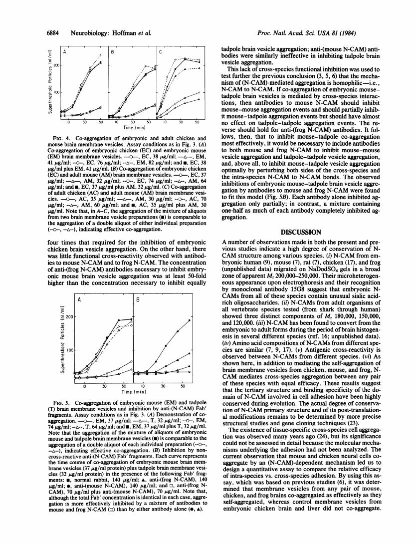
FIG. 4. Co-aggregation of embryonic and adult chicken andmouse brain membrane vesicles. Assay conditions as in Fig. 3. (A)Co-aggregation of embryonic chicken (EC) and embryonic mouse
(EM) brain membrane vesicles. -a--, EC, 38 j&g/ml; -A-, EM,41 jug/ml; --o--, EC, 76 /Ag/ml; --A--, EM, 82 j&g/ml; and *, EC, 38,ug/ml plus EM, 41 Ag/ml. (B) Co-aggregation of embryonic chicken(EC) and adult mouse (AM) brain membrane vesicles. 0---, EC, 37,ug/ml; - A-, AM, 32 ,ug/ml; --o--, EC, 74 ,ug/ml; --A--, AM, 64,ug/ml; andm, EC, 37 jug/ml plus AM, 32 ,ug/ml. (C) Co-aggregationof adult chicken (AC) and adult mouse (AM) brain membrane vesi-cles. -a--, AC, 35 ,ug/ml; -A-, AM, 30 ,ug/ml; --o--, AC, 70,ug/ml; --A--, AM, 60 tg/ml; and *, AC, 35 Ag/ml plus AM, 30,zg/ml. Note that, in A-C, the aggregation of the mixture of aliquotsfrom two brain membrane vesicle preparations (m) is comparable tothe aggregation of a double aliquot of either individual preparation(--v--, --A--), indicating effective co-aggregation.
four times that required for the inhibition of embryonicchicken brain vesicle aggregation. On the other hand, therewas little functional cross-reactivity observed with antibod-ies to mouse N-CAM and to frog N-CAM. The concentrationof anti-(frog N-CAM) antibodies necessary to inhibit embry-onic mouse brain vesicle aggregation was at least 50-foldhigher than the concentration necessary to inhibit equally
A B
cE 200-
'2 !1c00
I0 30 50 10 30 50
Time (min)
FIG. 5. Co-aggregation of embryonic mouse (EM) and tadpole(T) brain membrane vesicles and inhibition by anti-(N-CAM) Fab'fragments. Assay conditions as in Fig. 3. (A) Demonstration of co-
aggregation. -0---, EM, 37 ,g/ml; -A-, T, 32 ,tg/ml; --o--, EM,74 ,ug/ml; --A--, T, 64 ,ug/ml; and *, EM, 37 ,ug/ml plus T, 32 tug/ml.Note that the aggregation of the mixture of aliquots of embryonicmouse and tadpole brain membrane vesicles (m) is comparable to theaggregation of a double aliquot of each individual preparation (--a--,--A--), indicating effective co-aggregation. (B) Inhibition by non-
cross-reactive anti-(N-CAM) Fab' fragments. Each curve representsthe time course of co-aggregation of embryonic mouse brain mem-brane vesicles (37 ,ug/ml protein) plus tadpole brain membrane vesi-cles (32 ,ug/ml protein) in the presence of the following Fab' frag-ments: m, normal rabbit, 140 /Lg/ml; A, anti-(frog N-CAM), 140,ug/ml; *, anti-(mouse N-CAM), 140 tug/ml; and a, anti-(frog N-CAM), 70 Ag/ml plus anti-(mouse N-CAM), 70 tug/ml. Note that,although the total Fab' concentration is identical in each case, aggre-gation is more effectively inhibited by a mixture of antibodies tomouse and frog N-CAM (a) than by either antibody alone (e, A).
tadpole brain vesicle aggregation; anti-(mouse N-CAM) anti-bodies were similarly ineffective in inhibiting tadpole brainvesicle aggregation.
This lack of cross-species functional inhibition was used totest further the previous conclusion (3, 5, 6) that the mecha-nism of (N-CAM)-mediated aggregation is homophilic-i.e.,N-CAM to N-CAM. If co-aggregation of embryonic mouse-tadpole brain vesicles is mediated by cross-species interac-tions, then antibodies to mouse N-CAM should inhibitmouse-mouse aggregation events and should partially inhib-it mouse-tadpole aggregation events but should have almostno effect on tadpole-tadpole aggregation events. The re-verse should hold for anti-(frog N-CAM) antibodies. It fol-lows, then, that to inhibit mouse-tadpole co-aggregationmost effectively, it would be necessary to include antibodiesto both mouse and frog N-CAM to inhibit mouse-mousevesicle aggregation and tadpole-tadpole vesicle aggregation,and, above all, to inhibit mouse-tadpole vesicle aggregationoptimally by perturbing both sides of the cross-species andthe intra-species N-CAM to N-CAM bonds. The observedinhibitions of embryonic mouse-tadpole brain vesicle aggre-gation by antibodies to mouse and frog N-CAM were foundto fit this model (Fig. 5B). Each antibody alone inhibited ag-gregation only partially; in contrast, a mixture containingone-half as much of each antibody completely inhibited ag-gregation.
DISCUSSIONA number of observations made in both the present and pre-vious studies indicate a high degree of conservation of N-CAM structure among various species. (i) N-CAM from em-bryonic human (9), mouse (7), rat (7), chicken (17), and frog(unpublished data) migrated on NaDodSO4 gels in a broadzone of apparent Mr 200,000-250,000. Their microheterogen-eous appearance upon electrophoresis and their recognitionby monoclonal antibody 15G8 suggest that embryonic N-CAMs from all of these species contain unusual sialic acid-rich oligosaccharides. (it) N-CAMs from adult organisms ofall vertebrate species tested (from shark through human)showed three distinct components of M, 180,000, 150,000,and 120,000. (iii) N-CAM has been found to convert from theembryonic to adult forms during the period of brain histogen-esis in several different species (ref. 16; unpublished data).(iv) Amino acid compositions ofN-CAMs from different spe-cies are similar (7, 9, 17). (v) Antigenic cross-reactivity isobserved between N-CAMs from different species. (Vi) Asshown here, in addition to mediating the self-aggregation ofbrain membrane vesicles from chicken, mouse, and frog, N-CAM mediates cross-species aggregation between any pairof these species with equal efficacy. These results suggestthat the tertiary structure and binding specificity of the do-main of N-CAM involved in cell adhesion have been highlyconserved during evolution. The actual degree of conserva-tion of N-CAM primary structure and of its post-translation-al modifications remains to be determined by more precisestructural studies and gene cloning techniques (23).The existence of tissue-specific cross-species cell aggrega-
tion was observed many years ago (24), but its significancecould not be assessed in detail because the molecular mecha-nisms underlying the adhesion had not been analyzed. Thecurrent observation that mouse and chicken neural cells co-aggregate by an (N-CAM)-dependent mechanism led us todesign a quantitative assay to compare the relative efficacyof intra-species vs. cross-species adhesion. By using this as-say, which was based on previous studies (6), it was deter-mined that membrane vesicles from any pair of mouse,chicken, and frog brains co-aggregated as effectively as theyself-aggregated, whereas control membrane vesicles fromembryonic chicken brain and liver did not co-aggregate.
Proc. Natl. Acad Sci. USA 81 (1984)
Proc. NatL Acad. Sci. USA 81 (1984) 6885
Cross-species co-aggregation occurred equally effectivelywith embryonic membranes, adult membranes, or mixturesof embryonic and adult membranes, in accord with previousresults indicating that the E -* A conversion in N-CAMstructure increases the rate of membrane vesicle aggregationbut does not alter its specificity.When mouse and chicken neural cells were coincubated,
co-aggregates formed that frequently contained clumps oftwo to four cells from the same species (Fig. 2 b and c). Atpresent, we cannot distinguish whether this effect is due tooccasional incomplete dissociation of the parent tissue, tospecies-specific adhesive mechanisms mediated by mole-cules other than N-CAM, or to a higher intra-species N-CAM binding affinity. More sensitive assays than the vesicleco-aggregation assay might, in fact, reveal fine differences incross-species aggregation that were not detected in the pre-sent studies.The current observation that cross-species vesicle aggre-
gation is most effectively inhibited when antibodies againstN-CAM from both species are present further supports theconclusion that N-CAM mediates adhesion by a homophilicmechanism-i.e., N-CAM on one cell binds to N-CAM on asecond cell (6). In view of the symmetry requirements of ho-mophilic binding and the impossibility of mirror symmetry inproteins made of L-amino acids, it would seem that two dif-ferent complementary sites or subsites ought to exist withineach N-CAM binding domain to mediate N-CAM to N-CAMinteractions. Mutations altering either of these sites would,by symmetry, affect binding of the complementary site;those that diminished binding would therefore be particular-ly deleterious. The observed conservation of the specificityof N-CAM homophilic binding suggests that the structure ofeach of these complementary binding sites has been evolu-tionarily conserved.
Characteristic N-CAM polypeptides appeared in all of thevertebrate species examined here, including the cartilagi-nous fish, suggesting that (N-CAM)-like precursors werepresent on ancestor species arising at least 470 million yearsago (25). The fact that mouse and frog N-CAMs can bindeffectively to each other even though the ancestors of thesespecies diverged about 400 million years ago (25) indicatesthat the conservation of N-CAM function is almost equallyancient. Such observations do not rule out the possibilitythat N-CAM analogues exist in invertebrate species thatarose much earlier; indeed, in view of the relatively ancientorigin of neural tissue, we expect such molecules to befound.The results of the present study are consistent with the
idea that (N-CAM)-mediated morphogenetic events are fun-damental in developmental regulation and suggest that overrelatively long periods of evolutionary time little variation inthe specificity of those regions of N-CAM that mediate bind-ing and cell adhesion has occurred. In view of occasionallarge differences in morphology within nervous systems ofthe same and different animal classes, this, in turn, suggests
that differences in schedules of CAM gene expression (1)and CAM modulation (3) are likely to be found both withinand across different vertebrate classes, accounting in partfor their differences in form.
We thank Mr. George Eberhardt for help in collecting sharks andMs. Alison Schroeder and Ms. Elizabeth McAnaney for excellenttechnical assistance. This work was supported by U.S. PublicHealth Service Grants HD-09635, HD-16550, AI-11378, and AM-04256 and by a fellowship to S.H. from R. J. Reynolds Industries.
1. Edelman, G. M. (1984) Proc. Natl. Acad. Sci. USA 81, 1460-1464.
2. Edelman, G. M. (1984) Annu. Rev. Neurosci. 7, 339-377.3. Edelman, G. M. (1983) Science 219, 450-457.4. Thiery, J.-P., Brackenbury, R., Rutishauser, U. & Edelman,
G. M. (1977) J. Biol. Chem. 252, 6841-6845.5. Rutishauser, U., Hoffman, S. & Edelman, G. M. (1982) Proc.
Natl. Acad. Sci. USA 79, 685-689.6. Hoffman, S. & Edelman, G. M. (1983) Proc. Natl. Acad. Sci.
USA 81, 5762-5766.7. Chuong, C.-M., McClain, D. A., Streit, P. & Edelman, G. M.
(1982) Proc. Nail. Acad. Sci. USA 79, 4234-4238.8. Sadoul, R., Him, M., Deagostini-Bazin, H., Rougon, G. &
Goridis, C. (1983) Nature (London) 304, 347-349.9. McClain, D. A. & Edelman, G. M. (1982) Proc. Nadl. Acad.
Sci. USA 79, 6380-6384.10. Fraser, S. E., Murray, B. A., Chuong, C.-M. & Edelman,
G. M. (1984) Proc. Natl. Acad. Sci. USA 81, 4222-4226.11. Thiery, J.-P., Duband, J.-L., Rutishauser, U. & Edelman,
G. M. (1982) Proc. Natl. Acad. Sci. USA 79, 6737-6741.12. Edelman, G. M., Gallin, W. J., Delouvee, A., Cunningham,
B. A. & Thiery, J.-P. (1983) Proc. Natl. Acad. Sci. USA 80,4384-4388.
13. Rothbard, J. B., Brackenbury, R., Cunningham, B. A. &Edelman, G. M. (1982) J. Biol. Chem. 257, 11064-11069.
14. Edelman, G. M. & Chuong, C.-M. (1982) Proc. Nail. Acad.Sci. USA 79, 7036-7040.
15. Rougon, G., Deagostini-Bazin, H., Him, M. & Goridis, C.(1982) EMBO J. 1, 1239-1244.
16. Chuong, C.-M. & Edelman, G. M. (1984) J. Neurosci. 4, 2354-2368.
17. Hoffman, S., Sorkin, B. C., White, P. C., Brackenbury, R.,Mailhammer, R., Rutishauser, U., Cunningham, B. A. & Edel-man, G. M. (1982) J. Biol. Chem. 257, 7720-7729.
18. Brackenbury, R., Thiery, J.-P., Rutishauser, U. & Edelman,G. M. (1977) J. Biol. Chem. 252, 6835-6840.
19. Brackenbury, R., Rutishauser, U. & Edelman, G. M. (1981)Proc. Nail. Acad. Sci. USA 78, 387-391.
20. Burt, R. & Gierer, A. (1979) Wilhelm Roux' Arch. Entwick-lungsmech. Org. 187, 367-373.
21. Gallin, W. J., Edelman, G. M. & Cunningham, B. A. (1983)Proc. Nail. Acad. Sci. USA 80, 1038-1042.
22. Lowry, 0. H., Rosebrough, N. J., Farr, A. L. & Randall,R. J. (1951) J. Biol. Chem. 193, 265-275.
23. Murray, B. A., Hemperly, J. J., Gallin, W. J., MacGregor,J. B., Edelman, G. M. & Cunningham, B. A. (1984) Proc.Nail. Acad. Sci. USA 81, 5585-5589.
24. Moscona, A. (1957) Proc. Natl. Acad. Sci. USA 43, 184-194.25. Romer, A. S. (1968) The Procession of Life (World Press,
Cleveland, OH).
Neurobiology: Hoffman et aL