Embed Size (px)
Citation preview
Evolution of predetermined germ cells in vertebrate embryos:
implications for macroevolution
Andrew D. Johnson,a,b,* Matthew Drum,b Rosemary F. Bachvarova,c Thomas Masi,b Mary E. White,d
and Brian I. Crotherd
aDivision of Genetics, University of Nottingham, Queen’s Medical Centre, Nottingham NG7 2UH, UKbDepartment of Biological Science, Florida State University, Tallahassee, FL 32306, USAcDepartment of Cell and Developmental Biology, Weill Medical College of Cornell University, 1300 York Avenue,
New York, NY 10021, USAdDepartment of Biological Science, Southeastern Louisiana University, Hammond, LA 70402, USA*Author for correspondence (e-mail: [email protected])
SUMMARY The germ line is established in animal embryoswith the formation of primordial germ cells (PGCs), which giverise to gametes. Therefore, the need to form PGCs can act asa developmental constraint by inhibiting the evolution ofembryonic patterning mechanisms that compromise theirdevelopment. Conversely, events that stabilize the PGCsmay liberate these constraints. Two modes of germ celldetermination exist in animal embryos: (a) either PGCs arepredetermined by the inheritance of germ cell determinants(germ plasm) or (b) PGCs are formed by inducing signalssecreted by embryonic tissues (i.e., regulative determination).Surprisingly, among the major extant amphibian lineages, onemechanism is found in urodeles and the other in anurans. Inanuran amphibians PGCs are predetermined by germ plasm;in urodele amphibians PGCs are formed by inducing signals.
To determine which mechanism is ancestral to the tetrapodlineage and to understand the pattern of inheritance in highervertebrates, we used a phylogenetic approach to analyzebasic morphological processes in both groups and correlatedthese with mechanisms of germ cell determination. Our resultsindicate that regulative germ cell determination is a property ofembryos retaining ancestral embryological processes,whereas predetermined germ cells are found in embryoswith derived morphological traits. These correlationssuggest that regulative germ cell formation is an importantdevelopmental constraint in vertebrate embryos, actingbefore the highly conserved pharyngula stage. Moreover,our analysis suggests that germ plasm has evolvedindependently in several lineages of vertebrate embryos.
INTRODUCTION
Variations in adult morphology arising during evolution are
attributable to changes in developmental processes. The
embryos of many vertebrates pass through a morphologically
similar stage, the pharyngula, occurring after the completion
of neurulation, which is characterized by visible somites,
prominent pharyngeal arches, and ventral flexure occurring
at the position of the neck (i.e., cervical flexure). Embryo-
logical variation occurring after the pharyngula stage plays a
major role in diversification at higher order taxonomic levels
(Richardson 1999), and understanding the molecular me-
chanisms underlying such diversification is currently an area
of intense research. However, it is likely that variation of
earlier developmental processes, occurring before and
during the pharyngula stage, will result in the most pro-
found morphological variation, even though these events
may also be the most highly constrained and resistant to
change (Raff 1996). In this review we consider the evolution
of an early developmental process, the germ cell deter-
mining mechanisms among vertebrates, and its relation
to developmental processes occurring before and after
the pharyngula stage. Throughout we use the two major
amphibian groups, the anurans (frogs and toads) and the
urodeles (salamanders and newts), as the prime examples for
discussion, but we also consider several issues at the level of
the vertebrates.
Anuran and urodele embryos begin to diverge before
gastrulation, at least a day before the pharyngula stage. When
compared within a phylogenetic context, we note that in all
cases early (prepharyngula) anuran development appears to
be more derived and shows significant variability among
individual species, whereas urodele development retains
ancestral features and is less variable within the various
urodele lineages that have been examined. Because the germ
line develops by very different mechanisms in anuran and
urodele embryos, we next considered if this is related to the
observed differences in morphogenesis in these groups.
EVOLUTION & DEVELOPMENT 5:4, 414–431 (2003)
& BLACKWELL PUBLISHING, INC.414
In anuran embryos primordial germ cells (PGCs), the cells
that give rise to gametes, are of endodermal origin, and they
are specified by the differential distribution of maternally
deposited germ cell determinants (known as germ plasm) to
the presumptive germ line blastomeres. Thus, from the
inception of development, anuran PGCs are considered to
be predetermined by germ plasm. In urodele embryos PGCs
derive from lateral plate mesoderm. Urodele embryos do not
contain germ plasm, and so PGCs are specified later
in development than in anurans. In urodeles PGCs form in
response to extracellular inducing signals, not unlike those
that produce other mesodermal cell types. This is considered
regulative germ cell specification. When compared within a
phylogenetic context, again the anuran mode of development
appears to be derived, whereas the urodele mechanism
appears to be primitive to the tetrapods. Furthermore, when
considering the phylogeny of animals with and without germ
plasm, the data suggest that germ plasm has evolved
independently in several lineages of vertebrate embryos.
Development of the notochord is a fundamental aspect of
vertebrate embryogenesis. We identified a strong correlation
between the position of the notochord at the completion of
gastrulation and the mode of germ cell specification carried
out by embryos. Thus, regulative germ cell specification is
correlated, in diverse groups, with the ancestral mode of
notochord development. In contrast, a derived process of
notochord development is observed in embryos that develop
with a predetermined germ line. Within this context, we
discuss how the emergence of germ plasm within an individual
lineage may alleviate developmental constraints that are
imposed on morphogenetic movements by the process of
regulative germ cell specification and that regulative germ cell
specification may cause the retention of ancestral embryolog-
ical characters. In this way germ plasm may facilitate the
evolution of novel embryological features and thereby
contribute to macroevolutionary divergence of adult form.
EARLY MORPHOGENESIS DIVERGES BEFORETHE PHARYNGULA STAGE IN AXOLOTLSAND XENOPUS
Nothing in biology makes sense except when seen in the light of
evolution.
T. Dobzhansky (1973)
The fact that vertebrate embryos from diverse taxa and very
different early embryological forms (such as fish, amphibians,
and mammals) converge on a similar morphology at the
pharyngula stage has long been recognized (Von Baer 1828),
and it has been postulated that passage through the
pharyngula stage serves as a constraint on the potential for
morphological variation. Indeed, after the pharyngula stage
embryonic form diverges in a species-specific manner, leading
to the view that passage through the pharyngula stage
represents a developmental bottleneck (for discussions of the
‘‘hourglass model’’ describing the pharyngula stage as a
developmental bottleneck, see Elinson 1987; Raff 1996;
Gerhart and Kirschner 1997). Changes in developmental
processes occurring before the pharyngula stage, however,
may result in profound alterations to body structure, that is,
macroevolution (Raff 1996). An excellent example of early
developmental changes correlating with differences in adult
morphology are found in the major amphibian lineages.
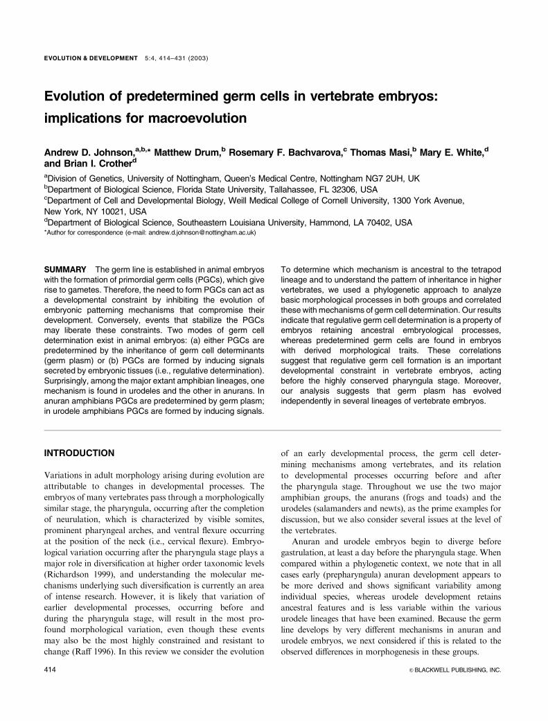
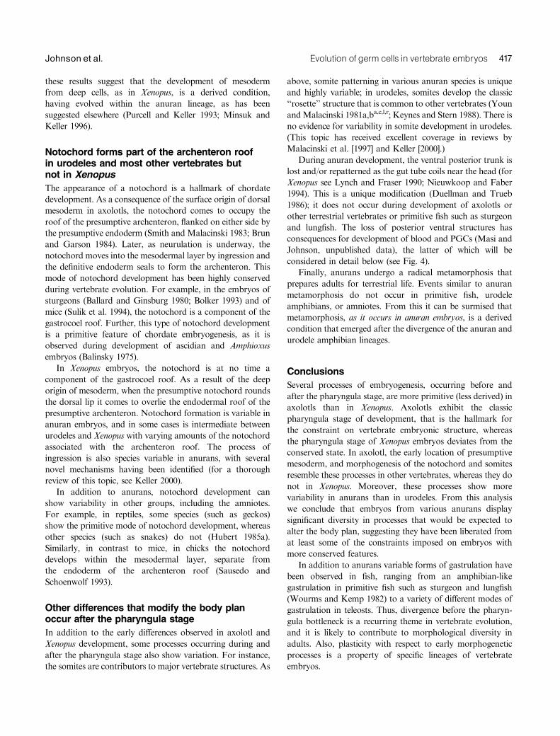
Pharyngula stage embryos and adults of Xenopus laevis (a
frog) and axolotl (Ambystoma mexicanum, a salamander) are
compared in Figure 1. Note that inXenopus embryos (Fig. 1A)
the cervical region is reduced and does not undergo flexure
(marking the future position of the neck) as it does in axolotls
(Fig. 1B, arrow) or other vertebrates. X-rays of adult Xenopus
and axolotl females show the vastly different skeletal structure
of these animals. The axolotl (Fig. 1, E and F) has a skeletal
structure similar to that of most vertebrates, including two
cervical vertebrae in the neck and 18 total vertebrae (arrow);
Xenopus (Fig. 1, C and D), in contrast, has a much shortened
vertebral column with only eight total vertebrae and one
cervical vertebra. In addition, Xenopus also has the expanded
pelvic girdle and lengthened hindlimbs typical of most frogs.
Anurans and urodeles diverged from a common ancestor
about 250 million years ago (Cannatella and Hillis 1992;
Milner 1992), and therefore it is likely that only one of the
patterns of early development observed in the extant
amphibians reflects the ancestral pattern. The other mode
would be a derived pattern. The adult urodele body plan more
closely approximates that of the common amphibian ancestor
than does that of anurans (Duellman and Trueb 1986),
particularly that of Xenopus, which is highly derived
(Cannatella and De Sa 1993). Here we compare various
aspects of embryonic development. For clarification, the
anuran or urodele species referred to in each study are
identified in text by superscripted letters that are listed in
Table 1. We conclude that urodele embryos have retained
more features that can be considered primitive to vertebrates,
and anuran embryos are generally more derived. Although
comparative aspects of amphibian morphogenesis have been
reviewed before (Hanken 1986; Malacinski et al. 1997), this
has not been addressed within a framework assuming a
monophyletic origin of urodeles and anurans nor with
insights available from more recent work.
Origin of mesoderm from deep cells, as occursin Xenopus, is a derived condition, whereasurodeles retain a more conserved mode ofmesoderm morphogenesis
A variety of evidence demonstrates that in Xenopus embryos
only a very small amount of the dorsal mesoderm originates
Evolution of germ cells in vertebrate embryos 415Johnson et al.
from cells that are on the surface epithelium at the gastrula
stage. Almost all the mesoderm originates from deep cells
underlying the surface epithelium (Keller 1975, 1976; Smith
and Malacinski 1983; Minsuk and Keller 1996; for a review
see Keller 2000). Those surface cells that involute around the
blastopore lips give rise primarily to endoderm. In contrast, in
axolotls about half of the dorsal mesoderm originates within
the surface epithelium (Smith and Malacinski 1983). The
presumptive paraxial mesoderm moves out of the epithelial
layer as it rounds the lateral lips of the blastopore (Lundmark
1986), ingressing into the space between ectoderm and
endoderm in a manner resembling the movements around
the primitive streak in mammals (Shook et al. 2002).
Comparative analysis suggests that the derivation of
mesoderm from surface cells is the ancestral condition for
vertebrate development. For example, in addition to axolotls,
mesoderm originates from surface cells in all other urodele
amphibians that have been studied (Vogt 1929b; Pasteels
1942c; Delarue et al. 1992c; Shook et al. 2002d,e) and most
informatively in sturgeons, a primitive chondrostean fish
(Ballard and Ginsburg 1980; Bolker 1993). Indeed, the origin
of mesoderm only from deep cells appears limited even
among anurans, as many anuran species other than Xenopus
develop dorsal mesoderm from surface cells (Vogt 1929m;
Pasteels 1942n; Purcell and Keller 1993o; Delarue et al. 1994p;
Minsuk and Keller 1996q), similar to urodeles. Together
Fig. 1. Comparison of Xenopus and axolotl embryos and adults. (A) Xenopus embryos during ‘‘pharyngula’’ stages. (B) Axolotl embryosduring pharyngula stages. Note cervical flexure at position of arrow in axolotl. Xenopus embryos do not undergo the prominent cervicalflexure (compare A and B). (For both Xenopus and axolotl, top embryo at tailbud stage 26, bottom embryo at tailbud stage 28; Nieuwkoopand Faber [1994] stages for Xenopus. Bordzilovskaya et al. [1989] stages for axolotl. Staging is roughly equivalent for both species.) (C–F)X-rays taken through adult females to show skeletal structure. (C) Adult Xenopus female. Note highly irregular vertebral pattern, includingextended pelvic bones. Xenopus retains only eight presacral vertebrae. (D) Close-up of animal shown in C, showing neck region. Note singlecervical vertebra (position of arrow). (E) Adult axolotl female. Axolotls adults have 18 presacral vertebrae. (Bright regions in gut areundigested food.) (F) Close-up of neck region of animal in A showing two cervical vertebrae (arrow).
Table 1. Amphibian species for which data are discussedfor aspects of morphogenesis or germ cell formation are
referred to in the text by superscript letters
Urodeles Anurans
a. Ambystoma mexicanum l. Xenopus laevis
b. Triton m. Bombinator
c. Pleurodeles waltlii n. Discoglossus
d. Ambystoma maculatum o. Ceratophrys ornata
e. Taricha granulosa p. Rana pipiens
f. Hemidactylium scutatum q. Hymenochirus
g. Triturus viridescens r. Rana sphenocephala
h. Triturus torosus s. Rana temporaria
i. Ambystoma jeffersonianum t. Rana esculenta
j. Triturus alpestris u. Bufo bufo
k. Triturus cristatus carnifex
416 EVOLUTION & DEVELOPMENT Vol. 5, No. 4, July^August 2003
these results suggest that the development of mesoderm
from deep cells, as in Xenopus, is a derived condition,
having evolved within the anuran lineage, as has been
suggested elsewhere (Purcell and Keller 1993; Minsuk and
Keller 1996).
Notochord forms part of the archenteron roofin urodeles and most other vertebrates butnot in Xenopus
The appearance of a notochord is a hallmark of chordate
development. As a consequence of the surface origin of dorsal
mesoderm in axolotls, the notochord comes to occupy the
roof of the presumptive archenteron, flanked on either side by
the presumptive endoderm (Smith and Malacinski 1983; Brun
and Garson 1984). Later, as neurulation is underway, the
notochord moves into the mesodermal layer by ingression and
the definitive endoderm seals to form the archenteron. This
mode of notochord development has been highly conserved
during vertebrate evolution. For example, in the embryos of
sturgeons (Ballard and Ginsburg 1980; Bolker 1993) and of
mice (Sulik et al. 1994), the notochord is a component of the
gastrocoel roof. Further, this type of notochord development
is a primitive feature of chordate embryogenesis, as it is
observed during development of ascidian and Amphioxus
embryos (Balinsky 1975).
In Xenopus embryos, the notochord is at no time a
component of the gastrocoel roof. As a result of the deep
origin of mesoderm, when the presumptive notochord rounds
the dorsal lip it comes to overlie the endodermal roof of the
presumptive archenteron. Notochord formation is variable in
anuran embryos, and in some cases is intermediate between
urodeles and Xenopus with varying amounts of the notochord
associated with the archenteron roof. The process of
ingression is also species variable in anurans, with several
novel mechanisms having been identified (for a thorough
review of this topic, see Keller 2000).
In addition to anurans, notochord development can
show variability in other groups, including the amniotes.
For example, in reptiles, some species (such as geckos)
show the primitive mode of notochord development, whereas
other species (such as snakes) do not (Hubert 1985a).
Similarly, in contrast to mice, in chicks the notochord
develops within the mesodermal layer, separate from
the endoderm of the archenteron roof (Sausedo and
Schoenwolf 1993).
Other differences that modify the body planoccur after the pharyngula stage
In addition to the early differences observed in axolotl and
Xenopus development, some processes occurring during and
after the pharyngula stage also show variation. For instance,
the somites are contributors to major vertebrate structures. As
above, somite patterning in various anuran species is unique
and highly variable; in urodeles, somites develop the classic
‘‘rosette’’ structure that is common to other vertebrates (Youn
andMalacinski 1981a,ba,c,l,r; Keynes and Stern 1988). There is
no evidence for variability in somite development in urodeles.
(This topic has received excellent coverage in reviews by
Malacinski et al. [1997] and Keller [2000].)
During anuran development, the ventral posterior trunk is
lost and/or repatterned as the gut tube coils near the head (for
Xenopus see Lynch and Fraser 1990; Nieuwkoop and Faber
1994). This is a unique modification (Duellman and Trueb
1986); it does not occur during development of axolotls or
other terrestrial vertebrates or primitive fish such as sturgeon
and lungfish. The loss of posterior ventral structures has
consequences for development of blood and PGCs (Masi and
Johnson, unpublished data), the latter of which will be
considered in detail below (see Fig. 4).
Finally, anurans undergo a radical metamorphosis that
prepares adults for terrestrial life. Events similar to anuran
metamorphosis do not occur in primitive fish, urodele
amphibians, or amniotes. From this it can be surmised that
metamorphosis, as it occurs in anuran embryos, is a derived
condition that emerged after the divergence of the anuran and
urodele amphibian lineages.
Conclusions
Several processes of embryogenesis, occurring before and
after the pharyngula stage, are more primitive (less derived) in
axolotls than in Xenopus. Axolotls exhibit the classic
pharyngula stage of development, that is the hallmark for
the constraint on vertebrate embryonic structure, whereas
the pharyngula stage of Xenopus embryos deviates from the
conserved state. In axolotl, the early location of presumptive
mesoderm, and morphogenesis of the notochord and somites
resemble these processes in other vertebrates, whereas they do
not in Xenopus. Moreover, these processes show more
variability in anurans than in urodeles. From this analysis
we conclude that embryos from various anurans display
significant diversity in processes that would be expected to
alter the body plan, suggesting they have been liberated from
at least some of the constraints imposed on embryos with
more conserved features.
In addition to anurans variable forms of gastrulation have
been observed in fish, ranging from an amphibian-like
gastrulation in primitive fish such as sturgeon and lungfish
(Wourms and Kemp 1982) to a variety of different modes of
gastrulation in teleosts. Thus, divergence before the pharyn-
gula bottleneck is a recurring theme in vertebrate evolution,
and it is likely to contribute to morphological diversity in
adults. Also, plasticity with respect to early morphogenetic
processes is a property of specific lineages of vertebrate
embryos.
Evolution of germ cells in vertebrate embryos 417Johnson et al.
DEVELOPMENT OF THE GERM LINE
In bilateral animals PGCs originate outside of the gonads,
to which they are transported later in development. Typically,
PGCs arrive at the gonads after the rudiments of the
embryonic body plan have been established, and therefore
it is possible to have separate influences govern the
development of the germ cells and the somatic cells. Higher
animals have at least two very different means for specifying
PGCs. Some species have a predetermined germ cell lineage
that is specified by cytoplasmic germ cell determinants laid
down in the oocyte and then differentially distributed to
presumptive germ cells during embryogenesis. In other
species, germ cell determination occurs later during
development, and is not directly dependent on maternal
molecules. In these species germ cell determination is
governed by a regulative mode involving cell–cell interactions.
Interestingly, the anurans use one of these mechanisms and
urodeles the other.
A priori, it is not possible to predict which mechanism
of germ cell determination is ancestral to the tetrapod
lineage. Little descriptive data exist describing germ cell
development in vertebrate embryos that could lead to
robust conclusions concerning the mechanism in embryos
of the tetrapod ancestor (Nieuwkoop and Sutasurya 1979),
and experimental data from such species is nonexistent.
Nevertheless, as a first step toward understanding this
process, we discuss below available information concerning
how PGCs are formed in those terrestrial vertebrate species
about which germ cell development has been described in
some detail.
Development of PGCs in amphibians
Germ plasm and germ cell specification in frogembryos
Among anurans most is known about germ cell development
in X. laevis. However, because germ cell formation in Xenopus
is typical of anurans (Blackler 1958), it is likely that most of
the events characterized in Xenopus occur in other frogs as
well. During oogenesis in Xenopus, electron-dense material
known as germ plasm is synthesized and transported to the
vegetal cortex through a specific transport mechanism
involving association with the mitochondrial cloud or
Balbiani body (Heasman et al. 1984; King et al. 1999). Germ
plasm is inherited by the vegetal-most blastomeres of newly
fertilized embryos, within the presumptive endoderm, and
segregated into a few cells at the blastula stage (Houston and
King 2000b; Kloc et al. 2001). Those cells that inherit germ
plasm will give rise to PGCs, and sister cells that do not
inherit germ plasm become typical somatic endoderm. By the
time PGCs reach the gonad, material resembling the electron-
dense germinal granules of germ plasm, called nuage,
accumulates in a perinuclear position within the PGCs. By
tracing germ plasm, or nuage, the germ line can be followed
as an independent cell lineage from the inception of
development through adulthood (for a review, see Nieuw-
koop and Sutasurya 1979). A number of experiments in which
germ plasm was destroyed (Bounoure 1939s; Bounoure et al.
1954s; Padoa 1963t; Smith 1966p; Tanabe and Kotani 1974l;
Zust and Dixon 1975l; Ikenishi and Kotani 1979l) or
transplanted into recipient embryos (Smith 1966p; Wakahara
1977l) suggest that germ plasm contains determinants that
govern germ cell specification.
Recent work has identified molecules that comprise the
germ plasm in Xenopus, and these include germ cell specific
proteins and RNAs. Recent reviews detail the development of
germ plasm and germ cell development in Xenopus extensively
(Houston and King 2000b; Kloc et al. 2001).
PGC development in urodele embryos
Urodeles use mechanisms of germ cell determination that
appear to have little in common with the mechanisms acting
in frogs. In urodele embryos determination of germ cells is
regulated by the response of cells to extracellular signals (i.e.,
regulative germ cell specification).
In urodele embryos vegetal pole germ plasm is absent and
PGCs develop in the posterior-lateral plate mesoderm
(Humphrey 1925,f,g,h 1929d,i; Nieuwkoop 1947a; Smith
1964a; Maufroid and Capuron 1972c; Ikenishi and Nieuw-
koop 1978a), in contrast to the endodermal origin of PGCs in
anurans. Typical of amphibian mesoderm, PGCs can be
induced from cells in the animal cap region of blastula stage
urodele embryos, if these are exposed to signals from the
ventral vegetal blastomeres (Nieuwkoop 1969a; Kocher-
Becker and Tiedemann 1971g; Boterenbrood and Nieuwkoop
1973a; Sutasurja and Nieuwkoop 1974a,c,j,k; Michael 1984a,j;
Maufroid and Capuron 1985c). In addition to PGCs, these
signals induce a variety of somatic mesodermal cell types,
including mesenchyme, mesonephros, and blood, suggesting
the signals that induce germ cells and somatic cells are not
qualitatively different.
The possibility that in urodeles germ plasm is located in the
equatorial region of the egg and plays a role in the production
of PGCs has been discussed for many years (Nieuwkoop and
Sutasurya 1979; Smith et al. 1983; Wakahara 1996). In
Xenopus, expression of the dazl gene (Xdazl) is a marker for
germ plasm (Houston et al. 1998). Using the axolotl homolog
of this sequence, Johnson et al. (2001) were unable to find
localized germ plasm during oogenesis or embryogenesis in
axolotls. They concluded that an anuran-type germ plasm
does not direct the specification of PGCs in urodele embryos.
Rather, PGCs in urodeles are most likely to be induced from
typical embryonic cells, such as those that also produce
somatic derivatives. In support of this, axolotl PGCs
418 EVOLUTION & DEVELOPMENT Vol. 5, No. 4, July^August 2003
commence expression of germ cell-specific genes only after
they come into close contact with the gonadal rudiments
(Johnson et al. 2001), a time that closely coincides with the
appearance of nuage in this species (Ikenishi and Nieuwkoop
1978). Combined with the classic experiments on several
urodele species described above, we conclude that in
urodeles PGCs are determined by zygotic influences,
including posterior-ventral mesoderm inducing signals and
subsequent cell interactions, not maternal molecules, as
in anurans.
Do amniotes retain anuran or urodelegerm cell determining mechanisms?
In addition to amphibians, divergent patterns of germ
cell specification and development have also been identified
in amniote embryos. All amniotes share a common amphi-
bian ancestor, and therefore the embryos of the amphibian
ancestor to the amniote lineage most likely had features
similar to extant urodeles or extant anurans, but not both.
Whether the predetermined mechanism of germ cell determi-
nation in anurans or the regulative mechanism of urodeles is
retained in amniotes is considered below.
PGC development in mammalian embryos
The development of PGCs in mice occurs with striking
parallels to the mechanism in urodele amphibians. In mouse
embryos PGCs originate from cells located within the
proximal epiblast, and the germ cell precursors are clonally
related to the founder cells of the primitive blood and the
allantois (Lawson and Hage 1994). PGCs require signaling
from bone morphogenetic proteins (BMPs) (ventral mesoderm
inducing agents) for their production (Lawson et al. 1999;
Ying and Zhao 2001; Ying et al. 2001). Furthermore, recent
work suggests that interferons may trigger a cascade of gene
expression, leading to lineage restriction of the germ
cells (Saitou et al. 2002). The PGC precursors migrate
through the posterior primitive streak in association with
extraembryonic mesoderm into the allantois, a posterior
extraembryonic region (Ginsburg et al. 1990). Later they
become associated with the hindgut, and from there they
move through the dorsal mesentery to the genital ridges
(Anderson et al. 2000). A similar site of origin of PGCs has
been described for several mammalian species (Nieuwkoop
and Sutasurya 1979).
Germ plasm has not been found in early mouse embryos
(reviewed in Eddy 1975). On the contrary, any cell in the
epiblast of a mouse embryo can give rise to PGCs if placed
within the proper signaling context (Tam and Zhou 1996;
Yoshimizu et al. 2001), indicating that mouse PGCs are
not predetermined. Nuage appears after specification, when
PGCs are in the hindgut (Eddy 1974). More recently
transcription of the mouse vasa gene homolog, mvh
(the vasa gene family is discussed in detail below), has
been shown to commence after PGCs begin to colonize the
genital ridge (Fujiwara et al. 1994; Toyooka et al. 2000),
which is very similar to the timing of vasa transcriptional
activation in axolotls (Bachvarova and Johnson, unpublished
data). Taken together, the results summarized above indicate
that urodele and mouse embryos share similar mechanisms of
PGC development (for further discussion of this view, see
Wakahara 1996; McLaren 1999), as they do mesoderm
morphogenetic patterns.
PGC development in chicken embryos
The development of PGCs in chicks occurs very differently
than in mice. In chicks the PGCs are derived from cells on the
ventral surface of the area pellucida, a central region of the
epiblast; these cells migrate ventrally into the plane of the
hypoblast, a layer of extraembryonic endoderm (Eyal-Giladi
et al. 1981; Sutasurya et al. 1983; Karagenc et al. 1996). After
becoming associated with the hypoblast, the PGCs move
anteriorly to colonize a region known as the germinal
crescent, within the area opaca (on the periphery of the
epiblast), anterior to the cranial-most region of the embryo.
From the germinal crescent the PGCs enter the embryonic
circulation (Nieuwkoop and Sutasurya 1979). They later exit
the circulation in the vicinity of the gonad and are drawn to
the developing genital ridges by chemotactic attraction
(Kuwana et al. 1986).
Tsunekawa et al. (2000) recently isolated a chick homolog
of vasa and showed that vasa protein was present in germ
cells throughout their life cycle. Vasa protein is found in
specific structures associated with the mitochondrial cloud
(a structure involved in germ plasm formation; see below) in
oocytes and is segregated into a few cells during
cleavage. Vasa-containing cells of the area pellucida then
follow the path to the germinal crescent and the gonad
described above for PGCs. This expression pattern suggests
that germ cells are predetermined by maternally inherited
germ plasm in chicks.
PGC development in reptiles
Much less is known about germ cell development in the
embryos of reptiles than in widely studied experimental
systems. Germ plasm has never been identified in reptilian
eggs (Hubert 1985b). Further, indications are that different
species of reptiles use one or the other of the two amniote
patterns of PGC development described above or a com-
bination of both patterns. For instance, in several groups
(Gekkonidae, Iguanidae, Lacertidae, turtles) PGCs follow a
pattern of development very similar to mice, in that PGCs
apparently originate within the epiblast and assume a
posterior location in the mesoderm adjacent to the cloaca
(Hubert 1985a). PGCs in these species migrate to the gonad
Evolution of germ cells in vertebrate embryos 419Johnson et al.
via an interstitial route through the posterior lateral
mesoderm. Other groups (Scincidae, Chamaeleonidae,
Agamidae, Cordylidae, Anguidae, snakes) apparently use
a mechanism similar to the chicks, in which PGCs migrate
to the anterior region, which is presumably equivalent to
the germinal crescent, and reach the gonads through the
circulatory system. Finally, in tuataras (Sphenodon) both
modes may be used, some PGCs showing posterior interstitial
migration and other appearing anteriorly and moving
through the circulation (for reviews, see Nieuwkoop and
Sutasurya 1979; Hubert 1985a,b).
Conclusion
In amniotes, like amphibians, two different modes of germ
cell development have been reported, one in mice in which the
germ cells develop in response to extracellular signals, similar
to urodeles, and another in chicks in which the germ cells
enter the endoderm and are predetermined by maternal
molecules, similar to frogs. Various species of reptiles
apparently display one or the other of these processes. Thus,
within the context that all terrestrial vertebrates share a
common ancestor, two possibilities exist. First, perhaps PGCs
in the embryos of the ancestral tetrapod were predetermined.
In this case frog and chick embryos would represent the
primitive mechanism, and urodeles and mice would have
evolved novel strategies. Second, alternatively, the primitive
condition was retained by urodeles and mammals, and the
predetermined germ line in frogs, and in chicks, is newly
evolved. To address which of these models may be more
accurate requires an analysis of available information from
the embryos of more primitive species.
EVOLUTION OF GERM CELL DETERMININGMECHANISMS IN EMBRYOS
Nothing in evolution makes sense except when seen in the light of
phylogeny.
Jay Savage (1997)
Is the anuran mechanism of germ cellspecification a primitive or convergent trait:phylogenetic analysis of the presence of germplasm in different animals
Much of our knowledge about the molecular genetic
mechanisms that govern animal development has been
obtained from studies using as model systems two widely
divergent animals: X. laevis, representing vertebrates, and
Drosophila melanogaster, representing invertebrates. These
species share strikingly similar mechanisms in the way they
establish the germ line.
The posterior pole of Drosophila eggs contains material
that is structurally and functionally similar to anuran germ
plasm (Mahowald and Hennen 1971) (called pole plasm in
Drosophila). Drosophila pole plasm is inherited by PGCs
during the early stages of development, and it has been
shown to function directly as a determinant of germ cells
(Illmensee and Mahowald 1974; Okada et al. 1974).
More recently, at least one homologous molecule has been
identified in the germ (pole) plasm of Drosophila and Xenopus
(nanos-XCAT2; Mosquera et al. 1993; Forbes and Lehmann
1998). These observations have led to the conclusion that the
role of germ plasm in PGC development is a process that
has been conserved from insects to vertebrates. However,
this view has not been considered within a plausible
phylogenetic context.
As discussed above, germ plasm is present in birds and
frogs but not in mammals or urodeles. Thus, either germ
plasm or the regulative mode of germ cell determination has
evolved independently in more than one lineage. The
morphological evidence presented above suggests that ur-
odeles more closely resemble both the embryonic develop-
ment and adult morphology of the tetrapod ancestor than
frogs, arguing against the view that germ plasm is an ancestral
character of tetrapod embryos. If this is true, then similarities
shared between the mechanisms of germ cell determination in
anurans and chick (and higher insects as well) must be the
result of independently derived traits converging on a
developmental process. However, the likely nature of the
ancestral trait can only be inferred by examining the trait in a
sister-group. To examine this question, we constructed an
abbreviated phylogenetic tree of the deuterostomes showing
the distribution of animals that contain germ plasm in their
eggs and those that do not (Fig. 2A).
Evidence presented below indicates that the closest sister-
group to tetrapods, represented by the lungfish, does not
contain germ plasm (Fig. 3). Going further down the tree,
although the embryos of zebrafish and other teleosts are
known to contain germ plasm, the situation in more primitive
fishes, such as sturgeon, is unclear. Urochordate embryos
appear to contain germ plasm (Fujimura and Takamura
2000); however, they also appear to be capable of producing
germ cells by regulative mechanisms (Takamura et al. 2002).
For this reason they are considered indeterminate. Echino-
derms are a sister-group of the chordate phylum. Germ plasm
has not been identified in early echinoderm embryos,
although oocytes contain nuage (Eddy 1975; Holland 1978).
Furthermore, more recent evidence from deletion studies
indicates that PGCs in sea urchins are not predetermined;
rather, they most likely arise in response to regulative
influences during development (Ransick et al. 1996), suggest-
ing that the regulative mode is primitive. These data were also
subjected to optimization analysis with the program Mac-
Clade to reconstruct the character evolution using parsimony
and to infer the ancestral mode of germ cell determination. As
indicated in Figure 2A, the hypothesis inferred by this analysis
is that the induced or regulative mode is the ancestral
420 EVOLUTION & DEVELOPMENT Vol. 5, No. 4, July^August 2003
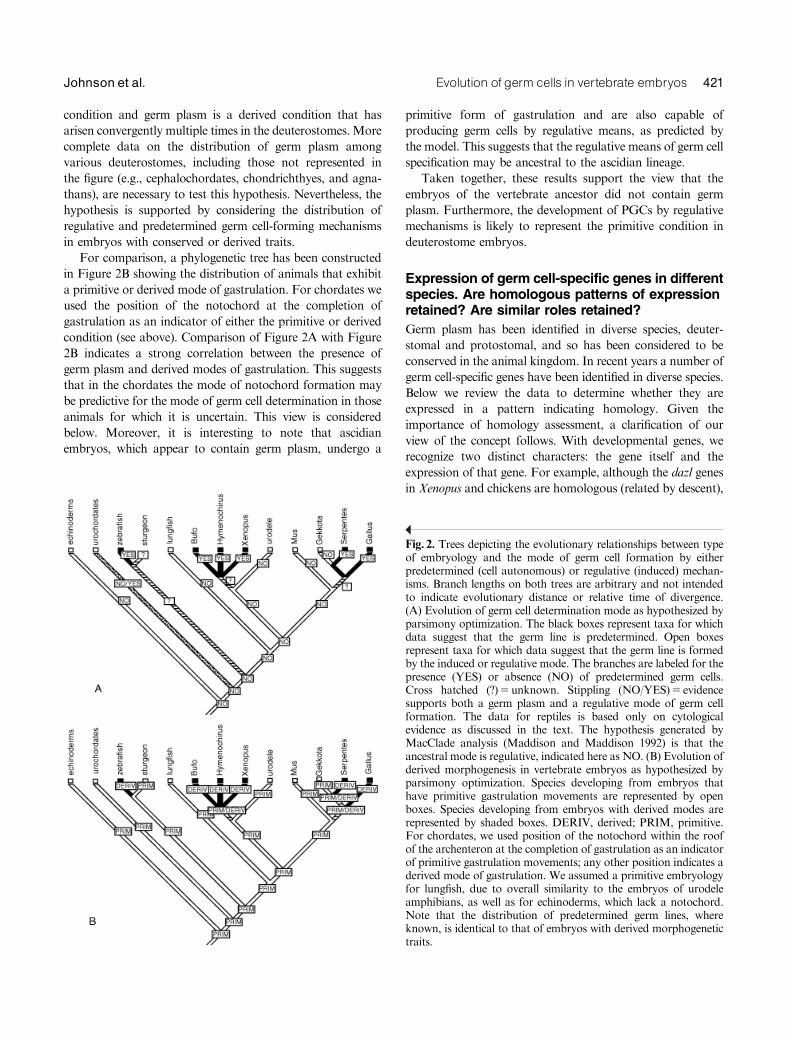
condition and germ plasm is a derived condition that has
arisen convergently multiple times in the deuterostomes. More
complete data on the distribution of germ plasm among
various deuterostomes, including those not represented in
the figure (e.g., cephalochordates, chondrichthyes, and agna-
thans), are necessary to test this hypothesis. Nevertheless, the
hypothesis is supported by considering the distribution of
regulative and predetermined germ cell-forming mechanisms
in embryos with conserved or derived traits.
For comparison, a phylogenetic tree has been constructed
in Figure 2B showing the distribution of animals that exhibit
a primitive or derived mode of gastrulation. For chordates we
used the position of the notochord at the completion of
gastrulation as an indicator of either the primitive or derived
condition (see above). Comparison of Figure 2A with Figure
2B indicates a strong correlation between the presence of
germ plasm and derived modes of gastrulation. This suggests
that in the chordates the mode of notochord formation may
be predictive for the mode of germ cell determination in those
animals for which it is uncertain. This view is considered
below. Moreover, it is interesting to note that ascidian
embryos, which appear to contain germ plasm, undergo a
primitive form of gastrulation and are also capable of
producing germ cells by regulative means, as predicted by
the model. This suggests that the regulative means of germ cell
specification may be ancestral to the ascidian lineage.
Taken together, these results support the view that the
embryos of the vertebrate ancestor did not contain germ
plasm. Furthermore, the development of PGCs by regulative
mechanisms is likely to represent the primitive condition in
deuterostome embryos.
Expression of germ cell-specific genes in differentspecies. Are homologous patterns of expressionretained? Are similar roles retained?
Germ plasm has been identified in diverse species, deuter-
stomal and protostomal, and so has been considered to be
conserved in the animal kingdom. In recent years a number of
germ cell-specific genes have been identified in diverse species.
Below we review the data to determine whether they are
expressed in a pattern indicating homology. Given the
importance of homology assessment, a clarification of our
view of the concept follows. With developmental genes, we
recognize two distinct characters: the gene itself and the
expression of that gene. For example, although the dazl genes
in Xenopus and chickens are homologous (related by descent),
Fig. 2. Trees depicting the evolutionary relationships between typeof embryology and the mode of germ cell formation by eitherpredetermined (cell autonomous) or regulative (induced) mechan-isms. Branch lengths on both trees are arbitrary and not intendedto indicate evolutionary distance or relative time of divergence.(A) Evolution of germ cell determination mode as hypothesized byparsimony optimization. The black boxes represent taxa for whichdata suggest that the germ line is predetermined. Open boxesrepresent taxa for which data suggest that the germ line is formedby the induced or regulative mode. The branches are labeled for thepresence (YES) or absence (NO) of predetermined germ cells.Cross hatched (?)5unknown. Stippling (NO/YES)5 evidencesupports both a germ plasm and a regulative mode of germ cellformation. The data for reptiles is based only on cytologicalevidence as discussed in the text. The hypothesis generated byMacClade analysis (Maddison and Maddison 1992) is that theancestral mode is regulative, indicated here as NO. (B) Evolution ofderived morphogenesis in vertebrate embryos as hypothesized byparsimony optimization. Species developing from embryos thathave primitive gastrulation movements are represented by openboxes. Species developing from embryos with derived modes arerepresented by shaded boxes. DERIV, derived; PRIM, primitive.For chordates, we used position of the notochord within the roofof the archenteron at the completion of gastrulation as an indicatorof primitive gastrulation movements; any other position indicates aderived mode of gastrulation. We assumed a primitive embryologyfor lungfish, due to overall similarity to the embryos of urodeleamphibians, as well as for echinoderms, which lack a notochord.Note that the distribution of predetermined germ lines, whereknown, is identical to that of embryos with derived morphogenetictraits.
Evolution of germ cells in vertebrate embryos 421Johnson et al.
the expression patterns of the genes need not be (they may be
convergent, independently evolved). The recognition of these
characters as distinct entities is critical to the discussion of the
evolution of germ cell specifying mechanisms which
follows. (For a discussion of this view of homology, see
Abouheif et al. 1997.)
DAZ-like genes
The DAZ (Deleted in Azoospermia) gene encodes an RNA
binding protein located on the Y chromosome of human
males, and mutations to DAZ are a major cause of male
sterility (Reijo et al. 1995). The dazl gene family was identified
on the basis of homology to DAZ. The dazl genes are
autosomal (Cooke et al. 1996; Reijo et al. 1996) and show
germ cell-specific expression in each of the species from which
members have been identified. Recent work demonstrates that
the dazl gene family is related to a Drosophila gene called
boule (Xu et al. 2001), which regulates late stages of gonadal
germ cell development in males. In fact, Drosophila mutants
for boule, in which germ cells arrest during meiotic progres-
sion (Eberhart et al. 1996), can be rescued by the product of
Xenopus Dazl (Houston et al. 1998), indicating conservation
of sequence between these genes. Nevertheless, during normal
Xenopus development the product of the dazl gene fulfills a
quite different function.
In Xenopus, Xdazl RNA is localized to the germ plasm in
eggs and embryos (Houston et al. 1998). Depletion studies
show that maternal Xdazl RNA is required for normal PGC
development (Houston and King 2000a), consistent with the
known role of germ plasm. Yet, this role for dazl RNA is not
conserved in amphibians. In axolotl eggs and embryos
maternal Dazl RNA is not localized (Johnson et al. 2001),
as would be suggested by the absence of germ plasm in this
species. The earliest cell specific expression of the axolotl dazl
gene is found in PGCs after they are in the vicinity of the
genital ridges (Johnson et al. 2001), suggesting that the gene is
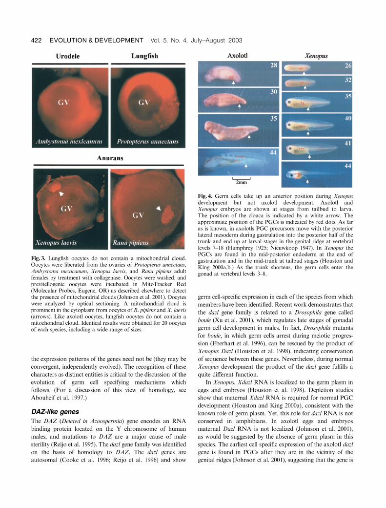
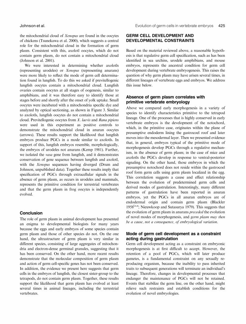
Fig. 3. Lungfish oocytes do not contain a mitochondrial cloud.Oocytes were liberated from the ovaries of Protopterus annectans,Ambystoma mexicanum, Xenopus laevis, and Rana pipiens adultfemales by treatment with collagenase. Oocytes were washed, andprevitellogenic oocytes were incubated in MitoTracker Red(Molecular Probes, Eugene, OR) as described elsewhere to detectthe presence of mitochondrial clouds (Johnson et al. 2001). Oocyteswere analyzed by optical sectioning. A mitochondrial cloud isprominent in the cytoplasm from oocytes of R. pipiens andX. laevis(arrows). Like axolotl oocytes, lungfish oocytes do not contain amitochondrial cloud. Identical results were obtained for 20 oocytesof each species, including a wide range of sizes.
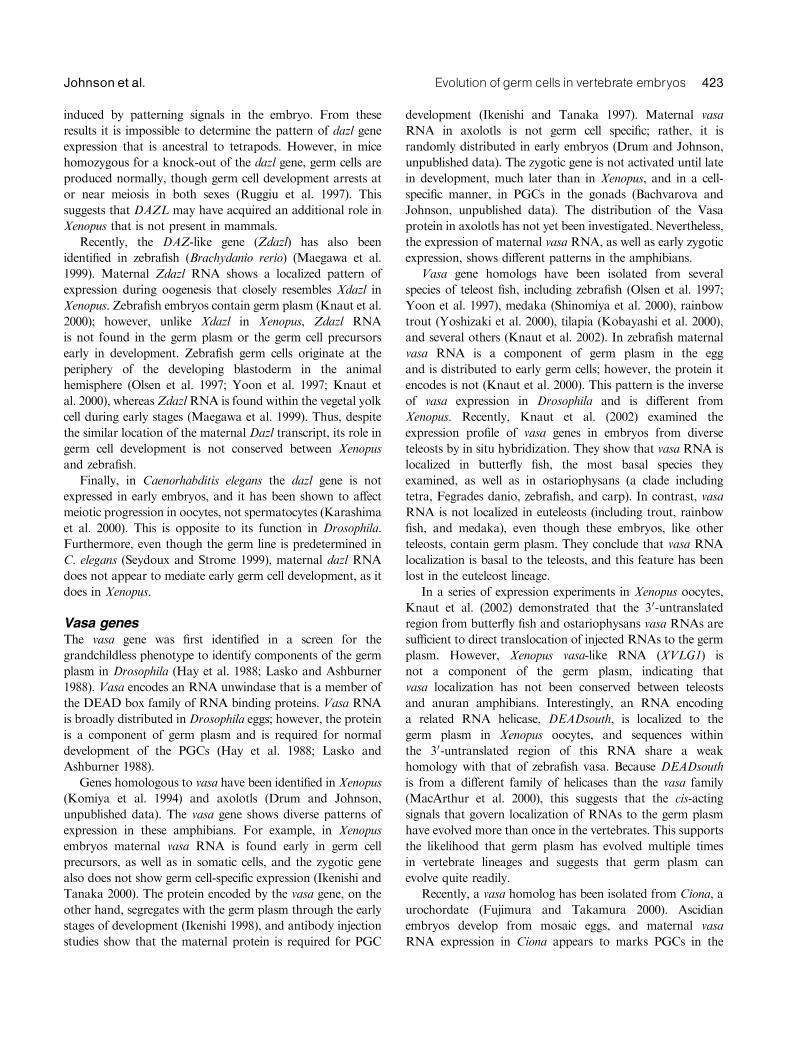
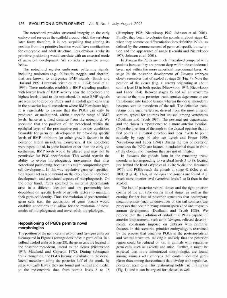
Fig. 4. Germ cells take up an anterior position during Xenopusdevelopment but not axolotl development. Axolotl andXenopus embryos are shown at stages from tailbud to larva.The position of the cloaca is indicated by a white arrow. Theapproximate position of the PGCs is indicated by red dots. As faras is known, in axolotls PGC precursors move with the posteriorlateral mesoderm during gastrulation into the posterior half of thetrunk and end up at larval stages in the genital ridge at vertebrallevels 7–18 (Humphrey 1925; Nieuwkoop 1947). In Xenopus thePGCs are found in the mid-posterior endoderm at the end ofgastrulation and in the mid-trunk at tailbud stages (Houston andKing 2000a,b.) As the trunk shortens, the germ cells enter thegonad at vertebral levels 3–8.
422 EVOLUTION & DEVELOPMENT Vol. 5, No. 4, July^August 2003
induced by patterning signals in the embryo. From these
results it is impossible to determine the pattern of dazl gene
expression that is ancestral to tetrapods. However, in mice
homozygous for a knock-out of the dazl gene, germ cells are
produced normally, though germ cell development arrests at
or near meiosis in both sexes (Ruggiu et al. 1997). This
suggests that DAZL may have acquired an additional role in
Xenopus that is not present in mammals.
Recently, the DAZ-like gene (Zdazl) has also been
identified in zebrafish (Brachydanio rerio) (Maegawa et al.
1999). Maternal Zdazl RNA shows a localized pattern of
expression during oogenesis that closely resembles Xdazl in
Xenopus. Zebrafish embryos contain germ plasm (Knaut et al.
2000); however, unlike Xdazl in Xenopus, Zdazl RNA
is not found in the germ plasm or the germ cell precursors
early in development. Zebrafish germ cells originate at the
periphery of the developing blastoderm in the animal
hemisphere (Olsen et al. 1997; Yoon et al. 1997; Knaut et
al. 2000), whereasZdazlRNA is found within the vegetal yolk
cell during early stages (Maegawa et al. 1999). Thus, despite
the similar location of the maternal Dazl transcript, its role in
germ cell development is not conserved between Xenopus
and zebrafish.
Finally, in Caenorhabditis elegans the dazl gene is not
expressed in early embryos, and it has been shown to affect
meiotic progression in oocytes, not spermatocytes (Karashima
et al. 2000). This is opposite to its function in Drosophila.
Furthermore, even though the germ line is predetermined in
C. elegans (Seydoux and Strome 1999), maternal dazl RNA
does not appear to mediate early germ cell development, as it
does in Xenopus.
Vasa genesThe vasa gene was first identified in a screen for the
grandchildless phenotype to identify components of the germ
plasm in Drosophila (Hay et al. 1988; Lasko and Ashburner
1988). Vasa encodes an RNA unwindase that is a member of
the DEAD box family of RNA binding proteins. Vasa RNA
is broadly distributed inDrosophila eggs; however, the protein
is a component of germ plasm and is required for normal
development of the PGCs (Hay et al. 1988; Lasko and
Ashburner 1988).
Genes homologous to vasa have been identified in Xenopus
(Komiya et al. 1994) and axolotls (Drum and Johnson,
unpublished data). The vasa gene shows diverse patterns of
expression in these amphibians. For example, in Xenopus
embryos maternal vasa RNA is found early in germ cell
precursors, as well as in somatic cells, and the zygotic gene
also does not show germ cell-specific expression (Ikenishi and
Tanaka 2000). The protein encoded by the vasa gene, on the
other hand, segregates with the germ plasm through the early
stages of development (Ikenishi 1998), and antibody injection
studies show that the maternal protein is required for PGC
development (Ikenishi and Tanaka 1997). Maternal vasa
RNA in axolotls is not germ cell specific; rather, it is
randomly distributed in early embryos (Drum and Johnson,
unpublished data). The zygotic gene is not activated until late
in development, much later than in Xenopus, and in a cell-
specific manner, in PGCs in the gonads (Bachvarova and
Johnson, unpublished data). The distribution of the Vasa
protein in axolotls has not yet been investigated. Nevertheless,
the expression of maternal vasa RNA, as well as early zygotic
expression, shows different patterns in the amphibians.
Vasa gene homologs have been isolated from several
species of teleost fish, including zebrafish (Olsen et al. 1997;
Yoon et al. 1997), medaka (Shinomiya et al. 2000), rainbow
trout (Yoshizaki et al. 2000), tilapia (Kobayashi et al. 2000),
and several others (Knaut et al. 2002). In zebrafish maternal
vasa RNA is a component of germ plasm in the egg
and is distributed to early germ cells; however, the protein it
encodes is not (Knaut et al. 2000). This pattern is the inverse
of vasa expression in Drosophila and is different from
Xenopus. Recently, Knaut et al. (2002) examined the
expression profile of vasa genes in embryos from diverse
teleosts by in situ hybridization. They show that vasa RNA is
localized in butterfly fish, the most basal species they
examined, as well as in ostariophysans (a clade including
tetra, Fegrades danio, zebrafish, and carp). In contrast, vasa
RNA is not localized in euteleosts (including trout, rainbow
fish, and medaka), even though these embryos, like other
teleosts, contain germ plasm. They conclude that vasa RNA
localization is basal to the teleosts, and this feature has been
lost in the euteleost lineage.
In a series of expression experiments in Xenopus oocytes,
Knaut et al. (2002) demonstrated that the 30-untranslated
region from butterfly fish and ostariophysans vasa RNAs are
sufficient to direct translocation of injected RNAs to the germ
plasm. However, Xenopus vasa-like RNA (XVLG1) is
not a component of the germ plasm, indicating that
vasa localization has not been conserved between teleosts
and anuran amphibians. Interestingly, an RNA encoding
a related RNA helicase, DEADsouth, is localized to the
germ plasm in Xenopus oocytes, and sequences within
the 30-untranslated region of this RNA share a weak
homology with that of zebrafish vasa. Because DEADsouth
is from a different family of helicases than the vasa family
(MacArthur et al. 2000), this suggests that the cis-acting
signals that govern localization of RNAs to the germ plasm
have evolved more than once in the vertebrates. This supports
the likelihood that germ plasm has evolved multiple times
in vertebrate lineages and suggests that germ plasm can
evolve quite readily.
Recently, a vasa homolog has been isolated from Ciona, a
urochordate (Fujimura and Takamura 2000). Ascidian
embryos develop from mosaic eggs, and maternal vasa
RNA expression in Ciona appears to marks PGCs in the
Evolution of germ cells in vertebrate embryos 423Johnson et al.
endodermal strand posterior cells (Takamura et al. 2002).
However, extirpation studies demonstrate thatCiona embryos
can compensate for the loss of early PGCs by producing them
anew within the embryo. Thus, ascidian embryos may have
both predetermined and regulative germ cells. Because PGCs
cannot be replaced upon removal in vertebrate embryos
(Nieuwkoop and Sutasurya 1979), it is uncertain how this
process relates to vertebrate germ cell development.
Finally, in amniotes, chick vasa protein is maternally
inherited and segregates with the germ cells from the inception
of their development, as described above. In mouse embryos
vasa RNA and protein are not maternally inherited; they are
first expressed in PGCs after they colonize the genital ridge
(Fujiwara et al. 1994; Toyooka et al. 2000). Also, homo-
zygotes for a null allele of the mouse vasa gene, mvh,
demonstrate that vasa gene products are not required to
produce PGCs. Rather, mvh is required only in male gonadal
germ cells to complete meiosis (Tanaka et al. 2000). There-
fore, the role of the vasa genes from mouse and Xenopus has
not been conserved.
Nanos genes
The nanos gene family encodes zinc finger RNA binding
proteins. Nanos was originally described in Drosophila, where
it is required for posterior embryonic development (Irish et al.
1989). Maternal Nanos protein is incorporated into germ cells
early in embryogenesis, and germ cells of embryos deficient
for maternal Nanos protein display a variety of defects,
including failure to complete migration to the gonads
(Kobayashi et al. 1996; Forbes and Lehmann 1998).
However, nanos is not required for the specification of germ
cells. Similarly, RNAs from nanos homologs of C. elegans
segregate with the germ cell progenitors and are also required
for germ line maintenance after specification (Subramaniam
and Seydoux 1999). Furthermore, nanos-deficient embryos
display a phenotype similar to Drosophila in that germ
cells fail to incorporate into the gonad. Interestingly, a
nanos homolog has been identified in the leech Helobdella
robusta (Pilon and Weisbat 1997), and its maternal transcript
is broadly distributed in the egg cytoplasm (Kang et al. 2002).
Its zygotic transcript only appears in presumptive germ cells
when they can first be identified in segmental mesoderm,
relatively late in development (Kang et al. 2002). Thus,
although nanos is likely to be involved in leech PGC
development, a role for maternal nanos transcripts has not
been conserved in protostomes.
Among vertebrates, nanos homologs have been identified
in zebrafish (Koprunner et al. 2001) and in Xenopus
(Mosquera et al. 1993). In both species maternal RNA
encoded by the homolog of the nanos gene becomes
associated with germ plasm, though this is regulated by
different mechanisms. In Xenopus, RNA encoding the nanos
homolog, XCAT2, is localized to the germ plasm during
early oogenesis, through the germ plasm specific localization
pathway mediated by the mitochondrial cloud (Zhou and
King 1996). Thus, XCAT2 RNA is a component of germ
plasm and is exclusive to the presumptive germ line
blastomeres from the inception of development in Xenopus.
In zebrafish, RNA from the nanos homolog, nos1, is
distributed throughout the oocyte cytoplasm. During early
embryogenesis a fraction of nos1 RNA becomes associated
with germ plasm (Koprunner et al. 2001). Nos1 transcripts
associated with the germ plasm are protected from
degradation, whereas RNA in other regions of cytoplasm
is destroyed. Eventually, by 50% epiboly, nos1 RNA is
found exclusively within the germ plasm, and it is then
partitioned to PGCs. Interestingly, nos1 is not required for
PGC specification but plays an essential role in migration of
PGCs to the gonad, similar to the phenotypes of nanos
mutants in Drosophila and C. elegans.
Although the biochemical functions of nanos RNAmay be
highly related, or equivalent, in diverse species, the expression
patterns of these genes have not been conserved in vertebrate
embryos. Apparently, the association of nanos RNA with
germ plasm has evolved along separate pathways in Xenopus
and zebrafish. Nanos homologs have not been identified in
vertebrate species that develop without germ plasm, preclud-
ing comparison of nanos structure and function in the two
types of organisms.
Summary
Based on the data discussed above, we draw the following
two conclusions. First, for germ cell-specific genes there
are distinct differences among organisms in whether the
protein, the RNA, or neither is localized in the germ plasm,
indicating that germ plasm has a different molecular
makeup in different organisms. Second, there are differences
in the way that germ plasm is formed in the oocytes of
different species, as exemplified by Xenopus and zebrafish.
This suggests that germ plasm has not been conserved across
vertebrate phyla.
Did oocytes of the tetrapod ancestorcontain germ plasm?
To shed light on whether the presence of maternal germ plasm
is ancestral to terrestrial vertebrates, we analyzed oocytes of
the lungfish, the closest living relative of the tetrapod ancestor
(Zardoya et al. 1998), to determine whether they contain germ
plasm.
Much is known about how germ plasm is formed in
Xenopus oocytes. Heasman et al. (1984) showed that electron-
dense germinal granules become associated with an aggregate
of mitochondria, the mitochondrial cloud, in the cytoplasm of
previtellogenic oocytes. During later stages of oogenesis, the
germ plasm is transported along with the mitochondrial cloud
to the vegetal cortex (King et al. 1999). Structures similar to
424 EVOLUTION & DEVELOPMENT Vol. 5, No. 4, July^August 2003
the mitochondrial cloud of Xenopus are found in the oocytes
of chickens (Tsunekawa et al. 2000), which suggests a central
role for the mitochondrial cloud in the formation of germ
plasm. Consistent with this, axolotl oocytes, which do not
contain germ plasm, do not contain a mitochondrial cloud
(Johnson et al. 2001).
We were interested in determining whether axolotls
(representing urodeles) or Xenopus (representing anurans)
were more likely to reflect the mode of germ cell determina-
tion found in lungfish. To do this we asked if previtellogenic
lungfish oocytes contain a mitochondrial cloud. Lungfish
ovaries contain oocytes at all stages of oogenesis, similar to
amphibians, and it was therefore easy to identify those at
stages before and shortly after the onset of yolk uptake. Small
oocytes were incubated with a mitochondria specific dye and
analyzed by optical sectioning, as shown in Figure 3. Similar
to axolotls, lungfish oocytes do not contain a mitochondrial
cloud. Previtellogenic oocytes from X. laevis and Rana pipiens
were used in this experiment as positive controls to
demonstrate the mitochondrial cloud in anuran oocytes
(arrows). These results support the likelihood that lungfish
embryos produce PGCs in a mode similar to axolotls. In
support of this, lungfish embryos resemble, morphologically,
the embryos of urodeles not anurans (Kemp 1981). Further,
we isolated the vasa gene from lungfish, and it demonstrates
conservation of gene sequence between lungfish and axolotl,
with the Xenopus sequences having diverged (Drum and
Johnson, unpublished data). Together these results imply that
specification of PGCs through extracellular signals in the
absence of germ plasm, as occurs in urodeles and mammals,
represents the primitive condition for terrestrial vertebrates
and that the germ plasm in frog oocytes is independently
evolved.
Conclusion
The role of germ plasm in animal development has presented
an enigma to developmental biologists for many years
because the eggs and early embryos of some species contain
germ plasm and those of other species do not. On the one
hand, the ultrastructure of germ plasm is very similar in
different species, consisting of large aggregates of mitochon-
dria and electron-dense germinal granules, suggesting that it
has been conserved. On the other hand, more recent results
demonstrate that the molecular composition of germ plasm
and action of germ cell-specific genes has not been conserved.
In addition, the evidence we present here suggests that germ
cells in the embryos of lungfish, the closest sister-group to the
tetrapods, do not contain germ plasm. Together, these results
support the likelihood that germ plasm has evolved at least
several times in animal lineages, including the terrestrial
vertebrates.
GERM CELL DEVELOPMENT ANDDEVELOPMENTAL CONSTRAINTS
Based on the material reviewed above, a reasonable hypoth-
esis is that regulative germ cell specification, such as has been
identified in sea urchins, urodele amphibians, and mouse
embryos, represents the ancestral condition for germ cell
development during vertebrate embryogenesis. This raises the
question of why germ plasm may have arisen several times, in
different lineages of vertebrate eggs and embryos. We address
this issue below.
Absence of germ plasm correlates withprimitive vertebrate embryology
Above we compared early morphogenesis in a variety of
species to identify characteristics primitive to the tetrapod
lineage. One of the processes that is highly conserved in early
vertebrate embryos is the development of the notochord,
which, in the primitive case, originates within the plane of
presumptive endoderm lining the gastrocoel roof and later
moves into the mesodermal layer. Then we presented evidence
that, in general, embryos typical of the primitive mode of
morphogenesis develop PGCs through a regulative mechan-
ism, in the absence of germ plasm; in the case of mice and
axolotls the PGCs develop in response to ventral-posterior
signaling. On the other hand, those embryos in which the
presumptive notochord does not reside within the gastrocoel
roof form germ cells using germ plasm localized in the egg.
This correlation suggests a cause and effect relationship
between the evolution of predetermined germ cells and
derived modes of gastrulation. Interestingly, many different
patterns of gastrulation have been reported in anuran
embryos, yet the PGCs in all anuran embryos are of
endodermal origin and contain germ plasm (Blackler
1958l,s,u; Nieuwkoop and Sutasurya 1979). This suggests that
the evolution of germ plasm in anurans preceded the evolution
of novel modes of morphogenesis, and germ plasm may then
be a cause, not a consequence, of embryological variation.
Mode of germ cell development as a constraintacting during gastrulationGerm cell development acting as a constraint on embryonic
morphogenesis is at first difficult to accept. However, the
retention of a pool of PGCs, which will later produce
gametes, is a fundamental constraint on any sexually re-
producing organism, because the inability to pass inherited
traits to subsequent generations will terminate an individual’s
lineage. Therefore, changes in developmental processes that
endanger the maintenance of PGCs will not be retained.
Events that stabilize the germ line, on the other hand, might
relieve such restraints and establish conditions for the
evolution of novel embryologies.
Evolution of germ cells in vertebrate embryos 425Johnson et al.
The notochord provides structural integrity to the early
embryo and serves as the scaffold around which the vertebrae
later form; therefore, it is not surprising that altering its
position from the primitive location would have ramifications
for embryonic and adult structure. Less obvious is why its
primitive positioning would correlate with an ancestral mode
of germ cell development. We consider a possible reason
below.
The notochord secretes embryonic patterning signals,
including molecules (e.g., follistatin, noggin, and chordin)
that are known to antagonize BMP signals (Smith and
Harland 1992; Hemmati-Brivanlou et al. 1994; Sasai et al.
1994). These molecules establish a BMP signaling gradient
with lowest levels of BMP activity near the notochord and
highest levels distal to the notochord. In mice BMP signals
are required to produce PGCs, and in axolotl germ cells arise
in the posterior lateral mesoderm where BMP levels are high.
It is reasonable to assume that the PGCs can only be
produced, or maintained, within a specific range of BMP
levels, hence at a fixed distance from the notochord. We
speculate that the position of the notochord within the
epithelial layer of the presumptive gut provides conditions
favorable for germ cell development by providing specific
levels of BMP inhibitors (or other growth factors) in the
posterior lateral mesoderm. Conversely, if the notochord
were repositioned, in some location other than the early gut
epithelium, BMP levels would be altered and may not be
permissive for PGC specification. This would restrain the
ability to evolve morphogenetic movements that alter
notochord positioning, because this might compromise germ
cell development. In this way regulative germ cell specifica-
tion would act as a constraint on the evolution of notochord
development and associated aspects of morphogenesis. On
the other hand, PGCs specified by maternal determinants
arise in a different location and are presumably less
dependent on specific levels of growth factors to maintain
their germ cell identity. Thus, the evolution of predetermined
germ cells (i.e., the acquisition of germ plasm) would
establish conditions that allow for the evolution of novel
modes of morphogenesis and novel adult morphologies.
Repositioning of PGCs permits novelmorphologies
The position of the germ cells in axolotl and Xenopus embryos
is compared in Figure 4 (orange dots indicate germ cells). In a
tailbud axolotl embryo (stage 28), the germ cells are located in
the posterior mesoderm, lateral to the cloaca (Nieuwkoop
1947; Maufroid and Capuron 1972). During subsequent
trunk elongation, the PGCs become distributed in the dorsal
lateral mesoderm along the posterior half of the trunk. By
stage 40 (early larva), they are found just ventral and medial
to the mesonephric duct from somite levels 8 to 18
(Humphrey 1925; Nieuwkoop 1947; Johnson et al. 2001).
Finally, they begin to colonize the gonads at about stage 42,
when they commence differentiation into definitive PGCs, as
defined by the commencement of germ cell-specific transcrip-
tion and the appearance of nuage (Ikenishi and Nieuwkoop
1978; Johnson et al. 2001).
InXenopus the PGCs are much internalized compared with
axolotls because they are present deep within the endodermal
layer, not within the more superficial mesodermal layer. At
stage 26 the posterior development of Xenopus embryos
closely resembles that of axolotl at stage 28 (Fig. 4). Note the
position of the cloaca (Fig. 4, arrow) originating at about
somite level 18 in both species (Nieuwkoop 1947; Nieuwkoop
and Faber 1994). Between stages 35 and 42, all structures
ventral to the most posterior trunk somites degenerate or are
transformed into tailbud tissues, whereas the dorsal mesoderm
becomes somitic mesoderm of the tail. The definitive trunk
retains only eight vertebrae, derived from the most anterior
somites, typical for anurans but unusual among vertebrates
(Duellman and Trueb 1986). The postanal gut degenerates,
and the cloaca is repositioned to a more anterior location.
(Note the inversion of the angle to the cloacal opening that at
first points in a rostral direction and then inverts to point
caudally by stage 40 [also see Lynch and Fraser 1990;
Nieuwkoop and Faber 1994].) During the loss of posterior
structures the PGCs are located in endodermal tissue in front
of the cloaca, and therefore they are not eliminated.
In Xenopus the gonads form in the remaining trunk
mesoderm (corresponding to vertebral levels 3 to 8), located
just behind the head (Wylie et al. 1976; Wylie and Heasman
1976), and PGCs reach the gonads at stage 42 (Kloc et al.
2001) (Fig. 4). Thus, in Xenopus the gonads are found at a
much more anterior level (vertebral levels 3 to 8) than in the
axolotl.
The loss of posterior-ventral tissues and the tight anterior
coiling of the gut tube during larval stages, as well as the
ensuing further loss of posterior structures that accompany
metamorphosis (such as derivatives of the tail somites), are
processes that occur in many anuran species and are unique to
anuran development (Duellman and Trueb 1986). We
propose that the evolution of endodermal PGCs capable of
anterior displacement, such as in Xenopus, relieved develop-
mental constraints imposed on embryos with primitive
features. In this scenario, primitive embryology is restrained
by the process that generates PGCs in the posterior-lateral
and ventral structures, making it unlikely that the posterior
region could be reduced or lost in animals with regulative
germ cells, such as axolotls and mice. Further, it might be
expected that more anteriorized morphologies are found
among animals with embryos that contain localized germ
plasm than among those animals that develop with regulative,
posterior, germ cells. This relationship holds true in anurans
(Fig. 1), and it can be argued for teleosts as well.
426 EVOLUTION & DEVELOPMENT Vol. 5, No. 4, July^August 2003
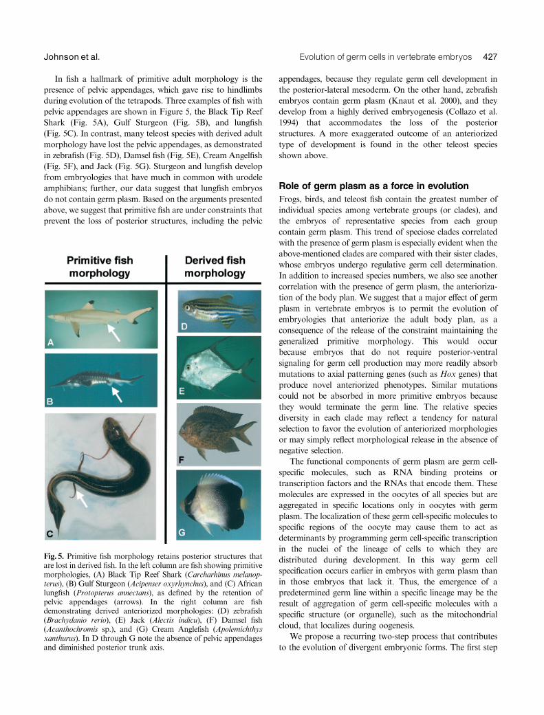
In fish a hallmark of primitive adult morphology is the
presence of pelvic appendages, which gave rise to hindlimbs
during evolution of the tetrapods. Three examples of fish with
pelvic appendages are shown in Figure 5, the Black Tip Reef
Shark (Fig. 5A), Gulf Sturgeon (Fig. 5B), and lungfish
(Fig. 5C). In contrast, many teleost species with derived adult
morphology have lost the pelvic appendages, as demonstrated
in zebrafish (Fig. 5D), Damsel fish (Fig. 5E), Cream Angelfish
(Fig. 5F), and Jack (Fig. 5G). Sturgeon and lungfish develop
from embryologies that have much in common with urodele
amphibians; further, our data suggest that lungfish embryos
do not contain germ plasm. Based on the arguments presented
above, we suggest that primitive fish are under constraints that
prevent the loss of posterior structures, including the pelvic
appendages, because they regulate germ cell development in
the posterior-lateral mesoderm. On the other hand, zebrafish
embryos contain germ plasm (Knaut et al. 2000), and they
develop from a highly derived embryogenesis (Collazo et al.
1994) that accommodates the loss of the posterior
structures. A more exaggerated outcome of an anteriorized
type of development is found in the other teleost species
shown above.
Role of germ plasm as a force in evolution
Frogs, birds, and teleost fish contain the greatest number of
individual species among vertebrate groups (or clades), and
the embryos of representative species from each group
contain germ plasm. This trend of speciose clades correlated
with the presence of germ plasm is especially evident when the
above-mentioned clades are compared with their sister clades,
whose embryos undergo regulative germ cell determination.
In addition to increased species numbers, we also see another
correlation with the presence of germ plasm, the anterioriza-
tion of the body plan. We suggest that a major effect of germ
plasm in vertebrate embryos is to permit the evolution of
embryologies that anteriorize the adult body plan, as a
consequence of the release of the constraint maintaining the
generalized primitive morphology. This would occur
because embryos that do not require posterior-ventral
signaling for germ cell production may more readily absorb
mutations to axial patterning genes (such as Hox genes) that
produce novel anteriorized phenotypes. Similar mutations
could not be absorbed in more primitive embryos because
they would terminate the germ line. The relative species
diversity in each clade may reflect a tendency for natural
selection to favor the evolution of anteriorized morphologies
or may simply reflect morphological release in the absence of
negative selection.
The functional components of germ plasm are germ cell-
specific molecules, such as RNA binding proteins or
transcription factors and the RNAs that encode them. These
molecules are expressed in the oocytes of all species but are
aggregated in specific locations only in oocytes with germ
plasm. The localization of these germ cell-specific molecules to
specific regions of the oocyte may cause them to act as
determinants by programming germ cell-specific transcription
in the nuclei of the lineage of cells to which they are
distributed during development. In this way germ cell
specification occurs earlier in embryos with germ plasm than
in those embryos that lack it. Thus, the emergence of a
predetermined germ line within a specific lineage may be the
result of aggregation of germ cell-specific molecules with a
specific structure (or organelle), such as the mitochondrial
cloud, that localizes during oogenesis.
We propose a recurring two-step process that contributes
to the evolution of divergent embryonic forms. The first step
Fig. 5. Primitive fish morphology retains posterior structures thatare lost in derived fish. In the left column are fish showing primitivemorphologies, (A) Black Tip Reef Shark (Carcharhinus melanop-terus), (B) Gulf Sturgeon (Acipenser oxyrhynchus), and (C) Africanlungfish (Protopterus annectans), as defined by the retention ofpelvic appendages (arrows). In the right column are fishdemonstrating derived anteriorized morphologies: (D) zebrafish(Brachydanio rerio), (E) Jack (Alectis indicu), (F) Damsel fish(Acanthochromis sp.), and (G) Cream Anglefish (Apolemichthysxanthurus). In D through G note the absence of pelvic appendagesand diminished posterior trunk axis.
Evolution of germ cells in vertebrate embryos 427Johnson et al.
involves the evolution of a localized functional germ plasm,
via the association of germ cell specific molecules with the
mitochondrial cloud, that can be transmitted to a specific set
of blastomeres in the embryo. Second, as a consequence of a
fixed (protected) germ line, the embryo can absorb mutations
to embryonic patterning genes that affect the morphogenetic
movements associated with gastrulation. The mutation
process will tend to give rise to diverse and derived adult
forms. Diversity can then be accomplished within individual
lineages by the accumulation of specific mutations that are
accommodated by prior steps in evolution. We suggest that
this embryologically based mechanism for variation in adult
morphology is a major contributor to species radiation and
morphological diversity in vertebrates and thus to the process
of macroevolution.
AcknowledgmentsWe thank Capitol Health Plan, Tallahassee, Florida for the generoususe of their X-ray facilities. A. D. J. thanks Peter Wainwright for firstsuggesting the possibility that urodeles might represent the primitivecondition. Joe Travis is thanked for suggesting the focal role ofprimitive fish and for encouraging writing of the manuscript in thefirst place. Many thanks to Roger Patient for help with the finalpreparation and toMaggie Walmsley for helpful suggestions. Finally,A. D. J. dedicates this article to Dr. Nancy Van Vessem, who fundedhis laboratory when external funding was not there and withoutwhom this entire project would never have been possible.
REFERENCES
Abouheif, E., Akam, M., Dickinson, W. J., Holland, P. W., Meyer, A.,Patel, N. H., et al. 1997. Homology and developmental genes. TrendsGenet. 13: 432–433.
Anderson, R., Copeland, T. K., Scholer, H., Heasman, J., and Wylie, C.2000. The onset of germ cell migration in the mouse embryo. Mech Dev91: 61–68.
Balinsky, B. I. 1975. An Introduction to Embryology. 4th Ed.W. B. Saunders, Philadelphia.
Ballard, W. W., and Ginsburg, A. S. 1980. Morphogenetic movements inacipenserid embryos. J. Exp. Zool. 213: 391–399.
Blackler, A. W. 1958. Contribution to the study of germ-cells in the Anura.J. Embryol. Exp. Morphol. 6: 491–503.
Bolker, J. A. 1993. Gastrulation and mesoderm morphogenesis in the whitesturgeon. J. Exp. Zool. 266: 116–131.
Bordzilovskaya, N. P., Dettlaff, T. A., Duhon, S. T., and Malacinski, G. M.1989. Developmental-stage series of axolotl embryos. In J. B.Armstrong, and G. M. Malacinski (eds.). Developmental Biology of theAxolotl. Oxford University Press, New York, pp. 210–219.
Boterenbrood, E. C., and Nieuwkoop, P. D. 1973. The formation of themesoderm in urodelean amphibians. V. Its regional induction by theendoderm. Wilhelm Roux Arch. 173: 319–332.
Bounoure, L. 1939. L’origine des cellules reproductrices et le probleme de lalignee germinale. Gauthier-Villars, Paris.
Bounoure, L., Aubry, R., and Huck, M. L. 1954. Nouvelles recherchesexperimentales sur les origines de la lignee reproductrice chez laGrenouille rousse. J. Embryol. Exp. Morphol. 2: 245–263.
Brun, R. F., and Garson, J. A. 1984. Notochord formation in the Mexicansalamander (Ambystoma mexicanum) is different from notochordformation in Xenopus laevis. J. Exp. Zool. 229: 235–240.
Cannatella, D. C., and Hillis, D. M. 1992. Amphibian relationships:phylogenetic analysis of morphology and molecules. HerpetolMonogr 6: 1–7.
Cannatella, D. C., and De Sa, R. O. 1993. Xenopus laevis as a modelorganism. System. Biol. 42: 476–507.
Collazo, A., Bolker, J., and Keller, R. 1994. A phylogenetic perspective onteleost gastrulation. Am. Nat. 144: 133–152.
Cooke, H. J., Lee, M., Kerr, S., and Ruggiu, M. 1996. A murine homologueof the human DAZ gene is autosomal and expressed only in male andfemale gonads. Hum. Mol. Genet. 5: 513–516.
Delarue, M., Johnson, K. E., and Boucaut, J. C. 1994. Superficial cells inthe early gastrula of Rana pipiens contribute to mesodermal derivatives.Dev. Biol. 165: 702–715.
Delarue, M., Sanchez, S., Johnson, K. E., Darribere, T., and Boucaut, J. C.1992. A fate map of superficial and deep circumblastoporal cells in theearly gastrula of Pleurodeles waltl. Development 114: 135–146.
Dobzhansky, T. 1973. Nothing in biology makes sense except when seen inthe light of evolution. Am. Biol. Teacher 35: 125–129.
Duellman, W. E., and Trueb, L. 1986. Biology of Amphibians. McGraw-Hill, New York.
Eberhart, C. G., Maines, J. Z., and Wasserman, S. A. 1996. Meiotic cellcycle requirement for a fly homologue of human Deleted in Azoospermia.Nature 381: 783–785.
Eddy, E. M. 1974. Fine structural observations on the form and distributionof nuage in germ cells of the rat. Anat. Rec. 178: 731–757.
Eddy, E. M. 1975. Germ plasm and the differentiation of the germ cell line.Intl. Rev. Cytol. 43: 229–280.
Elinson, R. P. 1987. Change in developmental patterns: embryos ofamphibians with large eggs. In R. A. Raff, and E. C. Raff (eds.).Develop-ment as an Evolutionary Process. Alan R. Liss, New York, pp. 1–21.
Eyal-Giladi, H., Ginsburg, M., and Farbarov, A. 1981. Avian primordialgerm cells are of epiblastic origin. J. Embryol. Exp. Morph. 65: 139–147.
Forbes, A., and Lehmann, R. 1998. Nanos and Pumilio have critical roles inthe development and function of Drosophila germline stem cells.Development 125: 679–690.
Fujimura, M., and Takamura, K. 2000. Characterization of an ascidianDEAD-box gene, Ci-DEAD1: specific expression in the germ cells and itsmRNA localization in the posterior-most blastomeres in early embryos.Dev. Genes Evol. 210: 64–72.
Fujiwara, Y., Komiya, T., Kawabata, H., Sato, M., Fujimoto, H., andFurusawa, M. 1994. Isolation of a DEAD-family protein gene thatencodes a murine homolog of Drosophila vasa and its specific expressionin germ cell lineage. Proc. Natl. Acad. Sci. USA 91: 12258–12262.
Gerhart, J. C., and Kirschner, M. W. 1997. Cells Embryos and Evolution.Blackwell Science, Oxford.
Ginsburg, M., Snow, M. H., and McLaren, A. 1990. Primordial germ cellsin the mouse embryo during gastrulation. Development 110: 521–528.
Hanken, J. 1986. Developmental evidence for amphibian origins. Evol. Biol.20: 389–417.
Hay, B., Jan, L. Y., and Jan, Y. N. 1988. A protein component ofDrosophila polar granules is encoded by vasa and has extensive sequencesimilarity to ATP-dependent helicases. Cell 55: 577–587.
Heasman, J., Quarmby, J., and Wylie, C. C. 1984. The mitochondrial cloudof Xenopus oocytes: the source of germinal granule material. Dev. Biol.105: 458–469.
Hemmati-Brivanlou, A., Kelly, O. G., and Melton, D. A. 1994. Follistatin,an antagonist of activin, is expressed in the Spemann organizer anddisplays direct neuralizing activity. Cell 77: 283–295.
Holland, N. D. 1978. The fine structure of Comanthus japonica(Echinodermata: Crinoidea) from zygote through early gastrula. TissueCell 10: 93–112.
Houston, D. W., and King, M. L. 2000a. A critical role for Xdazl, a germplasm-localized RNA, in the differentiation of primordial germ cells inXenopus. Development 127: 447–456.
Houston, D. W., and King, M. L. 2000b. Germ plasm and moleculardeterminants of germ cell fate. Curr. Top. Dev. Biol. 50: 155–181.
Houston, D. W., Zhang, J., Maines, J. Z., Wasserman, S. A., and King, M.L. 1998. A Xenopus DAZ-like gene encodes an RNA component of germplasm and is a functional homologue of Drosophila boule. Development125: 171–180.
Hubert, J. 1985a. Embryology of the squamata. In C. Gans, and F. Billet(eds.). Biology of the Reptilia. Vol. 15. Development B. John Wiley &Sons, New York, pp. 1–84.
428 EVOLUTION & DEVELOPMENT Vol. 5, No. 4, July^August 2003
Hubert, J. 1985b. Origin and development of oocytes. In Gans, C., andBillet, F. (eds.). Biology of the Reptilia. Vol. 14. Development a. JohnWiley & Sons, New York, pp. 41–74.
Humphrey, R. R. 1925. The primordial germ cells of Hemidactylium andother Amphibia. J. Morphol. Physiol. 41: 1–43.
Humphrey, R. R. 1929. The early position of the primordial germ cells inurodeles: evidence from experimental studies. Anat. Rec. 42: 301–314.
Ikenishi, K. 1998. Germ plasm in Caenorhabditis elegans, Drosophila andXenopus. Dev. Growth Differ. 40: 1–10.
Ikenishi, K., and Tanaka, T. S. 1997. Involvement of the protein of Xenopusvasa homolog (Xenopus vasa-like gene 1, XVLG1) in the differentiation ofprimordial germ cells. Dev. Growth Differ. 39: 625–633.
Ikenishi, K., and Kotani, M. 1979. Ultraviolet effects on presumptiveprimordial germ cells (pPGCs) in Xenopus laevis after the cleavage stage.Dev. Biol. 69: 237–246.
Ikenishi, K., and Nieuwkoop, P. D. 1978. Location and ultrastructure ofprimordial germ cells (PGCs) in Ambystoma mexicanum. Dev. GrowthDiffer. 20: 1–9.
Ikenishi, K., and Tanaka, T. S. 2000. Spatio-temporal expression ofXenopus vasa homolog, XVLG1, in oocytes and embryos: the presence ofXVLG1 RNA in somatic cells as well as germline cells. Dev. GrowthDiffer. 42: 95–103.
Illmensee, K., and Mahowald, A. P. 1974. Transplantation of posteriorpolar plasm in Drosophila. Induction of germ cells at the anterior pole ofthe egg. Proc. Natl. Acad. Sci. USA 71: 1016–1020.
Irish, V., Lehmann, R., and Akam, M. 1989. The Drosophila posterior-group gene nanos functions by repressing hunchback activity. Nature338: 646–648.
Johnson, A. D., Bachvarova, R. F., Drum, M., and Masi, T. 2001.Expression of axolotl DAZL RNA, a marker of germ plasm: widespreadmaternal RNA and onset of expression in germ cells approaching thegonad. Dev. Biol. 234: 402–415.
Kang, D., Pilon, M., and Weisblat, D. A. 2002. Maternal and zygoticexpression of a nanos-class gene in the leech Helobdella robusta: pri-mordial germ cells arise from segmental mesoderm. Dev. Biol. 245:28–41.
Karagenc, L., Cinnamon, Y., Ginsburg, M., and Petitte, J. N. 1996. Originof primordial germ cells in the prestreak chick embryo. Dev. Genet. 19:290–301.
Karashima, T., Sugimoto, A., and Yamamoto, M. 2000. Caenorhabditiselegans homologue of the human azoospermia factor DAZ is requiredfor oogenesis but not for spermatogenesis. Development 127: 1069–1079.
Keller, R. E. 1975. Vital dye mapping of the gastrula and neurulaof Xenopus laevis. I. Prospective areas and morphogenetic movements ofthe superficial layer. Dev. Biol. 42: 222–241.
Keller, R. E. 1976. Vital dye mapping of the gastrula and neurulaof Xenopus laevis. II. Prospective areas and morphogenetic movements ofthe deep layer. Dev. Biol. 51: 118–137.
Keller, R. 2000. The origin and morphogenesis of amphibian somites.Curr. Top. Dev. Biol. 47: 183–246.
Kemp, A. 1981. Rearing of embryos and larvae of the Australian lungfish,Neoceratodus forsteri, under laboratory conditions. Copeia 4: 776–784.
Keynes, R. J., and Stern, C. D. 1988. Mechanisms of vertebratesegmentation. Development 103: 413–429.
King, M. L., Zhou, Y., and Bubunenko, M. 1999. Polarizing geneticinformation in the egg: RNA localization in the frog oocyte. Bioessays21: 546–557.
Kloc, M., Bilinski, S., Chan, A. P., Allen, L. H., Zearfoss, N. R., and Etkin,L. D. 2001. RNA localization and germ cell determination in Xenopus.Intl. Rev. Cytol. 203: 63–91.
Knaut, H., Pelegri, F., Bohmann, K., Schwarz, H., and Nusslein-Volhard,C. 2000. Zebrafish vasa RNA but not its protein is a component of thegerm plasm and segregates asymmetrically before germline specification.J. Cell Biol. 149: 875–888.
Knaut, H., Steinbeisser, H., Schwarz, H., and Nusslein-Volhard, C. 2002.An evolutionary conserved region in the vasa 30UTR targetsRNA translation to the germ cells in the zebrafish. Curr. Biol. 12:454–466.
Kobayashi, S., Yamada, M., Asaoka, M., and Kitamura, T. 1996. Essentialrole of the posterior morphogen nanos for germline development inDrosophila. Nature 380: 708–711.
Kobayashi, T., Kajiura-Kobayashi, H., and Nagahama, Y. 2000.Differential expression of vasa homologue gene in the germ cells duringoogenesis and spermatogenesis in a teleost fish, tilapia, Oreochromisniloticus. Mech. Dev. 99: 139–142.
Kocher-Becker, U., and Tiedemann, H. 1971. Induction of mesodermal andprimordial germ cells in Triturus ectoderm by a vegetalizing factor fromchick embryos. Nature 233: 65–66.
Komiya, T., Itoh, K., Ikenishi, K., and Furusawa, M. 1994. Isolation andcharacterization of a novel gene of the DEAD box protein family whichis specifically expressed in germ cells of Xenopus laevis. Dev. Biol. 162:354–363.
Koprunner, M., Thisse, C., Thisse, B., and Raz, E. 2001. A zebrafish nanos-related gene is essential for the development of primordial germ cells.Genes Dev. 15: 2877–2885.
Kuwana, T., Maeda-Suga, H., and Fujimoto, T. 1986. Attraction of chickprimordial germ cells by gonadal anlage in vitro. Anat. Rec. 215:403–406.
Lasko, P. F., and Ashburner, M. 1988. The product of the Drosophilagene vasa is very similar to eukaryotic initiation factor-4A. Nature 335:611–617.
Lawson, K. A., and Hage, W. J. 1994. Clonal analysis of the origin ofprimordial germ cells in the mouse. Ciba Found. Symp. 182: 68–91.
Lawson, K. A., Dunn, N. R., Roelen, B. A., Zeinstra, L. M., Davis, A. M.,and Wright, C. V. 1999. Bmp4 is required for the generation ofprimordial germ cells in the mouse embryo. Genes Dev. 13: 424–436.
Lundmark, C. 1986. Role of bilateral zones of ingressing superficial cellsduring gastrulation of Ambystoma mexicanum. J. Embryol. Exp.Morphol. 97: 47–62.
Lynch, K., and Fraser, S. E. 1990. Cell migration in the formation of thepronephric duct in Xenopus laevis. Dev. Biol. 142: 283–292.
MacArthur, H., Houston, D. W., Bubunenko, M., Mosquera, L., andKing, M. L. 2000. DEADSouth is a germ plasm specific DEAD-boxRNA helicase in Xenopus related to eIF4A. Mech. Dev. 95: 291–295.
Maddison, W. P., and Maddison, D. R. 1992. MacCLade. Analysis ofPhylogeny and Character Evolution. Version 3.0. Sinauer Associates,Sunderland, MA.
Maegawa, S., Yasuda, K., and Inoue, K. 1999. Maternal mRNAlocalization of zebrafish DAZ-like gene. Mech. Dev. 81: 223–226.
Mahowald, A. P., and Hennen, S. 1971. Ultrastructure of the ‘‘germ plasm’’in eggs and embryos of Rana pipiens. Dev. Biol. 24: 37–53.
Malacinski, G. M., Bessho, T., Yokota, C., Fukui, A., and Asashima, M.1997. An essay on the similarities and differences between inductiveinteractions in anuran and urodele embryos. Cell Mol. Life Sci. 53:410–417.
Maufroid, J. P., and Capuron, A. P. 1972. Migration des cellules germinalesprimordiales chez l’Amphibien Urodele Pleurodeles waltlii Michah.Wilhelm Roux Arch. 170: 234–243.
Maufroid, J. P., and Capuron, A. P. 1985. A demonstration of cellularinteractions during the formation of mesoderm and primordial germ cellsin Pleurodeles waltlii. Differentiation 29: 20–24.
McLaren, A. 1999. Signaling for germ cells. Genes Dev. 13: 373–376.Michael, P. 1984. Are the primordial germ cells (PGCs) in urodela formed
by the inductive action of the vegetative yolk mass? Dev. Biol. 103:109–116.
Milner, A. R. 1992. The paleozoic relatives of the Lissamphibians.Herpetol.Monogr. 6: 8–26.
Minsuk, S. B., and Keller, R. E. 1996. Dorsal mesoderm has a dual originand forms by a novel mechanism inHymenochirus, a relative of Xenopus.Dev. Biol. 174: 92–103.
Mosquera, L., Forristall, C., Zhou, Y., and King, M. L. 1993. A mRNAlocalized to the vegetal cortex of Xenopus oocytes encodes a protein witha nanos-like zinc finger domain. Development 117: 377–386.
Nieuwkoop, P. D. 1947. Experimental investigations on the origin anddetermination of the germ cells and on the development of the lateralplates and germ ridges in the urodeles. Arch. Neer. Zool 8: 1–205.
Evolution of germ cells in vertebrate embryos 429Johnson et al.
Nieuwkoop, P. D. 1969. The formation of the mesoderm in urodeleanamphibians. I. Induction by the endoderm. Wilhelm Roux Arch. 162:341–373.
Nieuwkoop, P. D., and Faber, J. 1994. Normal Table of Xenopuslaevis.(Daudin). Garland Publishing, New York and London.
Nieuwkoop, P. D., and Sutasurya, L. A. 1979. Primordial Germ Cells in theChordates. Cambridge University Press, Cambridge.
Nieuwkoop, P. D., and Sutasurya, L. A. 1981. Primordial Germ Cells in theInvertebrates. Cambridge University Press, Cambridge.
Okada, M., Kleinman, I. A., and Schneiderman, H. A. 1974. Repair of agenetically-caused defect in oogenesis in Drosophila melanogaster bytransplantation of cytoplasm from wild-type eggs and by injection ofpyrimidine nucleosides. Dev. Biol. 37: 55–62.
Olsen, L. C., Aasland, R., and Fjose, A. 1997. A vasa-like gene in zebrafishidentifies putative primordial germ cells. Mech. Dev. 66: 95–105.
Padoa, E. 1963. Qualche precisazione sulla possibilita di distruggere conl’ultra-violetto il plasma germinale dela uova di Rana esculenta. Boll. Soc.Ital. Biol. Sper. 40: 272–275.
Pasteels, J. 1942. New observations concerning the maps of presumptiveareas of the young amphibian gastrula (Ambystoma and Discoglossus).J. Exp. Zool. 89: 255–281.
Pilon, M., and Weisblat, D. A. 1997. A nanos homolog in leech.Development 124: 1771–1780.
Purcell, S. M., and Keller, R. 1993. A different type of amphibianmesoderm morphogenesis in Ceratophrys ornata. Development 117:307–317.
Raff, R. A. 1996. The Shape of Life: Genes Development and the Evolution ofAnimal Form. University of Chicago Press, Chicago.
Ransick, A., Cameron, R. A., and Davidson, E. H. 1996. Postembryonicsegregation of the germ line in sea urchins in relation to indirectdevelopment. Proc. Natl. Acad. Sci. USA 93: 6759–6763.
Reijo, R., Lee, T. Y., Salo, P., Alagappan, R., Brown, L. G., andRosenberg, M., et al. 1995. Diverse spermatogenic defects in humanscaused by Y chromosome deletions encompassing a novel RNA-bindingprotein gene. Nat. Genet. 10: 383–393.
Reijo, R., Seligman, J., Dinulos, M. B., Jaffe, T., Brown, L. G., andDisteche, C. M., et al. 1996. Mouse autosomal homolog of DAZ, acandidate male sterility gene in humans, is expressed in male germ cellsbefore and after puberty. Genomics 35: 346–352.
Richardson, M. K. 1999. Vertebrate evolution: the developmental origins ofadult variation. Bioessays 21: 604–613.
Ruggiu, M., Speed, R., Taggart, M., McKay, S. J., Kilanowski, F., andSaunders, P., et al. 1997. The mouse Dazla gene encodes a cytoplasmicprotein essential for gametogenesis. Nature 389: 73–77.
Saitou, M., Barton, S. C., and Surani, M. A. 2002. A molecular programmefor the specification of germ cell fate in mice. Nature 418: 293–300.
Sasai, Y., Lu, B., Steinbeisser, H., Geissert, D., Gont, L. K., and DeRobertis, E. M. 1994. Xenopus chordin: a novel dorsalizing factoractivated by organizer-specific homeobox genes. Cell 79: 779–790.
Sausedo, R. A., and Schoenwolf, G. C. 1993. Cell behaviors underlyingnotochord formation and extension in avian embryos: quantitative andimmunocytochemical studies. Anat. Rec. 237: 58–70.
Savage, J. M. 1997. Society of Systematic Biologists Presidential Address.University of Colorado, Boulder.
Seydoux, G., and Strome, S. 1999. Launching the germline in Caenorhab-ditis elegans: regulation of gene expression in early germ cells.Development 126: 3275–3283.
Shinomiya, A., Tanaka, M., Kobayashi, T., Nagahama, Y., andHamaguchi, S. 2000. The vasa-like gene, olvas, identifies the migrationpath of primordial germ cells during embryonic body formation stage inthe medaka, Oryzias latipes. Dev. Growth Differ. 42: 317–326.
Shook, D. R., Majer, C., and Keller, R. 2002. Urodeles remove mesodermfrom the superficial layer by subduction through a bilateral primitivestreak. Dev. Biol 248: 220–239.
Smith, W. C., and Harland, R. M. 1992. Expression cloning of noggin- anew dorsalizing factor localized to the Spemann organizer in Xenopusembryos. Cell 70: 829–840.
Smith, J. C., and Malacinski, G. M. 1983. The origin of the mesoderm in ananuran, Xenopus laevis, and a urodele, Ambystoma mexicanum.Dev. Biol.98: 250–254.
Smith, L. D. 1964. A test of the capacity of presumptive somatic cells totransform into primordial germ cells in the Mexican axolotl. J. Exp.Zool. 156: 229–242.
Smith, L. D. 1966. The role of a ‘‘germinal plasm’’ in the formation ofprimordial germ cells in Rana pipiens. Dev. Biol. 14: 330–347.
Smith, L. D., Michael, P., and Williams, M. A. 1983. Does a predeterminedgerm line exist in amphibians? In McLaren, A., and Wylie, C. C. (eds.).Current Problems in Germ Cell Differentiation. Cambridge UniversityPress, Cambridge, pp. 19–39.
Subramaniam, K., and Seydoux, G. 1999. nos-1 and nos-2, two genesrelated to Drosophila nanos, regulate primordial germ cell developmentand survival in Caenorhabditis elegans. Development 126: 4861–4871.
Sulik, K., Dehart, D. B., Iangaki, T., Carson, J. L., Vrablic, T., andGesteland, K., et al. 1994. Morphogenesis of the murine node andnotochordal plate. Dev. Dynam. 201: 260–278.
Sutasurja, L. A., and Nieuwkoop, P. D. 1974. The induction ofthe primordial germ cells in the urodeles. Wilhelm Roux Arch. 175:199–220.
Sutasurya, L. A., Yasugi, S., and Mizuno, T. 1983. Appearance ofprimordial germ cells in young chick blastoderms cultured in vitro. Dev.Growth Differ. 25: 517–521.
Takamura, K., Fujimura, M., and Yamaguchi, Y. 2002. Primordial germcells originate from the endodermal strand cells in the ascidian Cionaintestinalis. Dev. Genes Evol. 212: 11–18.
Tam, P. P., and Zhou, S. X. 1996. The allocation of epiblast cells toectodermal and germ-line lineages is influenced by the position of thecells in the gastrulating mouse embryo. Dev. Biol. 178: 124–132.
Tanabe, K., and Kotani, M. 1974. Relationship between the amount of the‘‘germinal plasm’’ and the number of primordial germ cells in Xenopuslaevis. J. Embryol. Exp. Morphol. 31: 89–98.
Tanaka, S., Toyooka, Y., Akasu, R., Katoh-Fukui, Y., Nakahara, Y.,and Suzuki, R., et al. 2000. The mouse homolog of Drosophilavasa is required for the development of male germ cells. Genes Dev. 14:841–853.
Toyooka, Y., Tsunekawa, N., Takahashi, Y., Matsui, Y., Satoh, M., andNoce, T. 2000. Expression and intracellular localization of mouse Vasa-homologue protein during germ cell development. Mech. Dev. 93:139–149.
Tsunekawa, N., Naito, M., Sakai, Y., Nishida, T., and Noce, T. 2000.Isolation of chicken vasa homolog gene and tracing the origin ofprimordial germ cells. Development 127: 2741–2750.
Vogt, W. 1929. Gestaltunganalyse am Amphibienkeimmit oertlicherVitalfarbung. II. Teil. Gastrulation und mesodermbildung bei Urodelenund Anuren. Wilhelm Roux Arch. 120: 384–706.
Von Baer, K. E. 1828. Entwicklungsgeschichte der Thiere: Beobachtung undReflexion. Borntrager, Konigsberg.
Wakahara, M. 1977. Partial characterization of ‘‘primordial germ cell-forming activity’’ localized in vegetal pole cytoplasm in anuran eggs.J. Embryol. Exp. Morphol. 39: 221–233.
Wakahara, M. 1996. Primordial germ cell development: is the urodelepattern closer to mammals than to anurans? Intl. J. Dev. Biol. 40:653–659.
Wourms, J. P., and Kemp, A. 1982. SEM of gastrulation and developmentof the lungfish Neoceratodus. Am. Zool 22: 876.
Wylie, C. C., and Heasman, J. 1976. The formation of the gonadal ridge inXenopus laevis. I. A light and transmission electron microscope study.J. Embryol. Exp. Morphol. 35: 125–138.
Wylie, C. C., Bancroft, M., and Heasman, J. 1976. The formation of thegonadal ridge in Xenopus laevis. II. A scanning electron microscopestudy. J. Embryol. Exp. Morphol. 35: 139–148.
Xu, E. Y., Moore, F. L., and Pera, R. A. 2001. A gene family requiredfor human germ cell development evolved from an ancient meioticgene conserved in metazoans. Proc. Natl. Acad. Sci. USA 98:7414–7419.
Ying, Y., and Zhao, G. Q. 2001. Cooperation of endoderm-derived BMP2and extraembryonic ectoderm-derived BMP4 in primordial germ cellgeneration in the mouse. Dev. Biol. 232: 484–492.
Ying, Y., Qi, X., and Zhao, G. Q. 2001. Induction of primordial germ cellsfrom murine epiblasts by synergistic action of BMP4 and BMP8Bsignaling pathways. Proc. Natl. Acad. Sci. USA 98: 7858–7862.
430 EVOLUTION & DEVELOPMENT Vol. 5, No. 4, July^August 2003
Yoon, C., Kawakami, K., and Hopkins, N. 1997. Zebrafish vasahomologue RNA is localized to the cleavage planes of 2- and 4-cell-stage embryos and is expressed in the primordial germ cells. Development124: 3157–3165.
Yoshimizu, T., Obinata, M., and Matsui, Y. 2001. Stage-specific tissue andcell interactions play key roles in mouse germ cell specification.Development 128: 481–490.
Yoshizaki, G., Sakatani, S., Tominaga, H., and Takeuchi, T. 2000. Cloningand characterization of a vasa-like gene in rainbow trout and itsexpression in the germ cell lineage. Mol. Reprod. Dev. 55: 364–371.
Youn, B. W., and Malacinski, G. M. 1981a. Comparative analysis ofamphibian somite morphogenesis: cell rearrangement patternsduring rosette formation and myoblast fusion. J. Embryol. Exp.Morphol. 66: 1–26.
Youn, B. W., and Malacinski, G. M. 1981b. Somitogenesis in theamphibian Xenopus laevis: scanning electron microscopic analysis ofintrasomitic cellular arrangements during somite rotation. J. Embryol.Exp. Morphol. 64: 23–43.
Zardoya, R., Cao, Y., Hasegawa, M., and Meyer, A. 1998. Searchingfor the closest living relative(s) of tetrapods through evolu-tionary analyses of mitochondrial and nuclear data. Mol. Biol. Evol.15: 506–517.
Zhou, Y., and King, M. L. 1996. Localization of Xcat-2 RNA, a putativegerm plasm component, to the mitochondrial cloud in Xenopus stage Ioocytes. Development 122: 2947–2953.
Zust, B., and Dixon, K. E. 1975. The effect of u.v. irradiation of the vegetalpole of Xenopus laevis eggs on the presumptive primordial germ cells. J.Embryol. Exp. Morphol. 34: 209–220.
Evolution of germ cells in vertebrate embryos 431Johnson et al.

![Molecular specification of germ layers in vertebrate embryos...ducers in frog [19–22], fish [23], chick [24] and mouse embryos [25]. Blocking FGF function in these embryos leads](https://img.pdfslide.us/doc/110x75/60faba4f90a41b60861d0402/molecular-specification-of-germ-layers-in-vertebrate-embryos-ducers-in-frog.jpg)