Embed Size (px)
Citation preview


iii
“Every great advance in science has issued from a new audacity of imagination”
John Dewey

iv

v
À minha Mãe Virginia Rodrigues
À memória do meu avô Estêvão Rodrigues
À minha irmã Felicidade Lima e família
Para a Liliana

vi

vii
ACKNOWLEDGMENTS
AGRADECIMENTOS
À Professora Doutora Cecília Leão, Directora do Instituto de Ciências da Vida e da
Saúde da Escola de Ciências da Saúde da Universidade do Minho, agradeço o apoio e
o estímulo que sempre proporcionou para que este trabalho pudesse ser realizado.
Ao Jorge Correia-Pinto, orientador desta tese, uma palavra de admiração e apreço
pela dedicação e constante busca do conhecimento científico por pura paixão. Pela
amizade e companheirismo desde o primeiro ano da faculdade, obrigado.
À Carla Rolanda por um dia ter lido o artigo do Kalloo e al. e ter partilhado a sua
curiosidade, pelo companheirismo laboratorial de fins-de-semana e fins de dia.
Ao Dr. José Luís de Carvalho, ao David Silva, ao Tiago Henriques-Coelho, ao José M
Pêgo, ao Luís Osório e à Ivone Moreira pelo entusiasmo e espírito de equipa.
Uma palavra de grande apreço ao Dr. Filinto Marcelo, por me ter permitido
flexibilidade laboral e pelo incentivo.
Aos meus colegas do Serviço de Urologia do Hospital Geral de Santo António pela
compreensão e disponibilidade.
Um agradecimento especial ao meu companheiro das inovações cirúrgicas, José
Soares.
Ao Prof. Dr. La Fuente de Carvalho meu orientador do internato médico um
agradecimento especial.
Ao Dr. Araújo Milheiro e ao Dr. Adriano Pimenta pelas oportunidades dadas ao longo
da minha carreira médica.
Aos meus colegas na actividade lectiva Maria J Baptista, Rute Moura, Cristina Freitas,
Cristina Silva, Emanuel Dias, Gustavo Melo-Rocha, Sérgio Nabais e Susana Nunes,
bem como à Sílvia Gonzaga pela sempre pronta disponibilidade, colaboração e
amizade.
E por fim um grande obrigado pelos constantes incentivos por vezes bem inflamados
aos meus amigos e colegas de curso, Açores, Amândio, Carlinhos, Chico, João, Luís,
Quim Luís, Rui Carneiro e Tó Mané.

viii

ix
Desenvolvimento da Porta Transvesical para Cirurgia Sem Cicatriz
RESUMO
A cirurgia endoscópica transluminal por orifícios naturais (N.O.T.E.S.) é uma
área de investigação que está no limiar das novas técnicas cirúrgicas com aplicação em
Humanos. Após a descrição do acesso transgástrico, e do seu inesperado sucesso como
nova porta para acesso abdominal, duas evidências sobressaíram: i) a qualidade da
imagem fornecida pelos gastroscópios é suficiente; ii) mas, o conceito de instrumentos
usados através dos gastroscópios (flexíveis e paralelos) revelaram-se inapropriados.
Nesta sequência, levantámos a hipótese de usar um acesso abdominal pélvico com o
objectivo de superar muitas das limitações descritas para a porta transgástrica. Assim,
ao descrevermos a porta tranvesical por um método rápido e seguro, criámos as
condições necessárias para introduzir instrumentos rígidos de 5 mm na cavidade
peritoneal por um acesso estéril. Embora o tamanho da porta transvesical seja restrito, o
que nos limita o seu uso como porta isolada para realizar procedimentos cirúrgicos em
que seja necessário remover orgãos ou estruturas de maiores dimensões, a porta
transvesical revelou-se particularmente útil como porta acessória ao acesso
transgástrico. Tal permitiu-nos desenvolver o conceito de ‘acesso N.O.T.E.S.
combinado transgástrico-transvesical’, que nos permitiu executar procedimentos
experimentais moderadamente complexos, exclusivamente por N.O.T.E.S., tais como a
nefrectomia. Além disso, esta porta também foi testada como um acesso à cavidade
torácica, o que prevemos possa ser praticável após o desenvolvimento de instrumentos
mais apropriados (mais longos e articulados). Estas experiências demonstraram que
quase todos os procedimentos intra-abdominais são exequíveis por acesso N.O.T.E.S.
isolado ou combinado, mas demonstraram também, que não são facilmente aplicáveis
em Humanos presentemente, porque o encerramento das portas transgástrica,
transvesical e transcolónica não estavam ainda bem definidos. Nesta sequência, o nosso
grupo investigou um novo método de encerramento endoscópico de perforações
transvesicais usando ‘T-tags’ com suturas absorvíveis. A eficácia revelada por este
método de encerramento permite-nos concluir que a porta transvesical está total e
completamente caracterizada, havendo neste momento evidência suficiente para propor
a sua aplicação em Humanos.

x

xi
Development of Transvesical Port for Scarless Surgery
ABSTRACT
Natural Orifices Translumenal Endoscopic Surgery (N.O.T.E.S.) is an exciting
field that is clearly pushing the boundaries of surgery as we know it. After description
of the transgastric access and its unexpected success as a novel port for abdominal
surgery, two major conclusions come out: i) the quality of the image provided by the
gastroscopes is reasonable good for surgery; ii) but the concept of gastroscope-
instruments (flexible and parallel) is not appropriate. In this sequence, we hypothesized
that a lower abdominal acces such the transvesical approach would overcome many of
the transgastric limitations. Thus, we could describe an easy and safe method to create a
tranvesical port that allows the surgeons to introduce 5 mm rigid instruments into the
peritoneal cavity through a sterile pathway. Although the limited size of the transvesical
port does not allow us to use it as an isolated access for intra-abdominal procedures
where organ removal, it revealed particularly useful as an accessory port to the
transgastric access allowing us to develop the concept of ‘transgastric-transvesical
combined access’, which revealed efficacious to perform experimentally pure
N.O.T.E.S. scarless moderately complex procedures, such as nephrectomy. Moreover,
this port was even tested as an access to the thoracic cavity, what we predict to be more
feasible after development of proper instruments (longer and articulated). These
experiments had demonstrated that almost all intra-abdominal procedures are feasible
by pure isolated or combined N.O.T.E.S., but they also demonstrated that they are not
easily translated for Humans because both creation and mainly closure of transgastric,
transvesical and transcolonic ports are not well defined. In this sequence, we pursued
the idea to discover a novel method to closure the transvesical hole endoscopically. This
was reasonably achieved using T-fasteners with locking cinch using absorbable sutures.
This methodology increases not only the viability of transvesical port in N.O.TE.S., but
it also can have application in endoscopic urinary urology in Human field.

xii

xiii
INDEX OF CONTENTS
Chapter 1 General introduction------------------------------------------------------------15
Minimally invasive surgery: a brief revision
Basis for Natural Orifice Translumenal Endoscopic Surgery (NOTES)
Objectives of the Thesis
Chapter 2 Transvesical endoscopic port for abdominal surgery-----------------------25
Transvesical endoscopic peritoneoscopy: A novel 5 mm-port for
intra-abdominal scarless surgery
Chapter 3 Transvesical endoscopic port for thoracic surgery -------------------------31
Transvesical thoracoscopy: a natural orifice transluminal endoscopic
approach for thoracic surgery
Chapter 4 Nephrectomy by transvesical and transgastic ports------------------------39
Third-generation nephrectomy by natural orifice transluminal
endoscopic surgery
Chapter 5 Endoscopic closure of transvesical port-------------------------------------49
Endoscopic closure of transmural bladder wall perforations
Chapter 6 Discussion ----------------------------------------------------------------------61
xiv
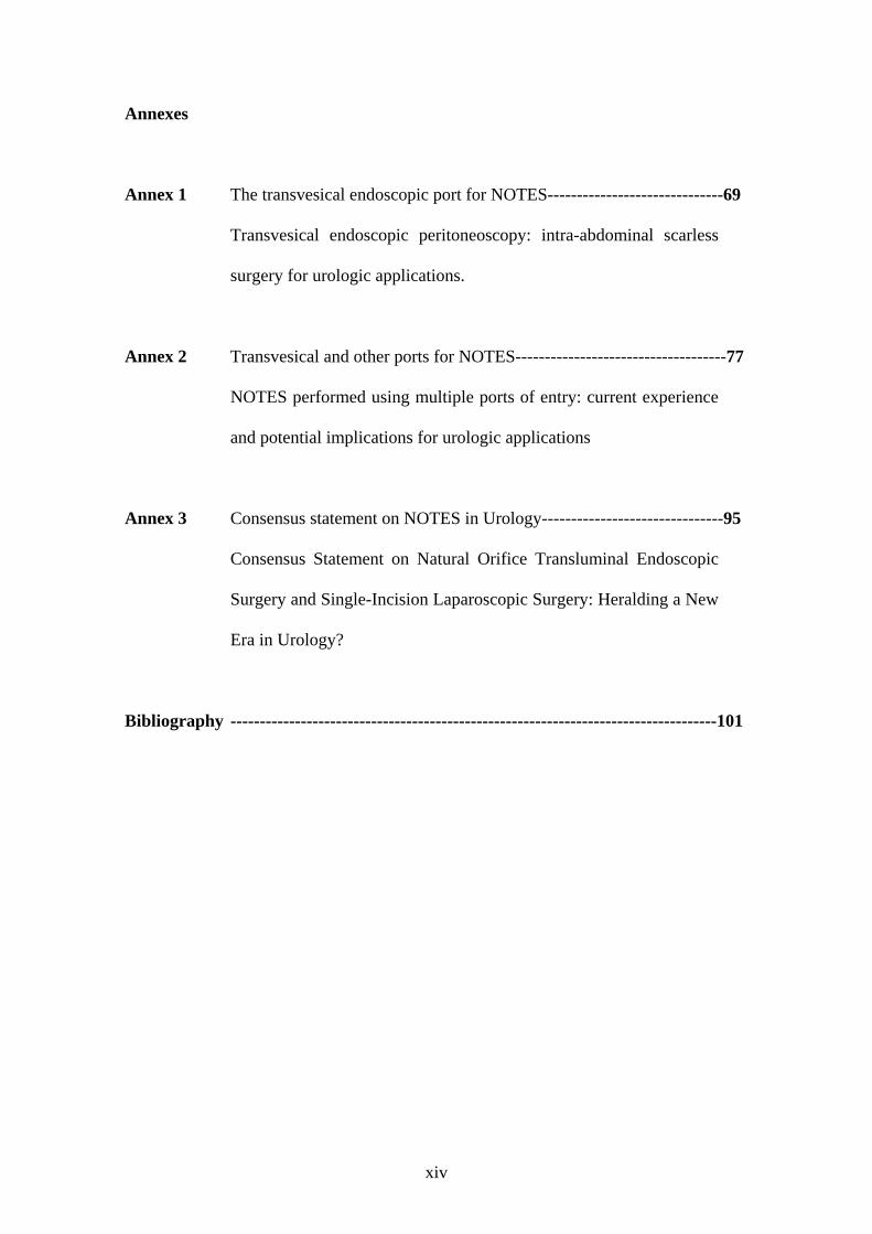
Annexes
Annex 1 The transvesical endoscopic port for NOTES------------------------------69
Transvesical endoscopic peritoneoscopy: intra-abdominal scarless
surgery for urologic applications.
Annex 2 Transvesical and other ports for NOTES------------------------------------77
NOTES performed using multiple ports of entry: current experience
and potential implications for urologic applications
Annex 3 Consensus statement on NOTES in Urology-------------------------------95
Consensus Statement on Natural Orifice Transluminal Endoscopic
Surgery and Single-Incision Laparoscopic Surgery: Heralding a New
Era in Urology?
Bibliography -----------------------------------------------------------------------------------101

15
Chapter 1
General Introduction
Minimally invasive surgery: a brief revision
The early history of laparoscopy is unknown to many surgeons, but endoscopy
was first described by Hippocrates in Greece (460-375 BC) (Rosin et al., 1993). He
made reference to a rectal speculum. The first simple speculum for gynecological
endoscopy dates from about the same time. The Talmud refers to a siphopherot made of
lead, bent at its tip with a mechul (wooden mandarin) used for inspection of the vagina.
Roman medicine also produced instruments with which they could inspect internal
organs (Bento and Hewlett, 1944). In Pompeii's ruins (70 AD), a three-bladed vaginal
speculum was discovered; this instrument was similar to the current vaginal speculum
(Gorden, 1993). Greek also transferred the teachings of medicine to the Arabs included
the use of speculums. The Arabian Abulkasim (936-1013 A.D.) improved upon
Hippocrates´ method by using reflected light to examine the cervix of utero (Bento and
Hewlett, 1944). The next evolutionary step was fostered by Tulio Caesare Aranzi in
1585. Aranzi was the first to use a light source for an endoscopic procedure, focusing
sunlight through a flask of water and projecting the light into the nasal cavity (Organ,
1933; Saleh, 1988; Gunning and Rosenzweing, 1991; Philipi et al., 1991). Thus, the
interest for physicians to look into the "internal organs" has existed since the early days
of medicine.
The credit for modern endoscopy belongs to Philipp Bozzini of Frankfurt -
German (1773-1809) (Bozzini, 1806). He developed a light conductor which he called
"Lichtleiter" to avoid the problems of inadequate illumination. This early endoscope
combined reflecting mirrors, candles, and an urethal cannula to direct light into the
internal cavities and then redirects it to the eye of the observer. This brilliant and
unconventional pioneer was officially reprimanded by the local medical community for
undue curiosity, having been so audacious as to place this instrument into the uretra of a

16
patient and attempt direct bladder inspection. Although he died at age 36 of typhoid
fever, Bozzini's invention established the principles that guided the development of
endoscopy, and it inspired others to forge ahead in this new field. John D. Fisher (1798-
1850) in Boston descrived an endoscope initially to inspect the vagina, but later he
modified it to examine the bladder and urethra (Picatoste et al., 1980). In 1853, Antoine
Jean Desormeaux, a French surgeon, invented the first endoscope that enabled the
physician to actually see (Desormeaux, 1867). For many he is considered the "Father of
Endoscopy." This instrument had a system of mirrors and lens, with a lamp flame as the
light source; the endoscope burned a mixture of alcohol and turpentine. Burns, as might
be imagined, were the major complication of these procedures. Desormeaux had
initially contemplated using electricity, but abandoned that idea. This endoscope was
mainly used for urologic cases. In 1869, Commander Pantaleoni used a modified
cystoscope to cauterize a hemorrhagic uterine growth. Pantaleoni thus performed the
first diagnostic and therapeutic hysteroscopy (Gunning and Rosenzweig, 1991).
Thomas Edison proved a new technologic breakthrough in 1879 with the
invention of the electric light bulb. This allowed Maximilian Nitze of Dresden - German
(1848-1906) and Josef Leiter of Vienna - Austria, in the same year, to introduce a rigid
cystoscope with a built-in light source formed from an electrically heated platinum wire,
a multilens system, and a separate water circulation system for cooling. Like the
Lichtleiter from Bozzini, this instrument was only used for urologic procedures (Rather,
1976; Hausmann, 1987). Then, Newman in 1883 described using a miniaturized version
of the incandescent bulb in a cystoscope and Boisseau de Rocher in 1890 introduced a
Mignon lamp cystoscope that offered the first double channel for ureteral
catheterization (Newman, 1888; De Rocher 1890).
In gastrointestinal field, in 1868, Kussmaul performed the first
esophagogastroscopy, initiating efforts at instrumentation of the gastrointestinal tract
(Haubrich, 1987). Mikulicz and Schindler, however, are credited with the advancement
of gastroscopy, having designed gastroscopes equipped with both an optical system and
a means to insufflate air (Haubrich, 1987). The use of rigid endoscopic instruments
severely limited access to the gastrointestinal tract. A flexible instrument was requied to
gain entrance into the duodenum or the colon proximal to the rectosigmoid junction.
The necessary technology took over 100 years to be developed until Georg Wolf and
Rudolph schindler (1932) introduced the first useful semi-flexible gastroscope to the

17
market ready for use (Haubrich, 1987). All these developments were restricted to the
inspection of organs through natural external access.
Laparoscopy has been evolving for more than 100 years, with the first
experimental laparoscopy reported in 1901 by George Kelling a German surgeon from
Dresden who was the first to apply Nitze´s cystoscope in combination with
pneumoperitoneum in peritoneal cavity of a living dog. The pneumoperitoneum was
created by injecting air through a sterile cotton filter in abdominal cavity with high-
pressure air insufflation with the goal of stopping intra-abdominal bleeding (ectopic
Pregnancy, bleeding ulcers, pancreatitis), but these studies did not find any response or
supporters.. He noted that the abdominal cavity could store more than 2.5 liters of blood
and he also considered intra-abdominal adhesions a contraindication for the procedure.
George Kelling coined the term "celioscopy" to describe the technique (Kelling, 1901).
Also in 1901, Dimitri Ott, working in Petrograd described “ventroscopy”, a technique in
which a speculum was introduced through an incision in the posterior vaginal fornix.
Ott wore head mirrors to reflect light and augment visualization (Ott, 1901).
During late 1910 and early 1911, Hans Jacobaeus, from Stockholm, used the
term "laparothoracoscopy" for the first time (Jacobeus, 1911). By 1912 he had
performed closed-cavity endoscopy with a Nitze cystoscope in over 100 patients with
ascite and also described liver pathology, peritoneal tuberculosis, and tumours. He also
suggested employing similar techniques to examine body cavities endoscopically.
Following this he devoted most of his attention to thoracoscopy. He published his report
on laparoscopy and thoracoscopy in humans in Münchener Medizinische Wochenschrift.
A response by Kelling appeared two months later in the same journal, disputing
Jacobaeus' claim to be the first to perform the procedure in humans, stating that he had
successfully used celioscopy in two humans between 1901-1910. Unfortunately, Kelling
had made a mistake: he did not publish his work. Interestingly, in Jacobaeus' paper in
1911, he viewed thoracoscopy as a more promising procedure than laparoscopy. He also
reported in 1923 the first bleeding complication requiring laparotomy (Harrell and
Heniford, 2005).
In 1911 Bertram Berheim, an assistant surgeon at Johns Hopkins, performed the
first laparoscopy in the United States, before he learned of the work of Kelling and
Jacobaeus. He published his laparoscopic experiences entitled, “organoscopy”, in the
Annals of Surgery (Bernheim, 1911). The instrument was a proctoscope of a half-inch
diameter, and he used ordinary light for illumination. Orndoff, an internist from

18
Chicago, reported the first large series of peritoneoscopies (42 cases) in the United
States in 1920 (Orndoff, 1920). One of his innovations was a sharp pyramidal trocar
point. In 1923, Kelling reported his 22 years of experience with laparoscopy to German
Surgical Society. Kelling became one of the earliest advocates of minimally invasive
surgery. He encouraged surgeons to use diagnostic laparoscopy in order to spare
patients the prolonged and costly stay of a laparotomy.
Over the next 40 years, specific instruments were developed to allow for an
accurate and complete examination of the peritoneal cavity. Pneumoperitoneum was
maintained initially through syringe injections until Otto Goetze developed a manual
insufflator with a foot pump in 1921 (Harrell and Heniford, 2005). Zollikofer a Swiss
gynecologist used carbon dioxide to obtain a pneumoperitoneum in 1924 because it was
absorbed faster and minimized the risk for explosion (Gaskin et al., 1991). In 1929
Heinz Kalk, a German gastroenterologist considered the founder of the German School
of Laparoscopy developed a 135-degree lens system and a dual trocar approach (Kalk,
1929). He used laparoscopy as a method of diagnosis for liver and gallbladder disease.
Therapeutic applications began in 1930. Fervers, a gynaecologist, initiated this process
with the laparoscopic adhesiolysis using electrocautery (Harrell and Heniford, 2005). In
1934 an American internist, John Ruddock, described laparoscopy as a good diagnostic
method, many times superior to laparotomy. His developed a specific lens system and
biopsy forceps to be used in laparoscopy (Ruddick, 1934). Then he reported his results
in 500 patients (Ruddick, 1937). Boesch of Switzerland performed a tubal ligation using
endoscopic electrocoagulation in 1936. Internists also appreciated the usefulness of this
technique to provide biopsy speciments of liver pathology (Harrell and Heniford, 2005).
A major advance occurred in 1938 when Janos Veress of Hungary developed the
spring-loaded needle for draining ascites and evacuating fluid and air from the chest. Its
main purpose was to perform therapeutic pneumothorax to treat patients suffering from
tuberculosis (Varess, 1938). He used it in over 2000 cases. He did not suggest that it be
used for laparoscopy. Its current modifications make the "Veress" needle a perfect tool
to achieve pneumoperitoneum during laparoscopic surgery. In 1944, Raoul Palmer of
Paris performed gynecological examinations using laparoscopy and placing the patients
in the Trendelenburg position so air could fill the pelvis (Palmer, 1947). He also
stressed the importance of continuous intra-abdominal pressure monitoring during a
laparoscopic procedure. Harold Hopkins was responsible for the two most important
inventions in endoscopy after World War II: the rod-lens system and fiberoptics. These
19
improvements allowed a brighness and the true color to the endoscopes that had not
been possible before. In addition, the light source was removed from the tip of the
insruments, and this decreased the risk for intra-abdominal thermal injuries (Harrell and
Heniford, 2005). Hasson, a gynecologist from the Grant Hospital of Chicago,
Augustana Hospital and Columbus-Cuneo Medical Center, published in the American
Journal of Obstetrics and Gynecology on July 15, 1971 his paper named: "A modified
instrument and method for laparoscopy." He developed a technique performing
laparoscopy through a miniature laparotomy incision (Hasson, 1971).
Despite such advances in laparoscopic imaging and technique, several
troublesome problems persisted. Bowel and vascular injuries during trocar insertion
continued to occur. No scientific knowledge existed regarding the dangers of increased
intraabdominal pressure. These dangers severely restricted the use of laparoscopy. Few
surgeons judged that the advantages of laparoscopy outweighed the inherent’s risks of
the technique. Additionally, remarkable discoveries were made that created modern
surgery in this same period of time. Anesthesia and antisepsis made elective surgery a
reality. Antibiotics decreased the mortality of surgeries and as a result complex
surgeries never before realistically attempted became possible. As the complexity of
surgeries increased, the doctrine of surgical exploration expanded exponentially. It was
the begining of the aforism “Big Surgeon Big Incision”.
During the 1960s, Kurt Semm, a German gynecologist, invented the automatic
insufflator for monitoring the pressure of CO2 pneumoperitoneum. His experience with
this new device was published in 1966 (Harrell and Heniford, 2005). But, Semm also
made and designed many new instruments included tissue morcellators, suction-
irrigation systems, laparoscopic endocoagulation, and laparoscopic scissors. He played
a major role in the development of laparoscopy. He called his procedure "Pelviscopy” to
avoid the negative connotations laparoscopy seemed to be attracting. He performed the
first laparoscopic appendectomy in 1983 during a gynecological procedure and opened
a large door for a new surgery, although he was almost removed from the Germany
Physician Society because of that procedure (Semm, 1983). Although Semm was not
recognized in his own land, on the other side of the Atlantic, both American physicians
and instrument makers valued the Semm insufflator for its simple application, clinical
value, and safety.
In England, in 1980, Patrick Steptoe started to perform laparoscopic procedures
in the operating room under sterile conditions. In 1981, rules and requirements to

20
perform laparoscopy were adopted by many hospitals and surgical societies. The
American Board of Obstetrics and Gynecology made laparoscopy training a required
component of residency training (Phillips et al., 1974; Reich, 1989).
In 1985, Erich Muhe reported the first laparoscopic cholecystectomy but the
procedure was rejected by the German Surgical Society, and scepticism remained until
1993 when his pioneering efforts finally were recognized formally by the German
Surgical Society (Reynolds, 1989; Litynski, 1998).
The first solid state camera was introduced in 1982. This was the start of "video-
laparoscopy." The modern era of laparoscopy really started when the French general
surgeon Philippe Mouret realized that laparoscopy could be used for more than just
exploration, and in 1987 he performed the first video-guided laparoscopic
cholecystectomy (Mouret, 1996). Within a year, Dubois (Paris), Perissat (Bordeaux),
and Barry Mckernan and William Saye (United States) had performed laparoscopic
cholecystectomy at their respective institutions on both sides of the Atlantic (Dubois et
al., 1990; Mckernan, 1990; Litynski et al., 1999; Harrell and Heniford, 2005).
Only five years later, this procedure was acknowledged as the “gold standard”
technique for the removal of the gallbladder (NIH Consensus Conference, 1993).
Progress in acknowledging the laparoscopic potential in urology followed the same
course, although the time scale was markedly truncated. Schuessler, in Texas reported
his initial experience in pelvic lymphadenectomy (Schuessler et al., 1991). Although
laparoscopic explorations for undescended testis had been done previous to this
reported, its impact was marginal, with the procedure being dismissed as amenable to
few patients with little other clinical applicability (Silber and Cohen, 1980). Soon
afterward, techniques for the extraction of solid organs, including the adrenal, kidney,
prostate and bladder, soon were developed (Clayman et al., 1991; Gagner et al., 1992;
Schuessler et al., 1997; Guillonneau et al., 1999; Abbou et al., 2000; Gill et al., 2000;
Rassweiler et al., 2001; Turk et al., 2001). Nowadays, practically all surgeries with
exception of transplantation can be made by laparoscopy with advantages for the
patients.

21
Basis for Natural Orifice Translumenal Endoscopic Surgery (NOTES)
The introduction of laparoscopic surgery will stand as the revolutionary episode
that marked the end of the twentieth century. This new approach has completely
revolutionized modern surgical practices, significantly changing the surgical way of
thinking, surgical techniques, and all other aspects of modern surgical patient care
(Harrell and Heniford, 2005).
Simultaneously, the endoscopy has changed progressively and dramatically over
the last few decades and the evolution of this technique has been from simple diagnostic
procedures to progressively more invasive one (Vitale et al., 2005). In this sequence,
one of the last achievements of surgical endoscopy was the aggressive and efficacious
gastric endoscopic mucosal resection. These procedures were progressively invading the
visceral wall, and some anecdotally reports began to describe safe endoscopic
procedures beyond the visceral wall, ie. transgastric drainage of pancreatic pseudocyst
(Vitale et al., 2005). Supported by these reports, Reddy and Rao, and Kalloo et al
described a new port to the peritoneal cavity through a transgastric approach in humans
and pigs, respectively (Kalloo et al., 2004; Rattner and Kalloo, 2006). This was the
dawn of a new era that open new perspectives for those dealing with abdominal
minimally invasive surgery, making them to pursue novel audacious goals such as
avoidance of incisions and even pain. In fact, the visceral wall is no longer a barrier for
endoscopic intervention.
Subsequently, various authors described more complex intra-abdominal
procedures such as ligation of fallopian tubes, cholecystectomy and cholecystogastric
anastomosis, gastrojejunostomy, partial hysterectomy and oophorectomy (Jagannath et
al., 2005; Park et al., 2005; Kantsevoy et al., 2005; wagh et al., 2005). The continuous
evolution of new instruments and the unexpected success reported by those authors in
porcine models with transgastric surgery seems to open a new era in surgical field:
Natural Orifice Translumenal Endoscopic Surgery (N.O.T.E.S.). These techniques are
likely less invasive than even laparoscopy because we don't have to cut through the skin
and muscle of the abdomen, and it may prove a viable alternate to existing surgical
procedures.
In this sequence, the development of other transvisceral approaches remained to
be defined. This was the call for our experiments on transvesical surgery with or
without combination with other natural orifices approaches in order to help the

22
implementation of new intra-abdominal scarless procedures in what seems to be the 3rd
generation surgery.

23
Objectives of the Thesis
This project aims to test the hypothesis that common abdominal and thoracic
surgical procedures might one day be performed by natural orifices. In addition to
avoidance of scars, the major rationale for this project emerges from the knowledge that
viscera walls do not have sensitivity to cut. Furthermore, the increasing development
image technology gives support to believe this goal is attainable soon. For all these
reasons, it make sense to explore the hypothesis to perform surgery by natural orifices
in what will be for sure a further step on minimally invasive surgical field. Thus, using
the pig model and current instruments from urology and gastroenterology fields, we
planned this PhD Thesis with the following objectives:
1) To develop novel strategies to introduce surgical instruments into peritoneal
cavity sparing abdominal wall through transvesical pathway;
2) To test the feasibility, usefulness and safety of this peritoneal access alone or
in combination to perform either abdominal or thoracic surgery;
3) To describe novels Natural Orifice Transluminal Endoscopic Surgical
techniques for commom procedures such as peritoneoscopy, liver biopsy,
thoracoscopy, lung biopsy and nephrectomy;
4) To assess the feasibility and safety of the new endoscopic closure method for
transvesical perforations.

24

25
Chapter 2
Transvesical endoscopic port for abdominal surgery
Lima E, Rolanda C, Pêgo JM, Henriques-Coelho T, Silva D, Carvalho JL, Correia-Pinto
J. Transvesical endoscopic peritoneoscopy: A novel 5 mm-port for intra-abdominal
scarless surgery. J Urol; 176: 802-805, 2006.

26

Transvesical Endoscopic Peritoneoscopy:A Novel 5 mm Port for Intra-Abdominal Scarless SurgeryEstevao-Lima, Carla Rolanda, José M. Pêgo, Tiago Henriques-Coelho, David Silva,José L. Carvalho and Jorge Correia-Pinto*From the Life and Health Sciences Research Institute, School of Health Sciences, University of Minho (EL, CR, JMP, DS, JCP),and Departments of Gastroenterology (CR) and Anesthesiology (JMP), Sao Marcos Hospital, Braga and Department of Urology,Santo António General Hospital (EL) and Department of Pediatric Surgery, Sao Joao Hospital (THC, JLC, JCP), Porto, Portugal
Purpose: Recently various groups reported successful attempts to perform intra-abdominal surgery through a transgastricpathway. We assessed the feasibility and safety of a novel transvesical endoscopic approach to the peritoneal cavity througha 5 mm port in a porcine model.Materials and Methods: Transvesical endoscopic peritoneoscopy was performed in 8 anesthetized female pigs, including 3nonsurvival and 5 survival animals. Under cystoscopic guidance a vesical hole was created on the ventral bladder wall withan open-ended ureteral catheter. An over tube with a luminal diameter of 5.5 mm was placed in the peritoneal cavity, guidedby a 0.035-inch guidewire. In all animals we performed peritoneoscopy of the entire abdomen as well as liver biopsy andfalciform ligament section. A vesical catheter was placed for 4 days in all survival animals, which were sacrificed by day 15postoperatively.Results: After a learning curve in the first 3 nonsurvival animals the creation of a vesical hole and placement of the over tubewere performed without complication in all survival animals. In these animals we easily introduced an EndoEYE™ into theperitoneal cavity, which provided a view of all intra-abdominal viscera, as well as a 9.8Fr ureteroscope, which allowed simplesurgical procedures without complications. In survival experiments all pigs recovered. Necropsy examination revealedcomplete healing of the vesical hole and no signs of infection or adhesions into the peritoneal cavity.Conclusions: Transvesical endoscopic peritoneoscopy was technically feasible and it could be safely performed in a porcinemodel. This study provides encouragement for additional preclinical studies of transvesical surgery with or without combi-nations with other natural orifices approaches to design new intra-abdominal scarless procedures in what seems to be thirdgeneration surgery.
Key Words: swine, endoscopy, laparoscopy, peritoneal cavity
Since the late 1980s, a revolution has begun with theimplementation of laparoscopic techniques that havegained progressive acceptance as the gold standard for
an increasing number of intra-abdominal procedures.1 Infact, there are many advantages to the laparoscopic ap-proach, such as smaller incisions, decreased postoperativepain and more rapid patient recovery.
Recently Kalloo et al described a new port to the perito-neal cavity through a transgastric approach in a porcinemodel.2 Subsequently various groups described more complexintra-abdominal procedures, such as fallopian tube ligation,cholecystectomy and cholecystogastric anastomosis, gastroje-junostomy, partial hysterectomy and oophorectomy.3–6 Thecontinuous evolution of new instruments and the unex-pected success reported by these investigators in porcine
Submitted for publication September 17, 2005.Study received approval from the ethical review boards, Minho
University, Braga, Portugal.Supported by Grants Bolsa de Investigação Básica JABA 2005 da
Associação Portuguesa de Urologia and POCTI/SAU-OBS/56428/2004 from FCT-Portugal.
* Correspondence: Instituto de Ciências da Vida e da Saúde, Es-cola de Ciências da Saúde, Universidade do Minho, Campus de
Gualtar, 4709-057 Braga, Portugal (telephone: �351 253 604 807;FAX: �351 253 604 831; e-mail: [email protected]).0022-5347/06/1762-0802/0THE JOURNAL OF UROLOGY®
Copyright © 2006 by AMERICAN UROLOGICAL ASSOCIATION
802
models with transgastric surgery seems to have opened anew era in the surgical field, that is endoscopic transvisceralsurgery.7–11
In this sequence the development of new transvisceralapproaches remains to be defined. In this pilot study weassessed the feasibility and safety of the transvesical endo-scopic approach to the peritoneal cavity with liver biopsyand falciform ligament section in a porcine model.
MATERIALS AND METHODS
This study was approved by the ethical review boards ofMinho University, Braga, Portugal. Transvesical procedureswere performed in 8 female pigs, including 3 in nonsurvivalstudies. Survival studies were done in 5 consecutive 35 to 45kg female pigs (Sus scrofus domesticus). These 5 pigs werefollowed after surgery for 15 days before sacrifice and nec-ropsy examination.
Pig PreparationThe animals were withdrawn from food for 24 hours andfrom water for 6 hours before the surgical procedure. Allprocedures were performed using general anesthesia with
6.0 mm endotracheal intubation with a Ruschelit® SuperVol. 176, 802-805, August 2006Printed in U.S.A.
DOI:10.1016/j.juro.2006.03.075

TRANSVESICAL PERITONEOSCOPY 803
Safety Clear Tracheal Tube and mechanical ventilation.Pre-anesthesia medication consisted of intramuscular injec-tion of 32 mg/ml azaperone (Stressnil®) reconstituted with 1mg/ml midazolam (Dormicum®) at dose of 0.15 to 0.2 ml/kg.
Venous access was obtained through an intravenous lineplaced in the marginal ear vein. Anesthesia was inducedwith 3 �g/kg fentanyl (Fentanest®), 10 mg/kg thiopentalsodium (Pentothal®) and 0.2 mg/kg vecuronium (Norcu-ron®). For infection prophylaxis all animals received anintramuscular injection of 1 gm ceftriaxone (Rocephin®) be-fore endoscopy. Anesthesia was maintained with 1.5% to 2%sevoflurane (Sevorane®) and a perfusion of 1 mg/kg per hourvecuronium (Norcuron®).
Surgical TechniqueAn Olympus® A2281 cystoscope with an A22001A telescopewas introduced through the urogenital sinus and urethrainto the bladder with hydrodistention. Before any furtherprocedure the bladder was emptied of urine and refilled withsaline. The vesicotomy site was selected on the ventral blad-der wall caudal to the bladder dome. Figure 1 shows theprocedures. The cystoscope was then replaced by an Olym-pus® A2942A ureteroscope guided through a Terumo®guidewire to achieve the bladder. A mucosal incision wasmade with Olympus® A2576 scissors introduced by theworking channel of the ureteroscope. Subsequently a 5FrSelectip™ open-ended ureteral catheter was pushed forwardthrough the incision into the peritoneal cavity (fig. 2).
A 0.035-inch flexible tip Terumo® guidewire was theninserted into the peritoneal cavity through the ureteral cath-eter lumen. Guided by the flexible tip guidewire the vesicalhole was enlarged with the dilator of a ureterorenoscopesheath (Microvasive-Boston Scientific Corp., Natick, Massa-
FIG. 1. A, vesical perforation with open-ended ureteral catheter, B,
guidewire passage. C, placing over tube in transvesical position. D,video telescope inside abdominal cavity, inserted through over tube.chusetts) enveloped by a flexible over tube. We designed thisequipment with a length of 25 cm, internal diameter of 5.5mm and wall thickness of 1 mm. Dilator passage over theguidewire and through the bladder wall spared muscle cut-ting. The ureteroscope was introduced into the peritonealcavity in the over tube and allowed to create pressure con-trolled CO2 pneumoperitoneum up to 12 mm Hg using anOlympus® UHI-3 Insufflator.
The peritoneal cavity was examined, biopsy specimenswere obtained from the liver with an Olympus® A2423 en-doscopic biopsy forceps and the falciform ligament was sec-tioned with an Olympus® A2576 device (fig. 3). The uretero-scope was withdrawn. It was then possible to introduce intothe peritoneal cavity a 5 mm Olympus® EndoEYE™ videotelescope with chip on the tip and a 0-degree direction ofview. The endoscope was removed and the peritoneal cavitywas decompressed through the over tube. At the end of thisprocedure the cystoscope was again introduced to observeinner bladder wall morphology. At the end of the operation a14Fr Foley catheter was inserted into the bladder and theballoon was inflated with 10 cm3 saline.
At the beginning of this protocol the first 3 animals weresacrificed immediately at the end of the procedure by anes-thetic overdose and the peritoneal cavity was examinedgrossly. In the 5 survival animals oral feeding was startedwithin the following 24 hours. They were evaluated dailyand followed for 15 days. The bladder catheter was removed
FIG. 2. Endoscopic view of vesical wall perforation with open-endedureteral catheter.
FIG. 3. Representative endoscopic views. A, transvesical liver bi-opsy. B, transvesical falciform ligament section.

TRANSVESICAL PERITONEOSCOPY804
4 days after surgery except in 1 animal, in which the vesicalcatheter exteriorized accidentally 12 hours after surgery.Necropsy was performed at the end of followup with partic-ular attention to the vesical incision site.
RESULTS
Through a rapid learning curve the first 3 nonsurvival pigswere used to acquire the necessary skills to perform trans-vesical endoscopic peritoneoscopy. In the subsequent sur-vival animals all procedures involved in creating the vesicalhole, including cystoscopy, bladder mucosal incision, vesicot-omy and transvesical over tube passage, were performedwithout complications. The ureteroscope was introducedeasily into the peritoneal cavity and CO2 insufflation wasperformed without incident. The view of the internal organsprovided by this instrument was reasonable, mainly for or-gans of the upper abdomen, including the liver, gallbladder,stomach, spleen and diaphragm. The length of the uretero-scope used allowed us to perform simple surgical procedures,such as liver biopsy and falciform ligament section, in allanimals without difficulty. The over tube allowed us to eas-ily introduce the EndoEYE™, which provided a detailedview of all intra-abdominal organs (fig. 4). After over tuberemoval the cystoscope showed obvious signs of contraction,making the vesical hole appear like a puncture hole. Oper-ative time from cystoscope introduction to the completion ofsurgery was between 20 and 40 minutes.
After recovery from anesthesia the pigs tolerated a regu-lar diet within 24 hours, ate heartily and thrived for the next14 days. Any adverse events occurred in the survival period.Until postoperative day 4 the pigs tolerated the bladdercatheter well. After its removal the pigs voided normally.
Necropsy 15 days after surgery revealed complete healingof the bladder wall incision. The vesical perforation locationwas detected on the inner surface of the bladder as a smalldimple (fig. 5). At necropsy the liver biopsy sites were com-pletely healed. There were no signs of infection or adhesionsin the peritoneal cavity.
FIG. 4. Endoscopic view of upper abdominal organs, that is liver andgallbladder, provided by video telescope with chip on tip inserted
through over tube. Over gallbladder note sectioned falciform liga-ment. Bleeding point on liver edge represents liver biopsy.DISCUSSION
Since Kelling used a Nitze cystoscope combined with pneu-moperitoneum to examine the peritoneal cavity in a dog in1902, many advances in endoscopic intra-abdominal surgeryhave occurred.1 In fact, the rationale for the current studycomes from constant technological advances making betterand more reliable instruments available for endoscopic pro-cedures and from the recent, unexpected success of trans-gastric surgery described in porcine models.2–6 These factsled us to predict that an additional transvisceral approachpositioned diametrically opposed to the stomach might behelpful for performing complex intraperitoneal endoscopicprocedures.
In this sequence we tested the possibility of creating atransvesical 5 mm port. Our study reveals that it was easy toperform it while handling the current instruments used forurological purposes. We began all procedures with a cysto-scope, mainly to facilitate the perception of anatomy, whichin the porcine model is different from that in humans sincethe urethra opens in a urogenital sinus. Additionally, thelarger vision field supplied by the cystoscope compared to aureteroscope allowed us to easily define the bladder point toperforate. We selected this point in the ventral wall toachieve a peritoneal cavity above the level of the bowel loops.Perforation of the bladder wall with the open-ended ureteralcatheter was rapid, efficient and safe. It should be empha-sized that the incision performed in the bladder mucosa wasessential to fix the catheter tip and prevent easy looping ofthe catheter inside the bladder. To date with this techniquewe have never injured any bowel loop since this catheter isnontraumatic.
Passage of the over tube with the ureteral dilator guidedby the guidewire was also an easy operation. The uretero-scope working channel allowed the creation of pressure con-trolled CO2 pneumoperitoneum. Although it has limitedwidth, the view provided by a 9.8Fr ureteroscope allowed usto perform simple procedures, such as liver biopsies andfalciform ligament section.
In our point of view a transvesical 5 mm port could beparticularly useful for complementing other transvisceralports. In fact, a group that performed complex abdominalprocedures through a transgastric pathway detected prob-lems in working with decreased triangulation and manytimes with retroflexion.4 The position of a transvesical portcould easily overcome some of these limitations with addi-tional advantages for performing those complex operations.In fact, it easily achieved by introducing a rigid 5 mm in-strument or a video telescope, which can be particularlyuseful in those procedures. It should be emphasized that the
FIG. 5. Bladder necropsy. A, external surface of air filled bladderwith closed hole (arrow). B, inner bladder surface with small resid-ual dimple at surgical perforation site (arrow).
transvesical port provides frontal access to the upper ab-

TRANSVESICAL PERITONEOSCOPY 805
dominal organs, allowing better instrument position forworking on these organs.
During creation of the vesical hole in our protocol the riskof bowel perforation was never neglected. However, in ourlimited number of experiments we never damaged any bowelloop. The explanation of these results might be related toseveral aspects. 1) Bowel loops in contact with the bladderwall are free into the abdomen, likely making them runahead of the ureteral catheter as it perforates the vesicalwall. 2) Subsequent procedures to position the over tubewere performed while guided by the hydrophilic guidewirewith atraumatic equipment.
Vesical perforation has potential complications, such asperitoneal urine leakage with secondary infection (peritoni-tis). This commonly occurs as a delayed complication ofundiagnosed vesical perforation in trauma or pathologicalbladder conditions, such as neoplasms.12 In fact, we hadreservations about leaving the transvesical entry site un-closed. Interestingly we did not note any of those complica-tions in our study, suggesting that suture or approximationof the vesical wall was unnecessary. In our understandingthis might be related to various reasons. 1) The techniquethat we used to perforate the bladder wall spared muscle fibersection. It seems that muscle fibers contract around the holeimmediately after over tube removal. At the end of the proce-dure we could see through the cystoscope that the vesicalhole became immediately and virtually closed. 2) Bladderdecompression provided by the vesical catheter surelyhelped prevent peritoneal leakage, enhancing all healingprocesses. In this regard we stress that in the last pig in ourstudy the vesical catheter exteriorized within 12 hours aftersurgery and even in these circumstances no intra-abdominalcomplications were observed. 3) We did not deal with abnor-mal bladder tissue invaded by neoplasm or infiltrated byinflammatory cells.
The possibility of installing a 5 mm port, which might allowthe introduction of a 5 mm rigid instrument or a video tele-scope with chip on the tip offering an excellent view of theupper abdominal organs, makes this approach appealing for acombination with a transgastric pathway for future intra-ab-dominal scarless surgery. In addition to cosmesis, we predictthat these procedures performed via a transvisceral pathwaymay have easier recovery than traditional surgery. We thinkthat postoperative pain might be significantly decreased sincethe density of pain receptors on the viscera is significantlylower than that in the abdominal wall.
CONCLUSIONS
This study demonstrates that the transvesical endoscopic
approach to the peritoneal cavity is feasible without anyfurther complications in a porcine model. This series pro-vides encouragement for additional preclinical studies oftransvesical surgery with or without combinations withother natural orifice approaches to design new intra-abdom-inal scarless procedures in what seems to be the third-generation surgery.
ACKNOWLEDGMENTS
André Oliveira and Mário Moita, Olympus-Portugal andManuel Silva contributed to this project.
REFERENCES
1. Harrell, A. G. and Heniford, T.: Minimally invasive abdominalsurgery: lux et veritas past, present, and future. Am J Surg,190: 239, 2005
2. Kalloo, A. N., Singh, V. K., Jagannath, S. B., Niiyama, H., Hill,S. L., Vaughn, C. A. et al: Flexible transgastric peritoneos-copy: a novel approach to diagnostic and therapeutic inter-ventions in the peritoneal cavity. Gastrointest Endosc, 60:114, 2004
3. Jagannath, S. B., Kantsevoy, S. V., Vaughn, C. A., Chung,S. S. C., Cotton, P. B., Gostout, C. J. et al: Per-oral trans-gastric ligation of fallopian tubes with long-term survival ina porcine model. Gastrointest Endosc, 61: 449, 2005
4. Park, P. O., Bergstrom, M., Ikeda, K., Fritscher-Ravens, A. andSwain, P.: Experimental studies of transgastric gallbladdersurgery: cholecystectomy and cholecystogastric anastomo-sis (videos). Gastrointest Endosc, 61: 601, 2005
5. Kantsevoy, S. V., Jagannath, S. B., Niiyama, H., Chung,S. S. C., Cotton, P. B., Gostout, C. J. et al: Endoscopicgastrojejunostomy with survival in a porcine model. Gas-trointest Endosc, 62: 287, 2005
6. Wagh, M. S., Merrifield, B. F. and Thompson, C. C.: Endoscopictransgastric abdominal exploration and organ resection:initial experience in a porcine model. Clin GastroenterolHepatol, 3: 892, 2005
7. Ponsky, J. L.: Gastroenterologists as surgeons: what they needto know. Gastrointest Endosc, 61: 454, 2005
8. Hochberger, J. and Lamade, W.: Transgastric surgery in theabdomen: the dawn of a new era? Gastrointest Endosc, 62:293, 2005
9. Vitale, G. C., Davis, B. R. and Tan, T. C.: The advancing artand science of endoscopy. Am J Surg, 190: 228, 2005
10. Weickert, U., Jakobs, R. and Riemann, J. F.: Diagnostic lapa-roscopy. Endoscopy, 37: 33, 2005
11. Liu, R., Chand, B. and Ponsky, J.: The future of surgicalendoscopy. Endoscopy, 37: 38, 2005
12. Cass, A. S. and Luxenberg, M.: Features of 164 bladder rup-
tures. J Urol, 138: 743, 1987
31
Chapter 3
Transvesical endoscopic port for thoracic surgery
Lima E, Henriques-Coelho T, Rolanda C, Pego JM, Silva D, Carvalho JL, Correia-Pinto
J. Transvesical thoracoscopy: a natural orifice transluminal endoscopic approach for
thoracic surgery. Surg Endosc; 21: 854-858, 2007.

32

Transvesical thoracoscopy: A natural orifice translumenal
endoscopic approach for thoracic surgery
Estevao Lima,1,2 Tiago Henriques-Coelho,3 Carla Rolanda,1,4 Jose M. Pego,1,4 David Silva,1 Jose L. Carvalho,3
Jorge Correia-Pinto1,3
1 Life and Health Sciences Research Institute (ICVS), School of Health Sciences, University of Minho, Braga, Portugal2 Department of Urology, Santo Antonio General Hospital, Porto, Portugal3 Department of Pediatric Surgery, S. Joao Hospital, Porto, Portugal4 Departments of Gastroenterology and Anesthesiology, S. Marcos Hospital, Braga, Portugal
Received: 4 February 2007/Accepted: 24 February 2007/Online publication: 4 May 2007
AbstractBackground: Recently there has been an increasingenthusiasm for using natural orifices translumenalendoscopic surgery (NOTES) to perform scarlessabdominal procedures. We have previously reported thefeasibility and safety of the transvesical endoscopicperitoneoscopy in a long-term survival porcine model asuseful for those purposes. Herein, we report our suc-cessful experience performing transvesical and transdi-aphragmatic endoscopic approach to the thoracic cavityin a long-term survival study in a porcine model.Methods: Transvesical and transdiaphragmatic endo-scopic thoracoscopy was performed in six anesthetizedfemale pigs. A 5 mm transvesical port was created on thebladder wall and an ureteroscope was advanced into theperitoneal cavity. After diaphragm inspection, weintroduced through the left diaphragmatic dome aureteroscope into the left thoracic cavity. In all animals,we performed thoracoscopy as well as peripheral lungbiopsy. Animals were sacrificed by day 15 postopera-tively.Results: We easily introduced a 9.8 Fr ureteroscope intothe thoracic cavity that allowed us to visualize thepleural cavity and to perform simple surgical proceduressuch as lung biopsies without complications. There wereneither respiratory distress episodes nor surgical com-plications to report. Postmortem examination revealedcomplete healing of vesical and diaphragmatic holes,whereas no signs of infection or adhesions were ob-served in the peritoneal or thoracic cavities.Conclusion: This study demonstrates the feasibility oftransvesical thoracoscopy in porcine model. However,although this study extends the potential applications ofNOTES to the thoracic cavity, new instruments andfurther work are needed to provide evidence that this
could be translated to humans and with advantages forpatients.
Key words: Swine — Endoscopy — Laparoscopy —Peritoneal cavity — Thoracic cavity
Background
Since the early 1990s, it has been proposed that thora-coscopy is less invasive than open procedures [6].Whereas its use as a definitive therapeutic tool in manypulmonary diseases is still controversial, its use as adiagnostic tool in pleural and pulmonary diseases is al-ready well established [2, 17]. Indeed many prospective,randomized studies confirmed that thoracoscopy re-duces postoperative pain, minimizes pulmonary dys-function and shortens hospital stay [1, 18, 21].
Recently, there is an increasing enthusiasm for usingnatural orifice approaches to perform abdominal scar-less surgery. In fact, Reddy and Rao (N. Reddy, V. G.Rao, oral communications, May 15 and 19, 2005; N.Reddy, oral communication, May 2004) and Kallooet al. [8] described a new port to the peritoneal cavitythrough a transgastric approach in humans and pigs,respectively. Subsequently, various authors describedmore-complex intra-abdominal procedures in porcinemodels, opening a new era in surgery that is beingconsidered third generation surgery: the natural orificetranslumenal endoscopic surgery (NOTES) [7, 9, 11–14,19–20]. Building on these findings, our group describedtransvesical endoscopic peritoneoscopy as a comple-mentary and safe approach to the abdominal cavity in aporcine model [10, 16].
The aim of this study was to assess the feasibilityand the safety of transvesical endoscopic approach toCorrespondence to: Jorge Correia-Pinto
Original Articles
Surg Endosc (2007) 21: 854–858
DOI: 10.1007/s00464-007-9366-x
� Springer Science+Business Media, LLC 2007
the thoracic cavity with lung biopsy in a porcinemodel.
Materials and methods
The ethical review board of Minho University (Braga, Portugal) ap-proved this study. Survival studies of thoracoscopy by transvesicalapproach were performed in six consecutive 15–20 kg female pigs (Susscrofus domesticus). Pigs were followed after surgery for 15 days beforesacrifice and postmortem examination.
Pig preparation
The animals were restrained from food (24 hours) and water (6 hours)before the surgical procedure. All procedures were performed undergeneral anesthesia, with 6.0-mm endo-tracheal intubation (SuperSafety clear tracheal tube, Ruschelit�) and mechanical ventilation. Pre-anesthesia medication consisted of an intramuscular injection of32 mg/ml azaperone (Stressnil�, Esteve Farma) reconstituted with1 mg/ml midazolam (Dormicum�, Roche) at dose of 0.15–0.2 ml/kg.
Venous access was obtained through an IV line placed in themarginal ear vein. Anesthesia was induced with 3 lg/kg fentanyl(Fentanest�, Janssen-Cilag), 10 mg/kg thiopental sodium (Pentothal�,Abbott) and 1 mg/kg vecuronium (Norcuron�, Organon). For infec-tion prophylaxis, all animals received intramuscular injection of 1 gceftriaxone (Rocephin�, Roche) before endoscopy. Anesthesia wasmaintained with 1.5–2% sevoflurane (Sevorane�, Abbott) and a per-fusion of 1 mg/kg/h vecuronium (Norcuron�, Organon).
Surgical technique
An ureteroscope (Olympus A2942A) was introduced through theurogenital sinus and urethra into the bladder with hydro-distension.Before any further procedure, the bladder was emptied from urine andrefilled with saline. The vesicotomy site was carefully selected on thebladder dome. A mucosal incision was made with scissors (OlympusA2576) introduced through the working channel of the ureteroscope.Subsequently, a 5 Fr open-ended ureteral catheter (Selectip, 62450200;Angiomed, Bard) was pushed forward through the incision into theperitoneal cavity. A 0.035-inch flexible-tip guide-wire (TerumoCorporation) was then inserted into the peritoneal cavity through thelumen of the ureteral catheter. Guided by the flexible-tip guide-wire,the vesical hole was enlarged with a dilator of an ureteroscope sheath(Microvasive, Boston Scientific Corporation) enveloped by a flexibleover tube (equipment designed by the authors: 25 cm length, 5.5 mminternal diameter and 1 mm wall thickness). An ureteroscope wasintroduced into the peritoneal cavity through the over tube and al-lowed the creation of a pressure controlled CO2 pneumoperitoneum upto 12 mmHg (Olympus Insufflator UHI-3 A90120A). Upper abdomenexploration and careful inspection of the diaphragm was performedusing the ureteroscope.
The site of transdiaphragmatic approach was carefully chosen onthe muscular pars of the left diaphragmatic dome. Peritoneal incisionwas made with a scissor (Olympus A2576) and the muscle fibers dis-sected until reaching the parietal pleura. Finally, we created a smallhole in the parietal pleura and the ureteroscope was advanced into thethoracic cavity (Figure 1). In the thorax, CO2 was insufflated up to 6mmHg and the pleural cavity and lung surface were inspected. Underdirect endoscopic control, lung biopsies were obtained from the lowerleft lung lobe using biopsy forceps (Olympus A2423). The ureteroscopewas withdrawn from the thoracic cavity after CO2 removal andexpansion of the left lung. The peritoneal cavity was then decom-pressed through the over tube. At the end of the operation, a 14 FrFoley catheter was inserted into the bladder and the balloon was in-flated with 10 cm3 of saline.
In this study, oral feeding was started within the following 24hours. Pigs were evaluated daily and followed for 15 days. Bladdercatheter was removed four days after surgery. Necropsy examinationwas then performed with particular attention to the site of vesical anddiaphragmatic incision.
Results
Transvesical and transdiaphragmatic endoscopic thora-coscopy with lung biopsy was performed on six pigs. Aswe have previously described, all procedures involved inthe creation of the vesical hole (cystoscopy, bladdermucosal incision, vesicotomy, transvesical overtubepassage) were performed without complications. Theureteroscope was introduced into the peritoneal cavityand the diaphragm was easily identified. The length ofthe ureteroscope allowed us to cross through the dia-phragm (small incision in the parietal peritoneum, dis-section of muscle fibers and incision of the parietalpleura) in all animals without difficulty (Figure 2). Theureteroscope working-channel allowed creating a pres-sure-controlled CO2-pneumothorax up to 6 mmHg. Inthe thorax, the visualization provided by the uretero-scope was limited to the lower half-cavity but allowed useasily to perform lower lobe lung biopsy (Figure 3). Atthe end of the procedure, it was easy to expand the lungand the ureteroscope showed signs of contraction of thesmall hole created in the diaphragm. The operative timefrom bladder-wall incision to completion of the surgeryvaried between 10 and 15 minutes.
Fig. 1. Schematic drawing of transvesical and transdiaphragmaticapproach.
855
After recovery of anesthesia, the pigs tolerated aregular diet started the morning after surgery and theyambulated freely, exhibiting normal behavior. No ad-verse event occurred during the survival period. Untilday 4 post-operative, pigs tolerated well the bladdercatheter. After its removal, pigs began to void normally.
The postmortem examination 15 days after surgeryrevealed complete healing of the vesical and diaphrag-matic incision (Figure 4). At necropsy, the lung biopsieswere completely healed. There were no signs of infectionor adhesions in both thoracic and peritoneal cavities.
Discussion
The widespread acceptance of minimally invasive tech-niques has revolutionized the practice of surgeryincluding thoracic surgery. Within a short period oftime, video-assisted thoracic surgery has become anacceptable approach with multiple studies confirmingthe advantages of thoracoscopy when compared to openprocedure, which include shorter hospital stay, rapidrecovery and return to physical activity, and excellentcosmetic results [1, 18, 21]. Recently, innovative naturalorifices endoscopic techniques using per-oral transga-stric surgery appear to further minimize surgicalaggression in what has been designated as third gener-ation surgery [10, 16]. In fact, several studies have con-firmed the technical feasibility of transgastric diagnosticand therapeutic peritoneoscopy, including liver biopsy,
tubal ligation, gastrojejunostomy, cholecystectomy,splenectomy, oophorectomy and partial hysterectomy[7–9, 11–14, 19–20]. In this regard, we showed for thefirst time that transvesical endoscopic approach to theperitoneal cavity is also feasible and particularly usefulto inspect diaphragm and upper abdominal organs [10,16]. Moreover, this approach revealed particularly use-ful to complement transgastric port, becoming chole-cystectomy by natural orifices a reliable procedure, atleast in the porcine model [16]. The feasibility and use-fulness of transvesical endoscopic peritoneoscopy forupper abdominal procedures raised the rationale to ex-tend its application to the thoracic cavity.
The aim of this study was to determine the technicalfeasibility and the safety of the transvesical approach tothe thoracic cavity with lung biopsy in a porcine modelin a long-term survival study. These experiments dem-onstrate that a transdiaphragmatic hole can be per-formed safely without injuring the lung or pericardium.The transdiaphragmatic incision could be made usingthe rigid ureteroscope and the scissors introducedthrough the working-channel. Interestingly, with ourapproach, we could easily identify the whole diaphragmand select the precise site of the incision. We could notreach the entire thoracic cavity mainly due to limitationsof current instruments (rigid and not long enough).Anyway, Lower lobe lung biopsies were also performedwithout any complications.
This study provides the first successful evidence thatthe thoracic cavity might also be reached throughNOTES. We anticipate some theoretical advantages to atransvesical-diaphragmatic approach to the thoraciccavity such as less pain and certainly a better cosmeticeffect than open or thoracoscopic procedures. We pre-dict less pain because, according to pain physiology,visceral wall has much less nervous terminal endingsthan skin, fascia and muscles. Transbronchic proceduresmight also share from these advantages but they areavailable only for peri-hilar lesions. In fact, althoughour approach might have some limitations (the risk ofabdominal infection or malignant cell spillage from thethoracic cavity), there is a vast number of interstitialpulmonary diseases where bronchoscopy with trans-
Fig. 2. Endoscopic views of a small hole on the muscular pars of leftdiaphragmatic dome. A Incision in diaphragmatic peritoneum.B Muscular hole being constructed in diaphragm pars muscularis.
Fig. 3. Representative endoscopic views of lung biopsy. A Edge of leftlower lung lobe; B lung biopsy being performed.
Fig. 4. Postmortem examination of diaphragm. Please note the smallscar in the diaphragm pars muscularis (arrow).
856
bronchic biopsy is unviable. For these conditions, wecould predict that transvesical-diaphragmatic approachmight have a potential role. Similarly, with the appro-priate instruments, pleural interventions like thoracicsympathectomy for the treatment of palmar hyperhi-drosis and Raynauds disease might also appear feasiblewith transvesical-diaphragmatic approach avoidingthoracic incisions [3]. Another potential application oftransvesical approach to the thoracic cavity is theimplantation of phrenic nerve pacing electrodes fordiaphragm stimulation [4], or just diaphragm pacing asrecently described by transgastric approach [12]. Re-cently, Fritscher-Ravens et al. reported the feasibilityand safety of a variety of interventional trans-esopha-geal cardiac procedures using endoscopic ultrasound inlive porcine model. In those experiments, the proceduresstudied included needle biopsies, contrast mediuminjections into atrium and coronary arteries, direct intra-cardiac recording of ECG, cardiac conductive tissueablation and direct cardiac pacing [5]. However, thesuperb view of the pericardium and the proximity ofcardiac structures by transvesical approach might allowa variety of transdiaphragmatic interventional proce-dures to be performed under direct control or underendoscopic ultrasound control without the risks oftrans-esophageal approach such as infection or absenceof direct control. Moreover, the transvesical approachcould be useful in the treatment of pericardial effusionand tamponade, especially those with malignant effu-sions. In fact, in these cases, the creation of a pericar-dioperitoneal window seems to be the most effectivemethod of drainage [15].
In our experiments, we did not place any pleuraldrain at the end of the examination and no complica-tions were reported, namely pneumothorax. In fact, weavoid this complication because: (i) we created a pres-sure-controlled CO2-pneumothorax only up to 6 mmHg;(ii) we promoted re-expansion of the lung during thewithdrawing of the ureteroscope; (iii) of the small size oflung biopsy. The vesical 5 mm hole has been well testedin our previous survival porcine study and had also noproblems of closure [10]. In fact, we did not observe anyside effects or complications over a two-week follow-upperiod. All pigs thrived after surgery and postmortemexamination did not reveal any signs of infection oradhesions in the thoracic cavity.
This preliminary study was only designed to assessthe technical feasibility of transvesical and transdia-phragmatic thoracoscopy. The ultimate implications forhumans can only be assessed with the development ofendoscopes with some flexibility and length to reachoverall thoracic cavity. However, our current studyclearly demonstrates that the transvesical and transdia-phragmatic approach to the thoracic cavity is feasibleand safe in a porcine model with long-term survivalassessment.
Conclusions
This study demonstrates that endoscopic transvesical-diaphragmatic thoracoscopy with lung biopsies is tech-
nically feasible in a porcine model. However, althoughthis study extends the potential applications of NOTESto the thoracic cavity, new instruments and much workare needed to provide evidence that this could betranslated for humans with advantages for patients.
Acknowledgements. Supported by Grants Bolsa de Investigacao BasicaJABA 2005 da Associacao Portuguesa de Urologia and POCI/SAU-OBS/56428/2004 from FCT-Portugal. Paulo Pereira and Jose Brag-anca, Ethicon Endo-Surgery – Portugal; Hospitais Privados de Por-tugal; Alexandre Rocha, Olympus Portugal, S.A. contributed for thisproject.
References
1. Ayed AK, Raghunathan R (2000) Thoracoscopy versus open lungbiopsy in the diagnosis of interstitial lung disease: a randomisedcontrolled trial. J R Coll Surg Edinb 45: 159–163
2. Blanc FX, Atassi K, Bignon J, Housset B (2002) Diagnostic valueof medical thoracoscopy in pleural disease. A 6-year retrospectivestudy. Chest 121: 1677–1683
3. Dewey TM, Herbert MA, Hill SL, Prince SL, Mack MJ (2006)One-year follow-up after thoracoscopic sympathectomy forhyperhidrosis: outcomes and consequences. Ann Thorac Surg 81:1227–1232
4. DiMarco AF (2005) Restoration of respiratory muscle functionfollowing spinal cord injury. Review of electrical and magneticstimulation techniques. Respir Physiol Neurobiol 147: 273–287
5. Fritcher-Ravens A (2006) EUS – experimental and evolvingtechniques. Endoscopy 38: 95–99
6. Hazelrigg SR, Landreneau RJ, Mack M, Acuff T, Seifert PE, AuerJE, Magee M (1993) Thoracoscopic staped resection for sponta-neous pneumothorax. J Thorac Cardiovasc Surg 105: 389–393
7. Jagannath SB, Kantsevoy SV, Vaughn CA, Chung SSC, CottonPB, Gostout CJ, Hawes RH, Pasricha PJ, Scorpio DG, MageeCA, Pipitone LJ, Kalloo AN (2005) Per-oral transgastric ligationof fallopian tubes with long-term survival in a porcine model.Gastrointest Endosc 61: 449–453
8. Kalloo AN, Singh VK, Jagannath SB, Niiyama H, Hill SL,Vaughn CA, Magee CA, Kantsevoy SV (2004) Flexible transga-stric peritoneoscopy: a novel approach to diagnostic and thera-peutic interventions in the peritoneal cavity. Gastrointest Endosc60: 114–117
9. Kantsevoy SV, Hu B, Jagannath SB, Vaughn CA, Beitler DM,Chung SSC, Cotton PB, Gostout CJ, Hawes RH, Pasricha PJ,Magee CA, Pipitone LL, Talamini MA, Kalloo AN (2006)Transgastric endoscopic splenectomy. Is it possible? Surg Endosc20: 522–525
10. Lima E, Rolanda C, Pego JM, Henriques-Coelho T, Silva D,Carvalho JL, Correia-Pinto J (2006) Transvesical endoscopicperitoneoscopy: A novel 5 mm-port for intra-abdominal scarlesssurgery. J Urol 176: 802–805
11. Merrifield BF, Wagh MS, Thompson CC (2006) Peroral transga-stric organ resection in the abdomen: feasibility study in pigs.Gastrointest Endosc 63: 693–697
12. Onders R, McGee MF, Marks J, Chak A, Schilz R, Rosen MJ,Ignagni A, Faulx A, Elmo MJ, Schomisch S, Ponsky J (2006)Diaphragm pacing with natural orifice transluminal endoscopicsurgery: potential for difficult-to-wean intensive care unit patients.Surg Endosc 21: 475–479
13. Pai RD, Fong DG, Bundga ME, Odze RD, Rattner DW,Thompson CC (2006) Transcolonic endoscopic cholecystectomy: aNOTES survival study in a porcine model (with video). Gastro-intest Endosc 64: 428–434
14. Park PO, Bergstrom M, Ikeda K, Fritscher-Ravens A, Swain P(2005) Experimental studies of transgastric gallbladder surgery:cholecystectomy and cholecystogastric anastomosis (videos).Gastrointest Endosc 61: 601–606
15. Rodriguez MI, Ash K, Foley RW, Liston W (1999) Pericardioperitoneal window:aparoscopic approach. Surg Endosc 13: 409–411
857

16. Rolanda C, Lima E, Pego JM, Henriques-Coelho T, Silva D,Carvalho JL, Correia-Pinto J (2007) Third Generation Cholecys-tectomy by Natural Orifices: Transgastric and Transvesical Com-bined Approach. Gastrointest Endosc 65: 111–117
17. Roviaro GE, Varoli F, Vergani C, Maciocco M (2002) State of theart in thoracoscopic surgery. A personal experience of 2000 vid-eothoracoscopic procedures and an overview of the literature.Surg Endosc 16: 881–892
18. Santambrogio L, Nosotti M, Bellavit N, Mezzetti M (1995) Vid-eothoracoscopy versus thoracotomy for diagnosis of the indeter-minate solitary pulmonary nodule. Ann Thorac Surg 59: 868–870
19. Wagh MS, Merrifield BF, Thompson CC (2005) EndoscopicTransgastric Abdominal Exploration and Organ Resection: InitialExperience in a Porcine Model. Clin Gastroenterol and Hepatol 3:892–896
20. Wagh MS, Merrifield BF, Thompson CC (2006) Survival studiesafter endoscopic transgastric oophorectomy and tubectomy in aporcine model. Gastrointest Endosc 63: 473–478
21. Waller DA, Forty J, Morritt GN (1994) Video-assisted thoraco-scopic surgery versus thoracotomy for spontaneous pneumotho-rax. Ann Thorac Surg 58: 372–476
858

38

39
Chapter 4
Nephrectomy by transvesical and transgastric ports
Lima E, Rolanda C, Pêgo JM, Henriques-Coelho T, Silva D, Osório L, Moreira I,
Carvalho JL, Correia-Pinto J. Third-generation nephrectomy by natural orifice
transluminal endoscopic surgery. J Urol; 178: 2648-2654, 2007.

40
Third-Generation Nephrectomy byNatural Orifice Transluminal Endoscopic SurgeryEstevao Lima, Carla Rolanda, José M. Pêgo, Tiago Henriques-Coelho, David Silva, Luís Osório,Ivone Moreira, José L. Carvalho and Jorge Correia-Pinto*From the Life and Health Sciences Research Institute, School of Health Sciences, University of Minho (EL, CR, JMP, DS, JCP) andDepartments of Gastroenterology and Anesthesiology, S. Marcos Hospital (CR, JMP), Braga, Department of Urology, St. António GeneralHospital (EL, LO) and Department of Pediatric Surgery, S. Joao Hospital (THC, JLC, JCP), Porto and Department of Oncology, Sra.Oliveira Hospital (IM), Guimarães, Portugal
Purpose: Recently there has been increasing enthusiasm for performing simple abdominal procedures by transgastricsurgery. We previously reported the usefulness of a combined transgastric and transvesical approach to cholecystectomy. Inthis study we assessed the feasibility of combined transgastric and transvesical approach for performing a more complexsurgical procedure, such as nephrectomy, in a porcine model.Materials and Methods: In a nonsurvival study combined transgastric and transvesical approaches were established in 6female pigs. Under ureteroscope guidance we installed a transvesical 5 mm over tube into the peritoneal cavity and a flexiblegastroscope was passed orally into the peritoneal cavity by a gastrotomy. We performed right or left nephrectomy withinstruments introduced by the 2 devices that worked in the renal hilum, alternating device intervention for dissection andretraction procedures.Results: Four right and 2 left nephrectomies were performed. There were no complications during the creation of trans-vesical and transgastric access. In all animals we visualized the 2 kidneys. The renal vessels and ureter were reasonablyindividualized and ligated separately with ultrasonic scissors, which were introduced through the transvesical port. In 2 earlycases mild hemorrhage occurred after ultrasonic ligation. To overcome this complication we applied clips successfully beforeultrasonic ligation in the remaining animals. Thus, complete renal release and mobilization to the stomach were achieved inall animals.Conclusions: Nephrectomy by natural orifices using the combined transgastric and transvesical approach is technicallyfeasible, although to our knowledge there is no reliable method for removing the specimen with current instruments.
Key Words: kidney; swine; nephrectomy; endoscopy; surgical procedures, minimally invasive
Renal surgery has its origin some 400 years B.C.E.with the drainage of abscesses and the removal ofcalculi from renal fistulas. In the early 19th century
kidneys were sometimes removed inadvertently during at-tempted ovarian surgery with the observation that the re-maining kidney continued to produce normal amounts ofurine. However, it was not until 1869 that Simon performedthe first planned nephrectomy.1 During the last centurythere was progressive development of the surgical tech-nique, aiming mainly at organ resection without apprehen-sion and associated morbidity.
With the first laparoscopic nephrectomy in 1990 per-formed by Clayman et al a revolution began with the imple-mentation of laparoscopic techniques that had become ac-
Submitted for publication February 13, 2007.Study received approval from ethical review boards at Minho
University, Braga, Portugal.Supported by Bolsa de Investigação Básica JABA 2006 da Asso-
ciação Portuguesa de Urologia, Bolsa de Investigação da SociedadePortuguesa de Endoscopia Digestiva 2006 and Grant POCI/SAU-OBS/56428/2004 from FCT-Portugal.
* Correspondence: Instituto de Ciências da Vida e Saúde, Escolade Ciências da Saúde, Universidade do Minho, Campus de Gualtar,
4709-057 Braga, Portugal (telephone: �351 253 604 872; FAX: �351253 604 831; e-mail: [email protected]).0022-5347/07/1786-2648/0THE JOURNAL OF UROLOGY®
Copyright © 2007 by AMERICAN UROLOGICAL ASSOCIATION
2648
cepted by the urological community worldwide, initially forbenign and more recently for malignant renal disease.2 Themain reasons that minimally invasive surgery increased inpopularity were the many proven advantages over tradi-tional open procedures, such as minimal scarring, decreasedpain and more rapid patient recovery.3
Currently NOTES is being studied as a potentially lessinvasive alternative to conventional laparoscopy for intra-abdominal surgery. In fact, there is increasing hope that wewill be able to perform the most common abdominal proce-dures in humans using this revolutionary technique thatseems to be third-generation surgery. After the developmentof transvaginal peritoneal access, mainly for specimen ex-traction,4 Gettman et al used this approach to perform ne-phrectomy.5 More recently transgastric access to the perito-neal cavity was described with unexpected success.6
Subsequently we had the opportunity to test the feasibilityand safety of a transvesical port to the peritoneal and tho-racic cavities.7,8 This port was revealed to be particularlyimportant because some procedures that appeared hazard-ous and not viable using an isolated transgastric port be-come feasible and safe when performed by a combined trans-gastric and transvesical approach, as we recently described
for cholecystectomy.9Vol. 178, 2648-2654, December 2007Printed in U.S.A.
DOI:10.1016/j.juro.2007.07.117

NEPHRECTOMY BY NATURAL ORIFICE TRANSLUMINAL ENDOSCOPIC SURGERY 2649
Before translation in humans additional preclinical stud-ies are still needed to increase our confidence with thesetechniques in high risk procedures such as nephrectomy.10
We report the feasibility of the combined transgastric andtransvesical approach for performing scarless natural orificenephrectomy in a porcine model.
MATERIALS AND METHODS
This study was approved by ethical review boards at MinhoUniversity, Braga, Portugal. After a surgical learning curveof 4 animals (data not shown) right or left nephrectomy wasperformed in 6 consecutive anesthetized female pigs (Susscrofus domesticus) weighing 25 to 30 kg. After the surgicalprocedures the animals were immediately sacrificed andnecropsy was performed.
Pig PreparationThe animals were fed liquids for 3 days and then weredenied food for 24 hours and water for 6 before surgicalintervention. All procedures were performed using generalanesthesia, as described previously.7
Surgical Technique and InstrumentsThe technique of performing nephrectomy by NOTES wasbegun using a transvesical port and subsequently a trans-gastric port (fig. 1). Through the transvesical port we useda rigid Olympus® A2942A ureteroscope, LCSC5L Ultra-Cision® Harmonic Scalpel® Long Shears ultrasonic scissorsor an EL5ML Ligamax™5 clip applicator. Through thetransgastric port an adult, forward viewing, double channelOlympus GIF-2T160 endoscope was introduced. Throughthe working channels of the 2 endoscopes we used certaininstruments, including 1) ureteroscope instruments (Olym-pus A2574 grasping forceps and Olympus A2576 scissors)and 2) gastroscope instruments (an Olympus KD-11Q-1 nee-dle knife, a Microvasive® 5156-01 guidewire, a Microvasive5837 through the scope balloon, Olympus FG-6L-1 and FG-47L-1 grasping forceps, a KD-16Q-1 papillotomy knife and aSensation™ M00562650 endoscopic snare). For cautery weused standard Olympus PSD 20 electrocautery equipment.
Transvesical AccessA transvesical port was established, as previously described byour group.7 Briefly, a ureteroscope was introduced into thebladder with CO2 distention. After making a small mucosalincision on the bladder dome we used a 5Fr open end 62450200Selectip™ ureteral catheter to perform cystotomy. Guided by a0.035-inch flexible tip RF*GA35153M Terumo® guidewire thevesical hole was enlarged with the dilator of a 250-105 Micro-vasive ureteroscope sheath, which was enveloped with a 5.5mm over tube. A rigid ureteroscope was introduced into theperitoneal cavity within the over tube, allowing the creation ofpressure controlled CO2 pneumoperitoneum as necessary. Theperitoneal cavity was thoroughly examined.
Transgastric AccessAs previously described,9 we introduced the gastroscope intothe peritoneal cavity through a gastrotomy established onthe anterior stomach wall. The gastric wall incision wasmade by a needle knife with cautery and it was then in-
creased using a papillotomy knife. All procedures were mon-itored using the view provided by the ureteroscope intro-duced through the transvesical port.
Nephrectomy ProcedureThe animals were placed in the lateral decubitus position atthe beginning of the procedure to expose the contralateralkidney. Subsequently the selected kidney and respectivehilum were immediately identified by the 2 endoscopes. Wethen mobilized the lower kidney pole by opening the parietalperitoneum with cautery using the needle knife introducedby the gastroscope. This procedure was helped by suspend-ing the peritoneum using grasping forceps introducedthrough the working channel of the ureteroscope. Subse-quently the peritoneum was reflected off the kidney hilumby serial combined actions of the grasping forceps and aneedle knife introduced through the ureteroscope and gas-troscope, respectively. This maneuver exposed the hilar el-ements, such as the renal vein, renal artery and urinaryexcretory structure. The renal vessels were then individual-ized and completely dissected from the surrounding tissuesusing instruments introduced through the 2 endoscopes,
FIG. 1. Instrument positioning for nephrectomy by combined trans-gastric and transvesical approach. a, gastroscope in retroflexionapproaching renal hilus. b, ureteroscope.
which worked in coordinated fashion.

NEPHRECTOMY BY NATURAL ORIFICE TRANSLUMINAL ENDOSCOPIC SURGERY2650
After the renal artery and vein were completely dissectedand circumferentially individualized the ureteroscope wasremoved from the peritoneal cavity. Subsequently 5 mmultrasonic scissors were introduced into the peritoneal cav-ity through the transvesical over tube and guided to therenal hilum using the gastroscopic image. The artery waspositioned between the blades of the ultrasonic scissors anddivided using level 1 for maximum coagulation. The renalvein was divided in a similar procedure. In 2 pigs the renalvessels were clipped before division by the ultrasonic scis-sors. It is worth mention that the renal vessels were dividedusing 5 mm transvesical instruments, which were used un-der gastroscope guidance. When the renal hilum was com-pletely free, we completed kidney dissection by isolating andmobilizing the upper pole using transvesical ultrasonic scis-sors or the needle knife, always under gastroscope guidance.Finally, we divided the ureter by half of its trajectory andthe kidney was dragged from its bed in the direction of thestomach, held by an endoscopic snare. However, the kidneywas left in the animal since we were not able to extract itwith the current instruments.
After removing the organ the hilar renal area waswashed and inspected for bleeding. Adjacent organs wereevaluated for evidence of laceration and perforation.
RESULTS
Nephrectomy by the combined transgastric and transvesicalapproach was done in 6 pigs. The procedures involved in thecreation of the vesical hole (cystoscopy, bladder mucosalincision, cystotomy and transvesical over tube passage) wereperformed easily and rapidly, and without complications inall animals. The ureteroscope was introduced in straight-forward fashion into the peritoneal cavity and CO2 insuffla-tion was performed without incident. The ureteroscopic im-age was particularly valuable for helping the gastroscopeoperator select the most appropriate point for gastrotomy onthe anterior gastric wall (fig. 2). Additionally, the externalview of the gastric wall provided by the ureteroscope allowedus to create the gastrotomy while preventing damage to themajor gastric vessels and adjacent structures (fig. 2, B). In fact,beginning access to the peritoneal cavity through the trans-vesical approach resulted in no complications during gastricincision and gastroscope entrance into the peritoneal cavity.
From the transgastric and transvesical ports it was pos-sible to easily find the selected kidney for nephrectomy
FIG. 2. Gastrotomy creation with papillotomy knife. A, gastroscopic
image shows internal view. B, ureteroscopic image shows externalview.(fig. 3). It should be stressed that the gastroscope was usedmost of the time in retroflexion. In fact, the kidney was moreeasily visualized by the gastroscope in the retroflexion posi-tion, directing its tip toward the upper quadrant. This al-lowed us to work with the gastroscope in a stable position.
Further procedures in the performance of nephrectomywere done using coordinated movements of the gastroscopeand ureteroscope operators, which increased during the ex-perimental protocol. Creation of a peritoneal window in thelower pole of the kidney and subsequent dissection to exposethe renal hilum were accomplished in all animals in a rapidand safe way (fig. 4). These procedures were done most of thetime by the gastroscope operator, whereas the ureteroscopeoperator grasped the peritoneum. Maneuvers to dissect andisolate the hilar vessels were done using gastroscope orureteroscope instruments, always in coordinated move-ments (fig. 5). Although it was time-consuming, vessel iso-lation was reasonably accomplished without hemorrhage inall animals.
To promote renal vessel ligation we always used the samesequence, characterized by removal of the ureteroscope andfollowed by the introduction of ultrasonic scissors into theperitoneal cavity through the transvesical over tube. Trans-vesical instrument exchange was always guided by the gas-troscopic image (fig. 6). In the first 2 animals ligation of theartery and vein with the ultrasonic scissors was efficientwith no evidence of hemorrhage (figs. 7 and 8). In animals 3and 4 ultrasonic ligation of the renal artery was insufficient,causing mild hemorrhage that significantly blurred the viewof renal hilar structures. Although in these cases hemor-rhage could be safely controlled with gastroscope instru-
FIG. 3. Renal identification before starting dissection. A, gastro-scopic image. B, ureteroscopic image.
FIG. 4. Creation of peritoneal window for hilar approach. A, openingperitoneum. B, visualizing hilar elements.

NEPHRECTOMY BY NATURAL ORIFICE TRANSLUMINAL ENDOSCOPIC SURGERY 2651
ments, such as grasping forceps followed by ultrasonic cau-tery reapplication, in the last 2 animals we successfullyapplied endoscopy clips before ultrasonic ligation to increasethe safety of vessel ligation. Clip application was easy and
FIG. 5. Renal vessel dissection. A, gastroscopic view of renal vein.B, gastroscopic view of renal artery. C, ureteroscopic view of renalartery. D, ureteroscopic view of renal vein.
FIG. 6. Gastroscopic view of entrance of ultrasonic scissors intoabdomen by transvesical port.
this approach was particularly successful with hemorrhagein none of these cases.
After vessel ligation ureteral dissection and section wereeasily accomplished in all animals (fig. 9). The kidney waseasily released by sectioning the peritoneum from the upperrenal pole and posterior perirenal tissue. For these proce-dures we used gastroscope cautery or transvesical ultrasonicscissors. The kidney was then mobilized from the renal bed,held with a gastroscopic snare and pulled to the stomach(fig. 10).
Nephrectomy was performed under pressure controlledCO2 pneumoperitoneum. However, it should be emphasizedthat most of the time the surgical procedure was performedunder a low CO2 pressure of around 3 mm Hg. In fact,pneumoperitoneal pressure was increased up to 12 mm Hgonly during gastric perforation and exchange of the trans-vesical surgical instrument.
Median time for the overall procedure, including estab-lishment of the transvesical and transgastric port, wasaround 120 minutes (range 90 to 150). Procedure time de-creased with experience.
All animals were sacrificed at the end of the nephrectomyprocedure, immediately after surgery. Necropsy did not re-veal any damage to the abdominal viscera that was relatedto transgastric and transvesical access to the peritonealcavity. It was possible to confirm nephrectomy and perfectligation of the renal vessels.
DISCUSSION
The current study confirms that nephrectomy is feasibleexclusively by NOTES. Moreover, the combined transgastricand transvesical approach was particularly useful for com-plete renal manipulation. Thus, renal intervention might beincluded in the list of potential clinical indications forNOTES.
This study describes a revolutionary surgical approach thatis being used for an increasing number of procedures, such as
FIG. 7. Gastroscopic image reveals renal artery ultrasonic ligation.A, starting ligation. B, finishing ligation. C, completed ligation.
FIG. 8. Gastroscopic image demonstrates renal vein ultrasonic liga-tion. A, dissected vein. B, starting ligation. C, finishing ligation.

NEPHRECTOMY BY NATURAL ORIFICE TRANSLUMINAL ENDOSCOPIC SURGERY2652
fallopian tube ligation, cholecystectomy, gastrojejunostomy,partial hysterectomy, oophorectomy and splenectomy essen-tially via the transgastric route.11–17 In fact, there are sev-eral theoretical advantages to NOTES. 1) There are noabdominal incisions and, therefore, abdominal wound infec-tions and incisional hernias are avoided, possibly resultingin less pain and certainly in a better cosmetic effect. 2) Theremay be potential advantages of a more rapid recovery, feweradhesions and less postoperative ileus. 3) The natural orificeapproach to the peritoneal cavity may be the ideal route inmorbidly obese patients.10
Believing in the potential benefits of natural orifices ap-proach to abdominal surgery, to our knowledge we were thefirst group to use a combined transgastric and transvesicalapproach to perform cholecystectomy, aiming to overcomemany limitations that were previously described for theisolated transgastric approach related to anatomy exposure,organ retraction, grasping and limited triangulation.9 Inter-estingly the combination of transgastric and transvesicalports was also particularly useful for approaching the kid-ney, making nephrectomy feasible by NOTES. We used thetransvesical port because it is diametrically opposed to thetransgastric port and seems to offer additional advantagesover transvaginal and transcolonic ports.18 In fact, althoughthe transvesical port does not support as large instrumentsas transvaginal and transcolonic ports, it is sterile, availablein the 2 genders and seems particularly safe, at least in aporcine model, even when left unclosed. Moreover, the blad-der dome offers the most anterior position in the lowerabdomen, allowing the introduction of surgical instrumentsabove the bowel loops.7,19 As proven by Gettman et al,5 thegreat advantage of the transvaginal approach is its avail-ability to remove the specimen.
Regarding the surgical technique, after a short learningcurve we could open the renal peritoneum and dissect therenal vessels and ureter in a safe way. To open the perito-neum and dissect the hilum the coordination of the gastro-scope and ureteroscope operators was vital. In fact, the 2operators were constantly alternating their intervention ondissection vs retraction procedures. Whereas the uretero-scope had the advantage of being rigid, it had a significantlimitation in width and image resolution as well as its frag-ile instruments. In contrast, the gastroscope had an enor-mous advantage in width and image quality but its flexibil-ity and unstable platform frequently compromised itsintervention. The gastroscope operator dealt with this limi-
FIG. 9. A, individualized ureter. B, ureteral ligation
tation, working frequently in retroflexion with the gastro-
scope loop supported on the abdominal walls. With thisapproach we could dissect the right or left kidney in a sim-ilar way. However, it might be emphasized that in pigs the2 kidneys are not hidden by the colon. In fact, predictingtranslation into humans, we should not neglect that thecolon loop might complicate any renal approach by NOTES.
Regarding vessel ligation, we refused the idea of using anendoscopic loop to ligate the renal vein and artery simulta-neously, as others described for splenectomy.17 We chose toindividualize each vessel, which we reasonably accom-plished in the majority of cases, ligating them individuallywith ultrasonic scissors. Ultrasonic ligation was sufficientfor most vessels but in 2 animals we observed mild hemor-rhage after ultrasonic ligation. In this sequence we success-fully applied surgical clips in some animals before ultrasonicligation. The ureter was also easily dissected and ligated inall cases. After complete kidney release we used an endo-scopic snare to hold up the organ and pull it out to thestomach.
During our experiments we realized that most of the timewe could work safely with a low CO2 pressure of approxi-mately 3 mm Hg. We have 2 major explanations for thisfinding. 1) The entrance of the instruments into the abdo-men is parallel to the abdominal wall. Thus, we do not needsignificant pneumoperitoneum because the instruments usethe abdominal wall as a fulcrum when mobilized. 2) Theclose-up properties of endoscopic instruments, particularlythe gastroscope, might be particularly relevant because, ifconfirmed in posterior experiments, it could mean that thesurgical stress of NOTES procedures might be significantlydecreased compared with that of laparoscopy. In fact, mostsurgical laparoscopic stress is related to CO2 pressure.20
A major limitation of the current technique is related toour inability to safely close the gastrotomy. In fact, a criticalelement of any transgastric procedure is the ability to se-curely close the gastrotomy site that is required for endo-scope passage and specimen removal. It is generally consid-ered that an appreciable increase in patient morbidity frompostoperative gastric leaks would expunge any patient ad-vantage of the transgastric approach. For NOTES to achievewidespread adoption gastrotomy closure must be completelyreliable. In this regard there are currently several endo-scopic suturing devices in development.21 Because we do nothave available at our laboratory a reliable device to close thegastrotomy, we did not enlarge the gastrotomy to pull thespecimen into the stomach. To promote its extraction we
FIG. 10. Completed nephrectomy. A, gastroscopic snare drags re-leased kidney. B, renal bed after kidney removal.

NEPHRECTOMY BY NATURAL ORIFICE TRANSLUMINAL ENDOSCOPIC SURGERY 2653
predicted that it would be removed after its division ormorcellation. Thus, we decided to sacrifice all animals afternephrectomy and necropsy revealed that the renal vesselswere completely sealed.
A common drawback of the NOTES approach is its limitedcapability to deal with perioperative complications. Althoughwe were able to control mild hemorrhage with current com-mercial instruments, we believe that new instruments anddevices are needed to increase our confidence in NOTES toperform complex intra-abdominal surgical procedures. Cur-rent ureteroscopes are highly developed for diagnostic andlimited therapeutic tasks in the urinary tract but they are farfrom the ideal design to be used in NOTES. They have severallimitations that limit their capabilities. 1) The image qualityof the ureteroscope is not similar to that of the gastroscope orlaparoscope and light intensity is also sometimes inade-quate. 2) Ureteroscopes usually have a diameter of between3.3 and 4.3 mm with 1 or 2 working channels of 4.2 Fr and6.6 Fr, respectively, which limits the size of instruments.The endoscope shaft should be 5 mm and it should contain alarger channel to introduce other instruments with betterefficiency. 3) Although the current rigid ureteroscope hassome advantage for NOTES, such as allowing vigorous or-gan retraction for exposure, its rigidity might be a limitationto achieve retroperitoneum and other organs that are not inthe axis of the bladder dome. Moreover, the tip of the ure-teroscope should have flexibility and the ability to maneuverin all planes, allowing better tissue manipulation. The idealdevice should allow complete rigidity for insertion and posi-tioning with subsequent rigidity of the shaft, allowing trac-tion/counter traction and continued flexibility of the tip,which should free the surgeon hands to manipulate differentorgans and tissues. 4) We should emphasize that the ultra-sonic shears and even the clips that we used in this study forhilar ligation are not consensually approved for human pur-poses.
Although these concepts of NOTES could seem futuristic,we believe that guidelines for NOTES have already beenestablished. Moreover, robotics and magnetic positioningtechnology can provide additional input for NOTES.22 Thefeasibility of nephrectomy by NOTES appears in our under-standing as the extreme of a large spectrum of renal proce-dures that can potentially be done by NOTES in the nearfuture. However, we might consider that much study is stillneeded to refine techniques, verify safety and documentefficacy before translation into humans to minimize unex-pected complications.
CONCLUSIONS
Right and left nephrectomy using NOTES was feasible in aporcine model. Our study also demonstrates the limitationsof the standard devices since we could not reliably achievegastrotomy closure and remove the specimen. This studyprovides encouragement to further innovative programs tocreate devices designed to advance the safety of NOTES.
ACKNOWLEDGMENTS
Paulo Pereira and José Bragança, Ethicon Endo-Surgery,Portugal and Alexandre Rocha, Olympus Portugal provided
assistance.Abbreviations and Acronyms
NOTES � natural orifice transluminal endoscopicsurgery
REFERENCES
1. Moll F and Rathert P: The surgeon and his intention: GustavSimon (1824–1876), his first planned nephrectomy and fur-ther contributions to urology. World J Urol 1999; 17: 162.
2. Clayman RV, Kavoussi LR, Soper NJ, Dierks SM, Meretyk S,Darcy MD et al: Laparoscopic nephrectomy: initial casereport. J Urol 1991; 146: 278.
3. Harrell AG and Heniford T: Minimally invasive abdominalsurgery: lux et veritas past, present, and future. Am J Surg2005; 190: 239.
4. Gill IS, Cherullo EE, Meraney AM, Borsuk F, Murphy DP andFalcone T: Vaginal extraction of the intact specimen follow-ing laparoscopic radical nephrectomy. J Urol 2002; 167:238.
5. Gettman MT, Lotan Y, Napper CA and Cadeddu JA: Trans-vaginal laparoscopic nephrectomy: development and feasi-bility in the porcine model. Urology 2002; 59: 446.
6. Kalloo AN, Singh VK, Jagannath SB, Niiyama H, Hill SL,Vaughn CA et al: Flexible transgastric peritoneoscopy: anovel approach to diagnostic and therapeutic interventionsin the peritoneal cavity. Gastrointest Endosc 2004; 60: 114.
7. Lima E, Rolanda C, Pêgo JM, Henriques-Coelho T, Silva D,Carvalho JL et al: Transvesical endoscopic peritoneoscopy:a novel 5 mm-port for intra-abdominal scarless surgery.J Urol 2006; 176: 802.
8. Lima E, Henriques-Coelho T, Rolanda C, Pego JM, Silva D,Carvalho JL et al: Transvesical thoracoscopy: a naturalorifice transluminal endoscopic approach for thoracic sur-gery. Surg Endosc 2007; 21: 854.
9. Rolanda C, Lima E, Pego JM, Henriques-Coelho T, Silva D,Carvalho JL et al: Third generation cholecystectomy bynatural orifices: transgastric and transvesical combined ap-proach (with video). Gastrointest Endosc 2007; 65: 111.
10. Rattner D and Kalloo A: ASGE/SAGES Working Group onnatural orifice transluminal endoscopic surgery. SAGES/ASGE Working Group on NOTES. Surg Endosc 2006; 20:329.
11. Jagannath SB, Kantsevoy SV, Vaughn CA, Chung SSC, CottonPB, Gostout CJ et al: Per-oral transgastric ligation of fal-lopian tubes with long-term survival in a porcine model.Gastrointest Endosc 2005; 61: 449.
12. Park PO, Bergstrom M, Ikeda K, Fritscher-Ravens A andSwain P: Experimental studies of transgastric gallbladdersurgery: cholecystectomy and cholecystogastric anastomo-sis (videos). Gastrointest Endosc 2005; 61: 601.
13. Kantsevoy SV, Jagannath SB, Niiyama H, Chung SSC, CottonPB, Gostout CJ et al: Endoscopic gastrojejunostomy withsurvival in a porcine model. Gastrointest Endosc 2005; 62:287.
14. Bergstrom M, Ikeda K, Swain P and Park PO: Transgastricanastomosis by using flexible endoscopy in a porcine model.Gastrointest Endosc 2006; 63: 307.
15. Wagh MS, Merrifield BF and Thompson CC: Survival studiesafter endoscopic transgastric oophorectomy and tubectomyin a porcine model. Gastrointest Endosc 2006; 63: 473.
16. Merrifield BF, Wagh MS and Thompson CC: Peroral trans-gastric organ resection in the Abdomen: feasibility studyin pigs. Gastrointest Endosc 2006; 63: 693.
17. Kantsevoy SV, Hu B, Jagannath SB, Vaughn CA, Beitler DM,Chung SSC et al: Transgastric endoscopic splenectomy. Is
it possible? Surg Endosc 2006; 20: 522.
NEPHRECTOMY BY NATURAL ORIFICE TRANSLUMINAL ENDOSCOPIC SURGERY2654
18. Fong DG, Pai RD and Thompson CC: Transcolonic endoscopicabdominal exploration: a NOTES survival study in a por-cine model. Gastrointest Endosc 2007; 65: 312.
19. Rolanda C, Lima E and Correia-Pinto J: Searching the bestapproach for third-generation cholecystectomy. Gastro-intest Endosc 2007; 65: 354.
20. Neudecker J, Sauerland S, Neugebauer E, Bargamaschi R,Bonjer HJ, Cuschieri A et al: European Association for
Endoscopic Surgery clinical practice guideline on thepneumoperitoneum for laparoscopic surgery. Surg Endosc2002; 16: 1121.
21. Sclabas GM, Swain P and Swanstrom LL: Endoluminal meth-ods for gastrotomy closure in natural orifice transentericsurgery (NOTES). Surg Innov 2006; 13: 23.
22. Park S, Bergs RA, Eberhart R, Baker L, Fernandez R andCadeddu JA: Trocar-less instrumentation for laparoscopy:magnetic positioning of intra-abdominal camera and re-
tractor. Ann Surg 2007; 245: 379.
48

49
Chapter 5
Endoscopic closure of transvesical port
Lima E, Rolanda C, Osório L, Pego J, Silva D, Henriques-Coelho T, Carvalho J,
Bergström M, Park PO, Mosse C, Swain P, Correia-Pinto J. Endoscopic closure of
transmural bladder wall perforations. Eur Urol 2008; Jun 13 [Epub ahead of print]

50

EURURO-2597; No of Pages 9
Endo-urology
Endoscopic Closure of Transmural Bladder Wall Perforations
Estevao Lima a,b, Carla Rolanda a,c, Luıs Osorio b, Jose M. Pego a,c, David Silva a,Tiago Henriques-Coelho f, Jose L. Carvalho f, Maria Bergstrom d, Per-Ola Park d,Charles A. Mosse e, Paul Swain e, Jorge Correia-Pinto a,f,*a Life and Health Sciences Research Institute (ICVS), School of Health Sciences, University of Minho, Braga, PortugalbDepartment of Urology, Santo Antonio General Hospital, Porto, PortugalcDepartments of Gastroenterology and Anesthesiology, Sao Marcos Hospital, Braga, PortugaldDepartment of Surgery, Sahlgrenska University Hospital/Ostra, Gothenburg, Swedene Imperial College of Science, Technology and Medicine, London, United KingdomfDepartment of Pediatric Surgery, Sao Joao Hospital, Porto, Portugal
e u r o p e a n u r o l o g y x x x ( 2 0 0 8 ) x x x – x x x
avai lab le at www.sciencedi rect .com
journa l homepage: www.europeanurology.com
Article info
Article history:Accepted June 3, 2008Published online ahead ofprint on June 13, 2008
Keywords:NOTESEndoscopyMinimally invasive surgerySuturingBladder
Abstract
Background: Traditionally, intraperitoneal bladder perforations caused by trauma or iatrogenic
interventions have been treated by open or laparoscopic surgery. Additionally, transvesical
access to the peritoneal cavity has been reported to be feasible and useful for natural orifice
translumenal endoscopic surgery (NOTES) but would be enhanced by a reliable method of
closing the vesicotomy.
Objective: To assess the feasibility and safety of an endoscopic closure method for vesical
perforations using a flexible, small-diameter endoscopic suturing kit in a survival porcine
model.
Design, setting, and participants: This pilot study was performed at the University of Minho,
Braga, Portugal, using six anesthetized female pigs.
Interventions: Closure of a full-thickness longitudinal incision in the bladder dome (up to 10 mm
in four animals and up to 20 mm in two animals) with the endoscopic suturing kit using one to
three absorbable stitches.
Measurements: The acute quality ofsealing was immediately tested bydistending the bladder with
methylene-blue dye under laparoscopic control (in two animals). Without a bladder catheter, the
animals weremonitoreddaily for 2 wk,and a necropsy examination wasperformed tocheck for the
signs of peritonitis, wound dehiscence, and quality of healing.
Results and limitations: Endoscopic closure of bladder perforation was carried out easily and
quickly in all animals. The laparoscopic view revealed no acute leak of methylene-blue dye after
distension of the bladder. After recovery from anaesthesia, the pigs began to void normally, and
no adverse event occurred. Postmortem examination revealed complete healing of vesical
incision with no signs of infection or adhesions in the peritoneal cavity. No limitations have
yet been studied clinically.
Conclusions: This study demonstrates the feasibility and the safety of endoscopic closure of
vesical perforations with an endoscopic suturing kit in a survival porcine model. This study
provides support for further studies using endoscopic closure of the bladder which may lead to a
new era in management of bladder rupture and adoption of the transvesical port in NOTES
procedures.
# 2008 European Association of Urology. Published by Elsevier B.V. All rights reserved.
* Corresponding author. Instituto de Ciencias da Vida e Saude (ICVS),Escola de Ciencias da Saude, Universidade do Minho, Campus de Gualtar,4709-057 Braga, Portugal. Tel. +351 253 604 910; Fax: +351 253 604 831.E-mail address: [email protected] (J. Correia-Pinto).
0302-2838/$ – see back matter # 2008 European Association of Urology. Published by Elsevier B.V. All rights reserved. doi:10.1016/j.eururo.2008.06.010
Please cite this article in press as: Lima E, et al., Endoscopic Closure of Transmural Bladder Wall Perforations, Eur Urol (2008),
doi:10.1016/j.eururo.2008.06.010

EURURO-2597; No of Pages 9
e u r o p e a n u r o l o g y x x x ( 2 0 0 8 ) x x x – x x x2
1. Introduction
Intraperitoneal bladder rupture may be caused bytrauma (blunt or penetrating) or by iatrogenicinterventions, particularly after resection followingbladder-tumour removal [1]. Traditionally, theseintraperitoneal perforations have been treated byopen surgery or recently by laparoscopy, and theresults of the prompt repair of the perforation sitehave been highly desirable, although others haveproposed a conservative approach, consisting ofeither simple bladder drainage or bladder drainagewith percutaneous drainage of the peritoneum,which also yields good results [2–4]. But for themajority of authors, urgent surgical closure remainsthe primary standard treatment for acute intraper-itoneal perforation [1,5]. However, surgical closure,whether accomplished by laparotomy or by laparo-scopy, is accompanied by risks of slow recoveryfrom surgery because of ileuses and other sequelae,especially in patients who undergo closure of aperforation after laparotomy. Endoscopic closure ofiatrogenic or traumatic vesical perforations mighthave the potential advantage of avoiding majoradditional trauma associated with laparotomy forsurgical closure, with quicker recovery and shorterhospitalization.
Recently, a transvesical approach to the perito-neal cavity has been described by Lima et al [6] as apotential way to perform natural orifice translume-nal surgery (NOTES). In experimental studies in pigsit was demonstrated that closure of a 5-mm bladderhole is not absolutely necessary if bladder drainageis assured. However, the development of an effec-
Fig. 1 – Endoscopic suturing kit. (A) The violet Monocryl holding
(D–F) sequence of macroimages of the locking mechanism.
Please cite this article in press as: Lima E, et al., Endoscopic Clos
doi:10.1016/j.eururo.2008.06.010
tive closure device might enable the widespreadadoption of the transvesical port in NOTES.
The aim of this study was to evaluate thefeasibility and the safety of endoscopic closure ofvesical perforations with an endoscopic suturing kitin a porcine model.
2. Methods
The ethical review board of Minho University (Braga, Portugal)
approved this study. Survival studies of endoscopic closure of
transmural bladder-wall perforations using endoscopic sutur-
ing kit were performed in six consecutive 20–30-kg female pigs
(Sus scrofus domesticus).
2.1. Pig preparation
The animals were restrained from food and water for 6 h
before the surgical procedure. As previously described in detail
[6], all procedures were performed under general anaesthesia,
with 6.0-mm endotracheal intubation (Super Safety clear
tracheal tube, Ruschelit, Rusch, Kernen, Germany) and
mechanical ventilation.
2.2. Endoscopic suturing
Paul Swain et al [7,8] developed a new device that has proven
to be versatile and reliable in a variety of applications. It
consists of three components (Fig. 1): (1) 3-0 thread attached to
a metal T-tag; (2) a 19-gauge needle catheter, a long hollow
needle enclosed in a plastic outer sheath, with a stylet to
dislodge the T-tag (Cook Endoscopy, Winston-Salem, NC,
USA); and (3) a thread lock-and-cut device for the thread
(Davol, CR Bard, Murray Hill, NJ, USA).
A metal tag with a 3-0 Monocryl thread (Ethicon, Somer-
ville, NJ) was loaded into the needle, and the needle point was
the metallic T-tag; (B) endoscopic needle; (C) locker device;
ure of Transmural Bladder Wall Perforations, Eur Urol (2008),

Fig. 2 – Diagram of the T-tags appliance and locking in the bladder wall. (A) Endoscopic needle approaching the edge of
vesicotomy; (B) release of the first T-tag; (C) endoscopic needle approaching of the other edge of the vesicotomy; (D) release
of the second T-tag; (E) lock advancement; (F) lock release.
e u r o p e a n u r o l o g y x x x ( 2 0 0 8 ) x x x – x x x 3
EURURO-2597; No of Pages 9
withdrawn into the protective sheath. The needle, loaded with
the tag and the thread, was passed through the working
channel of a cystoscope, and the needle tip was advanced by a
distance that penetrated through the bladder tissue into the
peritoneum parietal surface. The depth of suture placement
was controlled by extending the needle by a fixed distance in
its sheath, which in the current experiments was fixed at
10 mm. The sheath acted as a stop, so that the distance
penetrated by the needle could be controlled with some
precision. This protective sheath was used to act as a stop to
prevent the needle from passing too deeply. Once each pair of
stitches had been placed, the threads were locked together.
Subsequently, a combination device to lock and cut the thread
was used (Fig. 1D–E). First, the threads were passed through
holes in a cylinder with thread locks from a Bard (Davol)
EndoCinch kit (Bard Billerica, Boston, MA, USA) and advanced
until the lock was positioned snugly against the bladder wall.
The lock was closed by pushing a pin into a cylinder
compressing the two threads. The lock-and-cut device then
forced the threads against an edge, which caused the threads
to be cut. The suturing components were small enough to fit
through the working channel of a cystoscope.
2.3. Surgical technique
A continuous-flow rigid cystoscope (Olympus A20933A with
telescope A1931A) was introduced through the urogenital
sinus and urethra into the bladder with CO2 distension. Before
any further intervention, the bladder was emptied of urine and
Please cite this article in press as: Lima E, et al., Endoscopic Clos
doi:10.1016/j.eururo.2008.06.010
refilled with CO2. Then a full-thickness longitudinal incision of
10 mm (four animals) or 20 mm (two animals) at the bladder
dome was created with endoscopic scissors (Karl Storz
26168A) introduced through the working channel of the
cystoscope. Subsequently, the cystoscope was introduced
into the peritoneal cavity to confirm a full-thickness incision
of the bladder wall. All of the animals developed pneumoper-
itoneum, which was controlled with the insertion of a Veress
needle into the peritoneal cavity.
Three steps were involved in the endoscopic closure of the
perforation (Fig. 2): (1) With the animals in Trendelenburg
position, the needle punctured on the edge of the perforation.
(The 19-gauge needle was loaded with the metal T-tag
attached to a 3-0 violet Monocryl thread 90 cm in length
was advanced through the working channel of the cystoscope
and placed through the full thickness of one the edges of the
bladder wall.) By advancing the stylet, the T-tag and thread
were released from the needle and left in the exterior part of
the bladder. (2) Needle puncture of the opposite edge of the
perforation, followed by release of T-tag, was performed in a
similar way. (3) Knot tying was then accomplished, followed
by suture cutting with a lock-and-cut device which was
advanced to tie the threads together. The defect was closed by
pulling the threads on either side of the incision together until
they were snug against the lock and then closing the lock and
subsequently cutting the threads with the lock-and-cut device
and suture-cutting device. This resulted in a secure closure of
the perforation. The adequacy of immediate closure was
checked by performing a methylene-blue leak test under
ure of Transmural Bladder Wall Perforations, Eur Urol (2008),

Fig. 3 – Inside bladder view during T-tag application in a 20-mm vesicotomy. (A) Open vesicotomy; (B) One lock applied and
the positioning of an additional pair of threads (violet Monocryl) for a subsequent locking; (C) ending endoscopic suture of
the 20-mm vesicotomy closure (third pair of T-tags being applied).
Fig. 4 – Laparoscopic view after T-tag application and
distention of the bladder with saline. Twenty-millimeter
vesicotomy closure after application of six metallic T-tags
(arrow), which allowed three separated stitches.
e u r o p e a n u r o l o g y x x x ( 2 0 0 8 ) x x x – x x x4
EURURO-2597; No of Pages 9
laparoscopic control in two animals. In this procedure the
bladder was insuflatted with a hydrostatic column of up to
50 cm H2O.
All animals were allowed to recover from anaesthesia. No
catheter was left in the bladder. Oral feeding was started
within 24 h. All animals were evaluated daily for 15 d.
Necropsy examination was then performed with particular
attention to the site of vesical perforation.
3. Results
Six bladder perforations of 10 mm to 20 mm inlength were performed on six female pigs. Allprocedures involved in the creation of the vesicalhole (cystoscopy, bladder full-thickness incision,and introduction of stitches) were performed with-out complication. We confirmed the presence of theperforation when we felt the pneumoperitoneumincrease and saw the bowel loops inside theperitoneal cavity with the cystoscope.
The needle loaded with the T-tag punctured thebladder wall 5 mm to 10 mm from the edge of theperforation, and this component of the procedurewas easy, accurate, and reliable. After application ofeach pair of threads, the margins of the perforationswere closed by pulling smoothly on the threadswhich were positioned outside the cystoscope. Theneach pair of threads was held by applying thecylindrical locks (Fig. 3). The application of the lockand its release was easy and quick in all animals. Wewere able to close all of the perforations effectivelyusing one pair of threads in perforations �10 mm orthree pairs in perforations between 10 mm and20 mm. From the technical point of view, theendoluminal closure of the bladder perforationusing the endoscopic suturing kit was fairly simpleand straightforward. By controlling the pneumoper-itoneum pressure with intermittent switching,opening or closing the Veress needle introducedinto the peritoneal cavity transabdominally, we didnot experience any difficulty in maintaining patencyof the bladder lumen. The mean operative time from
Please cite this article in press as: Lima E, et al., Endoscopic Clos
doi:10.1016/j.eururo.2008.06.010
bladder-wall incision to completion of the closurewas around 15 min.
In two animals the entire procedure (perforation,application of T-tags, and locking) was monitored bylaparoscopy. In these animals we were able to fill thebladder with saline solution or solution colouredwith methylene-blue dye until the bladders werefully distended and no leaks were observed lapar-oscopically (Fig. 4).
After the surgical procedure no catheters wereleft in the bladders, and the animals were allowed torecover from anaesthesia. After this, the pigs beganto void urine normally and tolerated a regular dietwhich started the morning after surgery. Theyambulated freely, exhibiting normal behaviour for15 d, and no adverse event occurred during thissurvival period.
The postmortem examination 15 d after surgeryrevealed complete healing of the bladder wallincision. In the locale of vesical perforation, thesutures and locks were detected, and signs ofhealing without any evidence of transmural dehis-cence were observed in all pigs (Fig. 5). There were
ure of Transmural Bladder Wall Perforations, Eur Urol (2008),

Fig. 5 – Necropsy images from the bladder after closure of a 10-mm vesicotomy. (A) Inside view showing the lock (arrow);
(B) outside view showing metallic T-tags (arrow).
e u r o p e a n u r o l o g y x x x ( 2 0 0 8 ) x x x – x x x 5
EURURO-2597; No of Pages 9
no signs of infection, visible leakage, or adhesions inthe peritoneal cavity of any of the animals.
4. Discussion
This study describes a successful method forendoscopic closure of bladder perforations usingthe endoscopic suturing kit (T-fasteners with alocking cinch). Using a cystoscope with a regular7F working channel, this method was feasible, easy,and safe in a survival porcine model.
Bladder perforation is a relatively common find-ing during the clinical activity of a urologist. In fact,several causes can result in bladder perforation,such as iatrogenic damage during transurethralresection of bladder lesions, blunt or penetratingabdomen-pelvic trauma [1,5], and more recently theplanned vesicotomy described for NOTES proce-dures [6,9,10]. Until now, if we discovered a bladderrupture after transurethral bladder resection; weneeded to convert from endoluminal to laparoscopyor even laparotomy in order to repair the perforationsafely [1,5]. For small ruptures, bladder drainagemight have been enough, but we commonly neededto leave the bladder catheterized for several days.Also, for small perforations following resectionsafter removal of malignancies, bladder catheteriza-tion might have been be contraindicated in order toavoid intraperitoneal spillage of malignant cells.This happened because, until now, urologists havebeen unable to develop a reliable method for closingthese perforations endoluminally.
Interestingly, if we look for endoscopic methodsfor closing perforations from other viscera like thegastrointestinal tract, it is possible to find manymethods and devices designed for this purpose. Infact, one of the most highlighted challenges insurgery and gastroenterology is the endoscopicsuturing of intestinal perforations caused by dis-
Please cite this article in press as: Lima E, et al., Endoscopic Clos
doi:10.1016/j.eururo.2008.06.010
ease, inadvertent injury during endoscopy andendosurgery, and more recently, intentional per-forations created for NOTES purposes. Currently,three types of full-thickness suture devices areunder evaluation for perforation closure of stomachand colon: two through-the-endoscope devices suchas the endoscopic suturing kit (T-fasteners with alocking cinch) and the gProx (USGI Medical, SanClemente, CA, USA), and one over-the-endoscopedevice, the Eagle Claw Suturing Device (ApolloGroup and the Olympus Medical Systems, Tokyo,Japan) [11]. The endoscopic suturing kit has beenused successfully in different gastrointestinal pro-cedures (perforations after endoluminal gastric orcolon resections, transgastric gastrojejunostomy andpyloroplasty, closure of perforated gastric ulcer) bothin animals and humans [12–17]. Because this methoduses equipment that works through a 2.8-mmchannel of a standard gastroscope, we hypothesizedthat we could apply it through a 7F working channelof a cystoscope.
In this study, we applied the endoscopic suturingkit (T-fasteners with a locking cinch) method toclosure vesicotomies from 10 mm to 20 mm inlength, leaving the animals to survive without anybladder catheter. The method to apply the T-tagswas easy, quick, and reliable to close a vesicotomyindependent of its dimensions. In fact, we couldapply one to three T-tags without difficulty in amean time of �15 min.
This method uses a full-thickness stitch that waseasily applied in the bladder and does not requirecomplex technology. All animals survived withoutany problems. A potential concern of closure with afull-thickness, needle-puncture technique is the riskof injury to adjacent intra-abdominal viscera. Toavoid this, the depth of suture placement needs to becontrolled precisely by extending the needle by afixed distance in its sheath according the bladder-wall thickness that, in pigs, we found to be 10 mm.
ure of Transmural Bladder Wall Perforations, Eur Urol (2008),

e u r o p e a n u r o l o g y x x x ( 2 0 0 8 ) x x x – x x x6
EURURO-2597; No of Pages 9
Additionally, such complications can also beavoided using the Trendelenburg position and thepneumoperitoneum that is generated after bladderperforation, since we use CO2 during cystoscopy.These components of the procedure are importantin order to avoid inadvertent placement of T-tagsinto a contiguous bowel loop and may be the reasonsthat we did not experience any of these complica-tions in the postoperative period.
Regarding the appliance of T-tags in the bladder,it should be emphasized that for us, it seemed eveneasier than in the stomach or the colon because theinstruments involved in the T-tag application andlocking worked inside a rigid device (cystoscope)and the puncture was always done perpendicular tothe bladder wall. Moreover, we would highlight thatthis method allowed application of each stitchprecisely in place and always under good visualcontrol.
Based on our previous experience with thetransvesical approach to the peritoneal cavity usingan over-tube (NOTES techniques) that easily allowedcontrol of the pneumoperitoneum, we know that amajor concern during the planning of these studiesis the potential risk of a massive air leak through thebladder perforation into the peritoneal cavity caus-ing tension on the pneumoperitoneum, resulting incardiorespiratory compromise. To avoid this, weinserted percutaneously a Veress needle to relievepneumoperitoneal hypertension. By using thisdecompression technique immediately after thedevelopment of the pneumoperitoneum, none ofthe animals developed cardiorespiratory compro-mise, and the visibility of the perforation wasmaintained for successful closure. Moreover, inpractice, we did not observe problems with bladderdeflation, even in the pigs that had measuredevidence of a pneumoperitoneum before the intro-duction of Veress needle. Indeed, small, full-thick-ness, flexible endoscopic incisions in the bladder donot usually collapse the bladder completely.Another advantage of percutaneous insertion of aVeress needle into the peritoneal cavity is thepossibility of emptying the peritoneal cavity ofCO2 after bladder-wall closure.
A potential limitation of this method that leaves aforeign body in the bladder (the locks) might be thelithogenic stimulus that it can promote. It is wellknown that any nonabsorbable material left inbladder can start a lithogenic process, and in fact,the locks are made of PEEK (polyetheretherketone), anonabsorbable material. However, in our experi-ments we used for the first time T-tags held in placeby an absorbable stitch using a 3-0 Monocryl thread.Therefore, we can predict that after 4–6 wk the
Please cite this article in press as: Lima E, et al., Endoscopic Clos
doi:10.1016/j.eururo.2008.06.010
absorption of the thread releases the lock into thebladder cavity that, because of its small dimensions(3 mm � 1.5 mm), can be easily voided in the urinethrough the urethra. The application of T-tags withan absorbable suture is an innovative aspect of thisstudy.
Regarding the potential benefits of this method toclose iatrogenic bladder perforations endoscopi-cally, we predict that it may be possible to avoidthe additional trauma associated with laparotomyfor surgical closure, with consequent quicker recov-ery and shorter hospitalization. The ability to close aperforation immediately within 10 min to 15 minafter recognition during, for instance, a bladderresection limits the inevitable delay in treatmentand may minimize the morbidity associated withsurgical trauma. Moreover, it has the additionaladvantage of avoiding the risk of adhesions that maydevelop from peritoneal incision and dissectionoften involved with laparotomy and even laparo-scopy. The duration of hospitalization may beshortened by eliminating the risk of ileus thatfrequently complicates laparotomy. Patients withserious comorbid conditions may not tolerategeneral anaesthesia and additional trauma asso-ciated with the open surgery. This method can becompleted with the patient under raquianaesthesia,thereby eliminating the need for general anaesthe-sia and risks associated with it. Lastly, we predictthat the cost-effectiveness of this method whencompared with open or laparoscopic methods mightconstitute an additional advantage for its clinicalapplication. However, although the suturing kitalready has FDA (Food and Drug Administration[USA]) and CE (Conformite Europeenne [Europe])approval, it does not yet have a commerciallydefined price.
Although this study appears to provide enoughsupport for human application, further studiesmight be useful to define the role of endoluminalclosure of bladder perforations in terms of size(small vs large perforations), underlying bladderpathology (normal bladder vs inflamed bladder),and comparison with surgical closure. In contrastto our experiments, where we studied the closureof longitudinal perforations, rupture after traumaor iatrogenic are often not linear but irregular.Although the authors believe that this is notcritical or a limitation for the application ofT-tags, testing T-tags in these circumstancesmay be warranted.
Finally, despite the obvious utility of this methodin iatrogenic and traumatic bladder rupture, thisstudy strongly encourages application of the trans-vesical port in the emerging field of NOTES, which in
ure of Transmural Bladder Wall Perforations, Eur Urol (2008),
e u r o p e a n u r o l o g y x x x ( 2 0 0 8 ) x x x – x x x 7
EURURO-2597; No of Pages 9
general aims to minimize surgically associatedabdominal stress. In fact, after the description ofthe transvesical port and its usefulness to performseveral intra-abdominal procedures (ie, cholecys-tectomy and nephrectomy), it would be advanta-geous to develop a reliable closure method forvesicotomy which avoids bladder catheterization[18,19]. We believe the observations presented heremight be relevant to transluminal proceduresperformed during NOTES procedures using thetransvesical approach.
5. Conclusions
This study demonstrates the feasibility and thesafety of endoscopic closure of vesical perforationsusing an endoscopic suturing kit (T-fasteners witha locking cinch) in a survival porcine model. Thisstudy provides support for further studies whichassess the usefulness of clinical application ofT-fasteners in this novel endoscopic methodto close perforations in the bladder. This deve-lopment may open a new era in managementbladder rupture and transvesical port in NOTESprocedures.
Author contributions: Jorge Correia-Pinto had full access to all
the data in the study and takes responsibility for the integrity
of the data and the accuracy of the data analysis.
Study concept and design: Lima, Correia-Pinto.
Acquisition of data: Lima, Osorio, Pego, Silva, Henriques-Coelho,
Mosse.
Analysis and interpretation of data: Lima, Correia-Pinto.
Drafting of the manuscript: Lima, Correia-Pinto.
Critical revision of the manuscript for important intellectual content:
Bergstrom, Park, Mosse, Swain.
Statistical analysis: None.
Obtaining funding: Lima, Rolanda, Carvalho, Correia-Pinto.
Administrative, technical, or material support: Osorio, Silva, Hen-
riques-Coelho.
Supervision: Correia-Pinto.
Other (specify): None.
Financial disclosures: I certify that all conflicts of interest,
including specific financial interests and relationships and
affiliations relevant to the subject matter or materials dis-
cussed in the manuscript (eg, employment/affiliation, grants
or funding, consultancies, honoraria, stock ownership or
options, expert testimony, royalties, or patents filed, received,
or pending), are the following: Paul Swain is a named inventor
on patents relating to the devices used in this study. Maria
Bergstrom, Per-Ola Park, Charles A. Mosse, and Paul Swain act
as consultants for Ethicon Endosurgery but received no fee for
their contribution to this work. The remaining authors have no
financial or other interests in manufacture or distribution of
Please cite this article in press as: Lima E, et al., Endoscopic Clos
doi:10.1016/j.eururo.2008.06.010
any device mentioned in this article. The authors received no
consulting fees.
Funding/Support and role of the sponsor: None.
References
[1] Gomez RG, Ceballos L, Coburn M, et al. Consensus state-
ment on bladder injuries. BJU Int 2004;94:27–32.
[2] Figueiredo AA, Tostes JG, Jacob MV. Laparoscopic treat-
ment of traumatic intraperitoneal bladder rupture. Int
Braz J Urol 2007;33:380–2.
[3] Yee DS, Kalisvaart JF, Borin JF. Preoperative cystoscopy
is beneficial in selection of patients for laparoscopic repair
of intraperitoneal bladder rupture. J Endourol 2007;21:
1145–8.
[4] Pansadoro A, Franco B, Laurenti C, Pansadoro V. Conser-
vative treatment of intraperitoneal bladder perforation
during transurethral resection of bladder tumor. Urology
2002;60:682–4.
[5] Lynch TH, Martınez-Pineiro L, Plas E, et al. EAU guidelines
on urological trauma. Eur Urol 2005;47:1–15.
[6] Lima E, Rolanda C, Pego JM, et al. Transvesical endoscopic
peritoneoscopy: a novel 5-mm port for intra-abdominal
scarless surgery. J Urol 2006;176:802–5.
[7] Swain P, Park PO. Endoscopic suturing. Best Pract Res Clin
Gastroenterol 2004;18:37–47.
[8] Swain P. Endoscopic suturing: now and incoming. Gastro-
intest Endosc Clin N Am 2007;17:505–20.
[9] Lima E, Rolanda C, Pego JM, et al. Transvesical thora-
coscopy: a natural orifice translumenal endoscopic
approach for thoracic surgery. Surg Endosc 2007;21:854–8.
[10] Gettman MT, Blute ML. Transvesical peritoneoscopy:
initial clinical evaluation of the bladder as a portal for
natural orifice translumenal endoscopic surgery. Mayo
Clin Proc 2007;82:843–5.
[11] Sclabas GM, Swain P, Swanstrom LL. Endoluminal meth-
ods for gastrotomy closure in natural orifice transenteric
surgery (NOTES). Surg Innov 2006;13:23–30.
[12] Ikeda K, Mosse CA, Park PO, et al. Endoscopic full-thick-
ness resection: circumferential cutting method. Gastro-
intest Endosc 2006;64:82–9.
[13] Ikeda K, Fritscher-Ravens A, Mosse CA, et al. Endoscopic
full-thickness resection with sutured closure in a porcine
model. Gastrointest Endosc 2005;62:122–9.
[14] Park PO, Bergstrom M, Ikeda K, et al. Endoscopic pylor-
oplasty with full-thickness transgastric and transduode-
nal myotomy with sutured closure. Gastrointest Endosc
2007;66:116–20.
[15] Bergstrom M, Ikeda K, Swain P, Park PO. Transgastric
anastomosis by using flexible endoscopy in a porcine
model (with video). Gastrointest Endosc 2006;63:307–12.
[16] Raju GS, Shibukawa G, Ahmed I, et al. Endoluminal sutur-
ing may overcome the limitations of clip closure of a
gaping wide colon perforation (with videos). Gastrointest
Endosc 2007;65:906–11.
[17] Pham BV, Raju GS, Ahmed I, et al. Immediate endoscopic
closure of colon perforation by using a prototype
ure of Transmural Bladder Wall Perforations, Eur Urol (2008),
e u r o p e a n u r o l o g y x x x ( 2 0 0 8 ) x x x – x x x8
EURURO-2597.2; No of Pages 9
endoscopic suturing device: feasibility and outcome in a
porcine model (with video). Gastrointest Endosc 2006;64:
113–9.
[18] Rolanda C, Lima E, Pego JM, et al. Third generation cho-
lecystectomy by natural orifices: transgastric and trans-
Editorial Comment on: Endoscopic Closure ofTransmural Bladder Wall PerforationsRichard NasproUrology Unit, Humanitas Gavazzeni Hospital,Bergamo, [email protected]
Lima and coworkers present a well-conductedsafety and feasibility pilot study that was per-formed using a novel endoscopic kit for themanagement of bladder wall perforations in aporcine model [1]. The results are of potentialrelevance as they are presented by authors withrobust experience in the transvesical approach tothe peritoneal cavity during natural orifice trans-luminal endoscopic surgery (NOTES). The thrustbehind this paper seems to confirm the currenttrend of the surgical world and, in particular, ofurology to find new, minimally invasive, alter-native approaches for many surgical procedures[2,3].
As pointed out by the authors [1], the clinicalusefulness of this endoscopic kit can be found inthe apparently safe achievement of a water-tightbladder closure at the end of surgery; however,striving toward minimally invasive surgery mustnot distract the clinician from accurate andstandard clinical management. In this case, thefeeling is that the endoscopic closure of the bladdershould be reserved for cases in which a targetedperforation is performed and any other extravesi-cal problem has been previously assessed. When
Editorial Comment on: Endoscopic Closure ofTransmural Bladder Wall PerforationsHiten R.H. PatelSection of Laparoscopic Urology,Division of Surgery and Interventional Sciences,University College London Hospital, London,United [email protected]
NOTES (natural orifice transluminal endoscopicsurgery) is gaining worldwide attention among thepublic [1] and interventional doctors [2]. Develop-ment of the instruments for this approach are alimiting factor; thus, Lima et al [3] should be
Please cite this article in press as: Lima E, et al., Endoscopic Clos
doi:10.1016/j.eururo.2008.06.012
vesical combined approach. Gastrointest Endosc 2007;
65:111–7.
[19] Lima E, Rolanda C, Pego JM, et al. Third generation
nephrectomy by natural orifices translumenal endo-
scopic surgery. J Urol 2007;178:2648–54.
tackling abdominal trauma, for example, a laparo-scopic abdominal inspection can be a betterinvestigational tool. Anyhow, this study can indeedlead the way in suggesting an alternative whenperforming a novel and apparently valid method ina potentially catheter-free setting. As in most casesof uncomplicated bladder perforation, however,simple catheterization can be sufficient for heal-ing. The question of when to use the deviceremains unanswered. Many points raised by thispaper need to be further assessed, such as the cost–benefit ratio, the safety of the procedure, and therationale and the correct indications for its use [4].
References
[1] Lima E, Rolanda C, Osorio L, et al. Endoscopic closure of
transmural bladder wall perforations. Eur Urol. In press.
doi:10.1016/j.eururo.2008.06.010.
[2] Branco AW, Branco Filho AJ, Kondo W, et al. Hybrid
transvaginal nephrectomy. Eur Urol 2008;53:1290–4.
[3] Box GN, Lee HJ, Santos RJ, et al. Rapid communication:
Robot-assisted NOTES nephrectomy: initial report.
J Endourol 2008;22:503–6.
[4] Gettman MT, Box G, Averch T, et al. Consensus state-
ment on natural orifice transluminal endoscopic surgery
and single-incision laparoscopic surgery: heralding a
new era in urology? Eur Urol 2008;53:1117–20.
DOI: 10.1016/j.eururo.2008.06.011
DOI of original article: 10.1016/j.eururo.2008.06.010
congratulated for performing this feasibility studyusing a novel NOTES endoscopic suturing andcutting instrument (shown in Fig. 1 of their article)following intraperitoneal bladder rupture. Thiselegant system would appear to have a shortlearning curve and to be useful in clinical practice;however, as with any novel device, a few caveatsneed to be addressed.
Assuming a human model is used, there will onoccasion be intraperitoneal corruption of varyingdegrees, including surgery, perforation of thebowel, inflammatory disease, or systemic illness.Although transvesical intraperiteonoscopy hasbeen shown to be feasible in humans [4], the above
ure of Transmural Bladder Wall Perforations, Eur Urol (2008),
examples would all cause adhesions, making blindentry or manipulation in this area unsafe.
In these situations, when performing laparo-scopic surgery, we would advocate a retroperito-neal approach to avoid the corrupted areas [5]. Ifthe surgeon were unfamiliar with this approach, anopen technique (Hasson) would be used to safelyenter the intraperitoneal space, followed by laparo-scopic adhesiolysis.
Another issue would be the blind passing of theneedle and thread with clips into the peritonealcavity, which would increase the risk of bowelinjury and potentiate the formation of an enter-ovesical fistula or bowel leak (see Fig. 1 in MacRae[1]). The clips to hold the suture and the sutureitself are foreign bodies, so the risk of stoneformation is an issue; however, this future risk isrelatively small compared to performing an emer-gency laparotomy for the perforation.
Finally, if planning on performing this proce-dure, peritoneal inspection must be performed toreliably inspect the entire length of bowel. Aflexible camera could overcome this issue andallow an adequate inspection. Any bowel injurywould need suturing. The system described couldoversew any areas with diathermy injury or formalenterotomy, but the bowel would need steadying tocomplete the repair. Currently, the describedsystem cannot perform the latter.
If using this instrument in clinical practice, Iwould suggest that the patient be placed in steepTrendelenburg, followed by intraperitoneal pneu-moinsufflation to further delineate the bladderperforation from any organs, placement of a 5-mm
umbilical camera to inspect the bowel in detail, anddirect observation of the instrument suturing theperforation. If bowel is affected, two additional5-mm ports would suffice to safely repair anyenterotomies (and if needed, repair the bladder).
I hope to see further development of theseinstruments as we move forward into the new eraof NOTES.
References
[1] MacRae F. Surgeons remove man’s appendix through his
mouth in world first operation. Mail Online. April 19, 2008.
Available at www.dailymail.co.uk/news/article-560021/
Surgeons-remove-mans-appendix-mouth-world-
operation.html.
[2] Gettman MT, Box G, Averch T, et al. Consensus state-
ment on natural orifice transluminal endoscopic surgery
and single-incision laparoscopic surgery: heralding a
new era in urology? Eur Urol 2008;53:1117–20.
[3] Lima E, Rolanda C, Osorio L, et al. Endoscopic closure of
transmural bladder wall perforations. Eur Urol. In press.
doi:10.1016/j.eururo.2008.06.010.
[4] Gettman MT, Blute ML. Transvesical peritoneoscopy:
initial clinical evaluation of the bladder as a portal for
natural orifice translumenal endoscopic surgery. Mayo
Clin Proc 2007;82:843–5.
[5] Joseph JV, Patel HR. Extraperitoneal laparoscopy and
robotic surgery. New York: Springer. In press.
DOI: 10.1016/j.eururo.2008.06.012
DOI of original article: 10.1016/j.eururo.2008.06.010
e u r o p e a n u r o l o g y x x x ( 2 0 0 8 ) x x x – x x x 9
EURURO-2597.2; No of Pages 9
Please cite this article in press as: Lima E, et al., Endoscopic Closure of Transmural Bladder Wall Perforations, Eur Urol (2008),
doi:10.1016/j.eururo.2008.06.012

60

61
Chapter 6
Discussion
Since the introduction of laparoscopic surgery that has been shown that
reduction of surgical incisions reduces post-operative pain and recovery time.
Envisioning evolution Reddy and Rao from India performed a very controversial
procedure: Human transgastric appendectomy (Rattner and Kalloo, 2006). Recognizing
the potential benefits of this procedure, Kalloo and collaborators in 2004 pursued this
idea performing in a very well structured way testing in pigs the feasibility and
limitations of abdominal surgery without scars through a transgastric port (Kalloo et al.,
2004). This was the birth of Natural Orifice Translumenal Endoscopic Surgery
(NOTES). This approach was envisioned with several advantages once it would avoid
the abdominal incisions and all the consequences of them, such as incisional hernias and
infections of surgical wounds. Moreover, it has the theoretical potential to reduce even
more the postoperative pain and recovery time when compared with laparoscopy. This
makes sense because the factors that cause visceral pain are different from those that
cause somatic pain. In fact, it is well-known for several years that cutting the gut and
viscera rarely elicit pain.
Fascinated by this approach and predicting the emergence of what we saw as the
‘Third Generation Surgery’, we started with this venture project in 2005. At that time
there was a terrific debate about the potential benefits from transgastric access and the
several challenges that it was causing regarding the several limitations that were being
62
identified by the scarce groups that were testing the transgastric port experimentally. In
fact, there was a consensus that the transgastric access was not totally sterile and the
difficulties in its endoscopic closure could be cause of serious complications in
abdominal surgery. Despite the aggressive criticisms from the most conservative
surgeons and endoscopists, the possibility to perform scarless surgery nursed an
increasing number of dreamers and believers in NOTES. Thus, a joint effort from key
persons from the American Society for Gastrointestinal Endoscopy (ASGE) and Society
of American Gastrointestinal Endoscopic Surgeons (SAGES) collected the preliminary
data and summarized in a white paper the most important limitations and some potential
strategies to overcome them (Rattner and Kalloo, 2006). Interestingly, these strategies
were mainly based on increasing the size and complexity of gastroscopes with more
working-channels, tools and capabilities.
In this sequence, and believing in the rationale of NOTES, we also tried to
identify what would be the major limitations and what strategies we could launch with
our limited technological resources to solve those limitations. In our point of view, the
major limitations of isolated transgastric port were related with the nature of the
gastroscope instruments (flexible and parallel), which made the surgeons to loose some
important principles from classical and laparoscopic surgery during transgastric
procedures, such as: i) absence of triangulation; ii) poor retraction capability; iii) the
necessity to work frequently in retroflexion with an inverted image.
Thus, we hypothesized that the development of a lower abdominal port for
introduction of rigid instruments would be a simple and easy way to overcome most of
those limitations of the isolated transgastric port. Using current urologic instruments, we
planned an atraumatic method to create a transvesical port. In a preliminary surviving
experimental study, we demonstrated that the transvesical endoscopic approach to the

63
peritoneal cavity was feasible and easy to create without any further complications in a
porcine model even we left the vesicotomy open just with a bladder catheter (chapter 2)
(Lima et al., 2006).
The transvesical port revealed properties to become an excellent access to the
abdominal cavity. In fact, this access is naturally sterile, anatomically is the most
anterior lower abdominal port providing instrument access to the peritoneal cavity
above the bowel loops. Moreover, it allowed the introduction of rigid instruments into
the peritoneal cavity enhancing the possibility to retract structures in an easy way. The
only disadvantage was due to the diameter of the urethra limiting specimen retrieval and
the size of the instruments used by this approach (chapter 2) (Lima et al., 2006).
Given the unexpected good results from our first study, we felt encouraged to
test the possibility to reach even the thoracic cavity, what we could perform after
surpass the diaphragm. In this study, although we had been able to perform only limited
thoracoscopy and lung biopsies, it definitively extended the intervention field of
NOTES from peritoneal to thoracic cavity as well (chapter 3) (Lima et al, 2007).
Subsequently, other group from Harvard University developed the transcolonic
access as concurrent with the transvesical approach. This study confirmed the benefits
of a lower abdominal access namely the possibility to introduce rigid instruments and
direct image from the upper abdominal organs, what pushed them to perform
transcolonic cholecystectomy (Pai et al., 2006; Fong et al., 2007). However, the
transcolonic port retained many of the limitations previously described for the
transgastric port, because it is not sterile requiring a reliable and effective closure device
that was not available even at this moment.
Given the ongoing difficulties in finding safe devices for endoscopic closure,
several investigators tried to re-discovery the transvaginal access (posterior colpotomy),
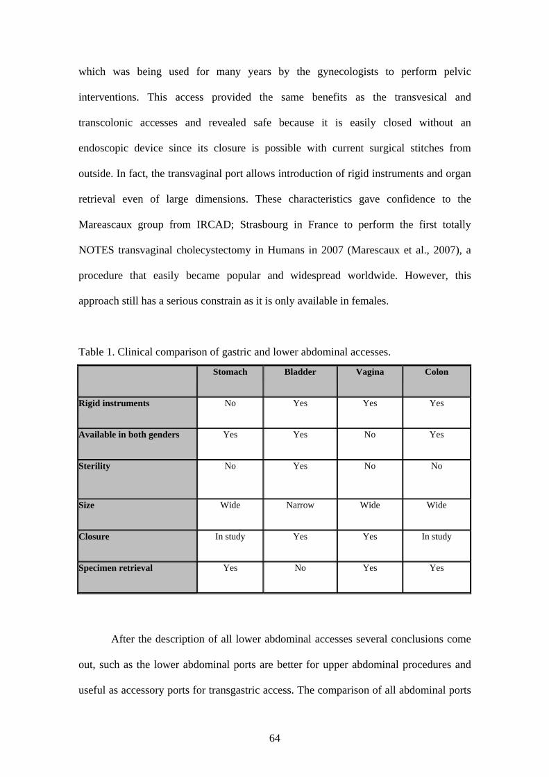
64
which was being used for many years by the gynecologists to perform pelvic
interventions. This access provided the same benefits as the transvesical and
transcolonic accesses and revealed safe because it is easily closed without an
endoscopic device since its closure is possible with current surgical stitches from
outside. In fact, the transvaginal port allows introduction of rigid instruments and organ
retrieval even of large dimensions. These characteristics gave confidence to the
Mareascaux group from IRCAD; Strasbourg in France to perform the first totally
NOTES transvaginal cholecystectomy in Humans in 2007 (Marescaux et al., 2007), a
procedure that easily became popular and widespread worldwide. However, this
approach still has a serious constrain as it is only available in females.
Table 1. Clinical comparison of gastric and lower abdominal accesses.
After the description of all lower abdominal accesses several conclusions come
out, such as the lower abdominal ports are better for upper abdominal procedures and
useful as accessory ports for transgastric access. The comparison of all abdominal ports
Stomach Bladder Vagina Colon
Rigid instruments No Yes Yes Yes
Available in both genders Yes Yes No Yes
Sterility No Yes No No
Size Wide Narrow Wide Wide
Closure In study Yes Yes In study
Specimen retrieval Yes No Yes Yes

65
(table 1) seems to select the transvaginal port as the most reliable for Human application
at this moment. In fact, the transvaginal access is the most commonly used in Human
field so far. However, it has a serious limitation since it is available only in females and
can raise serious psychological concerns that we are still far a way to fully know the
impact. Taking this into account, we believe that the transvesical port is likely the most
universal access from lower abdomen. However, the transvesical access had faced some
reluctance from Medical community since we did not describe a way to close it in our
first description. Thus, we checked all current methods to close visceral holes
endoscopically and we tested the usefulness of T-fasteners with a locking cinch system.
In this study, we could demonstrate the feasibility and the safety of endoscopic closure
of vesical perforations with an endoscopic suturing kit (T-fasteners with a locking
cinch) in a survival porcine model. These findings provide immediate support for
clinical application of this method to close bladder perforations both in management
bladder rupture and transvesical port in NOTES procedures (Chapter 5) (Lima et al.,
2008).
Confirming our initial hypothesis that the transvesical approach would overcome
some limitations of isolated transgastric access, we demonstrated that adding the
transvesical to the transgastric port provided the surgical team of a better surgical
triangulation and effective retraction. In fact, with this strategy we reported for the first
time the third generation nephrectomy by pure NOTES combined accesses (transgastric
and transvesical ports) launching the concept of combined or multiple ports for NOTES
(Chapter 4) (Lima et al., 2007).
Regarding the potential applications of the transvesical port in Humans given its
anatomic and physiological properties, we predict that the transvesical port can have
several types of application in Humans, such as: i) isolated port for both urological and
66
non-urologic procedures as deeply scrutinized in manuscripts from annexes 1 and 2; ii)
as an accessory port for combining with other ports as developed in manuscript from
annex 2; iii) in hybrid procedures with abdominal ports, namely with the single-port
access as advanced in manuscript from annexes 2 and 3 (Lima et al., 2008; Lima et al.,
2008; Gettman et al., 2008).
In conclusion, the thesis fully characterized experimentally the rationale, method
for creation, usefulness, limitations, and closure method of the transvesical port for
potential clinical application in NOTES. Moreover, the closure method can have other
applications in Humans namely as a reliable way to close endoscopically traumatic or
iatrogenic bladder ruptures.
Future Directions
Most criticism against NOTES ecoes what was said against those who were
pioneering laparoscopic surgery in the late 1980s. Laparoscopic surgery has now
become a gold standard in the treatment of many abdominal diseases. We should have
learned the lesson and look at new coming techniques with a good attitude.
Currently available instruments are designed to function inside the urinary and
gastrointestinal tract and have some limitations when used inside the peritoneal cavity.
Certainly the birth of NOTES is pushing researchs and surgeons to design a new
generation of instruments and equipment. Hybrid instruments that combine patterns
typical of rigid and flexible endoscopy will be the next genation technology to be used
in our future surgical rooms.
Advanced endoscopic skills are of paramout importance to perform NOTES. If
NOTES is to be adapted in humans the structure of training programs in Urology and
Surgery must change. Introducing and advanced tier of training in NOTES will

67
probably be a necessity.
The transvesical access to the peritoneal cavity was demonstrated to be feasible,
safe and useful for many intrabdominal procedures in a porcine model. The next logical
step is to get institutional review boards (IRB) approval for Human application of the
transvesical port. The first clinical trials should combine the transvesical port and
laparoscopy. This will provide a margin of confort in prenting and dealing with
complications as we progress to pure transvesical procedures.

68

69
Annex 1
The transvesical endoscopic port for NOTES
Lima E, Rolanda C, Correia-Pinto J. Transvesical endoscopic peritoneoscopy: intra-
abdominal scarless surgery for urologic applications. Curr Urol Rep; 9: 50-54, 2008.

70

Transvesical Endoscopic Peritoneoscopy: Intra-abdominal Scarless Surgery for
Urologic ApplicationsEstevao Lima, MD, FEBU, Carla Rolanda, MD,
and Jorge Correia-Pinto, MD, PhD
Corresponding authorJorge Correia-Pinto, MD, PhDInstituto de Ciências da Vida e Saúde (ICVS), Escola de Ciências da Saúde, Universidade do Minho, Campus de Gualtar; 4709-057 Braga, Portugal. E-mail: [email protected]
Current Urology Reports 2008, 9:50–54Current Medicine Group LLC ISSN 1527-2737Copyright © 2008 by Current Medicine Group LLC
For many abdominal procedures, advantages such as minimal scarring, reduced pain, and faster recovery have made laparoscopy the favored approach over tradi-tional open surgery. The most recent minimally invasive approach is natural orifice transluminal endoscopic surgery (NOTES), which limits morbidity because this surgery does not require incision. This article reviews the history, development, and current and future appli-cations of NOTES in the field of urology.
IntroductionThe introduction of laparoscopic surgery at the end of the 20th century revolutionized modern surgical practice, sig-nificantly changing surgical considerations, techniques, and all other aspects of modern surgical patient care [1]. Simultaneously, endoscopy evolved from simple diag-nostic to progressively invasive therapeutic procedures [2]. Historically, endoscopy has avoided gastrointestinal lumen perforation; although this principle mostly persists, recent anecdotal reports have described safe endoscopic procedures beyond the visceral wall, such as transgastric drainage of pancreatic pseudocyst [3]. Reddy and Rao [4], for example, performed transgastric appendectomy in humans cases, while Kalloo et al. [5••] demonstrated the feasibility and surgical utility of transgastric access to the peritoneal cavity in a porcine model.
These developments marked a new era in minimally invasive abdominal surgery, compelling novel, previously audacious goals, such as avoidance of incisions and pain. With the visceral wall no longer a barrier to endoscopic
intervention, various authors described novel, complex abdominal procedures [6–16]. In 2006, such reports motivated researchers in this emerging field to organize the Natural Orifice Surgery Consortium for Assessment and Research (NOSCAR), a joint initiative supported by the American Society for Gastrointestinal Endoscopy (AGES) and the Society of American Gastrointestinal and Endoscopic Surgeons (SAGES). Proceedings were reported in the Natural Orifice Transluminal Endoscopic Surgery (NOTES) white paper [17••]. European researchers also formed the European Association of Transluminal Surgery (http://www.eats.fr) and the EURO-NOTES Foundation (http://www.euro-notes.eu) to ease cooperation between the European Association for Endoscopic Surgery (EAES) and the European Society of Gastrointestinal Endoscopy (ESGE), which focuses on NOTES-related activities. As a result, an increasing number of researchers are committed to NOTES, publishing experimental reports and attend-ing industry-supported, international conferences where specialists confirm the approach’s feasibility and efficacy for abdominal surgery.
The use of natural orifices for abdominal surgery entrance ports subsequently lead to transvesical access, resulting in unexpected success for abdominal urologic and nonurologic procedures [18••].
This article reviews the current status of NOTES, focusing on new challenges and potential clinical applica-tions in urology.
Current Status of NOTESRationale and potential benefitsNOTES is a revolutionary peritoneal cavity intervention; a natural convergence of intraluminal (endoscopy) and extraluminal (laparoscopy) endoscopic techniques [19]; a third-generation surgery after open surgery (first gen-eration) and laparoscopy (second generation) [16,18••,20] that requires new equipment, special training, and often interdisciplinary collaboration. Advantages over other approaches include no scarring, less anesthesia and postoperative pain, no abdominal wound infection or

Transvesical Endoscopic Peritoneoscopy: Intra-abdominal Scarless Surgery Lima et al. 51
incisional hernia, fewer adhesions and postoperative ileus, and more rapid recovery [17••]. However, no studies have demonstrated advantages beyond cosmetic appear-ance. Studies need to compare the efficacy of NOTES and laparoscopy in terms of surgical stress prevention and morbidity. Impact on immune system also needs to be determined. Until these issues are addressed, potential benefits remain theoretical.
Experimental studiesNatural orifice surgery began in 1928 when Decker [21] performed the first culdoscopy. In 2002, Gettman et al. [22] reported a transvaginal laparoscopic nephrectomy in five female pigs using a single 5-mm abdominal trocar; however, limitations related to the porcine model and instrumentation made the procedure cumbersome. Kalloo et al. [5••] reported the first natural orifice endoscopic surgery using a transgastric approach in a porcine model in which they orally introduced a flexible endoscope into the peritoneal cavity to perform peritoneoscopy and liver biopsies. At procedure’s end, researchers closed the gastric wall with endoscopic clips. In five experiments, all pigs recovered and gained weight.
Several studies have since used the transgastric port for intraperitoneal abdominal procedures, such as fallo-pian tube ligation, cholecystectomy, gastrojejunostomy, lymphadenectomy, oophorectomy, partial hysterectomy, splenectomy, appendectomy, and diaphragmatic pacing [6–15,23]. Following the initial enthusiasm, however, abdominal procedures through isolated transgastric routes raised limitations that jeopardized application in humans. Potential barriers to clinical practice included safe access to peritoneal cavity; gastric closure; infection prevention; spatial orientation; stable multitasking platform to obtain adequate anatomy exposure, organ retraction, secure grasping, and triangulation; difficulty in controlling the pneumoperitoneum; and management of iatrogenic intra-peritoneal complications. These limitations are primarily related to the isolated transgastric approach and the char-acteristics of endoscopes such as flexibility [17••].
Lima et al. [18••] hypothesized that a port enabling rigid instrument use would be advantageous, and sub-sequently assessed the feasibility and safety of a 5-mm transvesical port for abdominal cavity access. This approach was feasible and safely performed in a survival porcine model [18••]. Recently, the same researchers used the transvesical port to perform thoracoscopy with lung biopsies through the diaphragm in a survival porcine model study [24]. A transcolonic port approach has also been reported involving several complications related to the nonsterile nature of the colon [25].
Cholecystectomy is the most challenging isolated transgastric approach. Using two endoscopes, or a single endoscope conjugated with a transabdominal trocar, Park et al. [7] and Swanstrom et al. [26] experienced significant difficulties performing cholecystectomy using
shape-lock technology. Rolanda et al. [20] introduced a combined transgastric and transvesical approach to effectively perform moderately complex upper-abdominal procedures such as cholecystectomy. More recently, Lima et al. [16] used the same combined approach to perform nephrectomy. In fact, this was the first group to combine distinct natural orifices approaches to perform complex abdominal surgery.
Human studiesReddy and Rao [4] proved the transgastric approach feasibility by performing a NOTES appendectomy in humans [4]. Recently, Marescaux et al. [27] performed a transvaginal cholecystectomy using a single abdominal 2-mm trocar. Although unpublished, Brazilian research-ers claim to have performed cholecystectomy by a hybrid transvaginal and transabdominal trocar approach. More recently, the first human cases of transgastric cholecystec-tomy have been presented using a single transabdominal trocar. Further, initial clinical evaluation of the bladder as a NOTES port has already been successfully attempted in humans [28].
Current limitationsNOTES is still in an investigational, developmental stage. Several limitations need to be addressed before these pro-cedures are safe and reliable. NOSCAR has established taxonomy, delineated the current limitations of NOTES, and instigated a unified plan of research to propel NOTES into human practice. Challenges include creating a safe, transmural access point; maintaining sterility of peritoneal cavity despite peroral instrument use; creating a pneu-moperitoneum to distend the peritoneal cavity and allow adequate visualization; ensuring availability of endoscopes and equipment for therapeutic procedures; and developing an entirely reliable means of securely closing the transmural access point [17••]. Of these barriers to clinical practice, gastrostomy closure is the most crucial to solve; various devices are being developed to aid closure [29]. Fast, simple, and effective endoscopic suturing devices will be important for anastomosis, closure of access incisions, and treatment of complications such as bleeding. Examples of endoscopic suturing devices include the prototype known as the Eagle Claw (Olympus America, Center Valley, PA); the novel endoscopic incision and closure device called the Stringer Device (LSI Solutions, Victor, NY); the g-Prox (USGI Medical, San Clemente, CA), which combines an aggres-sive grasper with a needle delivery device that delivers expandable baskets connected by permanent suture; and a prototype device that combines the well-known technology of T-fasteners with a locking cinch device (Johnson and Johnson, New Brunswick, NJ) [29,30]. Currently, endo-clips are the most commonly used method for gastrostomy closure [5••]. However, endoclips are designed for hemo-stasis and are not sited for approximating gastrostomy edges for incision closure.

52 New Techniques
Unless safe and simple devices with proven efficacy for endoscopic closure are available, NOTES will remain in the research field due to an unacceptable complication risk in general practice. Availability of a safe and simple gastrostomy closure device is therefore essential for wide-spread adoption.
The Role of Urology in NOTESBladder as natural orificeAs with the transgastric port innovation, the transvesical approach surpassed a classical urologic barrier, the uri-nary tract wall. Bladder wall perforation was considered a potential complication of urologic procedures. How-ever, with success similar to that of transgastric port, the bladder is an attractive port for NOTES procedures once sterilized because it allows passage of rigid instruments and it is the most anterior access (above bowel loops) from the lower abdomen [18••].
TechniqueUntil recently, experimental and human preliminary stud-ies reported that the transvesical port was easy to install, even with instruments for urologic purposes, using a technique based on the Seldinger principle. We use a ure-teroscope introduced through the urethra into the bladder with pneumodistension, emptying urine from the bladder and distending it with CO2. The vesicotomy site is care-fully selected on the bladder dome. A mucosal incision is made with scissors introduced through the working chan-nel of the ureteroscope. Subsequently, a 5 Fr open-ended ureteral catheter is pushed through the incision into the peritoneal cavity. A 0.035-inch flexible-tip guidewire is inserted into the peritoneal cavity through the ureteral catheter lumen. Guided by the flexible-tip guide-wire, a ureteroscope sheath dilator enlarges the vesical port with a flexible 5.5–mm overtube enveloping it. Introduction of a ureteroscope via overtube into the peritoneal cavity estab-lishes a pressure-controlled CO2 pneumoperitoneum. The ureteroscope is ultimately withdrawn from the abdominal cavity after CO2 removal and a Foley catheter is inserted into the bladder for 3 to 4 days [18••,24].
Advantages and limitationsLower abdominal access points, including the trans-vesical, transvaginal, and transcolonic ports, can be used as an isolated or complementary transgastric port. Several researchers who performed complex abdominal procedures via an isolated transgastric pathway have encountered problems with decreased triangulation and retroflexion [26]. Lower abdominal ports, however, may overcome some of these limitations, with added advantage in complex operations. Although lower abdominal ports provide frontal access to upper abdominal organs and enable improved instrument access, the transvesical port offers sterility and the anatomical advantage of the most
anterior lower abdominal access. Further, the transvesical port enables rigid instrument introduction and does not necessarily require closure [31].
Accessing the peritoneal cavity through a natural ori-fice risks damaging adjacent organs during visceral wall incision. Among natural orifice ports, transvesical port creation with a Seldinger-like technique may be the saf-est approach because bowel loops that contact the bladder wall are unrestricted in the abdomen, which make them run away from bladder instruments. Further, in proce-dures involving two natural orifices, such as transgastric and transvesical, the transvesical image can easily moni-tor the transgastric creation.
The transvesical port procedure involves a signifi-cant challenge related to instrument size, limiting organ retrieval through this port. Another concern related to transvesical port use is the necessity of bladder closure. Vesical perforation involves potential complications, such as peritoneal urine leakage with secondary infection (peri-tonitis), that commonly occur as delayed complications of undiagnosed traumatic vesical perforation or pathologi-cal bladder conditions, such as neoplasms. Experiments in pigs demonstrated that 5 mm bladder hole closure is not absolutely necessary if bladder drainage is assured [18••,24]. However, development of a closure device or method is imperative for the widespread adoption of transvesical port option in NOTES.
Applications in urologyNOTES’ urologic application may not be immediately apparent, considering the current acceptance of laparoscopic surgery. However, several groups are using natural orifices to perform complex procedures such as nephrectomy.
Gettman et al. [22] first performed a complete transvag-inal laparoscopic dissection and nephrectomy in a porcine model using a single, 5-mm abdominal trocar for visualiza-tion. A completely transvaginal laparoscopic nephrectomy was performed once, but limitations imposed by porcine anatomy and available instrumentation hindered the proce-dure [22]. More recently, Clayman et al. [32] performed a porcine nephrectomy with a single 12-mm trocar placed in the midline and the transvaginal introduction of a Trans-Port Multi-Lumen Operating Platform (USGI Medical, San Clemente, CA). This flexible device has four working channels and can be locked into position, creating a rigid multitasking platform that enables two-handed tissue manipulation [32]. Further, Lima et al. [16] demonstrated the feasibility of NOTES nephrectomy. In a nonsurvival study, combined transgastric and transvesical approaches were established in six female pigs. Under ureteroscope visualization through a 5-mm transvesical port, research-ers controlled the orally introduced flexible gastroscope by the gastrotomy into the peritoneal cavity. Right or left nephrectomy were performed using instruments introduced by devices that worked in the renal hilum, alternating intervention on dissection or retraction procedures. In all
Transvesical Endoscopic Peritoneoscopy: Intra-abdominal Scarless Surgery Lima et al. 53
animals, both kidneys were visualized, and the renal ves-sels and ureter were reasonably individualized and ligated separately with ultrasonic scissors introduced through the transvesical port [16].
Nephrectomy most likely will be one of the final renal procedures potentially performed by NOTES in the near future. However, much research is needed to minimize unexpected complications before NOTES can be applied in humans. Robotics and magnetically anchored instru-mentation used to perform single trocar laparoscopic nephrectomy may be logical steps in the development of scarless abdominal surgery by NOTES [33].
Beyond renal procedures, other potential human appli-cations of NOTES in urology include varicocelectomy, investigation of nonpalpable testis, and treatment of ura-chus remnants using transgastric or transvesical ports.
Involvement of urologists in other abdominal proceduresNOTES may present a tremendous challenge for urolo-gists in terms of technical demands. It may also involve multidisciplinary teams to deal with nonurologic clinical situations, considering the simplicity of accessing and viewing the upper abdominal organs via the transvesical port [15,24].
In animal and human settings, the transvesical port enables feasible and useful peritoneoscopy of all intra-abdominal viscera, mainly the upper abdominal organs [18••,24,28]. Further, the transvesical port is gaining a place in NOTES as a unique port associated with the transgastric port [16,20]. Rolanda et al. [20] demonstrated the utility of a combined transgastric and transvesi-cal approach, performing a reliable, feasible, exclusive NOTES cholecystectomy. This study emphasized the transvesical port’s ability to overcome limitations reported in a cholecystectomy performed exclusively through a transgastric port [20].
Although possibly difficult to accept clinically, the transvesical port provides exceptional access to the upper abdominal organs and may enable a transdiaphragmatic endoscopic approach to the thoracic cavity in a long-term survival study in a porcine model [21].
Most of these studies are preliminary and only represent the birth of NOTES in urology; however, they demonstrate a need for new instruments and further research that provides evidence that experimental successes may advan-tageously translate to clinical practice in humans.
ConclusionsIn many areas of medicine, divisions between specialties are blurring. For example, stent placement for carotid stenosis is now performed by neuroradiologists, interven-tional radiologists, vascular surgeons, and cardiologists. Similar reports commonly occur in other clinical areas. Currently, NOTES procedures and research are primarily performed by gastroenterologists and surgeons. However,
because the mouth and colon are not the only access points, urologists and gynecologists have approached the peritoneum through the bladder and vagina. Further, NOTES has enabled transvaginal nephrectomy, transgas-tric adrenalectomy (unpublished data), peritoneoscopy, and thoracoscopy using a transvesical approach, and cholecystectomy and nephrectomy using a combined transvesical and transgastric approach [16,18••,20,22,24, 28,32]. These procedures have mostly been performed in animal models, but human application is pending.
Urologists have been encouraged in this developing field to meet an especially great demand should NOTES develop as some investigators have proposed. Further, the future may require the development of new NOTES train-ing programs to address specific demands.
DisclosuresNo potential conflict of interest relevant to this article was reported.
References and Recommended ReadingPapers of particular interest, published recently, have been highlighted as:• Of importance•• Of major importance
1. Harrell AG, Heniford BT: Minimally invasive abdominal surgery: lux et veritas past, present, and future. Am J Surg 2005, 190:239–243.
2. Liu R, Chand B, Ponsky J: The future of surgical endos-copy. Endoscopy 2005, 37:38–41.
3. Vitale GC, Lawhon JC, Larson GM, et al.: Endoscopic drainage of the pancreatic pseudocyst. Surgery 1999, 26:616–623.
4. Richards WO, Rattner DW: Endoluminal and translumi-nal surgery: no longer if, but when. Surg Endosc 2005, 19:461–463.
5.•• Kalloo AN, Singh VK, Jagannath SB, et al.: Flexible transgastric peritoneoscopy: a novel approach to diagnostic and therapeutic interventions in the peritoneal cavity. Gastrointest Endosc 2004, 60:114–117.
The first description of transgastric surgery. This manuscript marks the birth of NOTES.6. Jagannath SB, Kantsevoy SV, Vaughn CA, et al.: Per-oral
transgastric ligation of fallopian tubes with long-term survival in a porcine model. Gastrointest Endosc 2005, 61:449–453.
7. Park PO, Bergström M, Ikeda K, et al.: Experimental stud-ies of transgastric gallbladder surgery: cholecystectomy and cholecystogastric anastomosis (videos). Gastrointest Endosc 2005, 61:601–606.
8. Kantsevoy SV, Jagannath SB, Niiyama H, et al.: Endoscopic gastrojejunostomy with survival in a porcine model. Gastrointest Endosc 2005, 62:287–292.
9. Bergström M, Ikeda K, Swain P, Park PO: Transgastric anastomosis by using flexible endoscopy in a porcine model. Gastrointest Endosc 2006, 63:307–312.
10. Fritscher-Ravens A, Mosse CA, Ikeda K, Swain P: Endoscopic transgastric lymphadenectomy by using EUS for selection and guidance. Gastrointest Endosc 2006, 63:302–306.
11. Wagh MS, Merrifield BF, Thompson CC: Survival studies after endoscopic transgastric oophorectomy and tubectomy in a porcine model. Gastrointest Endosc 2006, 63:473–478.

54 New Techniques
12. Merrifield BF, Wagh MS, Thompson CC: Peroral transgas-tric organ resection: a feasibility study in pigs. Gastrointest Endosc 2006, 63:693–697.
13. Kantsevoy SV, Hu B, Jagannath SB, et al.: Transgastric endoscopic splenectomy: is it possible? Surg Endosc 2006, 20:522–525.
14. Sumiyama K, Gostout CJ, Rajan E, et al.: Pilot study of the porcine uterine horn as an in vivo appendicitis model for development of endoscopic transgastric appendectomy. Gastrointest Endosc 2006, 64:808–812.
15. Onders R, McGee MF, Marks J, et al.: Diaphragm pacing with natural orifice transluminal endoscopic surgery: potential for difficult-to-wean intensive care unit patients. Surg Endosc 2007, 21:475–479.
16. Lima E, Rolanda C, Pêgo JM, et al.: Third generation nephrectomy by natural orifices translumenal endoscopic surgery. J Urol 2007, In press.
17.•• Rattner D, Kalloo A, and SAGES/ASGE working group on NOTES. ASGE/SAGES working group on natural orifice translumenal endoscopic surgery. Surg Endosc 2006, 20:329–333.
This interdisciplinary manuscript established the fundamental challenges to the safe introduction of NOTES.18.•• Lima E, Rolanda C, Pêgo JM, et al.: Transvesical
endoscopic peritoneoscopy: A novel 5 mm port for intra-abdominal scarless surgery. J Urol 2006, 176:802–805.
Includes the first description of the transvesical port approach and represents the beginning of the urology commitment to NOTES.19. Malik A, Mellinger JD, Hazey JW, et al.: Endoluminal and
transluminal surgery: Current status and future possibili-ties. Surg Endosc 2006, 20:1179–1192.
20. Rolanda C, Lima E, Pêgo JM, et al.: Third-generation cholecystectomy by natural orifices: transgastric and transvesical combined approach (with video). Gastrointest Endosc 2007, 65:111–117.
21. Decker A: Culdoscopy: a method for visual diagnosis of gynecologic disease. Clin Symp 1952, 4:201–210.
22. Gettman MT, Lotan Y, Napper CA, Cadeddu JA: Transvag-inal laparoscopic nephrectomy: development and feasibility in the porcine model. Urology 2002, 59:446–450.
23. Sumiyama K, Gostout CJ, Rajan E, et al.: Transgastric cholecys-tectomy: transgastric accessibility to the gallbladder improved with the SEMF method and a novel multibending therapeutic endoscope. Gastrointes Endosc 2007, 65:1028–1034.
24. Lima E, Henriques-Coelho T, Rolanda C, et al.: Transvesi-cal thoracoscopy: a natural orifice translumenal endoscopic approach for thoracic surgery. Surg Endosc 2007, 21:854–858.
25. Fong DG, Pai RD, Thompson CC: Transcolonic endoscopic abdominal exploration: a NOTES survival study in a porcine model. Gastrointest Endosc 2007, 65:312–318.
26. Swanstrom LL, Kozarek R, Pasricha PJ, et al.: Development of a new access device for transgastric surgery. J Gastroin-test Surg 2005, 9:1129–1137.
27. Marescaux J. Operation Anubis: A new step in NOTES his-tory! Available at http://www.websurg.com/notes. Accessed on September 13, 2007.
28. Gettman MT, Blute ML: Transvesical peritoneoscopy: initial clinical evaluation of the bladder as a portal for natural orifice translumenal endoscopic surgery. Mayo Clin Proc 2007, 82:843–845.
29. Sclabas GM, Swain P, Swanstrom LL: Endoluminal meth-ods for gastrotomy closure in natural orifice transenteric surgery (NOTES). Surg Innov 2006, 13:23–30.
30. Ryou M, Pai RD, Sauer JS, et al.: Evaluating an optimal gastric closure method for transgastric surgery. Surg Endosc 2007, 21:677–680.
31. Rolanda C, Lima E, Correia-Pinto J: Searching the best approach for third-generation cholecystectomy. Gastroin-test Endosc 2007, 65:354–355.
32. Clayman RV, Box GN, Abraham JB, et al.: Rapid communi-cation: transvaginal single-port NOTES nephrectomy: initial laboratory experience. J Endourol 2007, 21:640–644.
33. Zeltser IS, Bergs R, Fernandez R, et al.: Single trocar laparoscopic nephrectomy using magnetic anchoring and guidance system in the porcine model. J Urol 2007, 178:288–291.

76

77
Annex 2
Transvesical and other ports for N.O.T.E.S.
Lima E, Rolanda C, Correia-Pinto J. NOTES performed using multiple ports of entry:
current experience and potential implications for urologic applications. J Endourol
2008 (in press)

78

79
ABSTRACT
Background and purpose: Isolated transgastric port raises serious limitations in
performing natural orifice translumenal endoscopic surgery (NOTES) complex
procedures in urology field. Attempting to overcome these limitations several solutions
has been advanced, such as the hybrid approach (adding a single abdominal port access)
or the pure NOTES combined approach (joining multiple natural orifice ports). This
article reviews the current state of experimental and clinical results of multiple ports in
NOTES.
Patients and Methods: A literature search of PubMed was performed, seeking
publications from January 2002-to-2008 on NOTES. Additionally, we looked at
pertinent abstracts of annual meetings of the American Urological Association, the
European Association of Urology, and the World Congress of Endourology from 2007.
Results: In urology, multiple ports of entry seem to be necessary mainly for moderate
complex procedures. Thus, we could find studies using the hybrid approach
(combination of transgastric or transvaginal access with a single transabdominal port),
or using the pure NOTES combined approach (transgastric and transvesical,
transvaginal and transcolonic, or transgastric and transvaginal). Anyway there is still
limited experience in Humans using these approaches and no comparative studies exist
so far. Meanwhile, we are waiting for new equipments and instruments more
appropriate for these novel techniques.
Conclusions: In urology, it is predictable that for moderate complex procedures we will
need multiple ports being the most appealing approach the transvaginaltransabdominal
(hybrid approach), whereas in a pure NOTES perspective the transgastric-transvesical
approach seems to be the preferred approach.
Key Words: endoscopy; NOTES; transgastric surgery; transvesical surgery

80

81
INTRODUCTION
Minimally Invasive Surgery (MIS) is rapidly becoming the standard for surgical care. In
fact, refinements in laparoscopic surgery have progressed to the point that complex
surgical procedures can now be performed in a minimally invasive fashion.1 Recently,
natural orifice translumenal endoscopic surgery (NOTES) has emerged as a potential
exciting evolution of minimally invasive surgical care. This new approach was firstly
used by Reddy and Rao that carried out in a few humans cases appendectomy by a
transgastric access followed by Kalloo et al that demonstrated in a structured study the
feasibility and potential surgical usefulness of transgastric access to the peritoneal
cavity in a porcine model.2 Subsequently, others authors described more complex
intraabdominal procedures using isolated transgastric route.3-14 Following the initial
enthusiasm, the possibility to carry out abdominal procedures through isolated
transgastric route faced several limitations (White Paper. Surg Endosc 2006).15
Believing in the potential benefits of natural orifices approach for abdominal surgery,
Lima et al hypothesized that other ports would be advantageous. In this sequence, they
assessed the feasibility and safety of creating a transvesical port for the abdominal
cavity.16 As occurred with transgastric port, the transvesical approach also broke a
classical sanctuary of urology: the wall of urinary tract. In fact, the idea to perforate the
bladder wall was always feared as potential complication of urological procedures.
Following the unexpected success of transgastric port, the bladder comes out as an
attractive port for NOTES procedures once it is sterile, allows passage of rigid
instruments and appears anatomically as the most anterior lower abdominal access.16,17
RATIONALE FOR MULTIPLE PORT NOTES
Current laparoscopic surgery is using rigid instruments introduced at strategic anatomic
points through the abdominal wall. The positioning of the trocars is an important issue
since it can determine the feasibility of the procedure. This occurs because the correct
positioning of the trocars is essential for triangulation and many times for grasping and
efficient traction. In NOTES, the initial steps were carried out with common
gastroscopes and flexible instruments introduced through its parallel working channels.
Thus, after description of the first intra-abdominal moderate complex procedures (ie.
cholecystectomy) serious limitations related with absence of using rigid instruments and

82
lack of triangulation come out. For instances, Park et al conducted the first pilot study in
pigs applying NOTES to perform transgastric cholecystectomy. Using two endoscopes
or a single endoscope conjugated with a transabdominal trocar, cholecystectomy was
feasible, but important limitations were identified such as difficulty in controlling the
pneumoperitoneum, in obtaining a stable platform and adequate anatomy exposure,
organ retraction, secure grasping and triangulation of instruments.4 Swanström et al
attempted to overcome these limitations providing some rigidity to the gastroscope
using shapelock technology as a new over tube for transgastric surgery. However, even
with this equipment, isolated transgastric approach for gallbladder manipulation
remained a challenge with a success rate around 33%.18
In fact, most of these limitations namely the lack of triangulation, effective grasping and
traction, and the working difficulties in retroflexion were closely related with the use of
an isolated transgastric approach due to the characteristics of the gastroscopes such as
its flexibility. Attempting to overcome these limitations several solutions has been
advanced, such as the construction of more rigid transgastric platforms,19 the
combination of the transgastric access with a transabdominal port (hybrid approach
using for instances the umbilicus)20-21 or with a lower abdominal access (pure NOTES
combined approach) (fig. 1).22-24

83
Figure 1 – The rationale for combined accesses for pure NOTES.
THE TRANSVESICAL PORT AMONGST THE LOWER ABDOMINAL PORTS
For lower abdominal access several options exists. In fact, after the old description of
the transvaginal port in forties,25 the transvesical16 and the transcolonic approaches26
were recently described (Fig. 2). Despite these accesses share the possibility to
introduce rigid instruments into the abdomen, the transvesical port appears at the most
anterior positioning in the sagital plan and for this reason with less risk to damage
viscera. Moreover, introducing the surgical instruments from the bladder dome gives the
possibility to work above bowel loops, instead through among them.
Additionally, the transvesical port is the unique lower abdominal access that is naturally
sterile, and available in both genders in contrast to the transvaginal access.
Unfortunately, the transvesical port does not allow specimen retrieval as transvaginal
and transcolonic approaches (Table 1). For all these reasons, always it is needed the
combination of a lower abdominal access to the transgastric port, the transvesical port
seems quite attractive.

84
NOTES Access Evolution
Pai et al, 2006
Lima et al, 2006
Decker & Cherry, 1940
Sumiyama et al, 2007
Kalloo et al, 2004
Figure 2 – Drawing with NOTES accesses.
Table 1 - Comparison of lower abdominal accesses.

85
EXPERIMENTAL AND CLINICAL WORK-PROCEDURAL
The efficiency of the concept of combining transgastric (using flexible instruments) and
transvesical (using rigid instruments) ports was tested performing two moderately
complex procedures such as cholecystectomy and nephrectomy by pure NOTES
approach.22,24 Believing in the potential benefits of NOTES approach for abdominal
surgery, Rolanda et al and Lima et al used a transgastric and transvesical combined
approach to perform cholecystectomy and nephrectomy, respectively, aiming to
overcome many limitations previously described for isolated transgastric approach
related with anatomy exposure, organ retraction, grasping and limited triangulation.
This group used the transvesical port because it is diametrically opposed to the
transgastric port and seems to offer additional advantages over transvaginal and
transcolonic ports for the reasons cited above. However, recently there is a report of a
nephrectomy using the combination of transgastric and transvaginal approach.27 In this
study the transgastric port was introduced first followed by the gastroscope that guided
the introduction of a second transvaginal endoscope through a novel laparoscopic
trocar/endoscopic over tube device. The advantage of this approach was the possibility
to removal of the specimen through the vagina.27
In a reverse thinking, many authors are using the transvaginal approach for dissection,
replacing the transgastric by the transvaginal port for dissection and specimen retrieval.
Those that are using flexible instruments through vagina still need to use a rigid
instrument to provide effective retraction. In these studies, commonly the rigid
instrument(s) is (are) being introduced transabdominally. Anyway, this strategy allowed
performing safe transvaginal nephrectomy28,29 and cholecystectomy30 in the porcine
model22-23 and even the firsts Human cholecystectomies31-33 and nephrectomy.34
In the same way of thinking, Meining et al, described the combination of transsigmoid
approach using a specially designed trocar with a grasper introduced transabdominally
to perform cholecystectomy in an acute porcine model study.35 Recently, another group
described the transcolonic approach associated with the transvaginal port to perform
distal pancreatectomy in survival porcine model. In this study was used the combination
of an endoscope advanced into peritoneal cavity through an anterior colotomy with a
computer-assisted linear stapler introduced transvaginally.36
A alternative way is being developed by the University of Texas that use a magnetic
anchoring and guidance system to perform in porcine model nephrectomy with a single
86
transabdominal trocar and more recently cholecystectomy by pure NOTES through
vagina.37,38
TECHNIQUES FOR GAINING ABDOMINAL ACCESS THROUGH NATURAL
ORIFICES
As in laparoscopy we have discussion regarding the way to choose the first trocar to
start the pneumoperitoneum and to monitor the placement of the other ports, accessing
the peritoneal cavity through a natural orifice has always the risk of damaging the
adjacent organs during creation of the hole in the visceral wall. In fact, accessing the
peritoneal cavity in a blind way through the transgastric port can damage adjacent
organs as reported by many authors.15,39,40 Interestingly, the technique to establish the
transvesical port was described using neither cutting instruments nor cattery. This
makes the transvesical port as the safest port to start access to the peritoneal cavity and
particularly useful in procedures involving multiple natural orifices, since the
transvesical image can easily monitories the transgastric creation.
The placement of the transvesical port is based on the Seldinger principle.16,17,41
Currently, Lima et al performed it using an ureteroscope introduced through the urethra
into the bladder with pneumo-distension, the bladder is emptied from urine and
distended with CO2. The vesicotomy site is carefully selected on the bladder dome. A
mucosal incision is made with a scissors introduced through the working channel of the
ureteroscope. Subsequently, a 5 Fr open-ended ureteral catheter is pushed forward
through the incision into the peritoneal cavity. A 0.035-inch flexible-tip guide-wire is
then inserted into the peritoneal cavity through the lumen of the ureteral catheter.
Guided by the flexible-tip guide-wire, the vesical hole is enlarged with a dilator of an
ureteroscope sheath enveloped by a flexible 5.5 mm over tube. An ureteroscope is
introduced into the peritoneal cavity through the over tube and allows the creation of a
pressure controlled CO2 pneumoperitoneum.16-17
To establish the transgastric port several variants had already been described.2-14
Rolanda et al suggested to establish it under gastric outside view provided by a
transvesical scope in order to avoid damage of gastric wall vessels or adjacent organs.
The gastrotomy site is carefully chosen on the anterior wall. A gastric-wall incision is
made by pushing forward a needle knife, followed by its sheath with cautery under a 12
mmHg CO2 pneumoperitoneum (induced through the transvesical port). The

87
needleknife sheath is then used for positioning a guidewire. The puncture dilation is
performed with an 18-mm through-the-scope balloon over the guidewire or by
extending the gastrotomy with a sphincterotom. Then, the gastroscope is pushed
forward and passed into the peritoneal cavity. All the procedures become safer if they
are monitored by a scope positioned through a lower abdominal access or
transabdominally. As an alternative method, a PEG-like transgastric approach to the
peritoneal cavity appears technically simple and safe.39
The transcolonic port is also being developed. As it was described it is established using
a sterile dual-channel endoscope was introduced through the anus and advanced 15 to
20 cm from the anal verge. A needle knife is used to make a subcentimeter linear
incision. Once the incision is complete, the needle knife is retracted, and the catheter is
advanced through the incision into the peritoneum and the endoscope is then advanced
through the colonic wall into the peritoneal cavity.26
The transvaginal port was already described since the forties25 and had already been
used for several intra-abdominal procedures by gynecologists. Recently, there is a
recrudescent enthusiasm with the transvaginal port for upper abdominal procedures
since it does not have associated the risks of the transgastric port.31-34 The method is
easy to establish. Briefly, the peritoneal cavity is entered through an incision in the
posterior vaginal cul-de-sac.
POTENTIAL RISKS AND BENEFITS TO USING MULTIPLE PORTALS
Until now the most tested associations are the hybrid methods
(transvaginaltransabdominal,33-33 transcolonic-transabdominal35 and transgastric-
transabdominal20,21) and the pure NOTES association transgastric-transvesical,22,24 but
other associations had also been reported as transvaginal-transcolonic36 and the
transgastric-transvaginal.27
Always we establish a natural orifice port there are associated risks related with its
creation as well as risks associated with delayed leaking of leaving an ‘-otomy’ not
completely closed. Thus, it seems reasonably to expect that association of multiple
portals causes more risks.
However, current use of multiple portals association has also unravelled several
benefits. First of all, after creation of a port, it is possible to minimize the risk of the
second port as the first port can allow monitoring the creation of the second port.
88
Moreover, it is even predictable that the use of associated portals can enhance to find
better and more reliable methods to closure the ‘-otomies’. Anyway, the great advantage
to use multiple portals is related with the enhanced ability to increase triangulation,
grasping and retraction manoeuvres and consequent enhanced dissection.
Finally, the establishment of multiple portals associations commonly requires
multidisciplinary team involvement. This can be looked as a limitation or an advantage.
In our point of view, this constitutes an advantage once it brings for the some ‘battle
field’ resources and expertises from different interventional environments, what for sure
will help to achieve easier solutions and new methods and ideas.
OPINION REGARDING BENEFITS FOR UROLOGIC SPECIFIC NOTES
(POTENTIAL USES)
The appeal of NOTES in urology may not be immediately apparent, considering the
more or less mainstream acceptance of laparoscopic surgery. On the other hand, several
groups are using natural orifices to carry out complex procedures such as nephrectomy.
The first attempt was made by Gettman et al that performed complete transvaginal
laparoscopic dissection and nephrectomy in the porcine model using a single, 5-mm
abdominal trocar for visualization. A completely transvaginal laparoscopic nephrectomy
was performed once, but limitations imposed by the porcine anatomy and by the
currently available instrumentation made the procedure very cumbersome at that time.28
More recently, Clayman et al recovered this idea and carried out a porcine nephrectomy
recurring to a single 12-mm trocar placed in the midline and the new TransPort Multi-
Lumen Operating Platform (USGI Medical, San Clemente, CA) passed transvaginally.
This flexible device has four working channels and can be locked into position, thereby
creating a rigid multitasking platform that allows two-handed tissue manipulation.29
Stretching the limits, Lima et al demonstrated the feasibility of pure NOTES
nephrectomy. In a non-survival study, transgastric and transvesical combined
approaches were established in 6 female pigs. Under ureteroscope control, passed
through a 5 mm transvesical port, a flexible gastroscope was passed per-orally into
peritoneal cavity by a gastrotomy. Either right or left nephrectomy were carried out
using instruments introduced by both devices that worked in the renal hilum alternating
their intervention either on dissection or retraction procedures. In all animals, both
kidneys were visualized, and the renal vessels and ureter were reasonably

89
individualized, clipped and then ligated separately with ultrasonic scissors, which was
introduced through the transvesical port.24
The feasibility of nephrectomy by NOTES appears as the extreme of a large spectrum of
renal procedures that can potentially be done by NOTES in the near future. However,
we might take into account that much work is still needed to refine techniques, verify
safety, and document efficacy before Human translation in order to minimize
unexpected complications. For instances, magnetically anchored instrumentation as
suggested by Zeltser et al and Scott et al might be a good application and may be the
next logical step toward the development of truly scarless abdominal surgery by
NOTES.37,38
NOTES might appears as a tremendous challenge for the urologists. In addition to
technical demands to perform urological NOTES, urologists might be called to become
involved in multidisciplinary teams dealing with extra-urological clinical situations.
This might occur since the extraordinary simplicity to install the transvesical port and
the excellent view of upper abdominal organs that it provides.
Both in animals and human settings, the transvesical port revealed feasible and useful to
perform peritoneoscopy of all intra-abdominal viscera, mainly the upper abdominal
organs. Moreover, the transvesical port is gaining a place in NOTES as a unique port to
associate with the transgastric port in both genders.22,23 Although more hardly to accept
clinically, the exceptional opportunity that the transvesical port provides to view the
upper abdominal organs, it was even suggested that the transvesical port would make
possible transdiaphragmatic endoscopic approach to the thoracic cavity in a long-term
survival study in a porcine model.17
Most of these studies are still preliminary and embody the birth of NOTES. NOTES is
still in development and no well-established criteria are already defined. Anyway, we
predict that with the current concept of flexible scopes, always we advance for a
surgical procedure that in laparoscopy uses up to 3 trocars, in NOTES it will be feasible
through an isolated natural orifice (fig. 3); whereas always we are dealing with a
surgical procedure that use more than 3 trocars in laparoscopy, we predict that
association of multiple portals will be necessary (fig. 4). Always association is needed
the most appealing approaches are the transvaginal-transabdominal or in a pure NOTES
perspective the transgastric-transvesical approach.
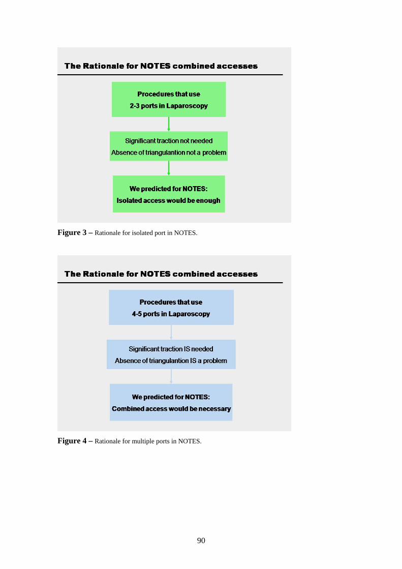
90
Figure 3 – Rationale for isolated port in NOTES.
Figure 4 – Rationale for multiple ports in NOTES.

91
CONCLUSION
In urology, it is predictable that for moderate complex procedures we will need multiple
ports being the most appealing approach the transvaginal-transabdominal (hybrid
approach), whereas in a pure NOTES perspective the transgastric-transvesical approach
seems to be the preferred approach.
REFERENCES
1.Harrell AG Harrell AG, Heniford T: Minimally invasive abdominal surgery: Lux et
veritas past, present, and future. Am J Surg 2005; 190: 239.
2.Kalloo AN, Singh VK, Jagannath SB, et al: Flexible transgastric peritoneoscopy: a
novel approach to diagnostic and therapeutic interventions in the peritoneal cavity.
Gastrointest Endosc 2004; 60: 114.
3.Jagannath SB, Kantsevoy SV, Vaughn CA, et al: Per-oral transgastric ligation of
fallopian tubes with long-term survival in a porcine model. Gastrointest Endosc 2005;
61: 449.
4.Park PO, Bergstrom M, Ikeda K, et al: cholecystectomy and cholecystogastric
anastomosis (videos). Gastrointest Endosc 2005; 61: 601.
5.Kantsevoy SV, Jagannath SB, Niiyama H, et al: Endoscopic gastrojejunostomy with
survival in a porcine model. Gastrointest Endosc 2005; 62: 287.
6.Bergstrom M, Ikeda K, Swain P, et al: Transgastric anastomosis by using flexible
endoscopy in a porcine model. Gastrointest Endosc 2006; 63: 307.
7.Wagh MS, Merrifield BF, Thompson CC: Survival studies after endoscopic
transgastric oophorectomy and tubectomy in a porcine model. Gastrointest Endosc
2006; 63: 473.
8.Merrifield BF, Wagh MS, Thompson CC: Peroral transgastric organ resection in the
Abdomen: feasibility study in pigs. Gastrointest Endosc 2006; 63: 693.
9.Kantsevoy SV, Hu B, Jagannath SB, et al: Transgastric endoscopic splenectomy. Is it
possible? Surg Endosc 2006; 20: 522.
10.Fritscher-Ravens A, Mosse CA, Ikeda K, et al: Endoscopic transgastric
lymphadenectomy by using EUS for selection and guidance. Gastrointest Endosc. 2006;
63: 302.

92
11. Sumiyama K, Gostout CJ, Rajan E, et al: Pilot study of the porcine uterine horn as
an in vivo appendicitis model for development of endoscopic transgastric
appendectomy. Gastrointest Endosc 2006; 64: 808.
12.Onders R, McGee MF, Marks J, et al: Diaphragm pacing with natural orifice
transluminal endoscopic surgery: potential for difficult-to-wean intensive care unit
patients. Surg Endosc 2007; 21: 475.
13.Hu B, Kalloo AN, Chung SS, et al: Peroral transgastric endoscopic primary repair of
a ventral hernia in a porcine model. Endoscopy 2007; 39: 390-393.
14.Perretta S, Dallemagne B, Coumaros D, et al: Natural Orifice Transluminal
Endoscopic Surgery. Transgastric Cholecystectomy in a survival porcine model. Surg
Endosc 2007; 11; [Epub ahead of print]
15.Rattner D, Kalloo A, and SAGES/ASGE working group on NOTES: ASGE/SAGES
Working group on natural orifice translumenal endoscopic surgery. Surg Endosc 2006;
20: 329.
16.Lima E, Rolanda C, Pêgo JM, et al: Transvesical endoscopic peritoneoscopy: A
novel 5 mm-port for intra-abdominal scarless surgery. J Urol 2006;176: 802.
17.Lima E, Henriques-Coelho T, Rolanda C, et al: Transvesical thoracoscopy: A natural
orifice translumenal endoscopic approach for thoracic surgery. Surg Endosc 2007; 21:
854.
18.Swanstrom LL, Kozarek R, Pasricha PF, et al: Development of a new access device
for transgastric surgery. J Gastrointest Surg 2005; 9: 1129.
19.Bardaro JS, Swanstrom L. Development of advanced endoscopes for natural orifice
translumenal endoscopic surgery (NOTES). Minimally invasive Therapy 2006; 15: 378.
20.Shih SP, Kantsevoy SV, kalloo AN, et al: Hybrid minimally invasive surgery-a
bridge between laparoscopic and translumenal surgery. Surg Endosc 2007; 21: 1450.
21.Mintz Y, Horgan S, Cullen J, et al: NOTES: the hybrid technique. J Laparoendosc
Adv Surg Tech A 2007; 17: 402.
22.Rolanda C, Lima E, Pêgo JM, et al: Third Generation Cholecystectomy by Natural
Orifices: Transgastric and Transvesical Combined Approach. Gastrointest Endosc 2007;
65: 111.
23.Rolanda C, Lima E, Correia-Pinto J: Searching the best approach for third-
generation cholecystectomy. Gastrointest Endosc 2007; 65: 354.
24.Lima E, Rolanda C, Pêgo JM, et al: Third generation nephrectomy by natural orifice
translumenal endoscopic surgery. J Urol 2007; 178: 2648.
93
25.Decker A: Culdoscopy: A method for visual diagnosis of gynecologic disease. Clin
Symp 1954; 6: 210.
26.Fong DG, Pai RD, Thompson CC: Transcolonic endoscopic abdominal exploration:
a NOTES survival study in a porcine model. Gastrointest Endosc 2007; 65: 312.
27.Ponsky L, Isariyawongse J, McGee M, et al: Pure natural orifice translumenal
endoscopic surgery (NOTES) nephrectomy using standard laparoscopic instruments in
the porcine model. J Endourol 2007; 21: A91.
28.Gettman MT, Lotan Y, Napper CA, et al: Transvaginal laparoscopic nephrectomy:
Development and feasibility in the porcine model. Urology 2002; 59: 446.
29.Clayman RV, Box GN, Abraham JB, et al: Rapid communication: transvaginal
single-port NOTES nephrectomy: initial laboratory experience. J Endourol 2007; 21:
640.
30.Zornig C, Emmermann A, Von Waldenfels HA, et al: Laparoscopic cholecystectomy
without visible scar: combined transvaginal and transumbilical approach. Endoscopy
2007; 39: 913.
31.Bessler M, Stevens PD, Milone L, et al: Transvaginal laparoscopically assisted
endoscopic cholecystectomy: a hybrid approach to natural orifice surgery. Gastrointest
Endosc 2007; 66: 1243.
32.Marescaux J, Dallemagne B, Perretta S, et al: Surgery without scars. Report of
transluminal cholecystectomy in a human being. Arch Surg 2007; 142: 823.
33.Zorron R, Maggioni LC, Pombo L, et al: NOTES transvaginal cholecystectomy:
preliminary clinical application. Surg Endosc 2007; 20; [Epub ahead of print]
34.Branco AW, Filho AJ, Kondo W, et al: Hybrid Transvaginal Nephrectomy. Eur Urol
2007; 5; [Epub ahead of print]
35.Meining A, Wilhelm D, Burian M, et al: Development, standardization, and
evaluation of NOTES cholecystectomy using a transsigmoid approach in the porcine
model: an acute feasibility study. Endoscopy 2007; 39: 860.
36.Ryou M, Fong DG, Pai RD, et al: Dual-port distal pancreatectomy using a prototype
endoscope and endoscopic stapler: a natural orifice transluminal endoscopic surgery
(NOTES) survival study in a porcine model. Endoscopy 2007; 39: 881.
37.Scott DJ, Tang SJ, Fernandez R, et al: Completely transvaginal NOTES
cholecystectomy using magnetically anchored instruments. Surg Endosc 2007; 21:
2308.

94
38.Zeltser IS, Bergs R, Fernandez R, et al: Single trocar laparoscopic nephrectomy
using magnetic anchosing and guidance system in the porcine model. J Urol 2007; 178:
288.
39.Kantsevoy SV, Jagannath SB, Niiyama H, et al: A novel safe approach to the
peritoneal cavity for per-oral transgastric endoscopic procedures. Gastrointest Endosc
2007; 65: 497.
40.Ko CW, Shin EJ, Buscaglia JM, et al: Preliminary pneumoperitoneum facilitates
transgastric access into the peritoneal cavity for natural orifice transluminal endoscopic
surgery: a pilot study in a live porcine model. Endoscopy 2007; 39: 849.
41.Gettman MT, Blute ML: Transvesical peritoneoscopy: initial clinical evaluation of
the bladder as a portal for natural orifice translumenal endoscopic surgery. Mayo Clin
Proc 2007; 82:843.

95
Annex 3
Consensus statement on N.O.T.E.S. in Urology
Gettman MT, Box G, Averch T, Cadeddu JA, Cherullo E, Clayman RV, Desai M, Frank
I, Gill I, Gupta M, Haber GP, Humphreys M, Kaouk J, Landman J, Lima E, Ponsky L.
Consensus Statement on Natural Orifice Transluminal Endoscopic Surgery and Single-
Incision Laparoscopic Surgery: Heralding a New Era in Urology? Eur Urol; 53: 1117-
1120, 2008.

96

e u r o p e a n u r o l o g y 5 3 ( 2 0 0 8 ) 1 1 1 7 – 1 1 2 0
avai lab le at www.sciencedi rect .com
journa l homepage: www.europeanurology.com
Editorial
Consensus Statement on Natural Orifice TransluminalEndoscopic Surgery and Single-Incision Laparoscopic Surgery:Heralding a New Era in Urology?
Matthew T. Gettman a,*, Geoffrey Box b, Timothy Averch c, Jeffrey A. Cadeddu d,Edward Cherullo e, Ralph V. Clayman b, Mihr Desai f, Igor Frank a, Indebir Gill f,Mantu Gupta g, Georges-Pascal Haber f, Mitchell Humphreys a, Jihad Kaouk f,Jaime Landman g, Estevao Lima h, Lee Ponsky e
aMayo Clinic, Department of Urology, Rochester, MN, United StatesbUniversity of California Irvine, CA, United StatescUniversity of Pittsburgh Medical Center, PA, United StatesdUniversity of Texas Southwestern Medical Center, Dallas, TX, United StateseCase Western Reserve University, Cleveland, OH, United StatesfCleveland Clinic, Cleveland, OH, United StatesgColumbia University Medical Center, New York, NY, United StateshUniversity of Minho, School of Health Science, Braga, Portugal
For decades, urologists have implemented technol-ogies that provide effective treatment while limitingmorbidity. In many instances, this has beenachieved by operating via natural body openings(eg, cystoscopy, transurethral resection, and ure-teroscopy) [1,2]. Urologists have also pioneerednovel techniques to address clinical situationswhere access through natural body openings wasimpossible, such as percutaneous stone surgery,laparoscopy, and robotics [3–5].
In the last 5 yr, exponential development oftherapeutic endoscopy has been realized. There isinterest now in developing surgical procedures thatenter the peritoneum or retroperitoneum throughhollow viscera that can be assessed via natural bodyopenings precluding skin incisions [6,7]. The newapproaches, coined natural orifice transluminalendoscopic surgery (NOTES), aim to further limittreatment morbidity, but studies addressing these
* Corresponding author. Mayo Clinic, Department of Urology, 200 FirTel. +1 507 281 9795; Fax: +1 507 284 4951.E-mail address: [email protected] (M.T. Gettman).
0302-2838/$ – see back matter # 2008 European Association of Urology. Publis
approaches have yet to be initiated because theclinical techniques for these approaches are beingdeveloped [6]. NOTES can be performed as a pureprocedure involving one portal of entry or as acombined procedure involving use of multiplebody openings. NOTES can also be performed ashybrid procedures in conjunction with conventionaltransabdominal ports. Closely related to NOTES isdevelopment of procedures performed through asolitary small transabdominal incision. These pro-cedures have been referred to as single-port access,single- port laparoscopy, or single-incision laparo-scopy; consensus on the most appropriate name forthe approach has not been achieved. More impor-tantly, the ‘‘it’s new, but is it better’’ question againhas yet to be addressed for these procedures,because they remain in their nascent forms.
Urology is favorably positioned for developmentof NOTES technology and introduction of new
st Street SW, Rochester, MN 55905, United States.
hed by Elsevier B.V. All rights reserved. doi:10.1016/j.eururo.2008.02.006

e u r o p e a n u r o l o g y 5 3 ( 2 0 0 8 ) 1 1 1 7 – 1 1 2 01118
surgical approaches given our training in opensurgical, endoscopic, and image-guided techniques.Urologists’ long history and experience with theseminimally invasive technologies give us an enor-mous advantage as we further develop and evaluateNOTES and single-incision procedures. In contrastto urology, other specialties are experiencing blur-ring of traditional procedural boundaries as newapproaches are introduced. Specialties preferen-tially interested in diagnostic procedures are nowinterested in therapeutic interventions. On the otherhand, specialties previously interested in predomi-nantly open therapeutic interventions are nowinterested in diagnostic techniques or minimallyinvasive therapies.
Experimentally, urologists have been at theforefront of NOTES and procedures performedthrough solitary small abdominal incisions. Theclinical event prompting experimental evaluation ofurologic NOTES was the description of vaginalextraction of an intact surgical specimen followinglaparoscopic radical nephrectomy [8,9]. In 2002,Gettman et al described the first experimentalapplication of natural orifice surgery when transva-ginal nephrectomy was successfully performedin the porcine model [10]. Indeed, this workpredated the acronym NOTES as well as the firstrecognized NOTES report by Kalloo et al in thegastroenterology literature, which was not reporteduntil 2004 [11]. Likewise, the bladder was success-fully used by Lima et al as a NOTES portal inexperimental models [12]. Combined NOTESapproaches involving transgastric and transvesicalaccess have also been reported experimentallyby the same group for nephrectomy and cholecys-tectomy [13,14] as well as thoracoscopy [15]. Inaddition, another combined NOTES nephrectomytechnique was recently described by Ponsky et alusing combined transgastric and transvaginalaccess [16]. NOTES operative platforms have alsobeen engendered and evaluated by urologists. Themagnetic anchoring and guidance system (MAGS)was introduced for active camera and instrumentcontrol. With MAGS, transvaginal nephrectomiesand cholecystectomies have been performed experi-mentally [17,18] A TransPort multilumen operatingplatform (USGI Medical, San Clemente, CA) hasalso been successfully used by Clayman et al forhybrid transvaginal NOTES nephrectomy in theporcine model [19]. In addition, transvaginal hybridNOTES nephrectomy has been performed experi-mentally with the da VinciS robot (Intuitive Surgical,Sunnyvale, CA) [20].
The new NOTES surgical techniques are alsogaining momentum clinically. Branco et al described
hybrid NOTES transvaginal nephrectomy involvingvaginal placement of an endoscope and two 5-mmabdominal trocars to remove a nonfunctioning rightkidney. Operative time was 170 min and no com-plications occurred [21]. Using novel instrumenta-tion, single-incision laparoscopic surgery hasemerged as another important clinical develop-ment. The pioneering work in urology was firstdescribed at the University of Texas SouthwesternMedical Center and soon after at Cleveland Clinic[22–24]. The first single-incision transumbilicalthree-port nephrectomy was performed at theUniversity of Texas Southwestern Medical Center.The first single-port transumbilical nephrectomywas performed at Cleveland Clinic. To date,researchers have routinely used this approachclinically for pyeloplasty and nephrectomy, andalso prostatectomy and ileal ureter substitution, anddonor nephrectomy (pers. comm., J. Kauok, Cleve-land, OH, USA). At Case Western Reserve University,urologists have performed single-access site laparo-scopic radical nephrectomy, whereby all instrumen-tation was placed through one 7-cm incision [25]. In2007, Gettman and Blute also reported the initialclinical NOTES experience in urology with transve-sical peritoneoscopy used to evaluate suprapubictube placement in conjunction with robotic prosta-tectomy [7].
The opportunity for the new NOTES approaches isenormous, yet the technology is still very much inevolution and methods themselves must be care-fully implemented and then scientifically evaluated.Active participation by urologists is mandatory inthis regard. To discuss the emerging roles in urologyfor NOTES and procedures performed throughsolitary small abdominal incisions, 16 members ofthe Endourological Society met in Cancun, Mexicoon November 1, 2007 during the 25th WorldCongress of Endourology. This was simply an adhoc group of interested individuals not necessarilyincluding all thought leaders in the field. In fact, thegroup welcomes additional participants at futuremeetings. Topics relevant to development of urolo-gic NOTES were discussed and a working groupnamed the Urology Working Group on NOTES wasformed. Stated working group goals are to:
� In
crease awareness of NOTES in urology � P rovide an outlet to share discoveries related tourologic NOTES
� G uide scientific evaluation and implementation ofurologic NOTES
� F acilitate learning opportunities with urologicNOTES
� D efine nomenclature of urologic NOTES
e u r o p e a n u r o l o g y 5 3 ( 2 0 0 8 ) 1 1 1 7 – 1 1 2 0 1119
The vision of the working group is to safely andsystematically implement NOTES in urology. We are
well aware of the often cited statement: ‘‘everythinggood was once new, but everything new is notnecessarily good.’’ The group remains firmly com-mitted to evaluating the new approach once it hasgone through formative technological and learningcurve stages.The working group will formally meet duringmajor urologic meetings sponsored by the Endour-ological Society, American Urological Association,and the European Association of Urology. Duringthese meeting, both didactic and research forumswill be held. Didactic sessions will be open to allconference participants; however, formal inclusionin the working group will mandate that one haspublished or presented experimental or clinicalwork related to NOTES. Members are expected toactively participate in the research forum byproviding brief summaries of their most recentNOTES activities. The first meeting of this naturehas been scheduled through the auspices of theEndourological Society and will take place at theAmerican Urological Association meeting on Satur-day May 17, 2008.
At the initial meeting it was critical to theworking group that urologic NOTES proceduresmust be performed by urologists, regardless of theportals of entry, because urologists are experts ofdiseases involving the urinary tract. Teams ofsurgeons are recommended for combined or hybridurologic procedures involving non-urologic portalsof entry, but the urologist should be the primarysurgeon; however, if a transgastric portal wasused, then the gastroenterologist would workwith the urologist to open and close the portal.Similarly, for combined, pure, or hybrid NOTESprocedures involving bladder access portals butnon-urologic indications, urologists should also bethe primary surgeon for access and closure of thebladder.
At the initial working group meeting, all parti-cipants agreed that pure NOTES (ie, transgastric,transcolonic, transvaginal, or transvesical) shouldbe further developed as a research topic in urologybefore widespread clinical implementation. Therationale was simply that more barriers exist withpure NOTES than the other single small incisiontechniques. In addition, any NOTES clinicalresearch at this time should be done underapproval of an institutional review board. Thegroup identifies lack of purpose-built endoscopes,instrumentation, and training as the most signifi-cant limitations presently for pure NOTES imple-mentation; however, the group is actively
addressing these concerns and developing solu-tions to these problems.
Based on the current state of the art, the workinggroup concluded that procedures performedthrough solitary small abdominal incisions areimmediately available for clinical implementation.The group recommends that outcomes be reportedas part of a multi-institutional clinical study. Sincethe vision of the working group is that newtechnologies should be safely and systematicallyimplemented, outcomes must be carefully tracked.To this end, a clinical registry and database are inthe process of being established as a requisite forNOTES working group members.
As a premise it is also thought that the newurologic NOTES techniques including the singlesmall abdominal incision techniques must matchor exceed the efficacy and safety of other acceptedminimally invasive techniques for the emergingapproaches to be justified. The realist in the mix willrightfully question the value of these newapproaches; each must prove itself to be equal orbetter than current laparoscopic approaches in therealms of efficiency, effectiveness, economy, andequanimity (ie, patient convalescence, morbidity,pain, and cosmesis). It is conceivable that such maynot be the case and that NOTES and single smallabdominal incision techniques may prove to be ablip rather than a staple in surgical evolution. Theworking group sees it as their responsibility toaccurately report and assess these techniques asapplied to each procedure such that what is of valuecan be identified and taught and what is of noproven benefit can be discarded.
Enthusiasm for the single small abdominal inci-sion techniques in urology is high given thesimilarities to traditional laparoscopic surgery. Forinstance, urologists are already familiar with accessand closure methodologies at the umbilicus as wellas management of complications related to con-ventional laparoscopy and robotics. In addition,urologists are already trained in surgical techniquesand surgical principles required for the newapproach. However, whether one port proves to bepreferable to four or five smaller ports remains to beproven.
In summary, NOTES and single small abdominalincision techniques are gaining momentum andmay represent the next frontier in minimallyinvasive surgery. Although many unanswered ques-tions persist, it is also critically important thaturologists maintain their pioneering spirit andevaluate these new approaches carefully and scien-tifically. Based predominantly on experimentalresults, pure NOTES should be actively pursued as

e u r o p e a n u r o l o g y 5 3 ( 2 0 0 8 ) 1 1 1 7 – 1 1 2 01120
a research topic. The single small abdominalincision techniques are immediately available forcareful clinical implementation and as such requireimmediate inclusion in a clinical registry to deter-mine their proper role in minimally invasiveurologic surgery, lest they be promoted before theyare tested and thus fall into the trap of other highlytouted but poorly tested urologic proceduresthat subsequently took years to ‘‘unmask’’ anddiscard, much to the public’s detriment. Indeed, atransition from standard laparoscopy to NOTES-type approaches is predicted only if this transition issupported by patient safety and clinical benefit. Atthe very least, evaluation of new technologiestargeted for NOTES may well advance other existingurologic technologies such as percutaneous renalsurgery, ureteroscopy, laparoscopy, or robotics.Furthermore, the working group adamantly sup-ports the concept that urologists are uniquelysituated to best evaluate NOTES approaches giventhe breadth of urologic training in open, endoscopic(cystoscopic, ureteroscopic, laparoscopic, percuta-neous, and robotic) and image-guided (ultrasoundand fluoroscopic) surgery.
Conflicts of interest
The authors have nothing to disclose.
References
[1] Rassweiler J, Teber D, Kuntz R, Hofmann R. Complications
of transurethral resection of the prostate (TURP)—inci-
dence, management, and prevention. Eur Urol 2006;50:
969–80.
[2] Geavlete P, Georgescu D, Mirciulescu V, Nita G. Uretero-
scopic laser approach in recurrent ureteropelvic junction
stenosis. Eur Urol 2007;51:1542–8.
[3] Michel MS, Trojan L, Rassweiller JJ. Complications in
percutaneous nephrolithotomy. Eur Urol 2007;51:899–906.
[4] Clayman RV, Kavoussi LR, Soper NJ, et al. Laparoscopic
nephrectomy: initial case report. J Urol 1991;146:278–82.
[5] Menon M, Shrivastava A, Kaul S, et al. Vattikuti Institute
prostatectomy: contemporary technique and analysis of
results. Eur Urol 2007;51:648–58.
[6] Rattner D, Kalloo A, ASGE/SAGES Working Group. ASGE/
SAGES working group on natural orifice transluminal
endoscopic surgery. Surg Endosc 2006;20:329–33.
[7] Gettman MT, Blute ML. Transvesical peritoneoscopy:
initial clinical evaluation of the bladder as a portal for
natural orifice transluminal endoscopic surgery. Mayo
Clinic Proc 2007;82:843–5.
[8] Gill IS, Cherullo EE, Meraney AM, et al. Vaginal extraction
of the intact specimen following laparoscopic radical
nephrectomy. J Urol 2002;167:238–41.
[9] Breda G, Silvestre P, Giunta A, et al. Laparoscopic
nephrectomy with vaginal delivery of the intact kidney.
Eur Urol 1993;24:116–7.
[10] Gettman MT, Lotan Y, Napper CA, Cadeddu JA. Transva-
ginal laparoscopic nephrectomy: development and feasi-
bility in the porcine model. Urology 2002;59:446–50.
[11] Kalloo AN, Singh VK, Jagannath SB, et al. Flexible trans-
gastric peritoneoscopy: a novel approach to diagnostic
and therapeutic interventions in the peritoneal cavity.
Gastrointest Endosc 2004;60:114–7.
[12] Lima E, Rolanda C, Pego J, et al. Transvesical endoscopic
peritoneoscopy: a novel 5 mm port for intra-abdominal
scarless surgery. J Urol 2006;176:802–5.
[13] Rolanda C, Lima E, Pego J, et al. Third-generation cholecys-
tectomy by natural orifices: transgastric and transvesical
combined approach. Gastrointest Endosc 2007;65:111–4.
[14] Lima E, Rolanda C, Pego J, et al. Third-generation
nephrectomy by natural orifice transluminal endoscopic
surgery. J Urol 2007;178:2648–54.
[15] Lima E, Henriques-Coelho T, Rolanda C, et al. Transvesical
thoracoscopy: a natural orifice translumenal endoscopic
approach for thoracic surgery. Surg Endosc 2007;21:
854–8.
[16] Ponsky L, Isariyawongse J, McGee M, et al. Pure natural
orifice transluminal endoscopic surgery (NOTES)
nephrectomy using standard laparoscopic instruments
in the porcine model. J Endourol 2007;21:A91.
[17] Zeltser IS, Bergs R, Fernandez R, et al. Single trocar laparo-
scopic nephrectomy using magnetic anchoring and
guidance system in the porcine model. J Urol 2007;178:
288–91.
[18] Scott DJ, Tang SJ, Fernandez R, et al. Completely transva-
ginal NOTES cholecystectomy using magnetically
anchored instruments. Surg Endosc 2007;21:2308–16.
[19] Clayman RV, Box GN, Abraham JB, et al. Rapid commu-
nication: transvaginal single-port NOTES nephrectomy:
initial laboratory experience. J Endourol 2007;21:640–4.
[20] Box GN, Lee HJ, Santo RJS, et al. Robot-assisted NOTES
nephrectomy: initial report. J Endourol. In press.
[21] Branco AW, Branco Filho AJ, Kondo W, et al. Hybrid trans-
vaginal nephrectomy. Eur Urol 2008;53:1290–4.
[22] Raman JD, Bensalah K, Bagrodia A, et al. Laboratory and
clinical development of single keyhole umbilical
nephrectomy. Urology 2007;70:1039–42.
[23] Desai MM, Rao PP, Aron M, et al. Scarless single port
transumbilical nephrectomy and pyeloplasty: first clini-
cal report. BJU Int 2008;101:83–8.
[24] Kaouk JH, Haber G-P, Goel R, et al. Single-port laparo-
scopic surgery in urology: initial experience. Urology
2007;71:3–6.
[25] Ponsky L, Cherullo E, Sawyer M, et al. Single access site
laparoscopic radical nephrectomy: initial clinical experi-
ence. J Endourol. In press.

101
Bibliography
General Bibliography
Abbou CC, Salomon L, Hoznek A, Antiphon P, Cicco A, Saint F, Alame W, Bellot J,
Chopin DK. Laparoscopic radical prostatectomy: preliminary results. Urology
2000;55:630-634.
Ayed AK, Raghunathan R. Thoracoscopy versus open lung biopsy in the diagnosis of
interstitial lung disease: a randomised controlled trial. J R Coll Surg Edinb
2000;45:159-163.
Bardaro SJ, Swanstrom L. Development of advanced endoscopes for natural orifice
translumenal endoscopic surgery (NOTES). Minim Invasive Ther Allied Technol.
2006;15:378-383.
Benton P, Hewlett JH. Surgery through the ages: a pictorial chronical. NY: Hastings
House Pub.,1944;31.
Bergström M, Ikeda K, Swain P, Park PO. Transgastric anastomosis by using flexible
endoscopy in a porcine model (with video). Gastrointest Endosc 2006;63:307-312.
Bernheim BM. Organoscopy: cystoscopy of the abdominal cavity. Ann Surg 1911;53:
764–767.
Bessler M, Stevens PD, Milone L, Parikh M, Fowler D. Transvaginal laparoscopically
assisted endoscopic cholecystectomy: a hybrid approach to natural orifice surgery.
Gastrointest Endosc 2007;66:1243-1245.
Blanc FX, Atassi K, Bignon J, Housset B. Diagnostic value of medical thoracoscopy in
pleural disease. A 6-year retrospective study. Chest 2002;121:1677–1683.

102
Box GN, Lee HJ, Santos RJ, Abraham JB, Louie MK, Gamboa AJ, Alipanah R, Deane
LA, McDougall EM, Clayman RV. Robot-assisted NOTES nephrectomy: initial report.
J Endourol 2008;22:503-506.
Bozzini PH. Lichtleiter, eine Erfindung zur Anschauung innerer Teile und Krankheiten.
J Prak Heilk 1806; & 24:107.
Branco AW, Filho AJ, Kondo W, Noda RW, Kawahara N, Camargo AA, Stunitz LC,
Valente J, Rangel M. Hybrid Transvaginal Nephrectomy. Eur Urol 2007;5;[Epub ahead
of print]
Breda G, Silvestre P, Giunta A, Xausa D, Tamai A, Gherardi L. Laparoscopic
nephrectomy with vaginal delivery of the intact kidney. Eur Urol 1993;24:116-117.
Clayman RV, Box GN, Abraham JB, Lee HJ, Deane LA, Sargent ER, Nguyen NT,
Chang K, Tan AK, Ponsky LE, McDougall EM. Rapid communication: transvaginal
single-port NOTES nephrectomy: initial laboratory experience. J Endourol
2007;21:640-644.
Clayman RV, Kavoussi LR, Soper NJ, Dierks SM, Meretyk S, Darcy MD, Roemer FD,
Pingleton ED, Thomson PG, Long SR. Laparoscopic nephrectomy: initial case report. J
Urol 1991;146:278-282.
De Rocher B. Cystoscopie et catheterisme des uretes. Ann Mal Org Genito-Urin
1890;10:413.
Decker A. Culdoscopy: A method for visual diagnosis of gynecologic disease. Clin
Symp 1952;6:201-210.
Desai MM, Rao PP, Aron M, Pascal-Haber G, Desai MR, Mishra S, Kaouk JH, Gill IS.
Scarless single port transumbilical nephrectomy and pyeloplasty: first clinical report.
BJU Int 2008;101:83-88.
Desormeaux AJ. The endoscope and its application to the diagnosis and treatment of
affections of the genitourinary passages. Chicago Med J 1867;24:177–194.
Dewey TM, Herbert MA, Hill SL, Prince SL, Mack MJ. One-year follow-up after
thoracoscopic sympathectomy for hyperhidrosis: outcomes and consequences. Ann
Thorac Surg 2006;81:1227–1232.
103
DiMarco AF. Restoration of respiratory muscle function following spinal cord injury.
Review of electrical and magnetic stimulation techniques. Respir Physiol Neurobiol
2005;147:273–287.
Dubois F, Icard P, Berthelot G, Levard H. Coelioscopic cholecystectomy. Ann Surg
1990;211:60–62.
Figueiredo AA, Tostes JG, Jacob MV. Laparoscopic treatment of traumatic
intraperitoneal bladder rupture. Int Braz J Urol 2007;33:380-382.
Fong DG, Pai RD, Thompson CC. Transcolonic endoscopic abdominal exploration: a
NOTES survival study in a porcine model. Gastrointest Endosc 2007;65:312-318.
Fritcher-Ravens A. EUS – experimental and evolving techniques. Endoscopy
2006;38:95–99.
Fritscher-Ravens A, Mosse CA, Ikeda K, Swain P. Endoscopic transgastric
lymphadenectomy by using EUS for selection and guidance. Gastrointest Endosc.
2006;63:302-306.
Gagner M, Lacroix A, Bolté E. Laparoscopic adrenalectomy in Cushing's syndrome
and pheochromocytoma. N Engl J Med 1992;327:1033.
Gaskin TA, Ische JH, Matthews JL, Winchester SB, Smith RJ. Laparoscopy and the
general surgeon. Surg Clin North Am 1991;71:1085–1097.
Gettman MT, Blute ML. Transvesical peritoneoscopy: initial clinical evaluation of the
bladder as a portal for natural orifice translumenal endoscopic surgery. Mayo Clin Proc
2007;82:843-845.
Gettman MT, Box G, Averch T, Cadeddu JA, Cherullo E, Clayman RV, Desai M, Frank
I, Gill I, Gupta M, Haber GP, Humphreys M, Kaouk J, Landman J, Lima E, Ponsky L.
Consensus Statement on Natural Orifice Transluminal Endoscopic Surgery and Single-
Incision Laparoscopic Surgery: Heralding a New Era in Urology? Eur Urol.
2008;53:1117-1120
Gettman MT, Lotan Y, Napper CA, Cadeddu JA. Transvaginal laparoscopic
nephrectomy: development and feasibility in the porcine model. Urology 2002;59:446-
450.

104
Gill IS, Cherullo EE, Meraney AM, Borsuk F, Murphy DP, Falcone T. Vaginal
extraction of the intact specimen following laparoscopic radical nephrectomy. J Urol
2002;167:238-241.
Gill IS, Fergany A, Klein EA, Kaouk JH, Sung GT, Meraney AM, Savage SJ, Ulchaker
JC, Novick AC. Laparoscopic radical cystoprostatectomy with ileal conduit performed
completely intracorporeally: the initial 2 cases. Urology 2000;56:26-29.
Gomez RG, Ceballos L, Coburn M, Corriere JN Jr, Dixon CM, Lobel B, McAninch J.
Consensus statement on bladder injuries. BJU Int 2004;94:27-32.
Gorden A. The history and development of endoscopic surgery. In Sutton C, Diamond
MP eds, Endoscopic Surgery for Gynaecologists. London: WB Saunders,1993:3–7.
Guillonneau B, Cathelineau X, Barret E, Rozet F, Vallancien G. Laparoscopic radical
prostatectomy: technical and early oncological assessment of 40 operations. Eur Urol
1999;36:14-20.
Gunning JE, Rosenzweig BA. Evolution of endoscopic surgery, In white RA, Klein SR
(eds): Endoscopic Surgery. Boston: Mosby Year Book, Inc.,1991;1-9.
Harrell AG, Heniford T. Minimally invasive abdominal surgery: lux et veritas past,
present, and future. Am J Surg 2005;190:239-243.
Hasson HM. A modified instrument and method for laparoscopy. Am J Obstet Gynecol
1971;110:886-887.
Haubrich WS. History of endoscopy. In: Sivak MV, ed. Gastroenterologic Endoscopy.
Philadelphia, PA: WB Saunders Co.,1987;2-19.
Hausmann VH. Maximilian Nitze (1848–1906) Seine Bedeutung fiir die Entwicklung
der Urologie. Z Urol Nephrol 1987; 80:539.
Hazelrigg SR, Landreneau RJ, Mack M, Acuff T, Seifert PE, Auer JE, Magee M.
Thoracoscopic staped resection for spontaneous pneumothorax. J Thorac Cardiovasc
Surg 1993;105:389–393.
Hochberger J, Lamade W. Transgastric surgery in the abdomen: the dawn of a new era?
Gastrointest Endosc 2005;62:293-296.
Hu B, Kalloo AN, Chung SS, Cotton PB, Gostout CJ, Hawes RH, Pasricha PJ,
Isakovich NV, Nakajima Y, Kawashima K, Kantsevoy SV. Peroral transgastric

105
endoscopic primary repair of a ventral hernia in a porcine model. Endoscopy
2007;39:390-393.
Ikeda K, Fritscher-Ravens A, Mosse CA, Mills T, Tajiri H, Swain CP. Endoscopic full-
thickness resection with sutured closure in a porcine model. Gastrointest Endosc
2005;62:122-129.
Ikeda K, Mosse CA, Park PO, Fritscher-Ravens A, Bergström M, Mills T, Tajiri H,
Swain CP. Endoscopic full-thickness resection: circumferential cutting method.
Gastrointest Endosc 2006;64:82-89.
Isariyawongse JP, McGee MF, Rosen MJ, Cherullo EE, Ponsky LE. Pure Natural
Orifice Transluminal Endoscopic Surgery (NOTES) Nephrectomy using Standard
Laparoscopic Instruments in the Porcine Model. J Endourol. 2008;17:[Epub ahead of
print]
Jacobeus HC. Uber Die Moglichkeit die Zystoskopie bei Untersuchung seroser
Hohlunger anzuwerder. Munch Med Wochenschr 1911;57:2090–2092.
Jagannath SB, Kantsevoy SV, Vaughn CA, Chung SSC, Cotton PB, Gostout CJ, Hawes
RH, Pasricha PJ, Scorpio DG, Magee CA, Pipitone LJ, Kalloo AN. Per-oral transgastric
ligation of fallopian tubes with long-term survival in a porcine model. Gastrointest
Endosc 2005;61:449–453.
Kalk H. Erfahrungen mit der lapprskopie (Zugleich mit Beschreibung Eines Nuen
Instrumentes). Z Klin Med 1929;111:303–348.
Kalloo AN, Singh VK, Jagannath SB, Niiyama H, Hill SL, Vaughn CA, Magee CA,
Kantsevoy SV. Flexible transgastric peritoneoscopy: a novel approach to diagnostic and
therapeutic interventions in the peritoneal cavity. Gastrointest Endosc 2004;60:114–
117.
Kantsevoy SV, Hu B, Jagannath SB, Vaughn CA, Beitler DM, Chung SSC, Cotton PB,
Gostout CJ, Hawes RH, Pasricha PJ, Magee CA, Pipitone LL, Talamini MA, Kalloo
AN. Transgastric endoscopic splenectomy. Is it possible? Surg Endosc 2006; 20:522–
525.
Kantsevoy SV, Jagannath SB, Niiyama H, Chung SS, Cotton PB, Gostout CJ, Hawes
RH, Pasricha PJ, Magee CA, Vaughn CA, Barlow D, Shimonaka H, Kalloo AN.
Endoscopic gastrojejunostomy with survival in a porcine model. Gastrointest Endosc

106
2005;62:287-292.
Kaouk JH, Haber GP, Goel RK, Desai MM, Aron M, Rackley RR, Moore C, Gill IS.
Single-port laparoscopic surgery in urology: initial experience. Urology 2008;71:3-6.
Kelling G. Die Tamponade der Speiseroehre und des magensmit beigsamen
instrumenten. Verdhandlungen der Gesellschaft Deutscher Naturtorscher und Aerzte.
Vogel verlag, Leipzig 1901;73:117–119.
Lima E, Henriques-Coelho T, Rolanda C, Pego JM, Silva D, Carvalho JL, Correia-
Pinto J. Transvesical thoracoscopy: a natural orifice transluminal endoscopic approach
for thoracic surgery. Surg Endosc 2007;21:854-854.
Lima E, Rolanda C, Correia-Pinto J. NOTES performed using multiple ports of entry:
current experience and potential implications for urologic applications. J Endourol
2008; (in press)
Lima E, Rolanda C, Correia-Pinto J. Transvesical endoscopic peritoneoscopy: intra-
abdominal scarless surgery for urologic applications. Curr Urol Rep. 2008;9:50-54.
Lima E, Rolanda C, Osório L, Pego J, Silva D, Henriques-Coelho T, Carvalho J,
Bergström M, Park PO, Mosse C, Swain P, Correia-Pinto C. Endoscopic closure of
transmural bladder wall perforations. Eur Urol 2008; Jun 13. [Epub ahead of print]
Lima E, Rolanda C, Pêgo JM, Henriques-Coelho T, Silva D, Carvalho JL, Correia-Pinto
J. Transvesical endoscopic peritoneoscopy: A novel 5 mm-port for intra-abdominal
scarless surgery. J Urol 2006;176:802–805.
Lima E, Rolanda C, Pêgo JM, Henriques-Coelho T, Silva D, Osório L, Moreira I,
Carvalho JL, Correia-Pinto J. Third-generation nephrectomy by natural orifice
transluminal endoscopic surgery. J Urol 2007;178:2648-2654.
Litynski GS, Mouret P, Dubois F, Perissat J. The laparoscopic breakthrough in Europe.
J Soc Laparosc Surg 1999;3:163–167.
Litynski GS. Erich Muhe and the rejection of laparoscopic cholecystectomy: a surgeon
ahead of his time. J Soc Laparosc Surg 1998;2:341–346.
Liu R, Chand B, Ponsky J. The future of surgical endoscopy. Endoscopy 2005, 37:38-
41.
Lynch TH, Martínez-Piñeiro L, Plas E, Serafetinides E, Türkeri L, Santucci RA,

107
Hohenfellner M; European Association of Urology. European Association of Urology.
EAU guidelines on urological trauma. Eur Urol 2005;47:1-15.
Malik A, Mellinger JD, Hazey JW, Dunkin BJ, MacFadyen BV Jr. Endoluminal and
transluminal surgery: current status and future possibilities. Surg Endosc 2006;20:1179-
1192.
Marescaux J, Dallemagne B, Perretta S, Wattiez A, Mutter D, Coumaros D. Surgery
without scars. Report of transluminal cholecystectomy in a human being. Arch Surg
2007;142:823-826.
Marescaux J. Operation Anubis: A new step in NOTES history! Available at
http://www.websurg.com/notes/.
McKernan JB. Origin of laparoscopic cholecystectomy in the USA: personal
experience. World J Surg 1990; 231:332–333.
Meining A, Wilhelm D, Burian M, Dundoulakis M, Schneider A, von Delius S,
Feussner H. Development, standardization, and evaluation of NOTES cholecystectomy
using a transsigmoid approach in the porcine model: an acute feasibility study.
Endoscopy 2007;39:860-864.
Merrifield BF, Wagh MS, Thompson CC. Peroral transgastric organ resection in the
abdomen: feasibility study in pigs. Gastrointest Endosc 2006;63:693–697.
Mintz Y, Horgan S, Cullen J, Ramamoorthy S, Chock A, Savu MK, Easter DW,
Talamini MA. NOTES: the hybrid technique. J Laparoendosc Adv Surg Tech A
2007;17:402-406.
Moll F, Rathert P. The surgeon and his intention: Gustav Simon (1824–1876), his first
planned nephrectomy and further contributions to urology. World J Urol 1999;17:162-
167.
Mouret P. How I developed laparoscopic cholecystectomy. Ann Acad Med Singapore
1996;25:744–747.
Neudecker J, Sauerland S, Neugebauer E, Bergamaschi R, Bonjer HJ, Cuschieri A,
Fuchs KH, Jacobi Ch, Jansen FW, Koivusalo AM, Lacy A, McMahon MJ, Millat B,
Schwenk W. The European Association for Endoscopic Surgery clinical practice

108
guideline on the pneumoperitoneum for laparoscopic surgery. Surg Endosc.
2002;16:1121-1143.
Newman D. lectures on surgical diseases of kidney. London, 1888.
NIH Consensus Conference. Gallstones and laparoscopic cholecystectomy. JAMA
1993;269:185-188.
Onders R, McGee MF, Marks J, Chak A, Schilz R, Rosen MJ, Ignagni A, Faulx A,
Elmo MJ, Schomisch S, Ponsky J. Diaphragm pacing with natural orifice transluminal
endoscopic surgery: potential for difficult-to-wean intensive care unit patients. Surg
Endosc 2006;21:475–479.
Organ CH. Historical contributions to gasless laparoscopy. In smith RS and Organ CH,
eds, Gasless laparoscopy with conventional instruments: the next phase in minimally
invasive surgery. San francisco: Norman Pub.,1933;1-10.
Orndoff RH. The peritonesocope in diagnosisof diseases of the abdomen. J Radiol
1920;1:307.
Ott DO. Ventroscopic illumination of the abdominal cavity in pregnancy. Z Akush
Zhenskikl Boleznei1901;15:7–8.
Pai RD, Fong DG, Bundga ME, Odze RD, Rattner DW, Thompson CC. Transcolonic
endoscopic cholecystectomy: a NOTES survival study in a porcine model (with video).
Gastrointest Endosc 2006;64:428–434.
Palmer R. Technique et instrumentation de la coelioscopie gynecologique. Gynecol
Obstet (Paris) 1947;46: 420.
Pansadoro A, Franco B, Laurenti C, Pansadoro V. Conservative treatment of
intraperitoneal bladder perforation during transurethral resection of bladder tumor.
Urology 2002;60:682-684.
Park PO, Bergström M, Ikeda K, Fritscher-Ravens A, Mosse S, Kochman M, Swain P.
Endoscopic pyloroplasty with full-thickness transgastric and transduodenal myotomy
with sutured closure. Gastrointest Endosc 2007;66:116-120.
Park PO, Bergstrom M, Ikeda K, Fritscher-Ravens A, Swain P. Experimental studies of
transgastric gallbladder surgery: cholecystectomy and cholecystogastric anastomosis
(videos). Gastrointest Endosc 2005;61:601–606.

109
Park S, Bergs RA, Eberhart R, Baker L, Fernandez R, Cadeddu JA. Trocar-less
instrumentation for laparoscopy: magnetic positioning of intra-abdominal camera and
retractor. Ann Surg 2007;245:379-384.
Perretta S, Dallemagne B, Coumaros D, Marescaux J. Natural Orifice Transluminal
Endoscopic Surgery. Transgastric Cholecystectomy in a survival porcine model. Surg
Endosc 2008;22:1126-1130.
Pham BV, Raju GS, Ahmed I, Brining D, Chung S, Cotton P, Gostout CJ, Hawes RH,
Kalloo AN, Kantsevoy SV, Pasricha PJ. Immediate endoscopic closure of colon
perforation by using a prototype endoscopic suturing device: feasibility and outcome in
a porcine model (with video). Gastrointest Endosc 2006;64:113-119.
Philipi CJ, Fitzgibbons RJ, Salerno GM. Historial review: Diagnostic laparoscopy to
laparoscopic cholecystectomy and beyond. In: Zucker KA, ed. Surgical laparoscopy. St
lous, MO: Quality Medical Publishing, Inc., 1991;3-21.
Phillips J, Keith D, Keith L. Gynecologic laparoscopy. J Reprod Med 1974;12:215–217.
Picatoste Y, Patino J, Martin GB, Hernandez RR. Cystoscopy (1879–1979): centennial
of a transcendental invention. Arch Esp Urol 1980;33:1–18.
Ponsky JL. Gastroenterologists as surgeons: what they need to know. Gastrointest
Endosc 2005;61:449-453.
Ponsky LE, Cherullo EE, Sawyer M, Hartke D. Single access site laparoscopic radical
nephrectomy: initial clinical experience. J Endourol 2008;22:663-666.
Raju GS, Shibukawa G, Ahmed I, Brining D, Poussard A, Xiao SY, Coe J, Cropper M,
Martin D, Hull J. Endoluminal suturing may overcome the limitations of clip closure of
a gaping wide colon perforation (with videos). Gastrointest Endosc 2007;65:906-911.
Raman JD, Bensalah K, Bagrodia A, Stern JM, Cadeddu JA. Laboratory and clinical
development of single keyhole umbilical nephrectomy. Urology 2007;70:1039-1042.
Rassweiler J, Sentker L, Seemann O, Hatzinger M, Stock C, Frede T. Heilbronn
laparoscopic radical prostatectomy. Technique and results after 100 cases. Eur Urol
2001; 40:54-64.
Rathert P. Max Nitze (1848–1906). Invest Urol 1967;5:327-530.
Rattner D, Kalloo A. ASGE/SAGES Working Group on natural orifice transluminal

110
endoscopic surgery. SAGES/ ASGE Working Group on NOTES. Surg Endosc
2006;20:329-333.
Reich H. New techniques in advanced laparoscopic surgery. Clin Obstet Gynecol
1989;3:655–681.
Reynolds W. Jr. The first laparoscopic cholecystectomy. J Soc Laparoendosc Surg
2001;5:89-94.
Richards WO, Rattner DW. Endoluminal and transluminal surgery: no longer if, but
when. Surg Endosc 2005,19:461-463.
Rodriguez MI, Ash K, Foley RW, Liston W. Pericardio peritoneal window: a
laparoscopic approach. Surg Endosc 1999;13:409–411.
Rolanda C, Lima E, Correia-Pinto J. Searching the best approach for third-generation
cholecystectomy. Gastrointest Endosc 2007;65:354.
Rolanda C, Lima E, Pêgo JM, Henriques-Coelho T, Silva D, Carvalho JL, Correia-Pinto
J. Third Generation Cholecystectomy by Natural Orifices: Transgastric and Transvesical
Combined Approach. Gastrointest Endosc 2007;65:111–117.
Rosin D. History. In Rosin D ed. Minimal Access Medicine and Surgery. Oxford:
Radcliffe Medical Press,1993:1–9.
Roviaro GE, Varoli F, Vergani C, Maciocco M. State of the art in thoracoscopic
surgery. A personal experience of 2000 videothoracoscopic procedures and an overview
of the literature. Surg Endosc 2002;16:881–892.
Ruddick JC. Peritoneoscopy. Surg Gynecol Obstet 1937;65:523–539.
Ruddick JC. Peritoneoscopy. West J Surg 1934;42:392–405.
Ryou M, Fong DG, Pai RD, Tavakkolizadeh A, Rattner DW, Thompson CC. Dual-port
distal pancreatectomy using a prototype endoscope and endoscopic stapler: a natural
orifice transluminal endoscopic surgery (NOTES) survival study in a porcine model.
Endoscopy 2007;39:881-887.
Ryou M, Pai RD, Sauer JS, Rattner DW, Thompson CC. Evaluating an optimal gastric
closure method for transgastric surgery. Surg Endosc 2007;21:677-680.
Saleh JW. Laparoscopy. Philadelphia: WB Saunders Co., 1988;vii-viii.

111
Santambrogio L, Nosotti M, Bellavit N, Mezzetti M. Videothoracoscopy versus
thoracotomy for diagnosis of the indeterminate solitary pulmonary nodule. Ann Thorac
Surg 1995;59:868–870.
Schuessler WW, Schulam PG, Clayman RV, Kavoussi LR. Laparoscopic radical
prostatectomy: initial short-term experience. Urology 1997;50:854-857.
Schuessler WW, Vancaillie TG, Reich H, Griffith DP. Transperitoneal endosurgical
lymphadenectomy in patients with localized prostate cancer. J Urol 1991;145:988-991.
Sclabas GM, Swain P, Swanstrom LL. Endoluminal methods for gastrotomy closure in
natural orifice transenteric surgery (NOTES). Surg Innov 2006;13:23-30.
Scott DJ, Tang SJ, Fernandez R, Bergs R, Goova MT, Zeltser I, Kehdy FJ, Cadeddu JA.
Completely transvaginal NOTES cholecystectomy using magnetically anchored
instruments. Surg Endosc 2007;21:2308-2316.
Semm K. Endoscopic appendicectomy. Endoscopy 1983;15:59–64.
Shih SP, Kantsevoy SV, Kalloo AN, Magno P, Giday SA, Ko CW, Isakovich NV,
Meireles O, Hanly EJ, Marohn MR. Hybrid minimally invasive surgery-a bridge
between laparoscopic and translumenal surgery. Surg Endosc 2007; 21:1450-1453.
Silber SJ, Cohen R. Laparoscopy for cryptorchidism. J Urol 1980;124:928-929.
Sumiyama K, Gostout CJ, Rajan E, Bakken TA, Deters JL, Knipschield MA, Hawes
RH, Kalloo AN, Pasricha PJ, Chung S, Kantsevoy SV, Cotton PB. Pilot study of the
porcine uterine horn as an in vivo appendicitis model for development of endoscopic
transgastric appendectomy. Gastrointest Endosc 2006;64:808-812.
Sumiyama K, Gostout CJ, Rajan E, Bakken TA, Knipschield MA, Chung S, Cotton PB,
Hawes RH, Kalloo AN, Kantsevoy SV, Pasricha PJ. Transgastric cholecystectomy:
Transgastric accessibility to the gallbladder improved with the SEMF method and a
novel multibending therapeutic endoscope. Gastrointes Endosc 2007,65:1028-1034.
Swain P, Park PO. Endoscopic suturing. Best Pract Res Clin Gastroenterol 2004;18:37-
47.
Swain P. Endoscopic suturing: now and incoming. Gastrointest Endosc Clin N Am
2007;17:505-520.
Swanstrom LL, Kozarek R, Pasricha PJ, Gross S, Birkett D, Park PO, Saadat V, Ewers

112
R, Swain P. Development of a new access device for transgastric surgery. J Gastrointest
Surg 2005;9:1129-1137.
Türk I, Deger S, Winkelmann B, Schönberger B, Loening SA. Laparoscopic radical
cystectomy with continent urinary diversion (rectal sigmoid pouch) performed
completely intracorporeally: the initial 5 cases. J Urol 2001;165:1863-1866.
Veress J. Neues Instrument Zur Ausfuhrung von brustoder Bachpunktionen und
Pneumothoraybehundlung. Deutsch Med Wochenschr 1938;64:1480–1481.
Vitale GC, Davis BR, Tan T C. The advancing art and science of endoscopy. Am J Surg
2005;190:228-233.
Vitale GC, Lawhon JC, Larson GM, Harrell DJ, Reed DN Jr, MacLeod S. Endoscopic
drainage of the pancreatic pseudocyst. Surgery 1999,26:616-623.
Wagh MS, Merrifield BF, Thompson CC. Endoscopic Transgastric Abdominal
Exploration and Organ Resection: Initial Experience in a Porcine Model. Clin
Gastroenterol and Hepatol 2005;3:892–896.
Wagh MS, Merrifield BF, Thompson CC. Survival studies after endoscopic transgastric
oophorectomy and tubectomy in a porcine model. Gastrointest Endosc 2006;63:473-
478.
Waller DA, Forty J, Morritt GN. Video-assisted thoracoscopic surgery versus
thoracotomy for spontaneous pneumothorax. Ann Thorac Surg 1994;58:372–476.
Weickert U, Jakobs R, Riemann JF. Diagnostic laparoscopy. Endoscopy 2005;37:33-37.
Yee DS, Kalisvaart JF, Borin JF. Preoperative cystoscopy is beneficial in selection of
patients for laparoscopic repair of intraperitoneal bladder rupture. J Endourol
2007;21:1145-1148.
Zeltser IS, Bergs R, Fernandez R, Baker L, Eberhart R, Cadeddu JA. Single trocar
laparoscopic nephrectomy using magnetic anchoring and guidance system in the porcine
model. J Urol 2007;178:288-291.
Zornig C, Emmermann A, von Waldenfels HA, Mofid H. Laparoscopic
cholecystectomy without visible scar: combined transvaginal and transumbilical
approach. Endoscopy 2007;39:913-915.

113
Zorron R, Maggioni LC, Pombo L, Oliveira AL, Carvalho GL, Filgueiras M. NOTES
transvaginal cholecystectomy: preliminary clinical application. Surg Endosc 2008;
22:542-547.