Embed Size (px)
Citation preview
22nd Annual Northeast Regional Nurse Practitioner Conference – May 6-8, 2015
Evaluation of the Injured Knee and ShoulderDouglas Moran, MD

D I S C L O S U R E S
• There has been no commercial support or sponsorship for this program.
• The planners and presenters have declared that no conflicts of interest exist.
• The program co-sponsors do not endorse any products in conjunction with any educational activity.

A C C R E D I TAT I O N
Boston College Connell School of Nursing Continuing Education Program is accredited as a provider of continuing nursing education by the American Nurses Association Massachusetts, an accredited approver by the American Nurses Credentialing Center’s Commission on Accreditation.
22nd Annual Northeast Regional Nurse Practitioner Conference – May 6-8, 2015
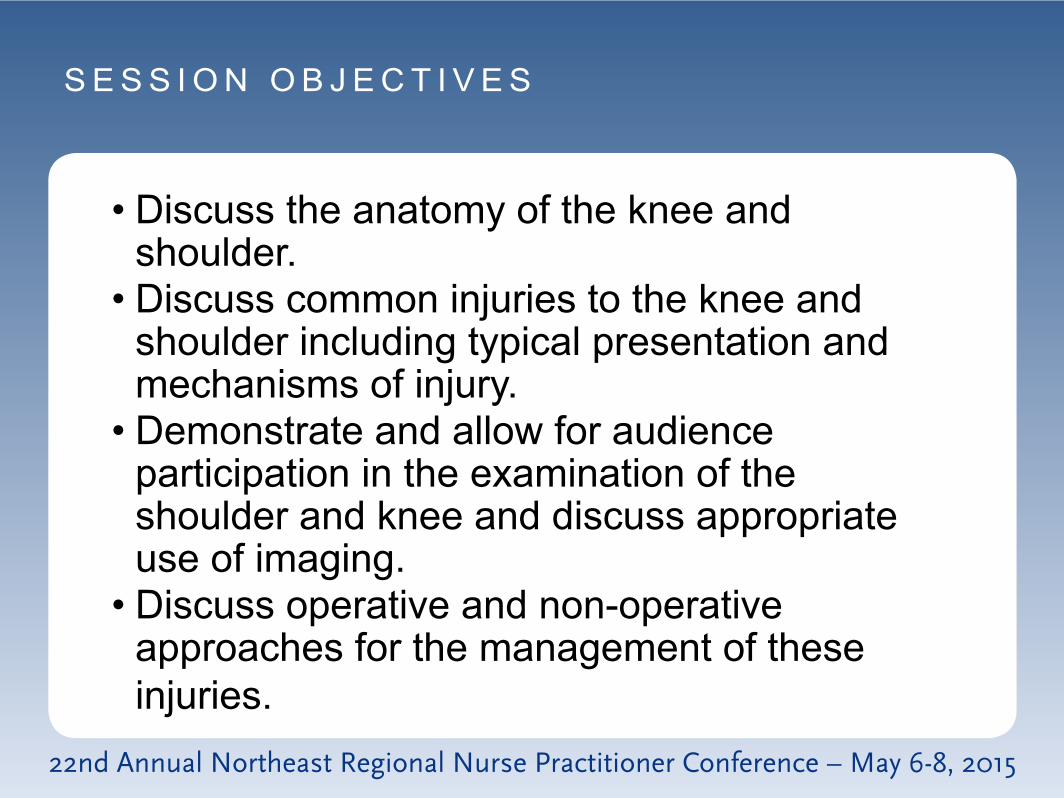
S E S S I O N O B J E C T I V E S
• Discuss the anatomy of the knee and shoulder.
• Discuss common injuries to the knee and shoulder including typical presentation and mechanisms of injury.
• Demonstrate and allow for audience participation in the examination of the shoulder and knee and discuss appropriate use of imaging.
• Discuss operative and non-operative approaches for the management of these injuries.

Weak in the Knees Acute Knee Injuries
Douglas J. Moran, MD Concord Orthopaedics
Northeast Regional Nurse Practitioner Conference

Knee Anatomy
Patellofemoral joint Articular cartilage
Tibiofemoral joint
Articular cartilage Meniscus Ligaments

Knee Anatomy
Ligaments: Anterior cruciate ligament (ACL) Medial collateral ligament (MCL) Posterior cruciate ligament (PCL) Lateral collateral ligament (LCL)

Knee Anatomy

Knee Anatomy Articular Cartilage
Normal
Abnormal

Acute Knee Injuries
When evaluating, timing is important
Earlier is better
Muscle spasm and swelling can make examination difficult

Acute Knee Injuries
Mechanism of Injury
Location of Pain
Effusion

Meniscus Injury

Meniscus Injury
Most common knee injury Bimodal distribution
Teenagers (usually with ACL tears) Those who think they are teenagers (Older
Crowd)

Meniscus Anatomy
Healthy Meniscus Torn Meniscus

Meniscus Anatomy
Vascular Supply At horn attaches to the
tibia Outer 1/3 or meniscal
rim

Meniscus Anatomy
Vascular Supply

Meniscus Injury
Episodic, sharp pain NOT a dull throbbing pain (articular
cartilage pain) Worse with deep bending Mechanical symptoms
Don’t confuse with patellofemoral

Meniscus Exam
Effusion Pain with hyperflexion Pain along joint line McMurray’s positive Ask them to squat or “duck walk”
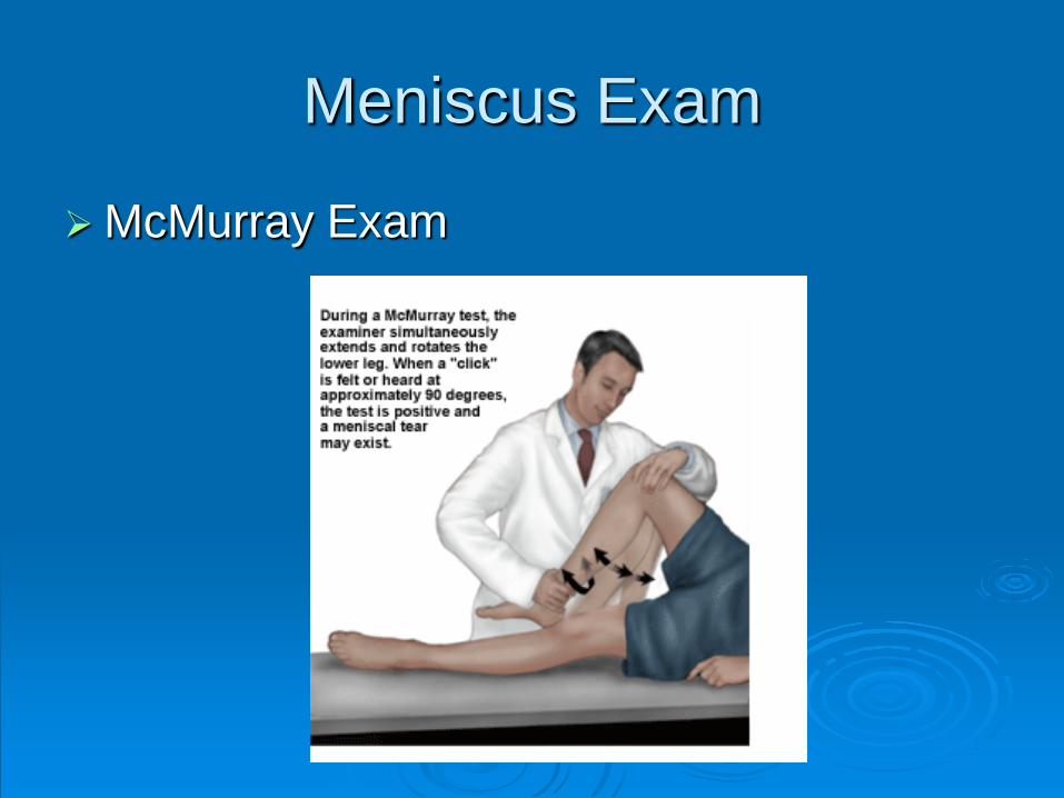
Meniscus Exam
McMurray Exam

Meniscus Tear MRI
MRI to confirm PE Tear pattern Concomitant injury
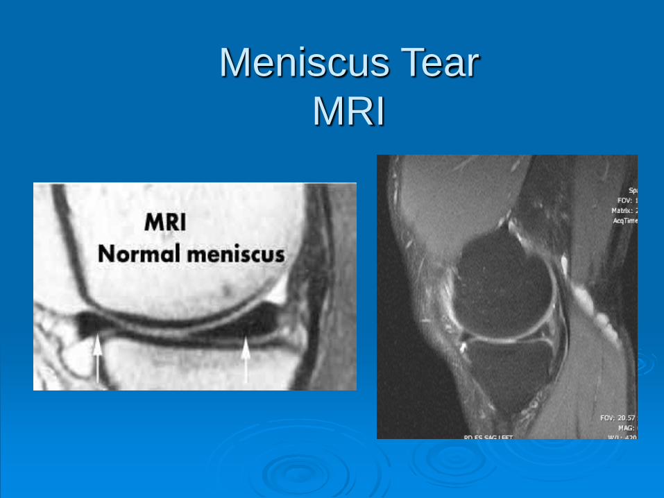
Meniscus Tear MRI

Meniscus Tear-Treatment
Surgical (arthroscopy) Ability to repair
based on tear pattern, size, location, blood supply

Anterior Cruciate Ligament Injury
(ACL)

ACL Tear-Mechanism
Contact Hit from either side with rotation
Non-contact Deceleration, rotation, hyperextension

ACL Tear

ACL Tear-History
Hear a “pop” Immediate large effusion (1 hour) Unable to return to play Patient feels like knee shifts

ACL Anatomy

Often limited by pain and guarding Effusion Hemarthrosis Lachman Anterior drawer Pivot shift
ACL Tear Examination

ACL Examination
Lachman Most sensitive Knee in 30 degrees of
flexion Hang heel off side of
bed Stabilize femur with
one hand Translate tibia with
other hand

ACL Examination
Anterior Drawer Knee flexed to 80
degrees Hamstrings are
palpated Proximal tibia moved
anteriorly Compare to
contralateral knee

ACL Examination Pivot Shift
Hardest to do Place a valgus stress Internal rotation of foot Flex knee beyond 30° Should feel the tibia
reduce from anteriorly subluxated position

ACL Tear - MRI
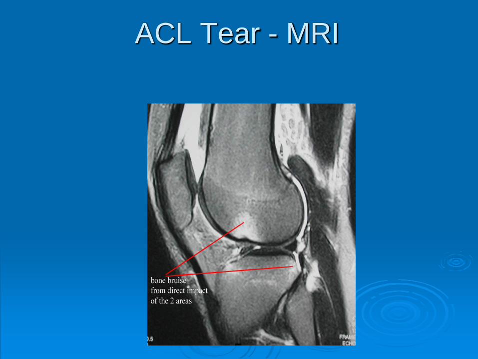
ACL Tear - MRI
ACL Tear- Treatment
Generally surgical Some exceptions
ACL Reconstruction

ACL Reconstruction
Patella Tendon

Posterior Cruciate Ligament Injury
(PCL)

PCL Tear History and Physical
Blow to the anterior knee with the knee flexed
Reports of instability are rare
Large effusion, positive posterior sag
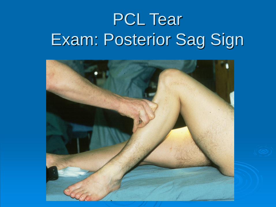
PCL Tear Exam: Posterior Sag Sign

PCL Tear - MRI
MRI to confirm diagnosis, check for concomitant ACL or posterolateral corner injury

Controversial Young active patients Avulsion injuries
Most treated non-operatively Concomitant posterolateral injury requires
surgery
PCL Tear - MRI

Medial Collateral Ligament Injury
(MCL)

Most common ligament injury
Usually caused by hit to the outside of the knee (valgus force)
MCL Injury

Patients often describe feeling something give, but not a true pop
Painful to flex the knee Ask them to point to location of pain -
often exquisitely tender over medial epicondyle
MCL Injury - History
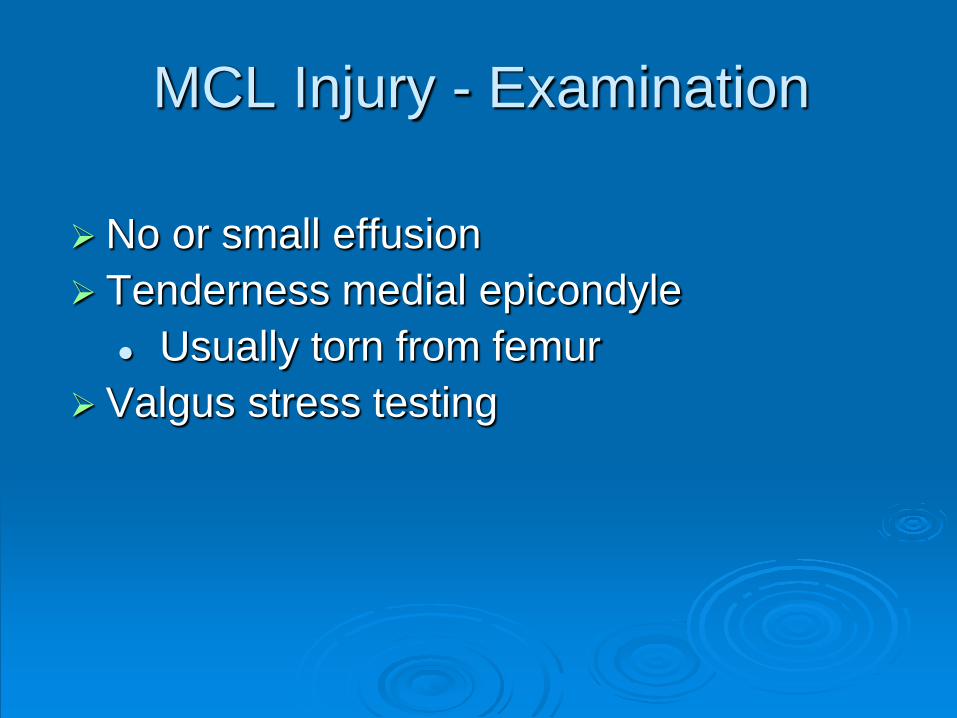
No or small effusion Tenderness medial epicondyle
Usually torn from femur Valgus stress testing
MCL Injury - Examination
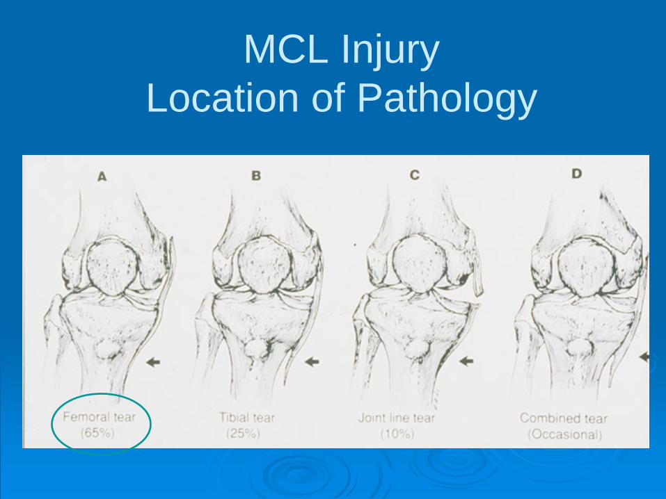
MCL Injury Location of Pathology

If questionable laxity
Large effusion Rule out other
ligament injury
MCL Injury - MRI
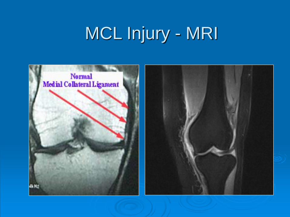
MCL Injury - MRI

Almost always non-surgical 95% will heal with support With concomitant ACL injury usually do
not need to fix MCL
MCL Injury - Treatment

Treatment based on severity Grade I: no brace, early rehab Grade II: brace 2-3 weeks Grade III: brace 4-6 weeks
MCL Injury - Treatment

Average return to full activity Grade I = 5 days Grade II = 17 days Grade III = 33 days
MCL Injury - Treatment

Patella Dislocation

Patella Dislocation
Usually twisting injury with valgus stress May have abnormal alignment Tear of medial patellofemoral ligament

Patella Dislocation History and Physical Exam
Patient describes kneecap out lateral Usually have a HUGE effusion
Won’t bend knee Tender medial epicondyle May be tender medial facet patella
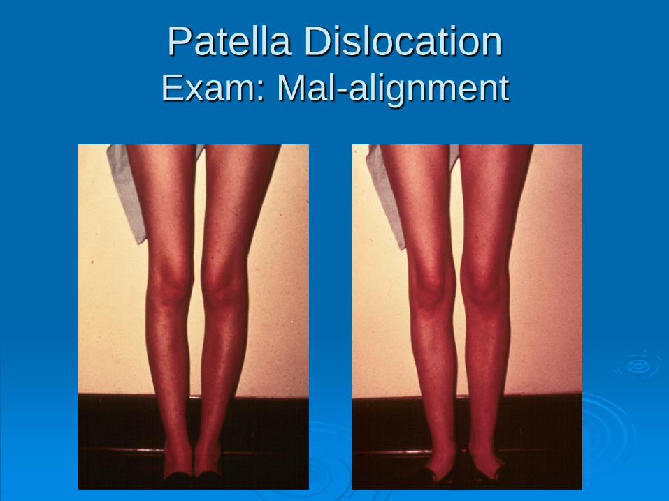
Patella Dislocation Exam: Mal-alignment

Patella Dislocation Exam: Apprehension Test

Patella Dislocation MRI

RICE Brace or tape to hold patella PT for quadriceps training Return to play after completing
functional rehab / running program
Patella Dislocation Treatment

1st time dislocation treat non-operatively 80% will heal
MRI to look for loose fragments in the
knee if persistent effusion
Patella Dislocation Treatment

Multiple dislocations will require operative treatment
Each time patella dislocates, the articular cartilage can be injured on the femur or patella
Patella Dislocation Treatment

Quadriceps Contusion

Quadriceps Contusion Direct blow to the thigh (quad) Hematoma in muscle Painful knee flexion Watch for compartment syndrome!
Quadriceps Contusion
Grade based on knee flexion at 24 hours Grade I: > 90 degrees Grade II: 45-90 degrees
Grade III: < 45 degrees

Ice Keep knee maximally flexed +/- compression wrap NSAIDs controversial
Quadriceps Contusion Treatment

Dreaded complication is myositis ossificans in quad muscle 9% incidence Higher when poor initial ROM, delayed
treatment Immobilization increases chance of it
Early ROM
Quadriceps Contusion Treatment

Iliotibial Band (ITB) Friction Syndrome
ITB Friction Syndrome History and Physical
Overuse injury Worse with increased activity Dull, throbbing pain No effusion Pain over lateral epicondyle Tight ITB

Popliteus tendonitis LCL sprain Lateral meniscus tear
ITB Friction Syndrome Differential Diagnosis

Rest Ice Anti-inflammatory medication Ultrasound or massage therapy Stretching Gradual return to exercise Correct any biomechanical/training
errors
ITB Friction Syndrome Treatment
Thank You! Questions?

EVALUATION OF ACUTE SHOULDER INJURIES
Douglas J. Moran, MD Orthopaedic Sports Medicine
Case Scenario
57 year old male
“I was attempting to pry a wheel off of a car at work, and the wheel sprung back…I felt a sudden pain in my right shoulder”

Where is the pain What is the quality of the pain Precipitating and alleviating factors Associated medical conditions and
social history Most importantly smoking!
History

Physical Examination Should be systematic!
Inspection Palpation ROM and Strength Special Tests Rotator Cuff Labrum and Biceps Instability AC Joint

Active and Passive ROM in BOTH shoulders
Strength
Physical Examination ROM and Strength

Neer and Hawkins Test Subacromial impingement a.k.a. bursitis
Jobe’s Test Rotator cuff tendonitis
Belly Press and Lift Off Test
Subscapularis
Special Tests Rotator Cuff

O’Brien Test Superior labrum
Speed Test Biceps or labrum
Special Tests Labrum and Biceps
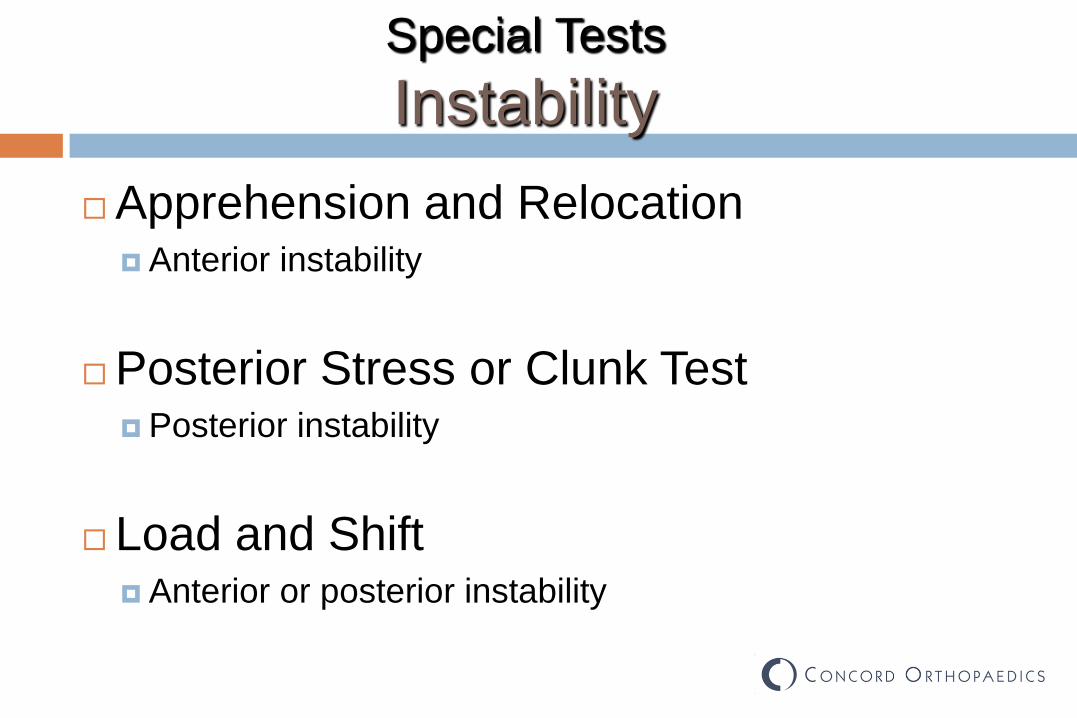
Apprehension and Relocation Anterior instability
Posterior Stress or Clunk Test Posterior instability
Load and Shift Anterior or posterior instability
Special Tests Instability
Pain is constant Radiates from superior shoulder through
upper arm to elbow Feels better in sling Worse with sleeping or any overhead
activity
Case Scenario

Case Scenario
Inspection normal No point tenderness ROM 170 / 50 / L3 vs. 180 / 60 / T8 No weakness with cuff testing + Neer + Hawkins + Jobe All other provocative testing negative

Management
Immediate MRI Indications Significant rotator cuff weakness
Everyone else… Rest, PT, medications, close F/U Consider injection if chronic in onset

Case Scenario No significant weakness Started PT for shoulder with diagnosis
rotator cuff strain
Follow up in two weeks

Case Scenario ROM 100 (130) / 40 / L3 + Neer + Hawkins + Jobe 4 out of 5 strength with rotator cuff
strength testing
MRI

Case Scenario MRI shows acute rotator cuff tear Proceed directly to surgical repair

Case Scenario # 2 47 YO male
“I was lifting a heavy bucket of mud
at work as a dry waller and felt a sudden tearing sensation in my left shoulder…”

Case Scenario # 2 Inspection normal Tenderness at greater tuberosity Active ROM 60 / 50 / L1 Passive ROM 180 / 50 / T12 + Neer + Hawkins + Jobe Rotator cuff strength 4+

Case Scenario # 3 58 YO female
“My right shoulder has been sore from
repetitive use at the deli counter”
Pain with repetitive use, significant night pain when sleeping on the right

Case Scenario # 3 Inspection normal No tenderness to palpation Active ROM = Passive ROM 170 / 40 / L2 + Neer + Hawkins – Jobe 5 of 5 rotator cuff strength All other provocative tests negative
Case Scenario # 3 No weakness Chronic onset of pain Therapy and injection If no improvement after 4 to 6 weeks
consider MRI to rule out underlying tear

Case Scenario # 4 26 YO male
“I was trying to take a keg down a flight of
stairs on a dolly, it slipped and my shoulder felt like it pulled out of the socket…”

Case Scenario # 4 Inspection normal Tender to palpation along anterior joint line Neer Hawkins Jobe negative O’Brien and Speeds positive Apprehension + relocation positive Posterior stress test negative Normal rotator cuff strength
Case Scenario # 4 Inspection normal Tender to palpation along anterior joint line Neer Hawkins Jobe negative O’Brien and Speeds positive Apprehension + relocation positive Posterior stress test negative Normal rotator cuff strength
Case Scenario # 4 Suspected labral tear No real reason for acute MRI
Management would not be changed with MRI
Start PT and anti-inflammatory If no improvement after 6 weeks MRI shows labral tear

Summary With careful history and physical
examination the diagnosis can be made in most cases…not everyone needs an MRI!
MRI if suspected large rotator cuff tear, or in patients who
fail to progress with other treatment

Summary Never too much of a downside to giving
someone 1-2 weeks of therapy or rest and re-examining the shoulder At least from a surgeon standpoint… Immediate MRI in everyone with work injury may lead to
incidental findings i.e. “What am I supposed to do with this information”