Embed Size (px)
Citation preview
1
Evaluation of the impact of excipients and an albendazole salt on albendazole
concentrations in upper small intestine using an in vitro biorelevant gastrointestinal
transfer (BioGIT) system
Alexandros Kourentas1, Maria Vertzoni1, Ibrahim Khadra2, Mira Symillides1, Hugh Clark3,
Gavin Halbert2, James Butler4, Christos Reppas1,*
1Faculty of Pharmacy, National and Kapodistrian University of Athens, Zografou, Greece
2Strathclyde Institute for Pharmacy and Biomedical Sciences, University of Strathclyde,
Scotland, UK
3Product Development, GlaxoSmithKline, Stevenage, UK
4Product Development, GlaxoSmithKline, Ware, UK
Running Title: Evaluation of albendazole concentrations in upper small intestine using BioGIT
*To whom correspondence should be addressed:
Christos Reppas PhD
Faculty of Pharmacy, School of Health Sciences,
National and Kapodistrian University of Athens,
Panepistimiopolis, 157 84 Zografou, Greece
Tel. 00 30 210 727 4678 / Fax: 00 30 210 727 4027
2
Abstract
An in vitro biorelevant transfer (BioGIT) system was assessed for its ability to mimic recently
reported albendazole concentrations in human upper small intestine after administration of
free base suspensions to fasted adults in absence and in presence of supersaturation
promoting excipients (hydroxypropylmethylcellulose and lipidic self-emulsifying vehicles).
The in vitro method was then used to evaluate the likely impact of using the sulfate salt on
albendazole concentrations in upper small intestine. In addition, BioGIT data were compared
with equilibrium solubility data of the salt and the free base in human aspirates and
biorelevant media. The BioGIT system adequately mimicked the average albendazole
gastrointestinal transfer process and in vivo concentrations in upper small intestine after
administration of the free base suspensions to fasted adults. However, the degree of
supersaturation seen initially in the duodenuum was greater in-vitro than in-vivo.
Albendazole sulfate resulted in minimal increase of albendazole concentrations in the
duodenal compartment of the BioGIT, despite improved equilibrium solubility observed in
human aspirates and biorelevant media, indicating that the use of a salt is unlikely to lead to
any significant oral absorption advantage for albendazole.
3
Keywords
BioGIT; gastrointestinal transfer; dissolution; supersaturation; poorly soluble weak base;
albendazole salts; lipid excipients; precipitation inhibitor; HPMC E5
4
Introduction
New drug candidates often have low aqueous solubility to an extent that require
administration in enabling formulations to promote supersaturation, in order to achieve
luminal concentrations to get most of the dose absorbed (e.g. Brouwers et al. 2009). In case
of a poorly soluble weak base, the impact of gastrointestinal transfer in the fasted state on
absorption rates may be substantial, because, due to the abrupt increase in pH, precipitation
in the upper small intestine is likely. The use of supersaturation promoting formulations may
then be required.
Various methodologies have been proposed to evaluate to impact of gastrointestinal
transfer on the performance of the dosage form and of the drug intraluminally (Kostewicz et
al. 2014). Recently, an in vitro biorelevant gastrointestinal transfer (BioGIT) system was
proposed for the evaluation of the impact of gastrointestinal transfer on concentrations in
upper intestinal lumen during the first hour, after oral administration of dispersing/solution
dosage forms of lipophilic weak bases in the fasted state (Kourentas et al. 2016).
In the present investigation, the impact of excipients and of free base vs. salt on
concentrations of lipophilic weak bases in upper small intestine was investigated with BioGIT
by using albendazole free base [ABZ, pka 2.80 (Jung et al 1998), logP 3.46 (Rivera et al.
2007)] as a model lipophilic weak base. There were three specific objectives of the present
study:
Firstly, to evaluate the usefulness of BioGIT in reproducing recently observed ABZ
concentrations and supersaturation of contents of the upper small intestine of healthy
5
adults after administration of various ABZ free base suspensions in absence and in presence
of supersaturation promoting excipients in the fasted state (Kourentas et al. submitted).
Secondly, to evaluate the impact of free base vs. salt on ABZ concentrations and degrees of
supersaturation of contents of the upper small intestine in absence and in presence of HPMC
E5 using BioGIT.
Thirdly, to compare BioGIT data with equilibrium solubility data (in human aspirates and in
biorelevant media) in order to evaluate the impact of free base vs. salt on ABZ behaviour in
the upper gastrointestinal lumen.
Since only the free base of ABZ is commercially available, a salt had to be prepared for the
needs of the present investigation. Although various salts of ABZ have been prepared and
characterized in the past (Paulekuhn et al. 2013), the relatively low pKa makes ABZ salts
sensitive to disproportionation (e.g. Stephenson et al. 2011). After considering various
counter-ions, the sulfate salt of ABZ was selected to be used, based on its comparatively
better crystallinity and stability characteristics.
6
Materials and Methods
Materials
Albendazole free base was from Sigma Aldrich (Saint Louis, U.S.A., 99% pure). Sodium
phosphate monobasic, sodium hydroxide, sodium chloride, ammonium formate, and pepsin
from porcine gastric mucosa (15.8% protein) were purchased from Sigma Aldrich (Saint
Louis, U.S.A.). Acetonitrile and water (HPLC grade) as well as tetrahydrofuran and ethanol
were also from Sigma Aldrich (Saint Louis, U.S.A.). n-butanol was purchased from Fluka (Neu-
Ulm, Germany). SIF® Powder Original was kindly donated by Biorelevant.com (Surrey, U.K.).
Hydrochloric acid was from Panreac Co. (Barcelona, Spain). Hydroxypropylmethylcellulose E5
was from JRS Pharma (Zacapu, Mexico). Miglyol 812N [Caprylic/Capric (C8–C10)
triglycerides] was received from Sasol Germany. Cremophor RH 40 was from BASF
(Ludwigshafen, Germany). Polysorbate 80 was from Sigma Aldrich (Saint Louis, U.S.A.).
Preparation of various ABZ salts and selection of the sulfate salt
Initially, the solubility of ABZ free base was assessed in tetrahydrofuran, ethanol, water, and
n-butanol at a range of temperatures (25-80 °C). In order to obtain albendazole
hydrochloride, albendazole sulfate and albendazole mesylate, 10, 20, 30, 40 and 50 mg of
ABZ free base were transferred into 2 ml HPLC vials followed by the addition of 1 ml of
solvent (tetrahydrofuran, water, ethanol or n-butanol) and the addition of an equi-molar
amount of acid (HCl, H2SO4 or methanesulfonic acid). The vials were placed in a Crystal 16
apparatus (Avantium Crystallization System, Amsterdam, Netherlands), temperature was
cycled with the maximum temperature set 10°C below the solvents boiling point and the salt
was formed at the end of the experiment. Two scale-up experiments were conducted using
7
25 ml and 50 ml of solvent, to ensure the scale-up procedure was viable. Equi-molar
amounts of ABZ free base and the acids (HCl, H2SO4, or methanesulfonic acid) were added to
100 ml glass beakers containing either 25 or 50 ml of solvent and the mixture heated to 10
degrees below the boiling point of the solvent. At the elevated temperature the ABZ salt was
completely dissolved; it was then left at room temperature to cool down and the salt
precipitated. Residual solvent was evaporated at room temperature and the material was
further dried at 40 °C, over 24 hours, and weighed. The formation of ABZ salts (as hydrates,
based on Karl-Fisher titration data) was confirmed using Nuclear Marnetic Resonance,
Differential Scanning Calorimetry (30 – 300 °C in 10 min, Mettler Toledo DSC 822e), and
Elemental Analysis for carbon, hydrogen and nitrogen (data on file). Based on X-ray
diffraction data, albendazole hydrochloride was poorly crystalline (unlike with a previous
report (Paulekuhn et al. 2013) whereas albendazole mesylate crystals and crystal stability
characteristics varied with the solvent used for their isolation. In contrast, albendazole
sulfate salt (ABZ sulfate) samples obtained with tetrahydrofuran and ethanol were
concordant (some differences in peak intensities may suggest small differences in
crystallinity). Also, based on the one-month stability data ABZ sulfate, peaks were
concordant at two tested conditions (40 oC / 75% humidity and 50 oC / ambient humidity)
with the initial analysis. Based on these data, ABZ sulfate obtained using tetrahydrofuran
was selected to be used in the present investigation.
Experiments with BioGIT
Methodology
BioGIT is an in vitro three compartment setup simulating the drug transfer from the stomach
into the fasted upper small intestine (Kourentas et al. 2016). The initial volume of the gastric
8
compartment is 250 ml. The duodenal volume is maintained at 40 ml during the entire
experiment. The mini - paddles in gastric and duodenal compartments rotate at 75 rpm. The
emptying of contents of gastric compartment (on a volume basis) follows first-order kinetics
with a half-life of 15 min. Experiments are performed using a three-channel peristaltic pump
(Reglo ICC pump, part ISM 4308, Ismatec®) for 45 min, after the initiation of an experiment.
Incoming flow rates are changed every 10 min and sampling is performed at midpoint
(Kourentas et al. 2016). In this study, experiments were performed in triplicate at 37 °C.
Contents of gastric compartment
Two aqueous suspensions of ABZ free base were tested. For the first, 50 mg ABZ free base
were mixed with 200 ml table water (Nera Kritis, Heraklion, Greece) under vigorous
magnetic stirring for 5 min at room temperature. Preparation of the second suspension was
achieved by dissolving 5 mg HPMC E5 in 200 ml table water, prior to mixing 50 mg ABZ free
base with the aqueous HPMC E5 solution. Regardless of the presence of HPMC E5, the
suspension was mixed with 50 ml concentrated Level III FaSSGF. The resulting 250 ml
suspension in Level III FaSSGF (Markopoulos, Andreas et al. 2015), Susp or Susp-HPMC, was
immediately added to the gastric compartment, and the transfer experiment was initiated.
Two lipid based suspensions of ABZ free base were tested, prepared from one Type IIIA and
one Type IV lipid based formulation (Pouton, 2006). The appropriate quantities of excipients
were weighed into a screw cap glass bottle of 50 ml capacity, i.e. 0.4 g Miglyol 812N, 0.2 g
Cremophor RH 40 and 0.4 g Tween 80 for Type IIIA lipid based formulation and 0.2 g
Cremophor RH40 and 0.4 g Tween 80 for Type IV lipid based formulation (Kourentas et al.
submitted). An accurately weighed quantity of 50 mg ABZ free base was added into the glass
bottle. The components were mixed using a magnetic stirrer for 1 h at room temperature.
9
The lipid based formulations of ABZ free base were stored at room temperature overnight
(Kourentas et al. submitted). Each lipid based formulation was transferred to a screw cap
glass bottle of 250 ml capacity by rinsing the 50 ml capacity glass bottle with small amounts
of table water (total volume of table water used for rinsing was 200 ml). The 250 ml capacity
bottle was then vigorously stirred using a magnetic stirrer for 5 min at room temperature
(Kourentas et al. submitted), 50 ml of concentrated Level III FaSSGF were added into the
bottle and mixed. The resulting 250 ml suspension in Level III FaSSGF, Susp-IIIA or Susp-IV,
was immediately added to the gastric compartment, and the experiment initiated.
Assuming that resting gastric volume in the fasted state is 10 ml (Psachoulias et al. 2011;
Vertzoni et al. 2012), volume and composition of contents in the gastric compartment when
using ABZ free base suspensions reflected the expected composition of gastric contents after
administration of the following suspensions to fasted adults (Kourentas et al. submitted):
- Suspension of ABZ free base in 240 ml table water (Susp)
- Suspension of ABZ free base in 240 ml table water in which HPMC E5 had been pre-
dissolved (Susp-HPMC)
- Suspension of a Type IIIA lipid based formulation of ABZ free base in 240 ml table water
(Susp-IIIA)
- Suspension of a Type IV lipid based formulation of ABZ free base in 240 ml table water
(Susp-IV)
Two aqueous suspensions of ABZ sulfate were then tested. For the first, 50 mg ABZ
equivalent were mixed with 200 ml table water under vigorous magnetic stirring for 5 min at
room temperature. Preparation of the second suspension was achieved by dissolving 5 mg
HPMC E5 in 200 ml table water, prior to mixing 50 mg ABZ equivalent with the aqueous
HPMC E5 solution. Regardless of the presence of HPMC E5, the suspension was mixed with
10
50 ml concentrated Level III FaSSGF. The resulting 250 ml suspension in Level III FaSSGF
(Markopoulos, Andreas et al. 2015), S-Susp or S-Susp-HPMC, was immediately added to the
gastric compartment, and the transfer experiment was initiated.
The composition of contents in the gastric compartment were designed to reflect
intragastric conditions using the same assumptions made for the ABZ free base suspensions
and mimicking administration of the following ABZ sulfate suspensions to fasted adults:
-Suspension of ABZ sulfate in 240 ml table water (S-Susp)
-Suspension of ABZ sulfate in 240 ml table water containing HPMC E5 (S-Susp-HPMC)
In all experiments (with ABZ free base and with ABZ sulfate), concentrated Level III FaSSGF
had pH 0.9 whereas concentrations of sodium chloride, pepsin, sodium taurocholate and
phosphatidylcholine were 5-fold higher than in Level III FaSSGF pH 1.6 (Markopoulos,
Andreas et al. 2015).
Contents of duodenal compartment at time zero and contents of reservoir compartment
Level II FaSSIF (Markopoulos, Andreas et al. 2015) was employed in the duodenal
compartment in order to simulate the fasted state conditions in upper small intestine. A
series of phosphate buffer solutions containing sodium chloride, bile salt and lecithin were
employed in the reservoir compartment so that the composition of contents in the duodenal
compartment (pH, buffer capacity, osmolality, bile salt and lecithin concentration) remains
unaltered during an experiment (Kourentas et al. 2016).
Sample treatment
11
Upon collection, each sample from the duodenal compartment was divided into two parts:
i) The first part was immediately filtered through regenerated cellulose filters (Titan 3, 17
mm, 0.45 μm, SUN SRi, Rockwood, U.S.A.) and the filtrate was divided in two portions:
- The first, after immediate dilution with mobile phase (so that precipitation
during subsequent handling is avoided), was used for measuring ABZ
concentration.
- The second portion was incubated (37 °C, 75 oscillations/min, model Unitronic
OR, j.p. Selecta, s.a., Barcelona, Spain) in presence of excess of ABZ free base or
ABZ sulfate solid material for 16 hours and, after filtration through a
regenerated cellulose 0.45 μm filter, equilibrium solubility was determined. The
duration of incubation for achieving equilibration had been evaluated with
preliminary experiments.
Adsorption of ABZ on to the regenerated cellulose filter had been evaluated and found to be
negligible.
ii) The second part was used for measuring total ABZ amount (solid and dissolved drug) per
volume, after appropriate dilution with mobile phase.
Equilibrium solubility of ABZ free base and ABZ sulfate in biorelevant media and in humans
aspirates
The solubility of ABZ free base and ABZ sulfate were measured in Level III FaSSGF, in Level II
FaSSIF (Markopoulos, Andreas et al. 2015) and in human aspirates collected from the
stomach and from the upper small intestine in the fasted state. Human aspirates had been
collected from healthy fasted adults (Petrakis et al. 2014) and pooled after one freeze-thaw
cycle. The pH after pooling was 1.6 for the gastric pooled sample (human gastric fluid, HGF)
and 7.4 for intestinal pooled sample (human intestinal fluid, HIF). An excess amount of ABZ
12
free base or ABZ sulfate solid material was added to glass vials containing the appropriate
medium. The vials were incubated at 37 °C (75 oscillations/min) until equilibrium was
reached. Equilibration times in human aspirates and in biorelevant media were evaluated
with preliminary experiments at 4h, 16h and 24h, after initiation of the incubation. For each
medium, concentrations were measured to be identical at all sampling times. For practical
reasons solubility values were measured at 16h in all media. At equilibrium, samples from
biorelevant media were filtered through 0.45 μm regenerated cellulose filters whereas HGF
and HIF samples were centrifuged (11800 g, 37 °C, 10 min). Filtrates and supernatants were
diluted with equal volume of mobile phase and samples were subjected to HPLC analysis. All
measurements were performed in triplicate. The adequacy of filtration vs. centrifugation to
separate dissolved from undissolved material had been evaluated with preliminary
experiments (Kourentas et al. submitted).
ABZ assay
ABZ assay was performed on a Spectra HPLC system consisting of a P1000 pump, an AS1000
autosampler, a UV2000 detector, and an SN4000 controller which was programmed by the
Chromquest® software (version 2.51, Thermoquest Inc., USA). Analysis of drug content
involved the use of a Fortis C18 column (3 μm, 150×3 mm) equipped with a BDS C18 (5 μm,
10×4.6 mm) pre-column. The mobile phase consisted of ammonium formate (50 mM):
acetonitrile 50:50 v/v, the flow rate was 0.5 ml/min, the wavelength was set at 292 nm and
the injection volume was 20 μl. In all cases, results are expressed as ABZ free base.
Quantification limit was 10 ng/ml.
Data Analysis
13
Raw data are presented as Box-Whisker plots showing the median value, the 10th, 25th, 75th,
and 90th percentiles, and the individual outlying data points.
Supersaturation in duodenal compartment was evaluated by estimating the Degree of
Supersaturation, DS:
ilitySomEquilibriu
ionConcentratDS
lub (Eq. 1)
DS>1 indicates supersaturation, DS=1 indicates saturation, and DS<1 indicates unsaturated
solution.
One-sample t–test (Sigmaplot 11.0, Systat Software, Inc., San Jose, California, USA) was
applied for evaluating whether a sample mean DS value comes from a population with mean
of 1. Differences between solubility in human fluids and biorelevant media and between ABZ
free base and ABZ sulfate were evaluated with unpaired t-test (Sigmaplot 11.0, Systat
Software, Inc., San Jose, California, USA). Maximum type I error had been set at 0.05, in all
cases.
14
Results and Discussion
BioGIT data vs. data in the upper small intestine after administration of ABZ free base
suspensions
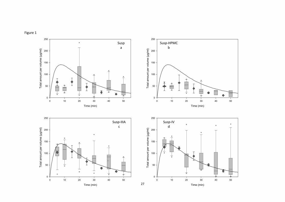
The total ABZ amount per volume in the duodenal compartment matched the average
luminal data during the first 50 min of gastric emptying of Susp, Susp-HPMC, Susp-IIIA and
Susp-IV (Figure 1). The BioGIT data are consistent with the lower total ABZ amount per
volume in upper small intestine observed after administration of Susp and Susp-HPMC than
after administration of Susp-IIIA or Susp-IV to fasted adults (diamonds and boxes in Figures
1a and 1b vs. diamonds and boxes in Figures 1c and 1d). As reported recently (Kourentas et
al. submitted), total ABZ amount per volume in upper small intestine, after administration of
Susp-IIIA and Susp-IV are in line with first-order gastric emptying kinetics and half-life of 15
minutes (Figure 1, continuous lines). Lower total ABZ amount per volume, after Susp or
Susp-HPMC could be attributed to the slower emptying of Susp and Susp-HPMC, due to
floating and/or adhesion of aggregates on to the walls of the gastric compartment (visually
observed in this study) and in stomach (Kourentas et al. submitted).
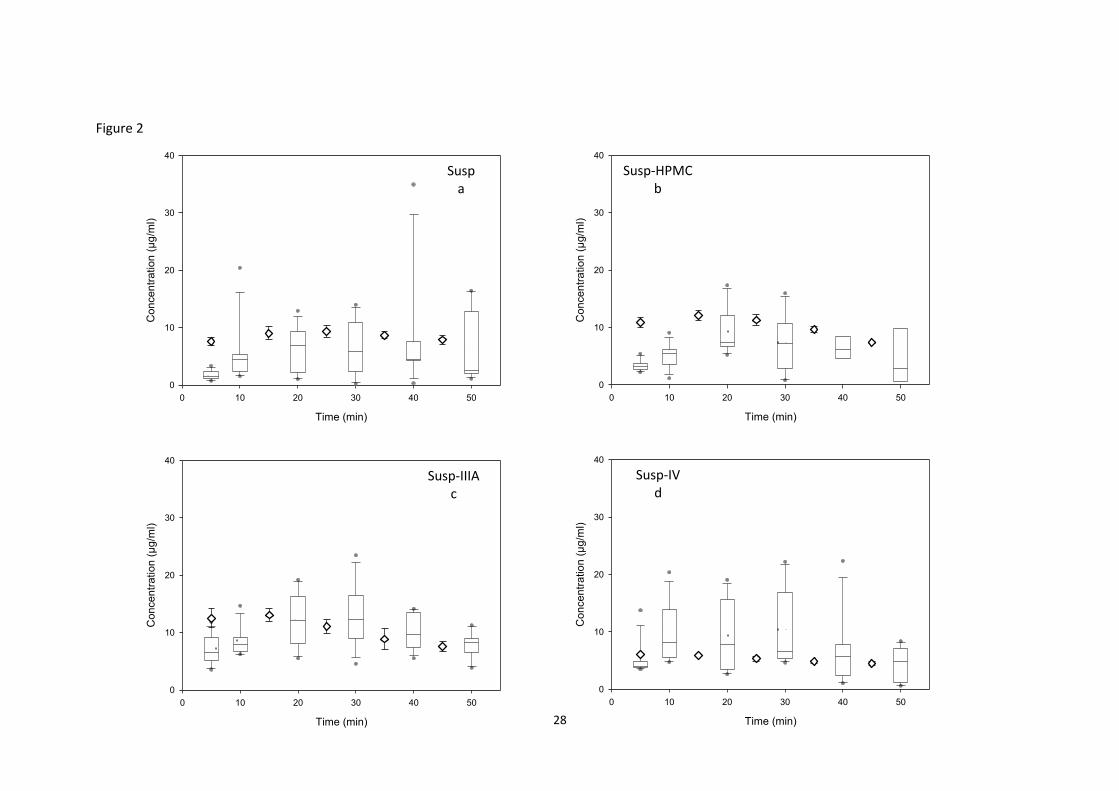
When using Susp or Susp-HPMC, ABZ concentrations in the duodenal compartment early, i.e.
about 5 minutes, after initiation of the experiment were somewhat higher than the average
concentrations in the upper small intestine (Figures 2a and 2b). This may be attributed to
the pH of contents in the gastric compartment (FaSSGF, pH 1.6) which reflects the average
pH in the stomach during the gastric emptying of glass of water in the fasted state, i.e. it is
lower than the pH expected to be in stomach early after administration of a glass of water
(about 2.5, e.g. Kalantzi et al. 2006). The lower than physiological pH at early times after
initiation of the experiment accelerates ABZ free base dissolution and, therefore,
15
concentrations in gastric and, subsequently, in duodenal compartment are increased,
compared with concentrations in the human upper gastrointestinal lumen at early times,
after administration. When using Susp-IIIA or Susp-IV, the discrepancy between in vitro and
luminal concentrations at early times was not apparent (Figures 2c and 2d). The fact that
ABZ concentration in Susp-IIIA and in Susp-IV administered to adults was 9 and 6 times,
respectively, higher than ABZ concentration in the administered Susp or Susp-HPMC
(Kourentas et al. submitted) could be a reason for the similarity of in vitro with luminal data
of Susp-IIIA and of Susp-IV at early times.
In all cases, ABZ concentrations in the duodenal compartment after about 5 minutes post
experiment start generally matched the average data in upper small intestine during the first
50 minutes, after administration of all four tested ABZ free base suspensions to fasted adults
(Figure 2).
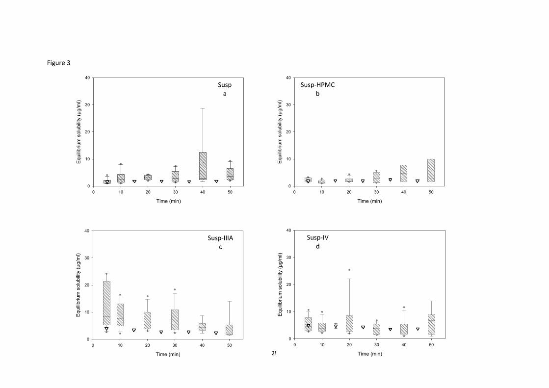
Solubility values in duodenal compartment of BioGIT were in line with previously measured
values in the lumen of upper small intestine of healthy adults (Figure 3). Occasionally,
limited underestimation was observed, primarily in the case of Susp-IIIA (Figure 3c).
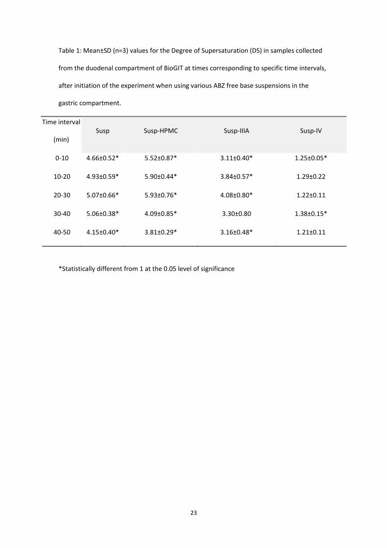
Contents of the duodenal compartment of BioGIT were significantly supersaturated in case
of Susp, Susp-HPMC, and Susp-IIIA at all sampling times but one, after initiation of the
experiment (Table 1). In case of Susp-IV, significant supersaturation was observed only
occasionally (Table 1). Recently reported data from the upper small intestine of healthy
adults suggest that significant supersaturation occurs only in case of Susp-HPMC, Susp-IIIA,
and Susp-IV and at specific times post administration (Kourentas et al. submitted). The
discrepancy between in vitro and luminal supersaturation could be attributed to the higher
concentrations in BioGIT at early times (diamonds vs. boxes in Figures 2a, 2b, and 2c), the
lower solubility in the duodenal compartment measured occasionally (Figure 3c), and the
16
high variability of luminal data which led to high average degrees of supersaturation but of
borderline statistical significance (Kourentas et al. submitted). Overall, the BioGIT data,
consistent with the human data (Kourentas et al. submitted) indicates that the lipid based
formulations may be slightly less effective that formulations containing HPMC E5 in
maintaining supersaturation in upper small intestine (Table 1).
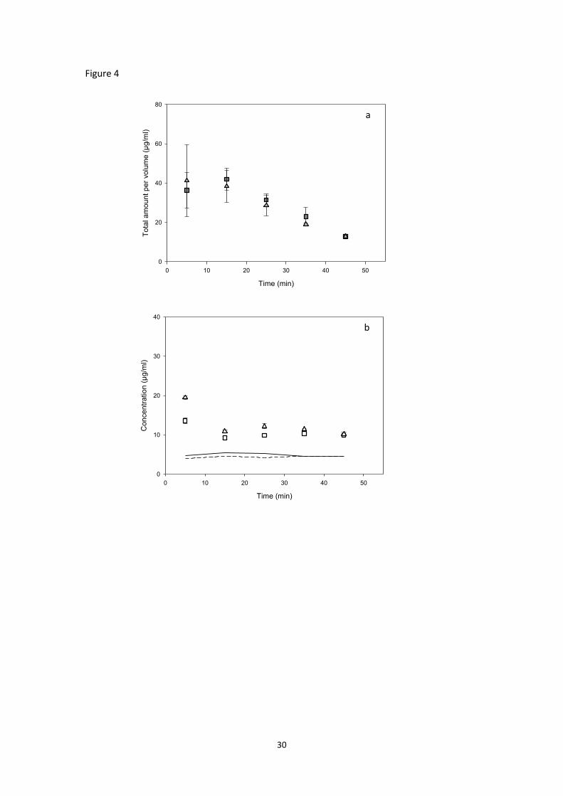
BioGIT data using ABZ free base and ABZ sulfate suspensions
Total ABZ amount per volume in the duodenal compartment of BioGIT did not change
substantially when S-Susp or S-Susp-HPMC was used in the gastric compartment (Figure 4a
vs. Figure 1a and 1b). ABZ concentrations in the duodenal compartment also remained
substantially unaffected, apart from at 5 minutes; when using S-Susp-HPMC concentration at
5 min was almost doubled, compared with the concentration measured when using Susp-
HPMC (Figure 4b vs. Figure 2b).
Since solubility of ABZ sulfate was not affected by the presence of HPMC E5 (Figure 4b), the
higher supersaturation when using S-Susp-HPMC could be attributed to the increased
concentrations in presence of HPMC E5, especially at early times, after initiation of the
experiment (Figure 4b). Although mean values of the degree of supersaturation were higher
with S-Susp-HPMC than with S-Susp (Table 2), values were smaller than those estimated
using Susp-HPMC and Susp, respectively (Table 2 vs. Table 1).
Regardless of the presence of HPMC E5, BioGIT data suggest that, compared with ABZ free
base, ABZ sulfate will not lead to significantly more drug available for absorption. This is in
line with literature data suggesting that usefulness of salts in increasing the absorption of
17
lipophilic weak bases has not been well documented in humans (Verbeeck et al. 2006) and
may be overestimated (Erceg et al. 2012; Dimopoulou et al. in press).
Εquilibrium solubility data vs. BioGIT data for evaluating the impact of free base vs. salt on
ABZ concentrations in upper small intestine
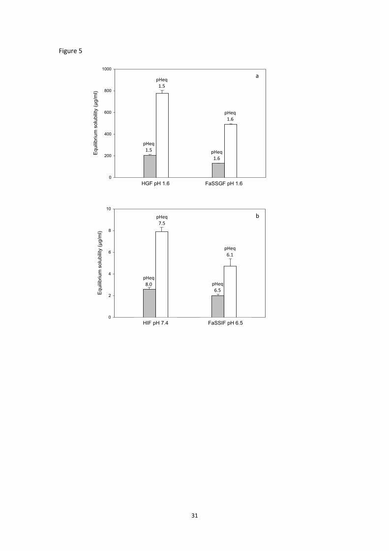
ABZ free base equilibrium solubility in Level III FaSSGF (mean: 128 g/ml, Figure 5a) is in line
with its pH - solubility profile (Paulekuhn et al. 2013). Solubilities in Level III FaSSGF and in
HGF as well solubilities in Level II FaSSIF and in HIF are significantly higher when ABZ sulfate
is used; compared with mean solubility of ABZ free base, mean solubility of ABZ sulfate is 3.8
times higher both in Level III FaSSGF and in HGF (Figure 5a). Similarly, compared with data
when ABZ free base was used, solubility in equilibrium with ABZ sulfate is 2.3 and 3.1 times
higher in Level II FaSSIF and in HIF, respectively (Figure 5b).
Although ABZ sulfate is more soluble in contents and in simulated contents of upper
gastrointestinal lumen (Figure 5), concentrations in duodenal compartment of BioGIT when
using Susp and when using S-Susp were similar (Figure 2a vs. Figure 4b).
18
Concluding remarks
BioGIT was demonstrated to be a useful tool for mimicking the average gastrointestinal
transfer process and concentrations in upper small intestine observed after administration
of various ABZ free base suspensions to fasted adults. However, compared with luminal
data, a greater extent of supersaturation in the duodenal compartment in the BioGIT was
typically observed.
BioGIT data indicate that ABZ sulfate, despite its improved equilibrium solubility in contents
of stomach and upper small intestine, leads to only a minimal, transient increase of
concentrations of ABZ in upper small intestine (only in the first 10 minutes after
administration and enhanced by the presence of HPMC E5). These data suggest that
equilibrium solubility data should be used to evaluate the luminal concentrations cautiously
in situations where supersaturation and precipitation is likely to occur.
19
Acknowledgments
This work was performed within the OrBiTo project (http://www.orbitoproject.eu) which is
funded by the Innovative Medicines Initiative Joint Undertaking under Grant Agreement No
115369. Part of the present work was presented as a poster at AAPS Annual Meeting,
November 2-6, 2014, San Diego, California, USA.
20
References
Brouwers J, Brewster ME, Augustijns P. Supersaturating drug delivery systems: the answer to
solubility-limited oral bioavailability? J Pharm Sci. 2009;98:2549-2572.
Dimopoulou M, Mourouti CS, Vertzoni M, Symillides M, Reppas C. In-vitro evaluation of
performance of solid immediate release dosage forms of weak bases in upper
gastrointestinal lumen: experience with miconazole and clopidogrel salts. J Pharm
Pharmacol. in press.
Erceg M, Vertzoni M, Cerić H, Dumić M, Cetina-Čižmek B, Reppas C. In vitro vs. canine data for
assessing early exposure of doxazosin base and its mesylate salt. Eur J Pharm Biopharm.
2012;80:402-409.
Jung H, Medina L, García L, Fuentes I, Moreno-Esparza R. Absorption studies of albendazole
and some physicochemical properties of the drug and its metabolite albendazole sulphoxide.
J Pharm Pharmacol. 1998;50:43-48.
Kalantzi L, Goumas K, Kalioras V, Abrahamsson B, Dressman JB, Reppas C. Characterization of
the human upper gastrointestinal contents under conditions simulating
bioavailability/bioequivalence studies. Pharm Res. 2006;23:165-176.
Kostewicz ES, Abrahamsson B, Brewster M, Brouwers J, Butler J, Carlert S, Dickinson PA,
Dressman J, Holm R, Klein S, Mann J, McAllister M, Minekus M, Muenster U, Müllertz A,
Verwei M, Vertzoni M, Weitschies W, Augustijns P. In vitro models for the prediction of in
vivo performance of oral dosage forms. Eur J Pharm Sci. 2014;57:342-366.
Kourentas A, Vertzoni M, Stavrinoudakis N, Symillidis A, Brouwers J, Augustijns P, Reppas C,
Symillides M. An in vitro biorelevant gastrointestinal transfer (BioGIT) system for forecasting
concentrations in the fasted upper small intestine: Design, implementation, and evaluation.
Eur J Pharm Sci. 2016;82:106-114.
21
Kourentas A, Vertzoni M, Symillides M, Goumas K, Butler J, Reppas C. Effectiveness of
supersaturation promoting excipients on luminal albendazole concentrations following
incomplete gastric dissolution in fasted healthy adults. Pharm Res. (Submitted).
Markopoulos C, Andreas C.J, Vertzoni M, Dressman J, Reppas C. In-vitro simulation of luminal
conditions for evaluation of performance of oral drug products: Choosing the appropriate
test media. Eur. J. Pharm. Biopharm. 2015;93:173-182.
Paulekuhn GS, Dressman JB, Saal C. Salt screening and characterization for poorly soluble,
weak basic compounds: case study albendazole. Pharmazie. 2013;68:555-564.
Petrakis O, Vertzoni M, Angelou A, Kesisoglou F, Bentz K, Goumas K, Reppas C. Identification
of key factors affecting the oral absorption of salts of lipophilic weak acids: a case example. J
Pharm Pharmacol. 2015;67:56-67.
Pouton CW. Formulation of poorly water-soluble drugs for oral administration:
physicochemical and physiological issues and the lipid formulation classification system. Eur
J Pharm Sci. 2006;29:278-287.
Psachoulias D, Vertzoni M, Goumas K, Kalioras V, Beato S, Butler J, et al. Precipitation in and
supersaturation of contents of the upper small intestine after administration of two weak
bases to fasted adults. Pharm Res. 2011;28:3145–3158.
Rivera JC, Yépez-Mulia L, Hernández-Campos A, Moreno-Esparza R, Castillo R, Navarrete-
Vázquez G, Fuentes-Noriega I, Jung-Cook H. Biopharmaceutic evaluation of novel
anthelmintic (1H-benzimidazol-5(6)-yl) carboxamide derivatives. Int J Pharm. 2007;343:159-
165.
Stephenson GA, Aburub A, Woods TA. Physical stability of salts of weak bases in the solid-
state. J Pharm Sci. 2011;100:1607-1617.
Verbeeck RK, Kanfer I, Walker RB. Generic substitution: the use of medicinal products
containing different salts and implications for safety and efficacy. Eur J Pharm Sci. 2006;28:1-
6.
22
Vertzoni M., Koulouri C., Goumas K., Poulou A., Reppas C. Intragastric performance of solid
oral dosage forms is unlikely to be affected by gastritis or Crohn’s disease. AAPS Annual
Meeting, October 14-18, 2012, Chicago, Illinois, USA (poster).
23
Table 1: Mean±SD (n=3) values for the Degree of Supersaturation (DS) in samples collected
from the duodenal compartment of BioGIT at times corresponding to specific time intervals,
after initiation of the experiment when using various ABZ free base suspensions in the
gastric compartment.
Time interval
(min) Susp Susp-HPMC Susp-IIIA Susp-IV
0-10 4.66±0.52* 5.52±0.87* 3.11±0.40* 1.25±0.05*
10-20 4.93±0.59* 5.90±0.44* 3.84±0.57* 1.29±0.22
20-30 5.07±0.66* 5.93±0.76* 4.08±0.80* 1.22±0.11
30-40 5.06±0.38* 4.09±0.85* 3.30±0.80 1.38±0.15*
40-50 4.15±0.40* 3.81±0.29* 3.16±0.48* 1.21±0.11
*Statistically different from 1 at the 0.05 level of significance
24
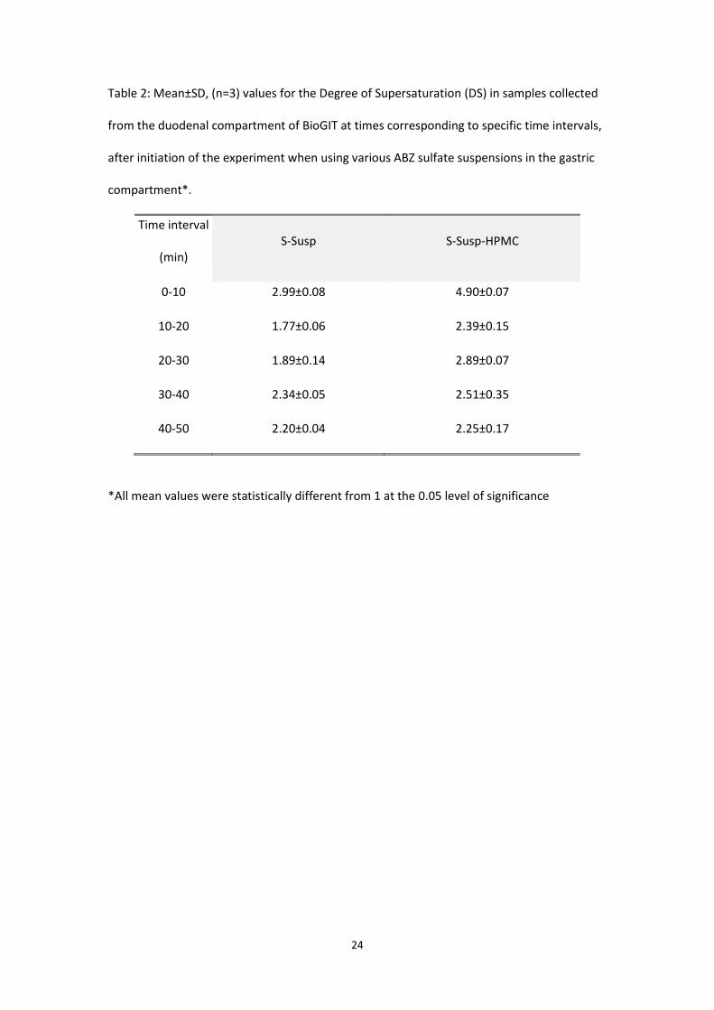
Table 2: Mean±SD, (n=3) values for the Degree of Supersaturation (DS) in samples collected
from the duodenal compartment of BioGIT at times corresponding to specific time intervals,
after initiation of the experiment when using various ABZ sulfate suspensions in the gastric
compartment*.
Time interval
(min) S-Susp S-Susp-HPMC
0-10 2.99±0.08 4.90±0.07
10-20 1.77±0.06 2.39±0.15
20-30 1.89±0.14 2.89±0.07
30-40 2.34±0.05 2.51±0.35
40-50 2.20±0.04 2.25±0.17
*All mean values were statistically different from 1 at the 0.05 level of significance
25
Figure captions
Figure 1
Mean±SD (n=3) values (diamonds) for the total ABZ amount per volume in the duodenal
compartment vs. time after initiation of the experiment with BioGIT when using Susp (a),
Susp-HPMC (b), Susp-IIIA (c) and Susp-IV (d) in the gastric compartment. For comparative
purposes, values estimated using the transfer model equation (continuous lines, Kourentas
et al. 2016), and recently reported total ABZ amount per volume in the contents of upper
small intestine, after administration of the corresponding suspensions to healthy adults are
also presented (box-whisker plots with dotted lines indicating the means, Kourentas et al.
submitted).
Figure 2
Mean±SD (n=3) values (diamonds) for ABZ concentration in the duodenal compartment vs.
time after initiation of the experiment with BioGIT when using Susp (a), Susp-HPMC (b),
Susp-IIIA (c) and Susp-IV (d) in the gastric compartment. For comparative purposes, recently
reported ABZ concentrations in the contents of upper small intestine of healthy adults, after
administration of the corresponding suspensions are also presented (box-whisker plots with
dotted lines indicating the means, Kourentas et al. submitted).
Figure 3
Mean±SD (n=3) values (triangles) for ABZ equilibrium solubility in the duodenal
compartment at various times after the initiation of the experiment with BioGIT using Susp
(a), Susp-HPMC (b), Susp-IIIA (c) and Susp-IV (d) in the gastric compartment. For comparative
purposes, recently reported ABZ equilibrium solubilities measured in the contents of upper
small intestine of healthy adults, after administration of the corresponding suspensions are
26
also presented (box-whisker plots with dotted lines indicating the means, Kourentas et al.
submitted).
Figure 4
(a) Mean±SD (n=3) values for total ABZ amount per volume in the duodenal compartment of
BioGIT; (b) Mean±SD (n=3) values for ABZ concentrations in duodenal compartment of
BioGIT (symbols) and mean (n=3) values for equilibrium solubility (lines).
Key: S-Susp, squares and continuous line; S-Susp-HPMC, triangles and dotted line.
Figure 5
Mean+SD (n=3) values for equilibrium solubility in (a) HGF and Level III FaSSGF, and (b) HIF
and Level II FaSSIF. Key: ABZ free base, grey bars; ABZ sulfate, white bars.
27
Time (min)
0 10 20 30 40 50
Tota
l am
ount
per
volu
me (
μg/m
l)
0
50
100
150
200
250
Time (min)
0 10 20 30 40 50
Tota
l am
ount
per
volu
me (
μg/m
l)
0
50
100
150
200
250
Time (min)
0 10 20 30 40 50
Tota
l am
ount
per
volu
me (
μg/m
l)
0
50
100
150
200
250
Time (min)
0 10 20 30 40 50
Tota
l am
ount
per
volu
me (
μg/m
l)
0
50
100
150
200
250
Susp a
Susp-HPMC b
Susp-IIIA c
Susp-IV d
Figure 1
28
Time (min)
0 10 20 30 40 50
Con
centr
ation
(μ
g/m
l)
0
10
20
30
40
Time (min)
0 10 20 30 40 50
Con
centr
ation
(μ
g/m
l)
0
10
20
30
40
Time (min)
0 10 20 30 40 50
Con
centr
ation
(μ
g/m
l)
0
10
20
30
40
Time (min)
0 10 20 30 40 50
Con
centr
ation
(μ
g/m
l)
0
10
20
30
40
Susp a
Susp-HPMC b
Susp-IIIA c
Susp-IV d
Figure 2
29
Time (min)
0 10 20 30 40 50
Equili
brium
solu
bili
ty (
μg/m
l)
0
10
20
30
40
Time (min)
0 10 20 30 40 50
Equili
brium
solu
bili
ty (
μg/m
l)
0
10
20
30
40
Time (min)
0 10 20 30 40 50
Equ
ilib
riu
m s
olu
bili
ty (
μg/m
l)
0
10
20
30
40
Time (min)
0 10 20 30 40 50
Equ
ilib
riu
m s
olu
bili
ty (
μg/m
l)
0
10
20
30
40
Susp a
Susp-HPMC b
Susp-IIIA c
Susp-IV d
Figure 3
30
Figure 4
Time (min)
0 10 20 30 40 50
To
tal a
mou
nt
pe
r vo
lum
e (
μg/m
l)
0
20
40
60
80
Time (min)
0 10 20 30 40 50
Co
nce
ntr
atio
n (
μg/m
l)
0
10
20
30
40
a
b
31
Figure 5
Equili
brium
solu
bili
ty (
μg/m
l)
0
200
400
600
800
1000
Equ
ilibrium
solu
bili
ty (
μg/m
l)
0
2
4
6
8
10
HGF pH 1.6 FaSSGF pH 1.6
pHeq 1.5
pHeq 1.5
pHeq 1.6
pHeq 1.6
a
HIF pH 7.4 FaSSIF pH 6.5
pHeq 8.0
pHeq 7.5
pHeq 6.5
pHeq 6.1
b