Embed Size (px)
Citation preview
37
ment to the masticatory muscles applying forces in different
vectors, its abrupt change in curvature, and the presence of
lower third molars3.
Hanson et al.1 have reported that the presence of a lower
third molar doubles the risk of angle fracture, and Rajandram
et al.4 showed the presence of lower third molars in 60.4%
of mandibular angle fractures. Partially erupted third molars
produce an increased risk of mandibular angle fracture. As a
result, previous studies have recommended that individuals
such as sports players, who are exposed to high risk of facial
trauma, wear protective facial gear and mouth guards4.
The extraction of the third molars in mandibular angle
fracture remains controversial. Some researchers support the
extraction of the tooth in the fracture line, as they believe it
decreases the risk of infection and the need for removal of the
third molars and plate in the future5. Other researchers have
shown that extraction of the tooth can lead to loss of bone,
I. Introduction
Mandibular angle fracture is the most common mandibular
fracture, accounting for 40% of all mandibular fractures1.
Mandibular angle fracture is defined as a fracture line be-
tween the anterior border of the mandibular ramus and the
body of the mandible2. The management of angle fracture is
difficult because of its thin cross-sectional area and its attach-
ORIGINAL ARTICLE
Sang-Jun ParkDepartment of Oral and Maxillofacial Surgery, Inje University Busan Paik Hospital, 75 Bokji-ro, Busanjin-gu, Busan 47392, KoreaTEL: +82-51-890-6369 FAX: +82-51-896-6675E-mail: [email protected]: http://orcid.org/0000-0002-6444-7560
This is an open-access article distributed under the terms of the Creative Commons Attribution Non-Commercial License (http://creativecommons.org/licenses/by-nc/4.0/), which permits unrestricted non-commercial use, distribution, and reproduction in any medium, provided the original work is properly cited.
CC
Evaluation of postoperative complications according to treatment of third molars in mandibular angle fracture
Hye-Youn Lim, Tae-Young Jung, Sang-Jun Park
Department of Oral and Maxillofacial Surgery, Inje University Busan Paik Hospital, Busan, Korea
Abstract (J Korean Assoc Oral Maxillofac Surg 2017;43:37-41)
Objectives: The aim of this study was to evaluate the implication of third molars in postoperative complications of mandibular angle fracture with open reduction and internal fixation (ORIF).Materials and Methods: Data were collected on patients who presented with mandibular angle fracture at our Department of Oral and Maxillofacial Surgery between January 2011 and December 2015. Of the 63 total patients who underwent ORIF and perioperative intermaxillary fixation (IMF) with an arch bar, 49 patients were identified as having third molars in the fracture line and were followed up with until plate removal. The complications of postoperative infection, postoperative nerve injury, bone healing, and changes in occlusion and temporomandibular joint were evaluated and analyzed using statistical methods.Results: In total, 49 patients had third molars in the fracture line and underwent ORIF surgery and perioperative IMF with an arch bar. The third molar in the fracture line was retained during ORIF in 39 patients. Several patients complained of nerve injury, temporomandibular disorder (TMD), change of occlusion, and postoperative infection around the retained third molar. The third molars were removed during ORIF surgery in 10 patients. Some of these patients complained of nerve injury, but no other complications, such as TMD, change in occlusion, or postoperative infection, were observed. There was no delayed union or nonunion in either of the groups. No statistically significant difference was found between the non-extraction group and the retained teeth group regarding complications after ORIF.Conclusion: If the third molar is partially impacted or completely nonfunctional, likely to be involved in pathologic conditions later in life, or possible to remove with the plate simultaneously, extraction of the third molar in the fracture line should be considered during ORIF surgery of the mandible angle fracture.
Key words: Mandibular fracture, Third molar, Open reduction[paper submitted 2016. 10. 4 / revised 2016. 12. 7 / accepted 2017. 1. 4]
Copyright Ⓒ 2017 The Korean Association of Oral and Maxillofacial Surgeons. All rights reserved.
https://doi.org/10.5125/jkaoms.2017.43.1.37pISSN 2234-7550·eISSN 2234-5930
J Korean Assoc Oral Maxillofac Surg 2017;43:37-41
38
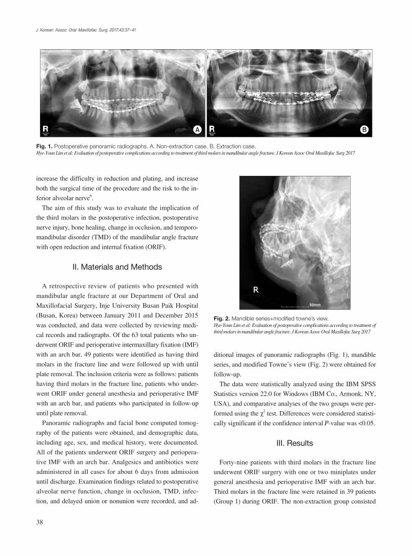
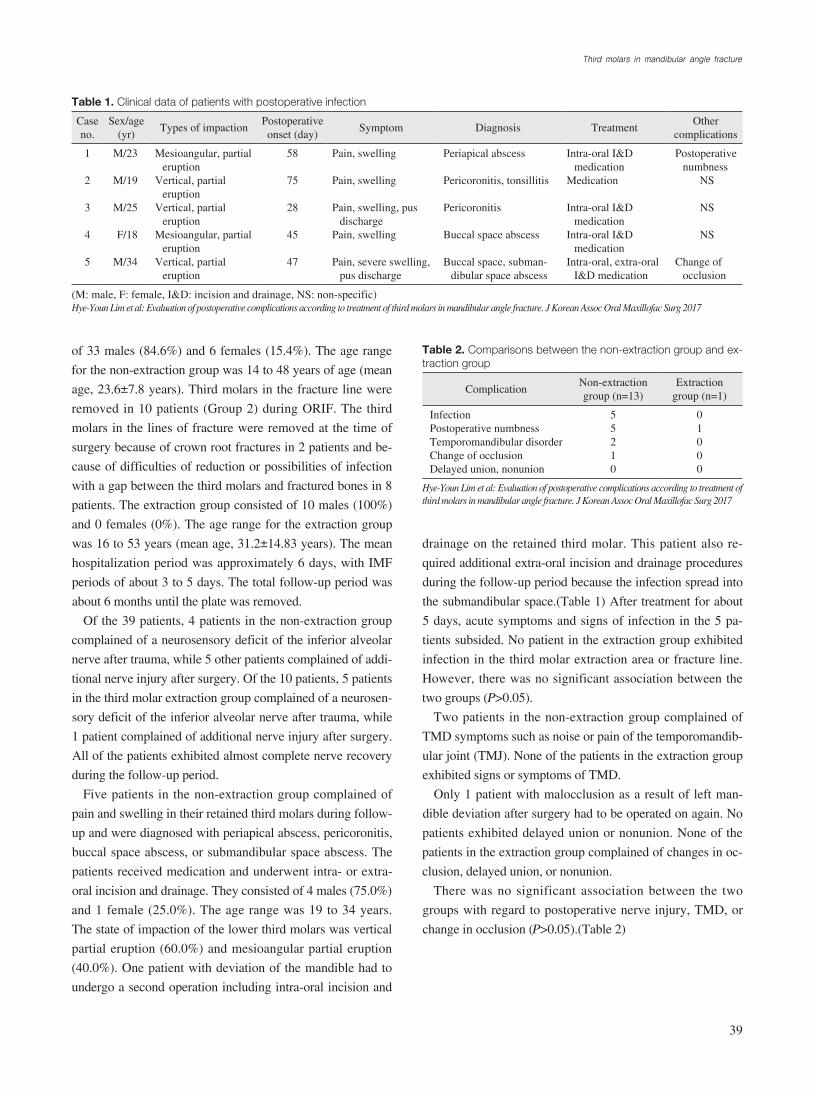
ditional images of panoramic radiographs (Fig. 1), mandible
series, and modified Towne’s view (Fig. 2) were obtained for
follow-up.
The data were statistically analyzed using the IBM SPSS
Statistics version 22.0 for Windows (IBM Co., Armonk, NY,
USA), and comparative analyses of the two groups were per-
formed using the χ2 test. Differences were considered statisti-
cally significant if the confidence interval P-value was <0.05.
III. Results
Forty-nine patients with third molars in the fracture line
underwent ORIF surgery with one or two miniplates under
general anesthesia and perioperative IMF with an arch bar.
Third molars in the fracture line were retained in 39 patients
(Group 1) during ORIF. The non-extraction group consisted
increase the difficulty in reduction and plating, and increase
both the surgical time of the procedure and the risk to the in-
ferior alveolar nerve6.
The aim of this study was to evaluate the implication of
the third molars in the postoperative infection, postoperative
nerve injury, bone healing, change in occlusion, and temporo-
mandibular disorder (TMD) of the mandibular angle fracture
with open reduction and internal fixation (ORIF).
II. Materials and Methods
A retrospective review of patients who presented with
mandibular angle fracture at our Department of Oral and
Maxillofacial Surgery, Inje University Busan Paik Hospital
(Busan, Korea) between January 2011 and December 2015
was conducted, and data were collected by reviewing medi-
cal records and radiographs. Of the 63 total patients who un-
derwent ORIF and perioperative intermaxillary fixation (IMF)
with an arch bar, 49 patients were identified as having third
molars in the fracture line and were followed up with until
plate removal. The inclusion criteria were as follows: patients
having third molars in the fracture line, patients who under-
went ORIF under general anesthesia and perioperative IMF
with an arch bar, and patients who participated in follow-up
until plate removal.
Panoramic radiographs and facial bone computed tomog-
raphy of the patients were obtained, and demographic data,
including age, sex, and medical history, were documented.
All of the patients underwent ORIF surgery and periopera-
tive IMF with an arch bar. Analgesics and antibiotics were
administered in all cases for about 6 days from admission
until discharge. Examination findings related to postoperative
alveolar nerve function, change in occlusion, TMD, infec-
tion, and delayed union or nonunion were recorded, and ad-
A B
Fig. 1. Postoperative panoramic radiographs. A. Non-extraction case. B. Extraction case.Hye-Youn Lim et al: Evaluation of postoperative complications according to treatment of third molars in mandibular angle fracture. J Korean Assoc Oral Maxillofac Surg 2017
Fig. 2. Mandible series+modified towne’s view.Hye-Youn Lim et al: Evaluation of postoperative complications according to treatment of third molars in mandibular angle fracture. J Korean Assoc Oral Maxillofac Surg 2017
Third molars in mandibular angle fracture
39
drainage on the retained third molar. This patient also re-
quired additional extra-oral incision and drainage procedures
during the follow-up period because the infection spread into
the submandibular space.(Table 1) After treatment for about
5 days, acute symptoms and signs of infection in the 5 pa-
tients subsided. No patient in the extraction group exhibited
infection in the third molar extraction area or fracture line.
However, there was no significant association between the
two groups (P>0.05).
Two patients in the non-extraction group complained of
TMD symptoms such as noise or pain of the temporomandib-
ular joint (TMJ). None of the patients in the extraction group
exhibited signs or symptoms of TMD.
Only 1 patient with malocclusion as a result of left man-
dible deviation after surgery had to be operated on again. No
patients exhibited delayed union or nonunion. None of the
patients in the extraction group complained of changes in oc-
clusion, delayed union, or nonunion.
There was no significant association between the two
groups with regard to postoperative nerve injury, TMD, or
change in occlusion (P>0.05).(Table 2)
of 33 males (84.6%) and 6 females (15.4%). The age range
for the non-extraction group was 14 to 48 years of age (mean
age, 23.6±7.8 years). Third molars in the fracture line were
removed in 10 patients (Group 2) during ORIF. The third
molars in the lines of fracture were removed at the time of
surgery because of crown root fractures in 2 patients and be-
cause of difficulties of reduction or possibilities of infection
with a gap between the third molars and fractured bones in 8
patients. The extraction group consisted of 10 males (100%)
and 0 females (0%). The age range for the extraction group
was 16 to 53 years (mean age, 31.2±14.83 years). The mean
hospitalization period was approximately 6 days, with IMF
periods of about 3 to 5 days. The total follow-up period was
about 6 months until the plate was removed.
Of the 39 patients, 4 patients in the non-extraction group
complained of a neurosensory deficit of the inferior alveolar
nerve after trauma, while 5 other patients complained of addi-
tional nerve injury after surgery. Of the 10 patients, 5 patients
in the third molar extraction group complained of a neurosen-
sory deficit of the inferior alveolar nerve after trauma, while
1 patient complained of additional nerve injury after surgery.
All of the patients exhibited almost complete nerve recovery
during the follow-up period.
Five patients in the non-extraction group complained of
pain and swelling in their retained third molars during follow-
up and were diagnosed with periapical abscess, pericoronitis,
buccal space abscess, or submandibular space abscess. The
patients received medication and underwent intra- or extra-
oral incision and drainage. They consisted of 4 males (75.0%)
and 1 female (25.0%). The age range was 19 to 34 years.
The state of impaction of the lower third molars was vertical
partial eruption (60.0%) and mesioangular partial eruption
(40.0%). One patient with deviation of the mandible had to
undergo a second operation including intra-oral incision and
Table 1. Clinical data of patients with postoperative infection
Case no.
Sex/age (yr)
Types of impactionPostoperative onset (day)
Symptom Diagnosis TreatmentOther
complications
1 2 3 4 5
M/23
M/19
M/25 F/18
M/34
Mesioangular, partial eruption
Vertical, partial eruption
Vertical, partial eruption
Mesioangular, partial eruption
Vertical, partial eruption
58 75 28 45 47
Pain, swelling Pain, swelling Pain, swelling, pus
dischargePain, swelling Pain, severe swelling,
pus discharge
Periapical abscess Pericoronitis, tonsillitis Pericoronitis Buccal space abscess Buccal space, subman-
dibular space abscess
Intra-oral I&D medication
Medication Intra-oral I&D
medicationIntra-oral I&D
medicationIntra-oral, extra-oral
I&D medication
Postoperative numbness
NS
NS
NS
Change of occlusion
(M: male, F: female, I&D: incision and drainage, NS: non-specific)Hye-Youn Lim et al: Evaluation of postoperative complications according to treatment of third molars in mandibular angle fracture. J Korean Assoc Oral Maxillofac Surg 2017
Table 2. Comparisons between the non-extraction group and ex-traction group
ComplicationNon-extraction group (n=13)
Extraction group (n=1)
InfectionPostoperative numbnessTemporomandibular disorderChange of occlusionDelayed union, nonunion
55210
01000
Hye-Youn Lim et al: Evaluation of postoperative complications according to treatment of third molars in mandibular angle fracture. J Korean Assoc Oral Maxillofac Surg 2017
J Korean Assoc Oral Maxillofac Surg 2017;43:37-41
40
risk to the inferior alveolar nerve. In their study, the incidence
of nerve injury was 16% for the retention group compared
to 39% for the removal group. Therefore, they stated that the
removal of third molars creates an additional risk of nerve in-
jury and increases operating time4. In this study, 4 patients in
the non-extraction group and 1 patient in the extraction group
complained of postoperative nerve injury, and there was no
significant difference between the groups. All of the patients
exhibited gradual recovery of the nerve during the follow-up
period.
The issue of postoperative infection has long been debated
and represents a common complication of mandible angle
fracture3. A 1994 study by Ellis and Walker12 found that the
extraction of third molars in fracture lines seemed to increase
postoperative infection3,4,13. In 2002, they proposed that the
risk of postoperative infection increased if teeth were left in
the fracture line3,4,13. Patel et al.3 defined infection as purulent
discharge from the intraoral incision through a sinus tract to
the skin, or a closed area of swelling that required incision
and drainage of purulent material. They found no statistically
meaningful relationship between tooth management in the
line of the fracture and rate of postoperative infection3. They
stated that the differences in rates of infection might be at-
tributed to inherent differences in the socioeconomic status
of patients, tobacco and alcohol use and abuse, nutritional
status, and other medical co-morbidities3. However, in this
study, the patients were mostly young and free of significant
medical history such as uncontrolled diabetes, alcohol abuse,
or severe smoking. Also, antibiotics were administered upon
admission to the hospital until about 6 days from admission
until discharge. As a result, there seemed to be no immedi-
ate infection or other complications. During the follow-up
period, 5 patients in the non-extraction group received medi-
cation and underwent intra or extra-oral incision and drain-
age because of infection. All third molars were partially im-
pacted. After treatment for 5 days, acute symptoms and sign
of infections subsided. All infected third molars underwent
extraction during the plate removal surgical procedure. None
of the patients in the extraction group exhibited infection in
the third molar extraction area or fracture line. This may be
attributed to standard protocol at the time of surgery such as
gentle curettage, copious irrigation, interrupted sutures offer-
ing strength and flexibility of the extraction site, and antibiot-
ics. Complications differed between the two groups, but there
was no significant difference.
Mandibular trauma was stated as the most frequent rea-
son for TMD. Baltrusaityte et al.14 reported that mandibular
IV. Discussion
This study included 49 patients, 43 males (87.8%) and 6
females (12.2%), divided into a non-extraction group and an
extraction group. Of these patients, 17 patients (34.7%) had
a mandible angle fracture on the right side, while 32 patients
(65.3%) had a mandible angle fracture on the left side. An-
other study on mandibular angle fracture reported similar epi-
demiologic data. Patel et al.3 showed that the overwhelming
majority of patients were men, comprising 85 of 103 patients
(82.5%), whereas women accounted for 18 of 103 patients
(17.5%) with mandibular angle fracture. Most fractures oc-
curred at the left mandibular angle (34.9%), followed by the
right side (34.9%) and both sides (5.8%).
In this study, all patients underwent IMF for the manage-
ment of mandibular angle fracture. IMF is a reliable tech-
nique for reduction and stabilization before plate fixation,
although the use of interdental wiring may have adverse ef-
fects on the teeth or surrounding tissue and require additional
time7-10. Bhagol et al.11 explained that although superior and
inferior plates are typically required for adequate fixation, a
superior border plate placed at the point of maximal tension
is sufficient for mandibular angle fracture. In this study, pre-
operative IMF using an arch bar was applied and used for ap-
proximately 3 to 5 days after surgery, with 1 or 2 miniplates
placed at the superior area during ORIF surgery. Only 1
patient with malocclusion because of left mandible deviation
after surgery required an additional operation. There were
no cases of delayed union or nonunion in either group. It is
speculated that closed reduction with IMF may reduce micro-
mobility and improve stability.
Management of the third molars in fracture lines has been
controversial. Yadavalli et al.6 proposed that the presence of
third molars in fracture lines may be of great value in the re-
positioning of fractures because the presence of third molars
prevents further injury to the bone tissue. The extraction of
third molars makes it difficult to reduce the contact between
fracture segments when the fragments are highly mobile6. Be-
cause extraction of the tooth increases the risk of contamina-
tion through the empty alveolus6,12, Yadavalli et al.6 used the
following criteria for surgical removal of the third molars in
the line of fracture: pericoronal or periodontal infection, cross
caries, extensive periapical lesions, mobility, or exposure of
the apical half or more of the root fracture.
McNamara et al.5 noted that the risks involved with extrac-
tion of third molars includes loss of bone, greater difficulty in
reduction and plating, increased surgical time, and increased
Third molars in mandibular angle fracture
41
fracture occurred various sings and symptoms of TMD. Al-
terations of the masticatory system caused by dislocation of
body fragments and conservative treatment of mandibular
fractures showed that inaccurate immobilization of the man-
dible after trauma resulted in a “new bite.” Consequently,
conditions for the development of TMJ dysfunction occurred
as a result of adapted dynamical masticatory movements and
new occlusion14,15. Instances of deviation upon opening and
the presence of joint sounds were higher in cases treated with
miniplate fixation and were more susceptible to development
of traumatic arthritis16. In this study, 2 patients in the non-
extraction group exhibited symptoms of TMD. One of the
patients complained of TMJ noise, while the other patient
complained of intermittent TMJ pain after surgery. However,
there was no statistically significant difference between the
two patients, and both of them improved during the follow-
up period.
One limitation of this study is the small number of patients
in the extraction group. Therefore, further studies are neces-
sary.
V. Conclusion
There was no statistically significant difference in post-
operative complications between the extraction and non-
extraction groups of third molars in the fracture line. If the
third molar is partially impacted or completely nonfunctional,
likely to be involved in pathologic conditions later in life, or
possible to remove it with the plate simultaneously, extrac-
tion of the third molar in the fracture line should be consid-
ered during ORIF surgery of the mandible angle fracture.
Conflict of Interest
No potential conflict of interest relevant to this article was
reported.
ORCID
Hye-Youn Lim, http://orcid.org/0000-0001-5846-3794Tae-Young Jung, http://orcid.org/0000-0001-7131-358XSang-Jun Park, http://orcid.org/0000-0002-6444-7560
References
1. Hanson BP, Cummings P, Rivara FP, John MT. The association of third molars with mandibular angle fractures: a meta-analysis. J Can Dent Assoc 2004;70:39-43.
2. Al-Moraissi EA, Ellis E 3rd. What method for management of uni-lateral mandibular angle fractures has the lowest rate of postopera-tive complications? A systematic review and meta-analysis. J Oral Maxillofac Surg 2014;72:2197-211.
3. Patel N, Kim B, Zaid W. A detailed analysis of mandibular angle fractures: epidemiology, patterns, treatments, and outcomes. J Oral Maxillofac Surg 2016;74:1792-9.
4. Rajandram RK, Nabil S, Shareif MS, Ishak I, Marhazlinda J, Nor-din R, et al. Mandibular third molar and angle of mandible frac-tures: an unsolved clinical dilemma. Sains Malaysiana 2013;42:39-43.
5. McNamara Z, Findlay G, O'Rourke P, Batstone M. Removal ver-sus retention of asymptomatic third molars in mandibular angle fractures: a randomized controlled trial. Int J Oral Maxillofac Surg 2016;45:571-4.
6. Yadavalli G, Mythily PH, Shetty JN. Clinical evaluation of man-dibular angle fractures with teeth in fracture line, treated with stable internal fixation. Indian J Stomatol 2011;2:216-21.
7. Bell RB, Wilson DM. Is the use of arch bars or interdental wire fixation necessary for successful outcomes in the open reduction and internal fixation of mandibular angle fractures? J Oral Maxil-lofac Surg 2008;66:2116-22.
8. Fordyce AM, Lalani Z, Songra AK, Hildreth AJ, Carton AT, Hawkesford JE. Intermaxillary fixation is not usually neces-sary to reduce mandibular fractures. Br J Oral Maxillofac Surg 1999;37:52-7.
9. Dimitroulis G. Management of fractured mandibles without the use of intermaxillary wire fixation. J Oral Maxillofac Surg 2002;60:1435-8.
10. Kim MY, Kim CH, Han SJ, Lee JH. A comparison of three treat-ment methods for fractures of the mandibular angle. Int J Oral Maxillofac Surg 2016;45:878-83.
11. Bhagol A, Shigh V, Singhal R. Management of mandibular frac-tures. In: Motamedi MHK, ed. A textbook of advanced oral and maxillofacial surgery. 1st ed. Rijeka, Croatia: InTech; 2013:385-414.
12. Ellis E, Walker L. Treatment of mandibular angle fractures us-ing two noncompression miniplates. J Oral Maxillofac Surg 1994;52:1032-6.
13. Ellis E 3rd. Outcomes of patients with teeth in the line of mandibu-lar angle fractures treated with stable internal fixation. J Oral Max-illofac Surg 2002;60:863-5.
14. Baltrusaityte A, Surna A, Pileicikiene G, Kubilius R, Gleiznys A, Baltrusaitis M. Dynamical changes of occlusion and articulation during treatment of mandibular angle fractures. Stomatologija 2013;15:12-9.
15. Pullinger AG, Seligman DA. Trauma history in diagnostic groups of temporomandibular disorders. Oral Surg Oral Med Oral Pathol 1991;71:529-34.
16. Chaurasia NK, Guan J, Wang X, Sah G. Clinical analysis of changes in function of the temporomandibular joint after open re-duction and internal fixation of mandible fracture. Indian J Oral Sci 2015;6:60-4.


![Segmentation of Intracranial Arterial Calci cation with ... · 2 arteriosclerosis [1]. ICAC lesions are identi ed in non-contrast computed tomog-raphy (CT) images as groups of voxels](https://img.pdfslide.us/doc/110x75/5d0be54b88c993956c8b665b/segmentation-of-intracranial-arterial-calci-cation-with-2-arteriosclerosis.jpg)