Embed Size (px)
Citation preview

Pelvic imaging
Dr Abubakr
15.5.2008

Diagnostic imaging techniques are:
Radiographs: Plain radiographs Hysterosalpingography Arteriography
Ultrasonography Computed Tomography (CT) Magnetic Resonance Imaging
(MRI)
They are used to:
1. Diagnose pelvic disease and fracture.
2. Assess congenital anomalies in pelvis and pelvic organs.
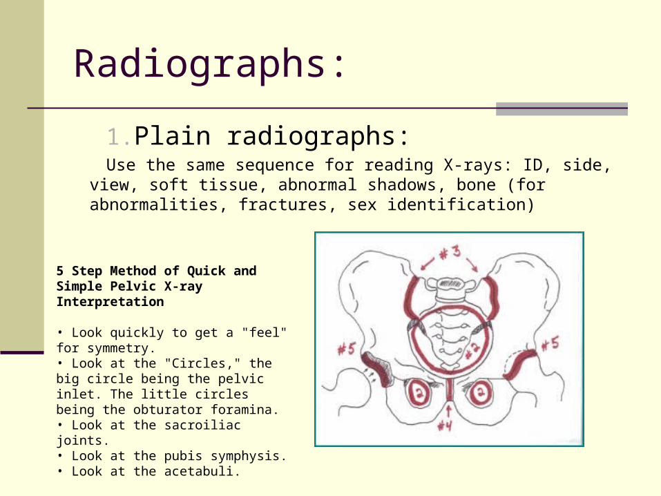
Radiographs:
1.Plain radiographs:Use the same sequence for reading X-rays: ID, side, view, soft tissue,
abnormal shadows, bone (for abnormalities, fractures, sex identification)
5 Step Method of Quick and Simple Pelvic X-ray Interpretation
• Look quickly to get a "feel" for symmetry. • Look at the "Circles," the big circle being the pelvic inlet. The little circles being the obturator foramina. • Look at the sacroiliac joints. • Look at the pubis symphysis. • Look at the acetabuli.
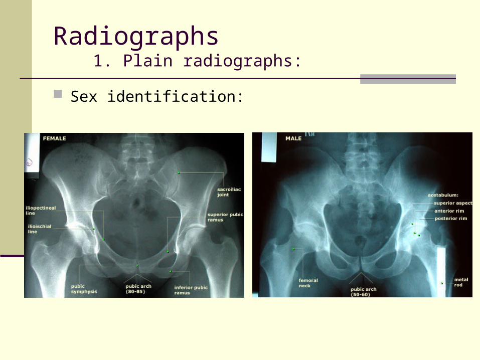
Radiographs1. Plain radiographs:
Sex identification:

Radiographs2. Hysterosalpingography :
Radiopaque dye is injected to the uterine cavity and tubes, is used to demonstrate anomy and patency of these organs, and for detecting any abnormality.
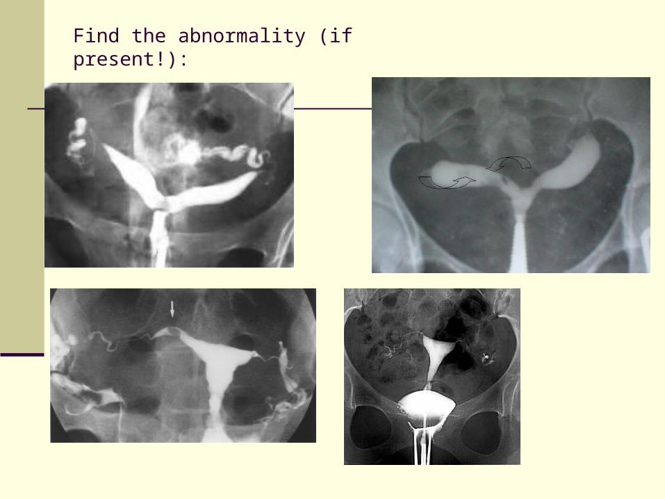
Find the abnormality (if present!):

?
vasogram
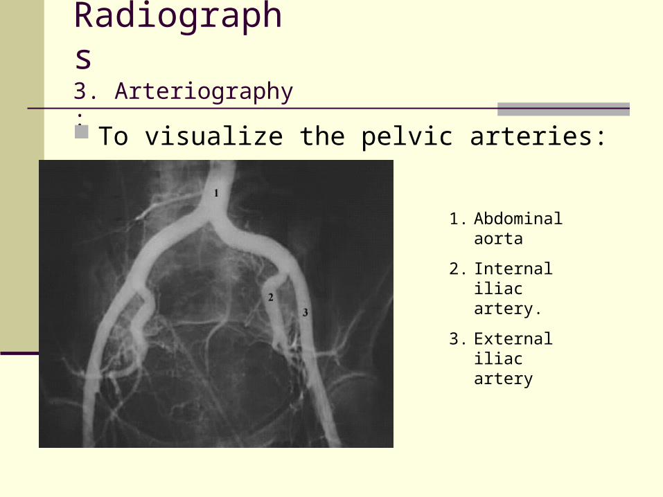
Radiographs3. Arteriography :
To visualize the pelvic arteries:
1. Abdominal aorta
2. Internal iliac artery.
3. External iliac artery

Ultrasonography Sound waves are used Solid & cystic tissues Safe for obstetrical scanning Transabdominal sonography, full
bladder Transrectal & tranvagianl and
more clear visualization of pelvic organs


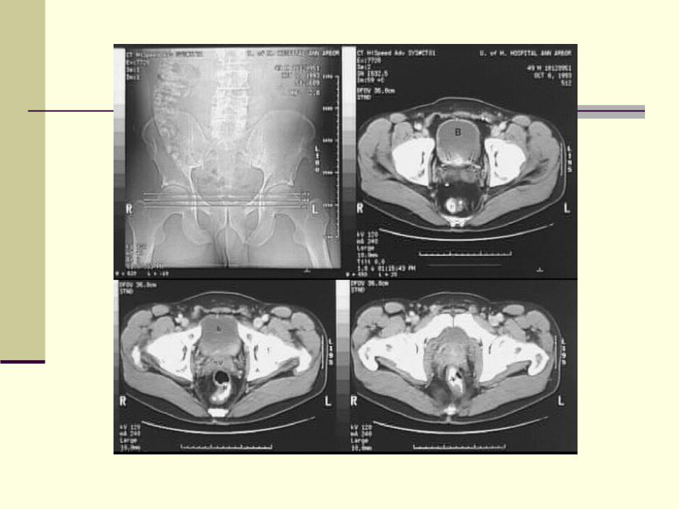
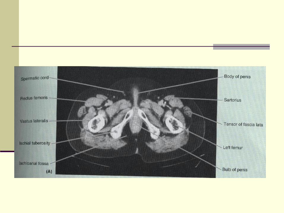
Computed Tomography (CT)


MRI:


Thanks