Embed Size (px)
Citation preview

EVALUATION OF MICROLEAKAGE OF THREE
DIFFERENT SINGLE-CONE OBTURATION
SYSTEMS BY QUANTITATIVE GLUCOSE
LEAKAGE MODEL – AN IN VITRO STUDY
Dissertation submitted to
THE TAMILNADU Dr. M.G.R. MEDICAL UNIVERSITY
In partial fulfillment for the Degree of
MASTER OF DENTAL SURGERY
BRANCH IV
CONSERVATIVE DENTISTRY AND ENDODONTICS
APRIL 2016


ACKNOWLEDGEMENT
I take this opportunity to sincerely thank my post graduate teacher
and my guide Dr.M. Rajasekaran M.D.S., Professor, Department of
Conservative Dentistry and Endodontics, Ragas Dental College and
Hospital, for his perseverance in motivating and supporting me throughout
my study period.
My sincere thanks to Dr. R. Indira, M.D.S., Professor and HOD,
Department of Conservative Dentistry and Endodontics, Ragas Dental
College and Hospital, who have helped me with her guidance, support and
constant encouragement throughout my study period wherever and
whenever needed.
My sincere thanks to Dr. S.Ramachandran, M.D.S., Professor &
Principal, Department of Conservative Dentistry and Endodontics, Ragas
Dental College and Hospital, who have helped me with his advice and
immense support throughout my post graduate curriculum.
I extend my sincere thanks to Dr.P.Shankar, M.D.S., Professor,
Ragas Dental College and Hospital, for his guidance, and constant
encouragement during the completion of my study.

My sincere thanks to Dr. R.Anil Kumar, M.D.S., Professor, Ragas
Dental College and Hospital, for his encouragement, support and guidance
all throughout my study period.
I extend my sincere thanks to Dr. C.S. Karumaran, M.D.S.,
Professor, for his constant encouragement throughout the completion of
this work.
I would like to solemnly thank Dr. Veni Ashok, M.D.S.,
Dr. Shankar Narayan, M.D.S., Dr.S.M. Venkatesan, M.D.S., Readers, for
all the help during my study period.
I would also like to thank Dr.Aravind, M.D.S., Dr. Sabari, M.D.S.,
Dr. B.Venkatesh, M.D.S., Senior lecturers for their friendly guidance and
support.
I also wish to thank the management of Ragas Dental College and
Hospital, Chennai for their help and support.
My sincere thanks Ganeshan, Omega labs, guindy for their help
and guidance in laboratory testing.
My sincere thanks to Mr. Ravanan., for his guidance in
biostatistics.
I remain ever grateful to all my batchmates, juniors and friends for
their support.
I would like to especially thank my parents, my sister and my wife
for their love, understanding, support and encouragement throughout these
years without which, I would not have never reached so far.
My sincere thanks to Mr.K.Thavamani and Miss.R.Sudha for their
guidance and support in DTP and Binding works.
Above all, I am thankful to God, who always guides me and has
given these wonderful people in my life.

CONTENTS
S. NO. INDEX PAGE. NO
1. INTRODUCTION 1
2. AIM AND OBJECTIVES 6
3. REVIEW OF LITERATURE 7
4. MATERIALS AND METHODS 28
5. RESULTS 35
6. DISCUSSION 37
7. SUMMARY 52
8. CONCLUSION 53
9. BIBLIOGRAPHY 54
10. ANNEXURE _

LIST OF TABLES
S.NO. TITLE
Table 1 MICROLEAKAGE IN GROUP I (GUTTA-PERCHA/AH PLUS)
Table 2 MICROLEAKAGE IN GROUP II (C-POINTS/BIO CERAMIC
SEALER)
Table 3 MICROLEAKAGE IN GROUP III (RESILON/EPIPHANY)
Table 4
KRUSKAL-WALLIS GLUCOSE LEAKAGE AT VARIOUS
INTERVALS OF TIME
Table 5 MANN-WHITNEY GLUCOSE LEAKAGE AT VARIOUS TIME
INTERVALS BETWEEN THE GROUPS

LIST OF GRAPHS
S.NO. TITLE
Graph I
GLUCOSE LEAKAGE AT VARIOUS TIME INTERVALS FOR
ALL THE THREE GROUPS
Graph II
GLUCOSE LEAKAGE AT VARIOUS TIME INTERVALS
BETWEEN GROUP I AND GROUP II
Graph III
GLUCOSE LEAKAGE AT VARIOUS TIME INTERVALS
BETWEEN GROUP II AND GROUP III
Graph IV
GLUCOSE LEAKAGE AT VARIOUS TIME INTERVALS
BETWEEN GROUP I AND GROUP III

LIST OF FIGURES
S.NO. TITLE
FIGURE 1 TOOTH SAMPLES
FIGURE 2 DECORONATION
FIGURE 3 RADIOGRAPHIC PICTURE OF THE TOOTH SAMPLES
FIGURE 4 INITIAL CLEANING AND SHAPING
FIGURE 5 IRRIGATION OF THE CANAL
FIGURE 6 ENDOACTIVATOR AND ENDOMOTOR FOR CLEANING AND
SHAPING
FIGURE 7 EXPERIMENTAL OBTURATING SYSTEMS
FIGURE 8 GUTTA PRECHA WITH AH PLUS SEALER
FIGURE 9 C-POINTS WITH BIOCREAMIC SEALER
FIGURE10 RESILON WITH EPIPHANY SEALER
FIGURE11 SAMPLES STORED AT 37℃ IN INCUBATOR
FIGURE12 AIR TIGHT GLASS BEAKER WITH RUBBER STOPPER
FIGURE13 GLASS TUBE CONNECTED TO TOOTH SAMPLE
FIGURE14 APPARATUS TO TEST MICROLEAKAGE
FIGURE15 1 MOL/L GLUCOSE SOLUTION
FIGURE16 0.2% SODIUM AZIDE
FIGURE17 GLUCOSE KIT

FIGURE18 SPECTROPHOTOMETER
FIGURE19 SPECTROPHOTOMETER

Introduction

Introduction
1
INTRODUCTION
Success of endodontic treatment largely depends on the three
dimensional obturation of the root canal system. The main objective of the
obturation is to achieve a hermatic seal, to be more precise it should have a
fluid impervious or bacterial tight seal.(18,10)
Adequate obturation of the root canal system following intracanal
preparation is a major objective of endodontic treatment. Different endodontic
filling materials and techniques have been introduced to the dental community
in an attempt to improve the apical seal. It is, therefore important to assess the
sealing quality of obturation materials.(43)
Ideally an obturating material should have good sealing ability to
prevent micro-leakage between the root canal filling and the canal walls. Thus
preventing bacterial invasion, that will adversely affect the outcome of root
canal treatment.(3,18)
When filling the root canal system, the sealer plays an important role
in reducing microleakage. To achieve this property it is important that the root
canal filling material should adhere to the root canal dentin. Good adhesion
eliminates any space that would allow any penetration and inhibition of
bacteria between the sealer and the wall thus, preventing failure of the
obturation.(43)

Introduction
2
According to Sousa-Neto et al. (2005), Adhesion of an endodontic
sealer is defined as its capacity to adhere to the root canal walls and promote
the union of the Gutta-percha cones to each other and to the dentine.
The most commonly used obturating technique is cold lateral
condensation because of its advantages of controlled placement of gutta-
percha in the root canal and low cost. The disadvantage of this technique was
its poor adaptation and inability to achieve a homogenous mass. This
technique is not suitable for curved canals.(40,42)
Various researchers attempted to overcome the drawback of this
technique, thus newer obturation techniques have been introduced. One such
obturation is the thermoplastic obturation technique which was introduced by
Schilder in 1967. This technique has shown better adaptation to the root canal
walls as compared to lateral condensation and at the same time it could
successfully obturate the lateral canals. However this new technique still could
not fulfill the three basic requisites of obturation.(40,42)
Advances in adhesive technology have reinforced the search for newer
means to minimize apical and coronal marginal leakage by improving sealer
adhesion to root canal walls. Therefore the mono-block concept has emerged,
where the core material, sealer and dentinal tubules becomes a single solid
structure. A thermoplastic synthetic polymer based root canal filling material
was introduced. The resin core filling material, Resilon (Resilon Research
Introduction
3
LLC, Madison, CT), handles like gutta-percha. Obturation with Resilon cones
is accomplished by use of Epiphany primer (Pentron Clinical Technologies,
LLC, Wallingford, CT) and Epiphany resin-based sealer (Pentron Clinical
Technologies).(3)
The thermoplasticity of Resilon is because of polycaprolactone, a
biodegradable polyester with a relatively low melting point, while its ability to
bond is derived from the inclusion of resin with methacryloxy groups. This
filling material also contains glass fillers and barium chloride as fillers, and is
capable of coupling to resin sealers, an example of which is Epiphany
(Pentron Clinical Technologies, Wallingford CT). Epiphany Root Canal
Sealant is a dual-curable resin composite containing a new redox catalyst, that
enables optimal auto-polymerization under acidic environments. (36)
With further progress in dentistry yet another new material found its
way into Endodontics namely the Smart Seal System. The system consists of
obturation points (C-points) containing a polyamide core with an outer bonded
hydrophilic polymer coating and an accompanying bio-ceramic sealer.
The endodontic points are designed to expand laterally without
expanding axially by absorbing residual water from the instrumented root
canal space and the naturally present moisture in the dentinal tubules. The
inner core of C-points is a mix of two nylon polymers, Trogamid T and
Trogamid CX. The polymer coating is a cross-linked copolymer of
Introduction
4
acrylonitrile and vinylpyrrolidone which has been polymerised and cross-
linked using allyl methacrylate and a thermal initiator.(8)
A great deal of attention has been given to the evaluation of sealing
ability of root canal filling materials and associated obturation techniques.
Various laboratory based experimental models are used to detect and measure
leakage along root fillings. Dye leakage, fluid transport and bacterial
penetration are currently the methods commonly used.
However, there was no standardization of methods, such as
measurement of time, the applied pressure, the diameter of the tube containing
bubble and the length of the bubble which might influence the results.(43)
Recently, Xu et al (2005) discussed a new model that measures the
leakage of glucose molecules and checks penetration of different tracers
through the root canal, assuming it travels along the canal and reaches the
apical region. Glucose has a low molecular size (MW=180 Da), and may be
used as an indication for toxins that might penetrate the canal. Shemesh et al.
(2006) described this model as a further development of the fluid
transportation concept that might be more sensitive than the measurement with
an air bubble.(22,43)
Therefore the aim of the present study was to evaluate microleakage
along root canal fillings using the said glucose leakage model by comparing
three single‐cone filling systems at different time intervals.

Aims and Objectives
Aims and Objectives
5
AIM AND OBJECTIVES
AIM: The aim of the present study was to compare the microleakage and the
sealing ability of three single cone obturating systems using a glucose leakage
model.
OBJECTIVES:
1. To evaluate the microleakage of the three obturating systems at
different time intervals.
2. To check the quantity of the glucose concentration leaked in each
group.
3. To compare the sealing ability of commonly used Gutta-percha/AH
Plus versus the recently introduced C-points/Bio ceramic sealer and
Resilon/ Epiphany systems.

Review of Literature

Review of Literature
6
REVIEW OF LITERATURE
Wu et al (1993)41
in their review on leakage studies, compared some
data on linear measurement of dye penetration following the cold lateral
condensation of gutta-percha. They evaluated various techniques and the cold
lateral condensation technique has been used as a standard control for
comparison. They concluded that more research should be done on leakage
study methodology, instead of continuing to evaluate the sealing ability of
different materials and techniques by methods that may give little relevant
information.
von Fraunhofer et al (2000)10
in their study evaluated the effects of
smear layer and canal instrumentation on leakage in root-filled teeth in an in-
vitro study on six groups of freshly extracted human canines and premolars
concluded that smear layer removal is beneficial to root canal sealing and
obturation with thermo-plasticized gutta-percha provides a superior seal whilst
canal instrumentation with engine-driven Ni-Ti files reduces the extent of
micro-leakage in root canals.
Kont Cobankara et al (2002)18
in their in-vitro study evaluated, the
micro-leakage of root fillings involving four root-canal sealers including AH
Plus , RoekoSeal , Ketac-Endo and Sultan using fluid filtration study on forty
extracted human maxillary anterior teeth. Preparation and obturation of the
teeth was done and a fluid filtration method was used for quantitative

Review of Literature
7
evaluation of apical leakage. They concluded that root fillings with RoekoSeal
in combination with cold lateral condensation technique showed better sealing
than those with Ketac-Endo, AH Plus and Sultan sealers after 21 days. The
fluid filtration test gave quantitative results and allowed nondestructive long
term evaluation of specimens.
Pommel et al (2003)24
did a study to evaluate the sealing properties of
four root canal sealers- Sealapex, Pulp Canal Sealer, AH 26, and Ketac on
forty eight maxillary central incisors. They measured the apical leakage using
fluid filtration method and concluded that the teeth filled with Sealapex
displayed a higher apical leakage than those filled with AH 26, Pulp Canal
Sealer or Ketac-Endo.
Tagger et al (2003)37
conducted a study to verify whether an
interaction existed between some sealers and different brands of gutta-percha
cones. Three brands of cones (Kerr,UDM,Beldent) were tested with three
types of endodontic sealers (AH 26, Apexit, Roth’s). They concluded that an
interaction between sealer and cones is present in some combinations.
According to the author it was not possible to ascribe a trend of greater effect
to a certain sealer, but Roth’s 811 had the least effect on flow.
Weis et al (2004)40
compared the average sealer cement film thickness
and the extent and pattern of sealer penetration into dentinal tubules in
association with four obturation techniques in curved root canals. Mesial
canals of 44 teeth were randomly divided among SimpliFill, continous wave,

Review of Literature
8
Thermafill and 0.04 matched taper lateral compaction obturation groups. They
concluded that sealer thickness was strongly dependent on obturation
technique. Assuming that minimal sealer thickness and fewer voids are good
measures of long-term sealing ability, Thermafil resulted in the best outcome.
Consistent, extensive sealer penetration into dentinal tubules was seen and was
unrelated to the obturation technique.
Tay et al (2005)36
compared the ultra-structural quality of the apical
seal achieved with Resilon/Epiphany an Gutta-percha/AH Plus. They
examined for gaps along canal walls using SEM, and for apical leakage using
transmission electron microscopy (TEM). SEM revealed both gap-free
regions, and gap-containing regions in canals filled with both materials. TEM
revealed the presence of silver deposits along the sealer-hybrid layer interface
in Resilon/Epiphany, and between the sealer and gutta-percha in the controls.
It was concluded that a complete hermetic apical seal cannot be achieved with
either root filling materials.
Xu et al (2005)43
did a study to introduce a new method for quantative
testing of endodontic leakage. Eighty straight maxillary anterior teeth were
divided into 3 groups. The conclusions drawn were that the quantitative
method is sensitive, nondestructive, and clinically relevant. Pulp Canal Sealer
EWT showed more leakage than Sealapex and AH Plus in most observation
time.

Review of Literature
9
Stratton et al (2006)34
compared the sealing ability of gutta-percha and
AH Plus sealer versus Resilon and Epiphany Resin Root Canal sealer using
three different final irrigants (5.25% NaOCl, 0.012% chlorhexidine (CHX), or
2% CHX) with the fluid filtration model using 140 teeth. The two-way
ANOVA analysis indicated significantly less leakage using Resilon with
Epiphany sealer compared to gutta-percha and AH Plus sealer. There was no
statistical significance between any of the irrigants used for either obturation
group.
Shemesh et al (2006)35
conducted a two month longitudinal study to
compare the leakage along apical root fillings with and without smear layer
using two different leakage models. 120 single rooted teeth were used in this
study. Under the conditions of this study, the glucose penetration model was
more sensitive in detecting leakage along root fillings. Removing the smear
layer before filling did not improve the sealing of the apical 4 mm of filling.
Resilon allowed more glucose penetration but the same amount of fluid
transport as the gutta-percha root fillings.
Veríssimo et al (2007)38
compared the level of apical leakage between
canals filled with gutta-percha/ AH-Plus (GP) and the Resilon/Epiphany
System (RES), when submitted to two filling techniques [lateral condensation
and Hybrid technique (HT)]. 70 extracted teeth were instrumented and
randomly divided into four groups in accordance with the materials and
techniques used. After 7 days in an oven the teeth were immersed in India ink

Review of Literature
10
and cleared. Leakage was measured by the NIH image program. The
comclusion was that there was no difference between the filling techniques,
but there was a statistically significant difference when RES was compared
with GP, which leaked more than RES. With RES, leakage was confined to
the apical third and HT could be used to thermoplasticize RES with
satisfactory results.
Wedding et al (2007)39
in their investigation compared micro-leakage
of teeth obturated with gutta-percha and teeth obturated with Resilon by using
a fluid filtration model.46 human single rooted mandibular premolars were
used. The results showed that Resilon is a suitable replacement for gutta-
percha as a root canal filling material on the basis of its increased resistance to
fluid micro-leakage.
Paque et al (2007)22
compared the long term apical sealing ability of
Resilon/Epiphany versus gutta-percha/AH Plus. The root canals of 90 single
rooted human mandibular premolars were prepared with ProFile 0.4 taper
instruments to apical size 40. The teeth were randomly divided into four
groups containing 20 teeth each.1o teeth were positive controls. The root
canals were filled with respective aterials and allowed to set for days at
3 c and 1 hu idity. Root canal aterials were re oved and fluid
movement was then measured using a fluid transportation model and re-
evaluated after 16-months of water storage. The results suggest that initially,
Resilon/Epiphany root fillings prevented fluid movement to the same degree

Review of Literature
11
as gutta-percha/ AH Plus counterparts, but showed more fluid movement
when tested at 16 months.
Patel et al (2007)23
compared penetration depth into dentinal tubules of
RealSeal with that of a well-established endodontic sealer (Tubliseal) by
means of confocal microscopy in 20 extracted teeth. Confocal microscopy was
used to assess the penetration depths of the sealers at three sites for each
specimen. The results are suggestive that the penetration depth of RealSeal
into the root dentinal tubules is significantly greater than that of Tubliseal.
Xu et al (2007)42
evaluated the sealing ability of 4 different obturation
techniques by using a glucose leakage test 0n 80 extracted single rooted
maxillary incisors. The teeth were de-coronated and the canals were prepared.
Then the teeth were randomly divided into4 groups and filled with cold lateral
compaction, warm vertical compaction, Thermafil, or the E & Q Plus. A
glucose leakage model was used for quantitative evaluation of the coronal-to-
apical micro-leakage at 24 hours,1,2,3,5,8 and 12 weeks. The authors
concluded that the warm vertical compaction, Thermafil, and the E & Q Plus
system showed a better sealing result than cold lateral compaction of gutta-
percha at extended observation periods.
Shemesh et al (2007)33
measured glucose penetration and fluid
transport through coronal root structure and compared it with leakage along
the coronal region of root fillings in 50 teeth and concluded that no leakage
Review of Literature
12
was observed through root structure. Filled canals were associated with
penetration of glucose regardless of the material used.
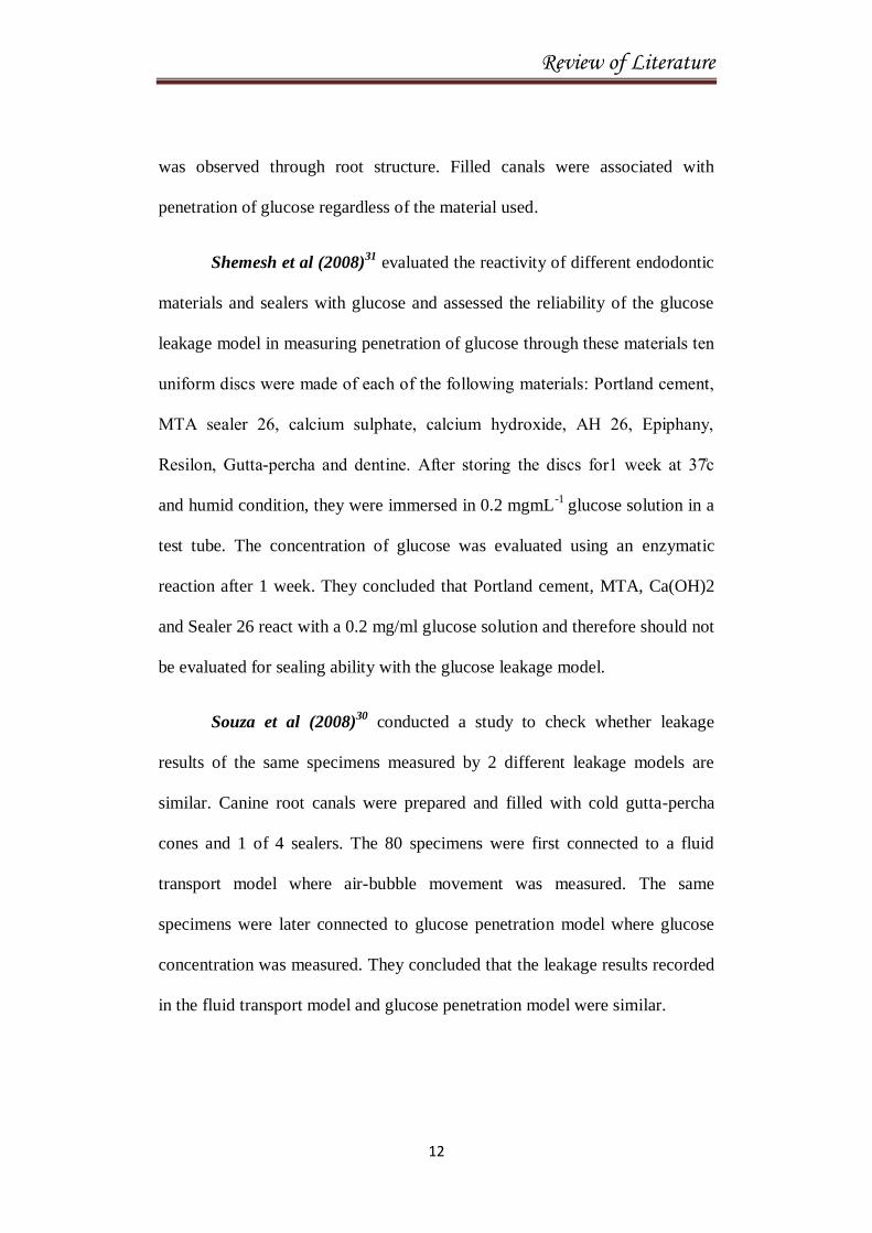
Shemesh et al (2008)31
evaluated the reactivity of different endodontic
materials and sealers with glucose and assessed the reliability of the glucose
leakage model in measuring penetration of glucose through these aterials ten
unifor discs were ade of each of the following aterials ortland ce ent,
TA sealer 26, calciu sulphate, calciu hydroxide, AH 26, piphany,
Resilon, utta-percha and dentine. After storing the discs for1 wee at 3 c
and humid condition, they were immersed in 0.2 mgmL-1
glucose solution in a
test tube. The concentration of glucose was evaluated using an enzymatic
reaction after 1 week. They concluded that Portland cement, MTA, Ca(OH)2
and Sealer 26 react with a 0.2 mg/ml glucose solution and therefore should not
be evaluated for sealing ability with the glucose leakage model.
Souza et al (2008)30
conducted a study to check whether leakage
results of the same specimens measured by 2 different leakage models are
similar. Canine root canals were prepared and filled with cold gutta-percha
cones and 1 of 4 sealers. The 80 specimens were first connected to a fluid
transport model where air-bubble movement was measured. The same
specimens were later connected to glucose penetration model where glucose
concentration was measured. They concluded that the leakage results recorded
in the fluid transport model and glucose penetration model were similar.

Review of Literature
13
Slutzky et al (2008)32
did a study to evaluate the antimicrobial effects
of root canal sealers - AH plus, Apexit Plus, Epiphany SE, and RoekoSeal
when in contact with Enterococcus faecalis. The direct contact test was used to
assess the anti microbial properties og the materials. The materials were
examined immediately after setting and 1,2, 7 and 14 days after aging in
phosphate buffered saline. The authors suggested that Apexit Plus had a
short-term antibacterial effect of 1 day on E. faecalis, whereas Epiphany SE
enhanced bacterial growth for at least 7 days. AH plus and RoekoSeal were
ineffective.
Alfredo et al (2008)3 evaluated the bond strength of AH Plus and
Epiphany sealers to human root canal dentine irradiated with a 980 nm diode
laser at different power and frequency parameters, using the push-out in 60
canine roots the specimens were prepared with a tapered bur and irrigated with
sodium hypochlorite, ethylene-di-aminetetraacetic acid and distilled water and
divided into five groups- one control and four experimental groups were
submitted to 980 nm diode laser irradiation at different power (1.5 and 3.0 W)
and frequency (continous wave and 100 Hz) parameters. Half of specimens in
each group had their canals filled with AH Plus sealer and half with Epiphany.
The push-out test was performed. The specimens were split longitudinally and
examined under SEM to assess the failure modes after sealer displacement.
The conclusion include that the 980 nm diode laser irradiation of root canal

Review of Literature
14
dentine increased the bond strength of AH Plus sealer, but did not affect the
adhesion of Epiphany sealer.
Bouillaguet et al (2008)4 evaluated the long-term sealing ability of
four contemporary endodontic sealers [Pulp Canal Sealer (PCS), AH-Plus,
GuttaFlow and Epiphany] using a fluid filtration technique in palatal roots of
40 human maxillary molar teeth. The root canals were prepared using a
crown-down technique. 24 hours after filling the roots were connected to an
automatic flow recording device filled with double distilled water under
pressure to measure leakage flow rates were assessed at 6, 12 or 24 hr and
after 1- year of storage. They concluded that GuttaFlow and Epiphany allowed
less fluid movement along filled straight roots.
Resende et al (2009)26
assessed the physicochemical properties and the
surface morphology of AH Plus, Epiphany, and Epiphany SE root canal
sealers. Five samples of each material were employed for each test according
to ANSI/ADA specification 57. The results suggest that the setting time, flow
and radiopacity tests conformed to ANSI/ADA standardization. The
dimensional change in all groups and the solubility of Epiphany were greater
than values considered acceptable, with higher amounts of calcium ion release.
Epiphany SE revealed more organized, compacted, and homogeneous
polymers in a reduced resin matrix when compared with the other groups.
Shanahan et al (2011)29
provided a review on Root canal filling using
Resilon stating that within the limit of the in-vitro studies Resilon appears to

Review of Literature
15
perform adequately in comparison to gutta-percha, however, as a result of the
questionable merit of such studies, it cannot presently be considered an
evidence-based alternative to the current gold standard gutta-percha.
Nawal et al (2011)20
evaluated the antimicrobial efficacy and flow
properties for Epiphany, Guttaflow and AH-Plus sealer with the use of
Enterococcus faecalis ATCC 29212 as a test organism. They concluded that
antimicrobial activity of the sealers was greatest for Epiphany followed by
AH-Plus sealer and Guttaflow. Epiphany sealer had the maximum flow
followed by AH-Plus sealer and Guttaflow.
L. Kqiku et al (2011)17
evaluated the active versus passive dye
microleakage and apical sealing ability of laterally condensed gutta-
percha/AH Plus versus Resilon/Epiphany in their in vitro study. One hundred
and twenty teeth were instrumented and divided into experimental, positive
and negative control groups. In group 1, the teeth were obturated with gutta-
percha/AH Plus and in group 2 the teeth were obturated with
Resilon/Epiphany. The apical seal was evaluated with a passive and active dye
penetration test. Absorbance of the extracted dye was determined with a
spectrophotometer. They concluded that canals obturated with
Resilon/Epiphany showed less apical leakage than those obturated with gutta-
percha/AH Plus, regardless of the type of dye penetration test used.
Assmann et al (2012)2 evaluated the bond strength to root dentin of 2
mineral trioxide aggregate (MTA)–based sealers (Endo-CPM sealer and MTA

Review of Literature
16
Fillapex) and of 1 epoxy resin–based sealer (AH Plus sealer). Forty-five
extracted human teeth with single roots were prepared by using the step-back
technique. Irrigation with 2.5% NaOCl and a final rinse with 17%
Ethylene-di-aminetetraacetic acid and distilled water were performed.
Canals were filled by using Endo- CPM sealer, MTA Fillapex, or AH Plus
sealer by means of the gutta-percha lateral condensation technique. After
7 days, the roots were sectioned perpendicularly to its long axis, and the push-
out test was carried out. From the results it can be concluded that Endo-CPM
sealer presented advantages when a post preparation was required. MTA
Fillapex presented acceptable resistance to dislodgement, which was similar to
that observed in samples filled with AH Plus sealer.
Economides et al (2012)9
evaluated ex vivo, the push-out bond
strength of a new filling material (Smart seal) compared with gutta-
percha/AH-26. A total of 40 extracted single-rooted human teeth were used.
After instrumentation using the ProTaper rotary system, the root canals were
filled as follows: Group 1, Smartseal sealer and a 0.06 taper Smartpoint
calibrated to apical tip size 30; Group 2, Smartseal sealer and an F3
SmartpointPT; Group 3, AH-26 sealer and a single F3 ProTaper gutta-percha
cone and Group 4, AH-26 sealer and gutta-percha using the cold lateral
condensation technique. Two successive disk shaped slices were cut from each
root sample and the bond strength was measured using the push-out test. The
author concluded that there was no difference in adhesion to dentine between

Review of Literature
17
the Smart seal system and gutta-percha/AH-26 applied using either the single
cone or lateral condensation technique.
Reddy et al (2013)25
carried out a study to determine the sealing ability of
four root end filling materials- Intermediate Restorative Material (IRM), Mineral
Trioxide Aggregate, Geristore and Retroplast using a glucose leakage model. 100
extracted teeth were used for this study. The teeth were divided into 6 groups – 4
experimental groups of 20 teeth each and 2 control groups of 10 teeth each. In the
positive control, no root end filling was done and in the negative control, the teeth
were completely coated with nail varnish. All the teeth were instrumented, their
apices were resected. 3mm deep root end preparations were prepared with retro tips.
The root end cavities of the experimental groups were filled with the retrograde
filling aterials. The aterials were anipulated according to the anufacturers’
instructions. Each tooth was mounted in a glucose leakage device as described by Xu
and coworkers. The amount of glucose was determined by a UV-VIS recording
spectrophotometer at 500-nm wavelength. According to the results of their study,
MTA showed the least leakage at both 7th and 14th days and hence can be considered
as the material of choice for root end filling.
Lumbini et al (2013)19
provided an overview about Smart seal- New
Age obturation stating that Smartseal is a recently introduced root canal
obturating system based on polymer technology. Its principle is based on the
hydrophilic nature of the obturating points which can absorb surrounding
moisture and expand resulting in filling of voids and spaces. According to the

Review of Literature
18
author since, its introduction, Smartseal has been widely reported to be
successfully used in endodontic therapy.
Didato et al (2013)6 evaluated the time-based lateral hygroscopic
expansion of a water-expandable endodontic obturation point. They compared
the time-based lateral expansion of two sizes and two batches of water-
expandable obturation points (CPoint, EndoTechnologies, LLC) and a similar-
sized gutta-percha point (control) at various distances from the point apex: 5,
10, and 15 mm. They concluded that when exposed to water, the lateral
expansion of a new hydrophilic endodontic obturation point significantly
increases in dimension within 20 min, whereas a conventional gutta-percha
point does not.
Eid et al (2013)8 conducted a study to evaluate the effects of C-Point
on the viability and mineralization potential of odontoblast-like cells. The
biocompatiability of CPoint and commercially available gutta-percha points
evaluated using rat odontoblast-like cell line. They concluded that the in vitro
biocompatibility of C-Point is comparable to gutta-percha with minimal
adverse effects on osteogenesis after elution of potentially toxic components.
Ruiz et al (2013)27
in their study evaluated the physical properties of
AH Plus alone and mixed with 1% or 2% chlorhexidine (CHX); 0.1%, 0.2%,
0.3%, and 0.5% of cetrimide (CTR); and combinations of both. Setting time,
flow, solubility, and radiopacity of AH Plus were evaluated following the
ANSI/ADA Specification No. 57/2000. Five samples of each material were

Review of Literature
19
tested for each property. They concluded that the addition of CHX, CPR, and
combinations of both to AH Plus did not alter the physical properties specified
by ANSI/ADA requirements.
Arora et al (2014)1 evaluated and compared a novel polyamide
polymer based obturating system and Gutta-percha and sealer in filling
simulated lateral canals and their homogeneity when used for obturating the
root canals using cone beam computed. A total of 60 freshly extracted human
single rooted teeth with fully formed apices were selected for this study. Teeth
were de-coronated, and roots were standardized to a working length of 15 mm.
Root canal preparation was carried out with rotary Protaper file system in all
groups. The specimens were then randomly divided into three groups A, B,
and C (n = 20). Ten samples from each group were decalcified and simulated
lateral canals were made at 2, 4, and 6 mm from the root apex. Remaining ten
samples from each group were maintained calcified. Group A was obturated
with SmartSeal system. Group B was obturated with sectional backfill method.
Group C was obutrated with cold lateral compaction method (control).
Decalcified samples from the respective groups were analyzed with digital
radiography and photography and the measurement of the linear extension and
area of lateral canal filling was done using UTHSCSA software. Calcified
samples were subjected to cone beam computed tomography image analysis
sectioned axially.They concluded that polyamide polymer obturation proved to
have greater efficiency when compared with Gutta-percha system, when used

Review of Literature
20
for obturation with regards to adaptation of the sealer and penetration into the
simulated lateral canals.
Cotti et al (2014)5 evaluated the cytotoxicity of the new experimental
self-adhesive, methacrylate-based hybrid root canal sealer XT and compared it
with the epoxy resin-based AH Plus Jet in their in vitro study published. The
cytotoxicity of the tested materials was evaluated after 1, 24, 48, and 72 hours
by using growing and confluent mouse fibroblast cell line L929. L929
fibroblasts were maintained in Dulbecco modified medium containing 10%
fetal calf seru at 3 C and 5% CO2. At confluence, cells were seeded in 24-
well plates at concentration of 1.5 × 10⁵ cells (growing cells) or 2.5 ×10
5
(confluent cells) for each well. An amount of 5 mL of each root sealer was
placed into individual wells containing a monolayer of L929 cells to mimic the
in vivo condition of the possible extrusion of sealer in the periapical tissues.
Neutral Red and [3-(4,5-dimethylthiazol-2-yl)- 2,5 diphenyl tetrazolium
bromide] were used for the cytotoxicity evaluation. Untreated cells were used
as control. Results were confirmed by examination with optical microscope.
They concluded that XT was less cytotoxic than AH Plus Jet as indicated by
viability and morphologic analyses, and its initial cytotoxicity decreased
progressively over time.
E. Iriboz et al (2014)16
evaluated the effectiveness of the ProTaper and
Mtwo retreatment systems for removal of resin-based obturation techniques
during retreatment. A total of 160 maxillary anterior teeth were enlarged to

Review of Literature
21
size 30 using ProTaper and Mtwo rotary instruments. Teeth were randomly
divided into eight groups. Resilon + Epiphany, gutta-percha + Epiphany,
gutta-percha + AH Plus and gutta-percha + Kerr Pulp Canal Sealer (PCS)
combinations were used for obturation. ProTaper and Mtwo retreatment files
were used for removal of root canal treatments. After clearing the roots, the
teeth were split vertically into halves, and the cleanliness of the canal walls
was determined by scanning electron microscopy. Specimens obturated with
gutta-percha and Kerr PCS displayed significantly more remnant obturation
material than did specimens filled with resin-based obturation materials. Teeth
prepared with Mtwo instruments contained significantly more remnant filling
material than did teeth prepared with ProTaper. ProTaper files were
significantly faster than Mtwo instruments in terms of the mean time of
retreatment and time required to reach working length. The Resilon +
Epiphany and AH Plus + gutta-percha obturation materials were removed
more easily than were the Epiphany + gutta-percha and Kerr PCS + gutta-
percha obturation materials. Thus, they concluded that although ProTaper
retreatment files worked faster than did Mtwo retreatment files in terms of
removing root canal obturation materials, both retreatment systems are
effective, reliable and fast.
Pawar et al (2014)21
in their in-vitro study evaluated and compared the
micro-leakage of three sealers; Endosequence bioceramic (BC) sealer, AH
Plus and Epiphany. Study was done on 75 extracted human single rooted

Review of Literature
22
permanent teeth, which were decoronated and the root canals were
instrumented. The specimens were randomly divided into three groups
(n = 25) and obturated by continuous wave condensation technique. Group A:
using Endosequence BC, Group B: using AH Plus sealer, Group C: using
Resilon Epiphany system. Micro-leakage was evaluated using dye penetration
method. Teeth were split longitudinally and then horizontally markings were
made at 2, 4 and 6 mm from the apex. Dye penetration evaluation was done
under stereomicroscope (30X magnification).The results suggested that newly
introduced BC sealer and Epiphany sealer sealed the root canal better
compared to AH Plus Sealer.
Souza et al (2014)28
evaluated and compared, by means of bacterial
infiltration, the quality of sealing obtained by Tagger’s hybrid (TH) and Single
Cone (SC) techniques, in association with AH Plus/Gutta-percha (AH) and
Epiphany/Resilon (ER). Palatal roots of 70 maxillary molars were
instrumented and divided randomly into six groups: G1, TH/AH; G2, SC/AH;
G3, TH/ER; G4, SC/ER; G5, negative control; G6, positive control. The roots
were sterilized and monitored for 56 days to detect bacterial leakage using
Enterococcus faecalis. From the results it can be concluded that none of the
groups were able to prevent bacterial leakage and the lowest ability to prevent
infiltration was obtained when applied SC/ER to filling the canal.
Elbatouty et al (2015)7 evaluated the push-out bond strength of
bioceramic root canal sealer (Endo Sequence BC) in comparison to a resin-

Review of Literature
23
based (AH Plus) sealer and a zinc oxide-eugenol-based (Kerr EWT) sealer.
Sixty-three roots were randomly divided into three groups (n = 21) according
to the root canal sealer: group 1, EndoSequence BC; group 2, AH Plus; and
group 3, Kerr EWT. 2mm thick horizontal sections from the coronal, middle,
and apical thirds of each root were sliced for the push-out bond strength
measurement using a universal testing machine after 7, 14 and 30 days. Modes
of failure were evaluated using a scanning electron microscope They
concluded that the EndoSequence BC samples showed the highest mean push-
out bond strength values after 1 and 4 weeks, followed by AH Plus and Kerr
EWT. After 2 weeks, the AH Plus samples showed the highest mean push-out
bond strength values followed by EndoSequence BC. The time after
obturation and the basic composition of the sealer are important factors in
determining the bond strength of the sealer to the root canal wall.
Hegde et al (2015)15
conducted a comparative assessment of apical
sealing ability of a novel hydrophilic vs. conventional hydrophobic obturation
systems- Smart-Seal System, Resilon, and conventional Gutta-Percha system
using a bacterial leakage. Seventy freshly extracted human single rooted teeth
with fully formed apices were randomly divided into three groups (20 each)
and two control groups (5 positive and 5 negative). Teeth were de-coronated,
and roots were standardized to a working length of 16 mm. Root canal
preparation was done with rotary pro-taper file system in all groups. Group A
was obturated using Smart-Seal system (Hydrophilic), Group B using

Review of Literature
24
Resilon/Epiphany system (Hydrophilic), and Group C using Gutta-Percha
(GP)/AH plus system (Hydrophobic) in a single cone technique. Using
Enterococcus faecalis, a split chamber bacterial leakage model was developed
to evaluate the sealing ability of three obturation systems. Samples will be
monitored every 24 hours for 60 days. They concluded that hydrophilic
obturations of the root canal shows a better resistance to bacterial leakage as
compared to hydrophobic obturations.
Hedge et al (2015)14
conducted a scanning electron microscopic push-
out bond strength study to evaluate the effect of different final irrigation
activation techniques affect the bond strength of self-expanding Smart-Seal
obturation at the different thirds of root canal space- manual dynamic
activation (MDA), Canal Brush activation, ultrasonic activation (UA) and
Endo-Activator. One hundred single-rooted human teeth were prepared using
the Pro-Taper system to size F3, and a final irrigation regimen using 3%
sodium hypochlorite and 17% EDTA was performed. The specimens were
randomly divided into five groups (n = 20) according to the final irrigation
activation technique used as follows: No activation (control), manual dynamic
activation (MDA), CanalBrush activation, ultrasonic activation (UA) and
EndoActivator. Five specimens from each group were subjected to scanning
electron microscopic observation for assessment of the smear layer removal
after the final irrigation procedures. All remaining roots were then obturated
with Smart-Seal obturation system. A push-out test was used to measure the

Review of Literature
25
bond strength between the root canal dentin and Smart-Seal paste. From the
study it was concluded that UA improved the bond strength of Smart-Seal
obturation in the coronal and middle third and MDA/Endo-Activator in the
apical third of the root canal space.
Hegde et al (2015)13
evaluated the Sealing ability of three hydrophilic
single cone obturation systems – single cone C-Points/smartpaste biosealer;
single cone bioceramic (BC) impregnated gutta-percha/endosequence BC
sealer; single cone Resilon/RealSeal SE using a glucose leakage. A total of 90
freshly extracted human maxillary single‑rooted teeth was selected, and their
crowns were cut. The root canal of each sample was instrumented using a
rotary crown down technique and then divided into four experimental (n = 20
each) and two control groups (n = 5 each). Samples in the experimental groups
were filled as follows: Group 1, cold lateral condensation using
gutta‑percha/AH Plus; group 2, single‑cone C‑points/smart‑paste bio‑sealer;
group 3, single‑cone bio‑ceramic (BC) impregnated gutta‑percha/
endo‑sequence BC sealer; group 4, single‑cone Resilon/ RealSeal SE after
7 days, the sealing ability of root canal fillings was tested at different time
intervals using glucose leakage model. Glucose leakage values were measured
using a spectrophotometer and statistically analyzed. They concluded that
CPoints/ smartpaste Bio and BC impregnated gutta-percha/endosequence BC
sealer combinations provided the superior sealing ability over the lateral
condensation technique.

Review of Literature
26
Hedge et al (2015)12
did a comparative assessment of fracture
resistance of roots obturated with three hydrophilic obturation systems- novel
C-Point system, Resilon/Epiphany system, and EndoSequence BC sealer; and
one hydrophobic gold standard gutta-percha/AH Plus system in ninety freshly
extracted, human, single rooted mandibular premolars. The specimens were
de-coronated and randomly divided into 6 groups. Specimens were prepared
and obturated with respective materials. Each group was then subjected to
fracture testing by using a universal testing machine. The force required to
fracture each material was recorded. They concluded that, in contrast to
hydrophobic systems, hydrophilic systems showed higher fracture resistance
in a single-rooted premolar.
Hegde et al (2015)11
evaluated the effects of calcium hydroxide (CH),
triple and double antibiotic pastes (DAPs) on the bond strength of Smart-Seal
obturation, C-points with Endosequence Bio-ceramic (BC) sealer to the root
canal dentin in sixty-four freshly extracted single-rooted human mandibular
premolars that were de-coronated and prepared using rotary Pro-taper system
with full sequence till F3. The specimens were randomly divided into a control
group and three experimental groups that received an intracanal dressing with
the materials. The dressing was removed after 3 weeks and then obturated
with C-points and Endosequence BC sealer. A push-out test was used to
measure the bond strength between the root canal dentine and the obturating
system. They concluded that the DAP and CH did not affect the bond strength
of the novel hydrophilic obutrating system. TAP improved the bond strength
of Smart-Seal system in the middle and apical thirds.

Materials and Methods

Materials and Methods
27
MATERIALS AND METHODS
ARMAMENTARIUM
Seventy extracted intact human mandibular first premolar teeth
Diamond points (Mani burs)
Endomotor (J.Morita, Japan)
Hyflex CM rotary file system (Coltene USA)
o Size 20/0.08%
o Size 20/0.04%
o Size 25/0.04%
o Size 30/0.04%
o Size 30/0.06%
Distilled water
2% Sodium Hypochlorite
17% EDTA
Syringe

Materials and Methods
28
Endoactivator (Dentsply, Tusla)
0.5% Chloramine T
C-Points size 30/ 0.06% taper (EndoTechnologies, LLC, Shrewsbury,
MA, USA)
Resilon cones – size 30/ 0.06% taper (SybronEndo, Orange, CA)
Gutta-percha cones - size 30/ 0.06% taper (Dentsply, Tusla)
Epiphany sealer (SybronEndo, Orange, CA)
Smart seal Bio (EndoTechnologies, LLC, Shrewsbury, MA, USA)
AH Plus sealer (Dentsply, Tusla)
Glass beaker, Rubber stopper, rubber tube, glass pipette
Micropipette
0.2% Sodium Azide (Chenchems, Chennai)
1 mol/L Glucose (Aspen laboratories, Delhi)
Equipment:
Glucose leakage model, Glucose kit (Coral clinical systems,
Goa )
Spectrophotometer (Shimadzu, Japan)

Materials and Methods
29
METHODOLOGY
The study was approved by the dissertation and ethical committee of
Ragas Dental College.
INCLUSION CRITERIA:
Seventy intact human mandibular first premolars with closed apices
and single canals were included for the study.
EXCLUSION CRITERIA:
Teeth with dental caries, cervical abrasion, calcifications, previous
restoration or endodontic manipulation, fractures or cracks, internal or external
resorption and dilacerations were excluded.
Seventy freshly extracted human mandibular first premolars extracted
for orthodontic purpose were selected according to the inclusion and exclusion
criteria and stored in 0.5% Chloramine t c for one month. The teeth were
de-coronated and root lengths were standardized to 15mm. A diamond bur was
used to gain a straight-line access to the root canal. A size 10 K-File was
inserted into the canal to verify the patency. The working length was
determined by subtracting 1mm from the total length of the root. The chemo-
mechanical preparation was done with Hyflex CM Ni-Ti files until size
30/0.06% taper using the J.Morita rotary system. Each canal was irrigated with
2% Sodium Hypochlorite with an Endoactivator after every instrument.
Materials and Methods
30
Copious irrigation of each root canal was carried out. The prepared teeth were
divided into five groups. Three experimental groups of twenty teeth each and
two groups of five teeth each, which will serve as positive and negative
control.
Group I (n=20)
After preparation was completed, the canals were rinsed with an
additional 5ml, 2% Sodium Hypochlorite solution followed by distilled water.
The teeth were further irrigated with 17% EDTA followed by irrigation with
distilled water. Each canal was dried using paper points.
AH Plus sealer was dispensed. It is a two paste system. A size
30/0.06% GP was taken and buttered with the AH Plus sealer and obturated.
Group II (n=20)
After preparation was completed, the canals were rinsed with an
additional 5ml, 2% Sodium Hypochlorite solution followed by distilled water.
The teeth were further irrigated with 17% EDTA followed by irrigation with
distilled water. Each canal was dried using paper points.
Smart seal-Bio sealer was dispensed. It is a single paste system, which
sets after coming in contact with water. A size 30/0.06% C-Point was taken
nd ‘buttered’ with Sm rt se l Bio se ler. A light pumping motion was used to

Materials and Methods
31
fill the canal with sealer. Adequate fit of the C-Point was verified. Excess of
the C-Point was sheared off.
Group III (n=20)
After preparation was completed, the canals were rinsed with an
additional 5ml, 2% Sodium Hypochlorite solution followed by distilled water.
The teeth were further irrigated with 17% EDTA followed by irrigation with
distilled water. Each canal was dried using paper points.
Epiphany is a single bottle methacrylate based resin sealer. The sealer
was placed into the canal. The size 30/0.06% Resilon cone was coated with
the sealer and placed into the prepared root canal and Cured.
Group IV –Positive Control (n=5)
The irrigation protocol for the control group was the same as for the
afore mentioned groups. The teeth that serve as positive control were
obturated with Gutta-Percha WITHOUT any sealer.
Group V – Negative Control (n=5)
The teeth that were used as negative control were not obturated and
coated completely with nail varnish.

Materials and Methods
32
The teeth in all the five groups were subjected to micro-leakage testing
using the Glucose leakage Model and the leakage was assessed at different
time intervals of 1,7,14,21 and 28 days.
DESCRIPTION OF GLUCOSE LEAKAGE MODEL:
All the teeth were coated with nail varnish except in the coronal and
apical region. The coronal 4mm of the root specimens were then embedded in
acrylic to form a cylinder around the root and enable intimate contact with the
rubber tube used to connect the specimen to the glucose leakage apparatus.
The apparatus is prepared by assembling a 5ml air tight glass glass jar fitted
with a rubber stopper. Two holes are prepared on the rubber stopper to allow
the 14 cm long glass tube which holds the tooth samples to pass into the glass
beaker and the other hole is to withdraw the 0.2% NaN3 present in the glass
jar. The apical 2mm of the root are immersed in the 0.2% NaN3 solution
present in the glass jar. 1mol/L glucose solution is passed through the 14cm
glass tube. In the Glucose leakage Model 10 µL of the sample was withdrawn
after 24 hours, followed by 10 µL at regular intervals with the help of a
micropipette. The sample withdrawn was then subjected to quantitative
glucose testing by Glucose oxidase-Peroxidase test using a spectrophotometer
at wavelength 505 . The 10 µL of sample withdrawn was replenished with
the same volume of 0.2% sodium azide.

Materials and Methods
33
Seventy single rooted human mandibular premolars with matured apex were
selected. Root lengths were standardized to 15mm
Instrumentation is completed with rotary Hyflex CM files using crown down
technique.
Irrigation with 2% sodium hypochloride and 17% EDTA
Teeth were divided into three experimental groups of 20 each and two control
groups of 5 each
Obturation done by using three different 6% single cones obturating material
with respective sealers for respective groups.
Experimental groups
Stored in incubator at 37oc for 1 week
The teeth in all the groups were subjected to micro-leakage testing using the
Glucose leakage Model. 10 L of the sample solution is withdrawn after 24
hours
Followed by 10 L at regular intervals with the help of a micropipette.
The 10 L of sample withdrawn were replenished with the same volume of
0.2% sodium azide
The samples withdrawn were subjected to quantitative glucose testing by
Glucose oxidase-peroxidase test using a spectrophotometer
Results recorded and subjected to statistical analysis
METHODOLOGY
Group – I
AH Plus / GP Group – II
SMART-SEAL
Group –III
REAL-SEAL

Figures

Figures
Fig 1: Tooth Samples (Mandibular Premolars)
Fig 2: Decoronation

Figures
Fig 3: Decoronated tooth samples
Fig 4: Initial cleaning and shaping

Figures
Fig 5: Irrigation of the canal
Fig 6: Endomotor and Endoactivator for cleaning and shaping

Figures
Fig 7: Experimental obturating systems
Fig 8: Gutta percha with AH Plus sealer

Figures
Fig 9: C-points with Bio ceramic sealer
Fig 10: Resilon with Epiphany

Figures
Fig 11: Samples stored at 37 in incubator
Fig 12: Air tight glass beaker with rubber stopper

Figures
Fig 13: Glass tube connecting the tooth sample
Fig 14: Apparatus to detect microleakage

Figures
Fig 15: 1 mol/L glucose solution
Fig 16: 0.2% NaN3

Figures
Fig 17: Glucose kit
Fig 18: Spectrophotometer

Figures
Fig 19: Spectrophotometer

Results
Results
34
RESULTS
The results of the present study were subjected to statistical analysis
with SPSS, Version 20 software to interpret the significant difference in the
microleakage between experimental groups using Kruskal–Wallis and Mann–
Whitney tests. To compare the leakage at different times within each group,
Freidman and Wilcoxon signed ranks tests were used.
All level of statistical significance was set at a P < 0.05.
For each day tested, the positive controls had immediate substantial
glucose leakage, which increased over time, where as the negative controls
showed no detectable glucose leakage. This indicates that the seal of the
glucose leakage system was effective and reliable.
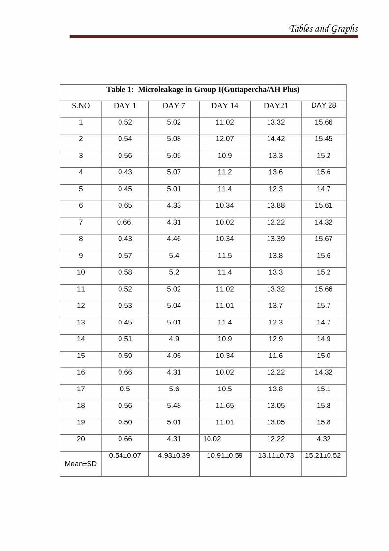
The mean values and statistical comparisions between the experimental
groups at each time interval were given in Table 1. The glucose leakage mean
value of Group I Gutta-percha/AH plus on day 1 was 0.54 0.07 mg/dl, at 7
days 4.93 0.39, at 14 days 10.91 0.59, at 21 days 13.11 0.73, at 28 days
15.21 0.52. The overall mean leakage value was 8.94 5.48. There was a
gradual increase of leakage from day 1 to day 28.
The mean values and statistical comparisions between the experimental
groups at each time interval were given in Table 2. Group II Smart seal
showed mean leakage value on day 1 0.30 0.16, on the 7th day 1.94 0.46,

Results
35
on the 14th day 5.35 0.77, on the 21 day 7.01 0.57 and on the 28
th day
7.81 0.49. The overall mean was 4.48 2.96. There was a gradual increase
of leakage from day 1 to day 28.
The mean values and statistical comparisions between the experimental
groups at each time interval were given in Table 3. Group III Real seal showed
mean leakage value on day 1 0.52 0.10, on the 7th day 3.21 0.48, on the
14th day 8.18 1.32, on the 21 day 8.81 1.10 and on the 28
th day 10.01
1.03. The overall mean was 6.14 3.77. There was a gradual increase of
leakage from day 1 to day 28.
INTERPRETAION OF RESULTS;
1. Glucose concentrations are seen more in Gutta-percha/AH Plus
samples compared to C-points/bio ceramic sealer and
Resilon/Epiphany systems
2. Sealing ability of C-Points with bio ceramic sealer was superior
compared to Resilon cone with Epiphany sealer and Gutta-percha with
AH-Plus.
3. Sealing ability of Resilon cone with epiphany sealer was better
compared to Gutta-percha with AH plus.
4. Sealing ability of Gutta-percha with AH-Plus sealer was considerable.
5. Sealing ability of Positive Control group was least effective.
6. No leakage was seen in negative control group.

Tables and Graphs
Tables and Graphs
Table 1: Microleakage in Group I(Guttapercha/AH Plus)
S.NO DAY 1 DAY 7 DAY 14 DAY21 DAY 28
1 0.52 5.02 11.02 13.32 15.66
2 0.54 5.08 12.07 14.42 15.45
3 0.56 5.05 10.9 13.3 15.2
4 0.43 5.07 11.2 13.6 15.6
5 0.45 5.01 11.4 12.3 14.7
6 0.65 4.33 10.34 13.88 15.61
7 0.66. 4.31 10.02 12.22 14.32
8 0.43 4.46 10.34 13.39 15.67
9 0.57 5.4 11.5 13.8 15.6
10 0.58 5.2 11.4 13.3 15.2
11 0.52 5.02 11.02 13.32 15.66
12 0.53 5.04 11.01 13.7 15.7
13 0.45 5.01 11.4 12.3 14.7
14 0.51 4.9 10.9 12.9 14.9
15 0.59 4.06 10.34 11.6 15.0
16 0.66 4.31 10.02 12.22 14.32
17 0.5 5.6 10.5 13.8 15.1
18 0.56 5.48 11.65 13.05 15.8
19 0.50 5.01 11.01 13.05 15.8
20 0.66 4.31 10.02 12.22 4.32
Mean±SD
0.54±0.07 4.93±0.39 10.91±0.59 13.11±0.73 15.21±0.52

Tables and Graphs
Table 2: Microleakage in Group II(Smart Seal)
S.NO DAY 1 DAY 7 DAY 14 DAY21 DAY 28
1 0 1.34 5.62 7.72 7.98
2 0.01 2.23 4.67 5.89 7.76
3 0.24 2.56 6.89 7.78 7.89
4 0.26 2.22 4.76 6.66 8.99
5 0.23 2.33 4.45 6.65 8.87
6 0.22 1.34 3.33 5.89 6.62
7 0.3 2.28 5.07 7.01 7.7
8 0.21 1.32 5.44 6.78 7.56
9 0.4 2.22 5.2 7.3 7.8
10 0.41 1.35 5.33 4.64 7.43
11 0.24 2.56 6.89 7.78 7.89
12 0.25 1.5 5.5 6.5 7.5
13 0.3 2.28 5.07 7.01 7.7
14 0.35 2.3 5.4 7.0 7.5
15 0.38 2.29 5.67 7.74 7.9
16 0.4 2.22 5.2 7.3 7.8
17 0.7 1.46 5.72 7.1 7.89
18 0.45 1.87 5.62 7.71 7.99
19 0.46 1.9 5.73 7.01 7.85
20 0.21 1.32 5.44 6.78 7.54
Mean±SD
0.30±0.16 1.94±0.46 5.35±0.77 7.01±0.57 7.81±0.49

Tables and Graphs
Table 3: Microleakage in Group III(Real seal)
S.NO DAY 1 DAY 7 DAY 14 DAY21 DAY 28
1 0.53 3.02 9.13 9.22 10.45
2 0.67 2.98 6.89 8.43 10.62
3 0.45 2.78 5.45 6.32 7.45
4 0.54 3.03 8.12 9.23 9.99
5 0.56 4.42 6.65 7.34 8.88
6 0.65 3.44 8.88 9.21 10.0
7 0.21 3.39 8.67 9.92 10.23
8 0.54 3.9 9.12 9.3 10.34
9 0.51 3.04 9.2 9.5 10.32
10 0.5 3.06 9.1 9.7 10.43
11 0.67 2.98 6.89 8.43 10.62
12 0.54 3.01 9.12 9.5 10.7
13 0.56 4.42 6.65 7.34 8.88
14 0.58 3.02 8.6 9.4 10.4
15 0.57 3.02 9.12 9.23 10.47
16 0.45 2.78 5.45 6.32 7.4
17 0.48 2.8 9.3 9.8 10.9
18 0.41 3.01 8.93 9.09 10.71
19 0.51 3.07 9.2 9.6 10.44
20 0.54 3.06 9.11 9.25 10.38
Mean±SD
0.52±0.10 3.21±0.48 8.18±1.32 8.81±1.10 10.01±1.03

Tables and Graphs
Table 4: Kruskal-Wallis test Glucose leakage at various time intervals
Group
P value Based on Kruskal-Wallis
test
Group I (AH Plus/G.P)
Group II (Smart Seal)
Group III (Real Seal)
Mean SD Mean SD Mean SD
1 .54 .07 .30 .16 .52 .10 <0.001**
7 4.93 .39 1.94 .46 3.21 .48 <0.001**
14 10.91 .59 5.35 .77 8.18 1.32 <0.001**
21 13.11 .73 7.01 .57 8.81 1.10 <0.001**
28 15.21 .52 7.81 .49 10.01 1.03 <0.001**
** Denotes significant at 1% confidence level
Table 5: Mann-Whitney test Glucose leakage at various time intervals
between groups
** Denotes significant at 1% confidence level
Days
Group
P value based on Mann-Whitney test
Group I (AH Plus/G.P)
Group II (Smart Seal)
Group III (Real Seal)
Group I and II
Group I and III
Group II and III
Mean SD Mean SD Mean SD
1 .54 .07 .30 .16 .52 .10
<0.001** 0.640 <0.001**
7 4.93 .39 1.94 .46 3.21 .48
<0.001** <0.001** <0.001**
14 10.91 .59 5.35 .77 8.18 1.32
<0.001** <0.001** <0.001**
21 13.11 .73 7.01 .57 8.81 1.10
<0.001** <0.001** <0.001**
28 15.21 .52 7.81 .49 10.01 1.03
<0.001** <0.001** <0.001**

Tables and Graphs
GRAPH I: Glucose leakage at various time intervals for All the three
groups
Group I - Gutta-percha with AH Plus
Group II – C- points/ Bioceramic sealer
Group III – Resilon/Epiphany
0
2
4
6
8
10
12
14
16
1 7 14 21 28
GLU
CO
SE P
ENET
RA
TIO
N
DAYS
Group I Mean
Group II Mean
Group III Mean

Tables and Graphs
GRAPH II: Glucose leakage at various time intervals between Group I
and Group II
Group I - Gutta-percha with AH Plus
Group II – C- points/ Bioceramic sealer
0
2
4
6
8
10
12
14
16
1 7 14 21 28
GLU
CO
SE P
ENET
RA
TIO
N
DAYS
Group I Mean
Group I Mean2

Tables and Graphs
GRAPH III: Glucose leakage at various time intervals between Group II
and Group III
Group II – C- points/ Bioceramic sealer
Group III – Resilon/Epiphany
0
2
4
6
8
10
12
1 7 14 21 28
GLU
CO
SE P
ENET
RA
TIO
N
DAYS
Group II Mean
Group III Mean2
Tables and Graphs
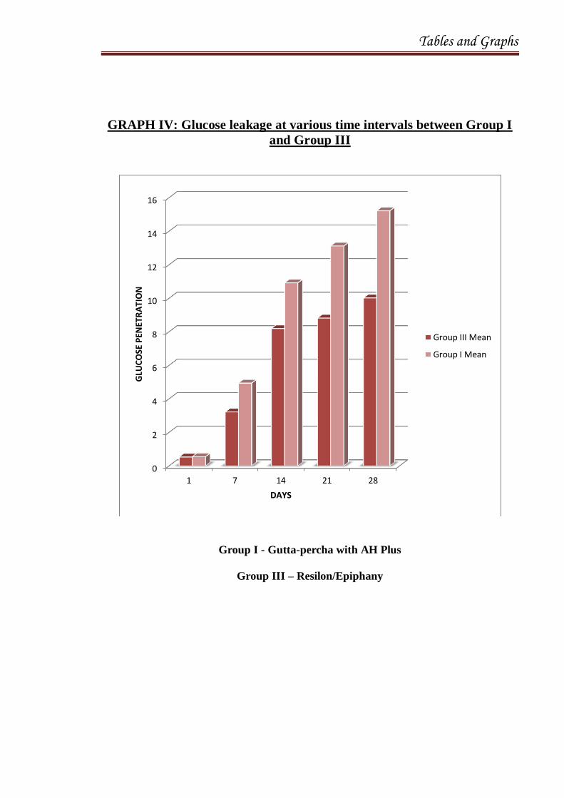
GRAPH IV: Glucose leakage at various time intervals between Group I
and Group III
Group I - Gutta-percha with AH Plus
Group III – Resilon/Epiphany
0
2
4
6
8
10
12
14
16
1 7 14 21 28
GLU
CO
SE P
ENET
RA
TIO
N
DAYS
Group III Mean
Group I Mean

Discussion

Discussion
36
DISCUSSION
The main objective of a root canal filling is to obturate the entire root
canal system and produce an impervious apical seal. This prevents the
penetration of micro-organisms and toxins from the oral cavity via the root
canal into the peri-radicular tissues by sealing the root canal system at both the
coronal and apical ends. Apical obturation prevents infection by anachoresis,
and also blocks the portal of exit to the periapex for organisms which have
survived, even after instrumentation and disinfection. To prevent the
reinfection the whole pulp space is filled, thus blocking the dentinal tubules
and accessory canals. A potential locus for multiplication of micro-organisms
and all portals of exit to the body is sealed by these means.(10,18)
Obturation of a root canal is done by two materials one being the core
and other is the sealer. Core material could either be cold or thermo
plasticized. Warm condensation technique is considered as “golden” standard
for endodontic treatment, that results in a friction fit, “cork-in-the-bottle” type
sealing.(21)
Sealers play an important role in root canal filling. The ideal root canal
sealer should be inert, dimensionally stable, and possess good antimicrobial
activity and low toxicity towards the surrounding tissues. A good sealer
adheres strongly to dentin and the core material.(32)

Discussion
37
Various types of sealers are available, like the traditionally used
Eugenol based, Non Eugenol, Calcium Silicate, Glass Ionomer sealers, Resin
based sealers and to the most recent being Bio-Ceramic sealers. Each of them
have their own inherent drawbacks. None of the traditionally available sealers
fulfill all the ideal requirements of a sealer.
According to Schafer et al. (2003) the quality of the seal obtained with
conventional Gutta-percha/ zinc oxide-eugenol is not perfect. Epoxy resin-
based cements perform well as root canal sealers. AH Plus has been shown to
have satisfactory physicochemical properties, low solubility and
disintegration, good adhesion to dentine, antimicrobial action and good
biological properties. Although AH-Plus has adequate long-term dimensional
stability, its sealing ability remains controversial partly because AH-Plus does
not bond to gutta-percha.(3,4)
According to Shipper et al. (2004) Epiphany is a dual-curing
dimethacrylate resin that uses a primer. With this material, a thermoplastic
core material is bonded to the resin-based sealer, root canals filled with
Epiphany exhibit less microleakage than roots filled with gutta-percha and
conventional sealers. Failures at the sealer–dentine interface may occur
because of the polymerization of the methacrylate-based resin sealer
immediately after its placement into the root canal. In addition, the coronal
photo-activation of the sealer, following the manufacturers instructions, may
reduce its flow and limit its contact with the primer and hence its penetration
Discussion
38
into the dentinal tubules. In a study Epiphany exhibited less antimicrobial
activity than other sealers, except for AH 26 due to its hydrophilic resin form.
All these studies prove beyond doubt that there is a no ideal sealer that
fulfills the requirements of a endodontic sealer henceforth we have selected a
new obturating system Smart seal (C-points/ Bio-ceramic sealer) to check for
its sealing ability, considering its unique property of self expanding in the root
canals. Recent literature and studies have been limited on the properties of the
C- points.
Didato et al (2013)6 evaluated the time-based lateral hygroscopic
expansion of a water-expandable endodontic obturation point. They compared
the time-based lateral expansion of two sizes and two batches of water-
expandable obturation points (CPoint, EndoTechnologies, LLC) and a similar-
sized gutta-percha point (control) at various distances from the point apex: 5,
10, and 15 mm. They concluded that when exposed to water, the lateral
expansion of a new hydrophilic endodontic obturation point significantly
increases in dimension within 20 min, whereas a conventional gutta-percha
point does not.
Eid et al (2013)8 conducted a study to evaluate the effects of C-Point
on the viability and mineralization potential of odontoblast-like cells. The
biocompatibility of C-Point and commercially available gutta-percha points
evaluated using rat odontoblast-like cell line. They concluded that the in vitro
biocompatibility of C-Point is comparable to gutta-percha with minimal

Discussion
39
adverse effects on osteogenesis after elution of potentially toxic components.
Literature evaluating the efficacy of C Point system as an obturation system is
limited.
This brings in the need to find a new core/sealer which fulfills the ideal
requirements. With this background the present study was contemplated with
the aim to compare the sealing ability of three single cone obturating systems
using a glucose leakage model.(24)
In the present study seventy human mandibular 1st premolars extracted
for orthodontic purpose were selected according to the inclusion and exclusion
criteria and were stored in 0.5% Chloramine at c for one month.
Chloramine T controls infection and does not show any adverse effect on the
organic phase of the dentin. The teeth were de-coronated and root lengths
were standardized to 15mm. A diamond bur was used to gain a straight-line
access to the root canal. A size 10 K-File was inserted into the canal to verify
the patency.
Weines method was used to determine the working length. The
working length was determined by subtracting 1mm from the total length of
the root. The chemo-mechanical preparation was completed with Hyflex CM
Ni-Ti files until size 30/0.06% taper using the J.Morita rotary system. After
preparation is completed, the canals were rinsed with 5ml, 2% Sodium
Hypochlorite solution using an endoactivator followed by distilled water. The
teeth were further irrigated with 17% EDTA to remove the smear layer

Discussion
40
followed by irrigation with distilled water. Each canal was dried using paper
points.
The teeth were coated with nail varnish except in the coronal and
apical region. The coronal 4mm of the root specimens were then embedded in
acrylic to form a cylinder around the root and enable intimate contact with the
rubber tube used to connect the specimen to the Glucose leakage Apparatus.
After the initial instrumentation was done, the teeth were assigned into
5 groups. The groups were as follows: Three groups with 20 teeth in each, 2
groups with 5 teeth in each, which served as positive and negative control.
The groups were allocated with following intervention materials.
Group I Gutta-percha with AH Plus sealer, Group II C Points with bioceramic
sealer and Group III Resilon cone with Epiphany sealer, Group IV –Positive
Control (n=5) obturated with Gutta-Percha WITHOUT any sealer and Group
IV – Negative Control (n=5) were not obturated and were coated completely
with nail varnish.
AH 26 is an epoxy resin recommended by Shroeder in 1957. This was
later modified to AH Plus which is a paste-paste system. AH Plus is a sealer
based on epoxy resin. According to the manufacturer, it has excellent sealing
properties without the release of formaldehyde.

Discussion
41
It consists of Epoxide paste Diepoxide, Calcium tungstate, Zirconium
oxide, Aerosil, Iron oxide pigments and Amine paste 1-adamantane amine, N,
N-dibenzyl-5-oxa nonandiamine, Calcium tungstate, Zirconium oxide,
Silicone oil. AH Plus is able to flow into the orifices of the dentinal tubules,
which is the reason for the comparatively good adhesion of AH Plus to
dentin. It has less fracture resistance when used with gutta percha as
compared to Resilon/Realseal. According to Almeida et al. leakage with AH
Plus was significantly less than that with the ZnOE sealer.(18)
The resin core filling material, Resilon (Resilon Research LLC,
Madison, CT), handles like gutta-percha. Obturation with Resilon cones were
accomplished by use of Epiphany primer (Pentron Clinical Technologies,
LLC, Wallingford, CT) and Epiphany resin-based sealer (Pentron Clinical
Technologies). The RealSeal sealer is a dual-curing, resin-based composite
sealer. The resin matrix is composed of bisphenol-A-glycidyl methacrylate
(BisGMA), ethoxylated BisGMA, urethane dimethacrylate (UDMA), and
hydrophilic difunctional methacrylate. The sealer with the aid of a primer
adheres to the core material and dentin.(29)
According to cornelis, one of the factors that was instrumental in the
development of resin-based sealers was the recognition that gutta-percha does
not bond to dentin or to any conventionally used sealer, such as zinc oxide-
eugenol (ZOE)-based cements and epoxy resins such as AH-26 or AH Plus.

Discussion
42
This combination supposedly forms a mono-block in the root canal
system. The Resilon material has been shown to be biocompatible,
nonmutagenic, noncytotoxic, resolvable. It also has properties similar to those
of gutta-percha, and is less irritating than epoxy resin or ZOE sealers. For
retreatment purposes it may be softened with heat, or dissolved with solvents
such as chloroform. The Epiphany Root Canal Sealant is a dual curable dental
resin composite sealer. Studies recommend that EDTA or chlorhexidine
(CHX) should be used as the final irrigant as sodium hypochlorite or hydrogen
peroxide may weaken the seal.(26,39)
The most recent advancement in endodontic obturating materials is the
evolution of Smart Seal system, a hydrophilic polymer. The system consists of
obturation points (C-points) containing a polyamide core with an outer bonded
hydrophilic polymer coating and an accompanying sealer which is further
provided with polymer powder to be incorporated during the manipulation of
the seal. The inner core of C‐points is a mix of two proprietary nylon
polymers: Trogamid T and Trogamid CX. The polymer coating is a
cross‐linked copolymer of acrylonitrile and vinylpyrrolidone which has been
polymerized and cross‐linked using allyl methacrylate and a thermal initiator.
The lateral expansion of C-points is claimed to occur non-uniformly with the
expandability depending on the extent to which the hydrophilic polymer is
prestressed. Radioopacity of both the core and polymer coating is provided
with the inclusion zirconia dioxide particles(15)

Discussion
43
Various methods have been developed to assess the sealing ability of
root canal filling materials. Methods such as dye leakage, fluid transport and
bacterial penetration, had been frequently used for evaluation of micro-
leakage. Other methods such as radio-labelled isotopes and electromechanical
test have also been described. However, these methods often yielded large
variations in the outcome and they are not considered to be reproducible and
comparable.(33,42)
Assessment of bacterial leakage is considered to be more biologically
relevant than that of dye or radioisotope penetration, but the conclusions might
vary with the bacterial species used. Maintaining aseptic conditions
throughout all steps of the experiment can be difficult. Radioisotope labeling
and electrochemical technique were less frequently employed because they
pose a radiation hazard and require sophisticated materials and apparatus.(43)
Several test methods have been described to evaluate the sealing quality
of filled root canals. The most popular methods are fluid transport model (Wu
et al. 1993) and the glucose leakage model (Xu et al. 2005). The latter can be
seen as a further development of the fluid transportation concept, both
measure passage of fluid along root filled teeth after subjecting them to
constant pressure.(35.42.43)
The fluid filtration method, which was developed by Derkson et al for
measuring dentin permeability,
and later modified by Wu et al to evaluate
Discussion
44
endodontic leakage, is gaining popularity because it is sensitive and
nondestructive and permits repeated observation of the same specimen over
times. These techniques do not provide any information about the volume of
tracer that penetrates which provides only semi-quantitative data with a high
level of variation. However, the glucose model allows measurements of
diffusion of the marker molecules as well. The glucose test might be more
sensitive than the measurement of air-bubble movement, not only because the
detected threshold measurement by eye is higher than that of the
spectrophotometer, but also because the convective fluid transport was
combined with glucose molecule diffusion.(35,42,43)
In the present study the glucose leakage model was used to completely
evaluate the volume of tracer penetration. The advantages of this model are
the relative ease of assembly and operation, the availability of the materials
and equipment and the great sensitivity of the test. Glucose was selected as the
tracer because of its small molecular size (MW = 180 Da) and is a nutrient for
bacteria. The choice of tracer should be carefully chosen because its size and
physicochemical properties may influence the result. The use of tracer of a
small molecular size was favored by the previous studies conducted to obtain a
relevant outcome.(31,35,43)
If the glucose could enter the canal from the oral cavity, bacteria that
might survive root canal preparation and obturation could multiply and
potentially lead to periapical inflammation. Glucose, therefore, was thought to

Discussion
45
be more clinically relevant than other tracers used in micro-leakage tests.
Quantitative analysis of leakage was possible by determining the
concentration of glucose in the apical reservoir that leaked through the filled
root canal.(43)
The teeth in all the five groups were subjected to micro-leakage testing
using the Glucose leakage Model. The samples were tested for leakage at 7
days intervals and the data were collected at baseline (day 1), 7th day, 14
th day,
21th day and 28th
day.
In the Glucose leakage Model 10 µL of the sample was withdrawn
after 24 hours, followed by 10 µL at regular intervals with the help of a
micropipette. The sample withdrawn was then subjected to quantitative
glucose testing by Glucose oxidase-Peroxidase test using a spectrophotometer
at wavelength 505 . The 10 µL of sample withdrawn was replenished with
the same volume of 0.2% sodium azide. The study model used in the present
study was similar to the technique used by Xu et al.(43)
To determine the concentration of glucose, the enzymatic glucose
oxidase method was chosen because it provided the ultimate degree of
specificity and high sensitivity when compared with other methods, such as
copper or ferricyanide methods. With this method, glucose is oxidized by the
enzyme glucose oxidase in the presence of oxygen to gluconic acid with the
formation of hydrogen peroxide. Then in the presence of a peroxidase enzyme,

Discussion
46
a chromogenic oxygen acceptor (4‐aminoantipyrine and phenol) is oxidized by
the hydrogen peroxide, resulting in the formation of a red product (oxidized
chromogen).(43)
The quantity of this oxidized chromogen is proportional to the glucose
present initially in the first reaction, which quantity is determined by
spectrophotometry. With this model, it was possible to quantify the endodontic
micro-leakage continuously over time. The amount of microleakage was the
cumulative value of leaked glucose. The reactivity of obturating materials with
glucose could affect the results of the glucose leakage test. The results of
Shemesh et al. indicated that all materials used in the current study did not
show glucose reactivity.(31,43)
The results of the current study clearly demonstrate that none of the
materials completely sealed the root apex in vitro (Table 1). Inadequate apical
seals could result from the technique used to fill the canal system; for
example, the use of a single-cone filling technique is often considered inferior
to more sophisticated 3D compaction techniques. In the single-cone technique,
the volume of sealer is high relative to the volume of the cone, and this ratio
promotes void formation and reduces the quality of the seal. However, it must
be noted that the concept of the single-cone technique has been recently re-
visited, and that the volume of the sealer used in the present study was
minimized because gutta-percha, C-point cones, Resilon cones were matched
to the preparation. Use of the single-cone technique also allowed a comparison

Discussion
47
of the performance of all materials under relatively standardized conditions.(4)
For each day tested, the positive controls had immediate substantial
glucose leakage, which increased over time, whereas the negative controls
showed no detectable glucose leakage. This indicates that the seal of the
glucose leakage system was effective and reliable.
The glucose leakage mean value of Group I gutta-percha/AH plus on
day 1 was 0.54 0.07 mg/dl, at 7 days 4.93 0.39, at 14 days 10.91 0.59,
at 21 days 13.11 0.73, at 28 days 15.21 0.52. Overall mean leakage value
was 8.94 5.48. There was a gradual increase of leakage from day1 to day 28.
In the present study micro-leakage was found to be least in Group II
and Group III. In Group II, Smart Seal showed mean leakage value on day 1
0.30 0.16, on the 7th day 1.94 0.46, on the 14
th day 5.35 0.77, on the 21
day 7.01 0.57, on the 28th day 7.81 0.49. The overall mean was 4.48
2.96. There was a gradual increase of leakage from day1 to day 28.
In Group III, Real Seal showed mean leakage value on day 1
0.52 0.10, on the 7th day 3.21 0.48, on the 14
th day 8.18 1.32, on the 21
day 8.81 1.10, on the 28th day 10.01 1.03. The overall mean was 6.14
3.77. There was a gradual increase of leakage from day 1 to day 28.
On comparison of micro-leakage among the three groups, Group II
which used Smart Seal showed the least mean glucose leakage.

Discussion
48
In Group I the disparity in the finding could be explained by the fast
setting and subsequent polymerization shrinkage of AH plus sealer, the lack of
bonding between this sealer and gutta‐percha, the low penetration ability of
this sealer within the dentinal tubules and its hydrophobic property that
prevents good adaptation of it in the incompletely dried canal.(38)
During root canal treatment, especially after rinsing the root canal
system, it is obvious that fluid droplets are retained in the dentinal tubules and
may not be completely removed through the use of paper points. Because the
sealer may be exposed to tissue fluid and exudate, water sorption and
solubility behaviour of the root canal sealers in the humid root canal system is
of considerable importance.(33)
According to Tat and pashley, the currently marketed dentine
adhesives, including the hydrophilic resin-based root canal sealer Epiphany,
contain hydrophilic and ionic monomers, making them highly susceptible to
water sorption and hydrolysis. This water sorption plasticizes polymers and
lowers their physical/mechanical properties which decrease the life expectancy
of the interfaces by hydrolysis and microcrack formation.(23)
According to Sano et al. one of the most important factors in the
strength and stability of the resin/dentine bond is the incomplete resin
infiltration into the demineralized dentine (hybrid layer). As a result, fluid
movement occurs. This nanoleakage, or ingress of oral fluid through nano-

Discussion
49
meter sized channels along collagen fibrils within the hybrid layer, is
considered to be detrimental to bond integrity. C‐factor can be more
preponderant. Hence, any polymerizing endodontic sealer would be subjected
to sizably voluminous polymerization stresses during the setting process,
resulting in debonding and gap formation along the periphery of the root
filling and thus can be a contributing factor for the increased leakage seen in
this group. Therefore, in spite of the hydrophilic nature of Resilon, leakage
was significantly more than other hydrophilic groups. According to Shipper et
al., this material has been shown to be more resistant to leakage than gutta-
percha for filling root canals.(38)
On comparison between the groups 1, 7, 14, 21 and 28 days we found
that Group II (SMART SEAL) showed least leakage followed by Group III
(REAL SEAL), Group I showed highest amount of glucose leakage. Statistical
difference between the groups were found to be highly significant (p<0.001).
Leakage cannot be totally eliminated from the fate of a root canal
treated teeth. Lateral canals, accessory canals and other anatomical variation
play an important role in this, with periapical pressure being the leading
factor.(21)
With the introduction of novel hydrophilic SmartSeal system
(C-points) over the conventional hydrophobic Gutta-percha system, has
widened our range of achieving a 3D seal.

Discussion
50
The present study was carried out on mandibular premolars with
straight canals. Hence, further studies has to be directed in teeth with
complicated anatomy and curved root canals to evaluate the microleakage in
the root canal system.

Summary
Summary
51
SUMMARY
This study was aimed to evaluate and compare the micro-leakage and
to assess the sealing ability of three obturating materials. Seventy teeth with
single canals (verified with radiograph) were selected for this study. Root
length was standardized to 15mm and were randomly divided into 5 groups.
Group I was Gutta-percha with AH-Plus sealer (n=20), Group II was C-Points
with Bio ceramic sealer (n=20), Group III was Resilon with Epiphany sealer
(n=20), Group IV (positive control)- Guttapercha without sealer (n=5) and
Group V (negative control)- Teeth without obturation, coated with nail varnish
(n=5)
Working length was obtained using 10 size stainless steel k-file.
Chemo-mechanical preparation was performed by Ni-Ti rotary Hy-flex CM
files(Size 20/0.08%,Size 20/0.04%,Size 25/0.04%,Size 30/0.04%,Size
30/0.06%) in crown down sequence with irrigation using 2% NaOCl and
17%EDTA with every change of each file. Irrigation was carried out passively
with a Endoactivator with tip being placed 1mm short of working length and
finally rinsed with distilled water.
The root canals were dried using paper points, obturated according to
c for 1 week.
After incubation samples were subjected to glucose leakage test, later the
solutions were subjected to spectrophotometer at 505 at different time
intervals (day 0, day 7, day 14, day 28).

Summary
52
The score values were recorded, tabulated and statistically analyzed by
Mann-Whitney Test and Wilcoxon signed-rank tests.

Conclusion

Conclusion
53
CONCLUSION
With the introduction of novel hydrophilic Smart Seal system
(C-points) over the conventional hydrophobic Gutta-percha system, has
widened our range of achieving a three dimentional seal.
Within the limitations of this in vitro study, it can be concluded that:
1. C-Points with bio ceramic sealer showed least glucose leakage values
hence proving superior sealing ability compared to other groups.
2. C-Points with bio-ceramic sealer showed superior sealing ability when
compared with Resilon/Epiphany, Gutta-percha/AH-Plus and control
groups.
From the above study it can be inferred that C-Points with bio ceramic
sealer can be considered as a potent alternative for Resilon Epiphany system
and Gutta-percha with AH-Plus sealer because of its superior sealing ability of
the root canal system and negligible micro-leakage.

Bibliography

Bibliography
BIBLIOGRAPHY
1. Arora S, Vibha Hegde:
Comparative evaluation of a novel smart seal obturating system and its
homogeneity of using cone beam computed tomography: In vitro
simulated lateral canal study.
Journal of Conservative Dentistry,2014;Vol 17( 4).
2. Assmann E, Roberta Kochenborger Scarparo:
Dentine bond strength of two mineral trioxide aggregate based and one
epoxy resin based sealers.
Journal of Endodontics 2012; 38:219-221.
3. Alfredo E, Silva S R C, Ozorio J E V:
Bond strength of AH Plus and epiphany sealers on root dentine
irradiated with 980 nm diode laser.
International Endodontic Journal,2008; 41:733-740.
4. Bouillaguet S, Shaw L, Barthelemy J:
Long term sealing ability of Pulp Canal Sealer, AH-Plus, Guttaflow
and Epiphany.
International Endodontic Journal,2008;41:219-226.
5. Cotti E, Petreucic V, Dino Re:
Cytotoxicity evaluation of a new resin-based hybrid root canal sealer:
An in vitro study
Journal of Endodontics 2014; 40:124-128.
54

Bibliography
6. Didato A, Eid A A, Levin M:
Time based lateral hygroscopic expansion of a water expandable
endodontic obturation point.
Journal Of Dentistry,2013;41:796-801.
7. Elbatouty K M, Ibrahim D Y, Youniss W N:
In vitro bond strength of bioceramic root canal sealer in comparision to
resin based and eugenol based root canal sealers.
Quintessenz Endo (Long Engl) 2015;9(1):59-63.
8. Eid A A, Nikonov S Y, Looney S W:
In vitro biocompatiability evaluation of a root canal filling material
that expands on water sorption.
Journal Of Endodontics,2013;39:883-888.
9. Economides N, Gogos C, Kodonas K:
An ex vivo comparision of the push-out bond strength of a new
endodontic filling material(smart seal) and various gutta-percha filling
techniques.
Journal of Odontology, 2012;100:187-191.
10. Fraunhofer J A, Fagundes D K, Mc Donald N J:
The effect of root canal preparation on microleakage within
endodontically treated teeth- an in vitro study.
International Endodontic Journal,2000;33:355-360.

Bibliography
11. Hegde V, Arora S:
Effect of intracanal medicaments on push-out bond strength of Smart-
Seal system
Journal of Conservative Dentistry,2015;Vol 18(5).
12. Hegde V, Arora S:
Fracture resistance of roots obturated with novel hydrophilic obturation
systems
Journal of Conservative Dentistry,2015; Vol 18(3).
13. Hegde V, Arora S:
Sealing ability of three hydrophilic single-cone obturation systems: In
vitro glucose leakage study.
Journal of Contemporary Clinical Dentistry,2015; Vol 6( 1).
14. Hegde V, Arora S:
Effect of advanced irrigation protocols on self-expanding Smart seal
obturation system: A scanning electron microscopic push-out bond
strength study.
Contemporary Clinical Dentistry,2015; Vol 6( 1).
15. Hegde V, Arora S:
Sealing ability of a novel hydrophilic versus conventional hydrophobic
obturation sytems: A bacterial leakage study.
Journal of Consevative Dentistry,2015; Vol 18( 1).
16. Iriboz E, Sazak.H

Bibliography
Comparision of protaper and Mtwo retreatment systems in the removal
of resin-based root canal obturating materials during retreatment
AustEndod J 2014;40: 6-11.
17. Kqiku L, Stadtler P, Gruber H J:
Active versus passive microleakage of Resilon/ Epiphany and
Guttapercha/ AH Plus.
AustEndod J 2011;37: 141-146.
18. KontCobankara F, Aadanir N, Belli S:
A quantitative evaluation of apical leakage of four root canal sealers.
International Endodontic Journal,2002;35:979-984.
19. Lumbini P, Karthik Krishna N:
Smart seal – new age obturation.
Annals of Dental Speciality,2013; volume 01(01).
20. Nawal P R, Parande M, Sehgal R:
A comparative evaluation of antimicrobial efficacy and flow properties
for Epiphany, Guttaflow and AH-Plus sealer.
International Endodontic Journal,2011;44:307-313.
21. Pawar S S, Pujar M A, Makandar S D:
Evaluation of the apical sealing ability of bioceramic sealer, AH Plus
& Epiphany: An in vitro study.
Journal of Conservative Dentistry,2014; Vol 17(6).

Bibliography
22. Paque F, Sirtes G:
Apical sealing ability of Resilon/Epiphany versus gutta-percha/AH
Plus: immediate and 16-months leakage.
International Endodontic Journal,2007;40:722-729.
23. Patel D V, Sherriff M, Ford T R P:
The penetration of RealSeal primer and Tubliseal into root canal
dentinal tubules: a confocal microscopic study.
International Endodontic Journal,2007;40:67-71.
24. Pommel L, About I, Pashley D:
Apical leakage of four endodontic sealers.
Journal of Endodontics, March,2003; Vol 29: No. 3.
25. Reddy S S, Sujatha V, Mahalaxmi S:
Sealing ability of four root end filling materials : a glucose penetration
leakage study.
Adv Hum Biol 2013;3(2):11-18.
26. Resende L M, Rached- Junior F J A, Versiani M A:
A comparative study of physicochemical properties of AH Plus,
Epiphany SE root canal sealers.
International Endodontic Journal,2009;42:785-793.
27. Ruiz-Linares M, Estela bailon-sanchez
Physical properties of AH Plus with chlorhexidine
Journal of Endodontics 2013; 39:1611-1614.

Bibliography
28. Souza P A R, Affonso P R:
Comparision of bacterial leakage between Epiphany and AH Plus
sealers using single cone and Tagger’s hybrid obturation techniques.
Rev Odonto Cienc,2014;29(3):87-91.
29. Shanahan D J, Duncan H F:
Root canal filling using Resilon: A review.
British Dental Journal,2011;Volume 211(2).
30. Souza E M, Wu M K, Shemesh H:
Comparability of results from two leakage models.
Oral Surg Oral Med Oral Pathol Oral RadiolEndod,2008;106:309-13.
31. Shemesh H, Souza E M, Wu M K:
Glucose reactivity with filling materials as a limitation for using the
glucose leakage model.
International Endodontic Journal,2008;41:869-872.
32. Slutzky- Goldberg I, Slutzky H, Solomonov M:
Antibacterial properties of four endodontic sealers.
Journal of Endodontics,2008; Volume 34(6).
33. Shemesh H, Van den Bos M, Wu M K:
Glucose penetration and fluid transport through coronal root structure
and filled root canals.
International Endodontic Journal,2007;40:866-872.
Bibliography
34. Stratton R K, Apicella M J, Mines P:
A fluid filtration comparison of Gutta-percha versus Resilon , a new
soft resin endodontic obturation system.
Journal of Endodontics,2006; Volume 32: No 7.
35. Shemesh H, Wu M K, Wesselink P R:
Leakage along apical root fillings with and without smear layer using
two different leakage models: a two month longitudinal ex vivo study.
International Endodontic Journal,2006;39:968-976.
36. Tay F R, Loushine R J, Weller R N:
Ultrastructural evaluation of the apical seal in roots filled with a
polycaprolactone- based root canal filling material.
Journal of Endodontics,2005; Volume 31,N0 7.
37. Tagger M, Greenberg B, Sela G:
Interaction between sealers and Gutta-percha cones.
Journal of Endodontics,2003;Vol 29: No.12.
38. Verissimo D M, Monica Sampaio do Vale, Monteiro A J:
Comparison of apical leakage between canals filled with Gutta-
percha/AH-Plus and the Resilon/Epiphany system, when submitted to
two filling techniques.
Journal of Endodontics,2007; Volume 33:no 3.
39. Wedding J R, Brown C E, Legan J J:
An in vitro comparison of microleakage between Resilon and Gutta-
percha with a fluid filtration model.

Bibliography
Journal of Endodontics,2007; Volume 33:no 12.
40. Weis M V, Parashos P, Messer H H:
Effect of obturation technique on sealer cement thickness and dentinal
tubule penetration.
International Endodontic Journal,2004;37:653-663.
41. Wu M K, Wesselink P R:
Endodontic leakage studies reconsidered. Part I. Methodology,
application and relevance.
International Endodontic Journal,1993;26:37-43.
42. Xu Qiong, Ling J, Cheung G S P:
A quantitative evaluation of sealing ability of 4 obturation techniques
by using a glucose leakage test.
Oral Surg Oral Med Oral Pathol Oral RadiolEndod 2007;104:e 109-e
113.
43. Xu Qiong, Fan M W, Fan B:
A new quantitative method using glucose for analysis of endodontic
leakage.
Oral Surg Oral Med Oral Pathol Oral RadiolEndod,2005 ;99:107-11.

Annexure
Annexure
ANNEXURE