Embed Size (px)
Citation preview
Tikrit Journal for Dental Sciences 1(2015)
Evaluation of Color Stability for Two Types of Denture Base
Materials: Heat Cured Acrylic and Flexible Resin.
Dr. Areej Shehab Ahmad M.Sc. in Prosthodontics. (1) Dr. Khalid Talib Bandar M.Sc. in Periodontology. (2) Alaa Ezat Abd Al-majeed M.Sc. in dental technologies. (3)
Introduction
Since the mid 1940s, most denture bases have
been fabricated using poly methyl-
methacrylate (PMMA) resins (1). It becomes
an increasingly popular choice, due to its
properties and ease of handling; though color
stability is still controversial (2) (3), While the
thermoplastic material for dental prosthesis
were first introduced to dentistry
in the end of 1959, rapid injection systems
currently known as the flexible company
introduced the first flexible thermoplastic
which was a flour polymer (A Teflon type
of plastic) (4) .The term thermoplastic
implies that a polymer softens on heating
and then hardens into the final shape upon
cooling; this is a desirable property for an
oral appliance because the final shape can be
individualized for each case by using a study
model (5).
Oral hygiene remains important even when
some or all teeth have been replaced with
Key words Color stability,
staining of denture,
heat cure acrylic,
Valplast
thermoplastic resin.
Abstract Commonly consumed beverages used by human being daily (Tea
& Cola) caused external staining. This study is done to evaluate
the color stability for two different types of denture base materials
against this external staining caused by (Tea & Cola). Sixty (60) samples were prepared, divided into two groups, 30
samples of heat cure acrylic material & 30 samples of flexible
resin material (Valplast). Each group divided into three subgroups
10 samples in each, according to the type of staining solution that
immersed in it, (synthetic saliva & tea), (synthetic saliva & Cola),
and (synthetic saliva alone as a control). Tea & Cola were mixed
with synthetic saliva in order to create intraoral environment to
certain extent. Color measurement was made at Baghdad
University, Collage of Engineering, using reflected
spectrophotometer before immersion (at baseline) & after
immersion, at intervals of (24 hours) & (1 week) respectively.
The result was a highly significant difference between, heat cure
acrylic & flexible resin at baseline & the (flexible samples) were
the higher value in discoloration than (heat cure acrylic samples)
regardless the type of staining solution. Maximum discoloration
was seen in (synthetic saliva & tea) solution for both denture base
materials. Followed by (synthetic saliva & Cola) & (synthetic
saliva alone) respectively.
Both denture base materials had a color changes after immersion
in staining solution (tea, Cola & synthetic saliva). The color
changes, for two materials, were increased with the increase of
immersion time.
.
(1 Lecturer in dental technologies department, Collage of Health and Medical Technologies
(2) Lecturer in dental technologies department, Collage of
Health and Medical Technologies (3) Lecturer in dental technologies department, Collage of
Health and Medical Technologies
)5(2011…. Evaluation of Color Stability
2
removable dentures (6). It is generally
recognized that dirty dentures may have
undesirable effects on the patient oral health
and ability to successfully wear dentures (7).
Denture cleanser is essential to prevent
malodor, poor aesthetics and the
accumulation of stains, plague and calculus
with its deleterious effects on the mucosa
(8). Denture base materials and denture
teeth collect deposits and stain in the same
manner, as do natural teeth (9). Soft debris
that clings to a denture can be removed
easily by light brushing followed by rinsing.
Hard deposits and stains such as those that
occur from tea, coffee, cola and tobacco tars
are much difficult to remove (10).
Crispin & Caputo in 1979 (11), studied the
color stability of different types of materials.
They found that methyl methacrylate
material exhibited the least darkening,
followed by ethyl methacrylate and another
types of thermoplastic acrylic materials. In-
spite of various studies being carried out to
study the color changes of different
provisional materials using different staining
solutions, still the literature on color stability
of these materials is limited (12). Thus, this
study was directed to determine color
stability for two different types of denture
base materials (Heat cure acrylic &
thermoplastic-flexible resins), after
immersion in most commonly used staining
solution (Tea, Cola & Synthetic saliva as
control), to find out the most color stable
between them.
Materials and Methods
Sixty (60) samples were prepared. They
divided into two main groups according to
the type of denture base material: 1st group:
30 samples of heat cure acrylic resin. Figure
(1)-A
2nd group: 30 samples of thermoplastic
(flexible) resin. Figure (1)-B
Each group was divided into three
subgroups according to the type of staining
solution that immersed in it, each subgroup
consist of 10 samples: 10 samples
immersion in synthetic saliva & tea, 10
samples immersion in synthetic saliva &
cola, & 10 samples immersion in synthetic
saliva only as control.
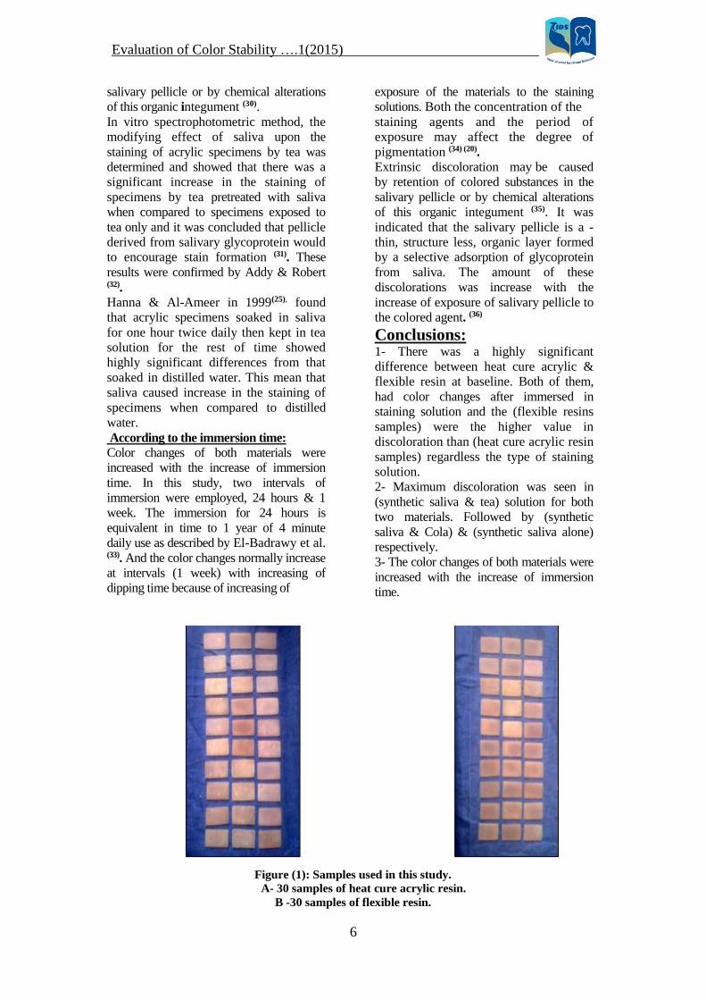
Mould Preparation: Metal mould made
from a brass in the form of a rectangular.
Figure (2)
The dimensions are (40 mm length, 20 mm
width & 0.8 mm thickness); these
dimensions are according to the
spectrophotometer device, “Macbeth Color
Eye 7000A” (Macbeth, USA)
Preparation of samples:
A- Preparation of the heat cured
acrylic samples: The conventional flasking technique for
complete denture was followed in mould
preparation. All material were mixed and
manipulated according to manufactures
instructions in each procedure, packing,
curing till to the finishing, polishing &
conditioning.
The 2 halves of the mould were coated with
separating medium (cold mold seal) and
allowed to dry biform investing them in the
lower half of the flask which contain stone
mix according to the manufacture
instruction and allow setting. The patterns
were inserted to one half of its depth. The
set lower half was coated with separating
medium and allowed dry and then the upper
half of the flask was assembled and filled
with stone mixture. After removal of the
metal patterns the two halves of the mold
were coated with a separating medium to be
ready for packing with acrylic dough. Figure
(3)
The mixed procedure was carried out in
glass jar with clean metal spatula; mixture
was then covered and left to second until it
reached a consistency suitable for packing
(dough stage).(13)
Packing for heat cure acrylic:- The acrylic resin was packed in the late
dough stage indicated by clean separation of
resin from the walls of glass mixing jar.
Acrylic resin dough was placed and the 2
halves were assembled and placed under the
press with gradual application of pressure to
allow even flow of the dough throughout the
mould space, then the pressure was relieved.
The flask was then opened and the over
flowed material surrounding the mould
space was removed with sharp knife. A
second trial closure was preformed that the 2
halves of the flask were finally closed under
pressure until metal to metal contact had
)5(2011…. Evaluation of Color Stability
3
been established and left under press (20
bar) for 5 mints before clamping was done
.(13)
Curing cycle of heat cure acrylic:- Curing cycles were followed used water
path, by use the rapid- cure method at
165f°for one hour and then boiled for half
one hour. After completion and curing, the
acrylic specimens were removed carefully
from stone mold. (13)
Finishing, Polishing & conditioning: All of the samples were carefully de-flask
and cleaned flashes of hot cure acrylic were
removed with acrylic bur. To get a smooth
surface, all samples were finished by stone
bur to remove all excessive materials for
two minute with low speed 1500 rpm and
low pressure then Tungsten carbide bur for
two minute with low speed 1500 rpm and
low pressure after that sand paper (120)
grain size, for one minute with low speed
1500 rpm and low pressure with continuous
water cooling. Figure (4) , finally the
samples were polished for two minute with
low speed 1500 rpm and low pressure .
Polishing was accomplished by using bristle
brush, (60 mm) diameter (Milano, Italy),
and rag wheel with pumice, fine grade (QD,
England), in lathe polishing machine. A
gloss surface was obtained by using dental
lathe using low speed (1500 rpm] with
regard to continuous cooling with water to
avoid overheating. (14)
B- Preparation and injection of flexible
resin: The conventional flasking technique for
complete denture was followed in the mold
preparation in the first part of flask then a
major sprue with (2.5 mm) in diameter &
(3cm) in length was attach to the middle of
specimen. Or two minor sprues, (l.5 mm) in
diameter, were attached to each other from
one end and other end attached to the
specimen Figure (5). Then the point that
attach to the specimen must be removed &
coated with a separated medium.
The upper half of the flask was placed in
position and filled with stone mixtures and
allowed to be hardened for(60) minutes the
flask was put in boiling water for 5 minutes
for wax elimination after that we put a
separating medium (15). Then the flask was
placed inside the special clamp in plastic
injection machine. Care should be taken that
the opening of the major sprue should meet
the opening of the clamp. The machine of
plastic injection is operated. The flask
should stay hot in temperature (70-100) 0C
on a special heater to avoid cooling of
injection material during injection. When
the temperature of machine is reach to (287)
C° place the capsule of flexible acrylic
inside the heater of machine for (12-15)
minutes and began the procedure of
injection of material with rapid pressure
applied about (4.5) Bar by hydraulic press.
(15)
All samples were finished by using: Stone
bur to remove all excessive materials,
Tungsten carbide bur, and Sand paper for
two minute with low speed 1500 rpm and
low pressure with continuous water cooling
manually.
Polishing was accomplished by using bristle
brush (60 mm) diameter (Milano, Italy), fine
grade (QD, England), in lathe polishing
machine with wet rag wheel. A gloss
surface was obtained by using dental lathe
using low speed (1500 rpm for two minutes.
(14)
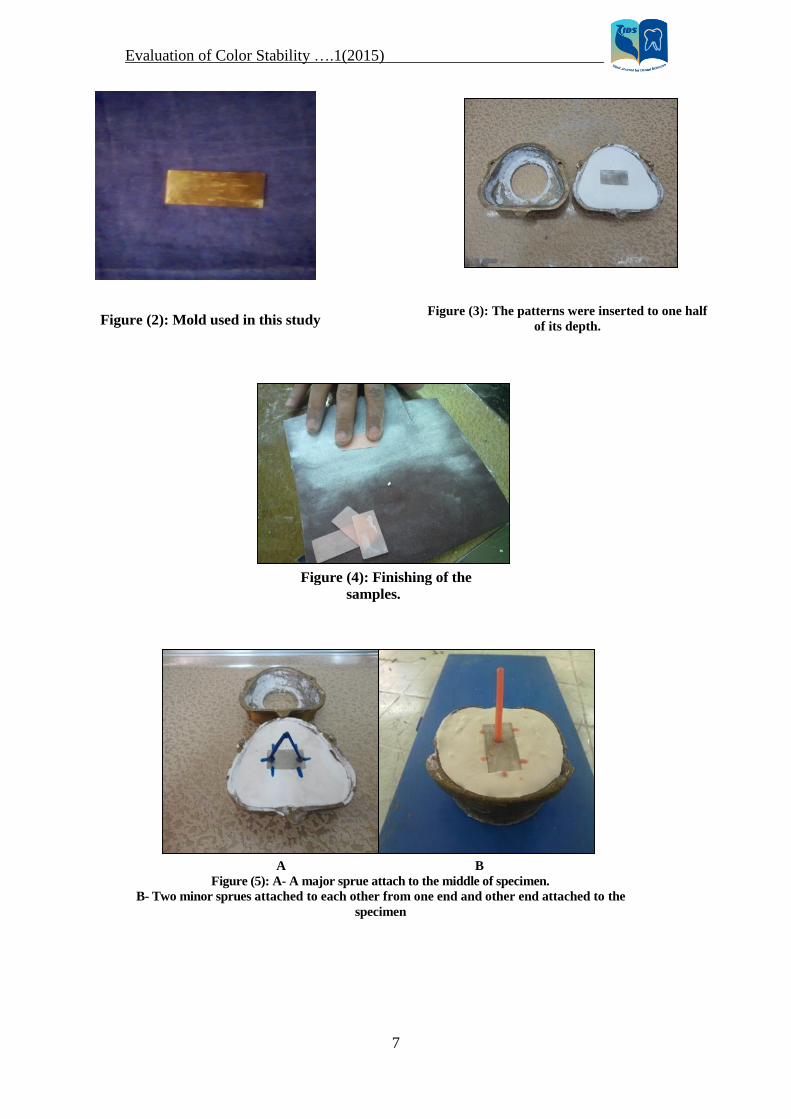
Preparation of Staining Solutions:
(Tea & Cola) solutions were mixed with
synthetic saliva in order to create intraoral
environment to certain extent. 250 ml test
solution of tea & synthetic saliva was
prepared in the ratio of 2:1. Tea solution
was prepared using 150 ml of boiling
distilled water, with tea bag, teaspoon of
sugar, and teaspoon of powdered milk,
immersed for 5 minute and then filtered
through a filter paper (Tea caused the
discoloration , milk &sugar are written here
as details of materials &methods). Figure
(6) Similarly, test solution of Cola beverage &
synthetic saliva, Figure (7) was prepared in
the ratio of 2:1. And finally a sample of 250
ml of synthetic saliva was taken as control
(16), Figure (8).
The synthetic saliva of Fusayama -Meyer
type was the electrolyte solution chosen for
testing in this study (17) (18). The
composition of synthetic saliva in gm/l:
0.4 NaCl; 0.4 KCl; 0.795 NaCl2.2H20;
0.69 NaH2Po4; 1.0 Urea; 1000 ml distilled
water.
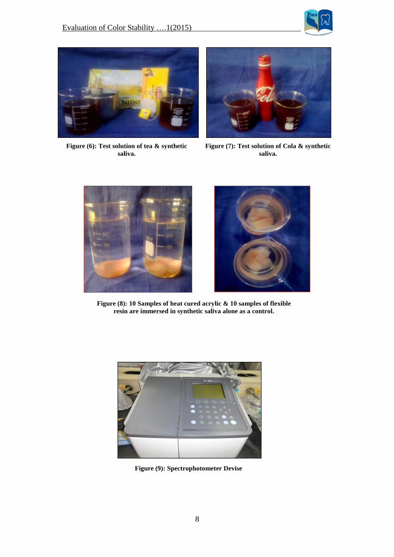
The color measurement was made using
reflectance spectrophotometer at baseline
(before immersion), at intervals of 24 hours,
)5(2011…. Evaluation of Color Stability
4
and 1 week, respectively. Immersion in
staining solutions of synthetic saliva & tea
for three times per day for ten minutes each,
in synthetic saliva & Cola beverage for one
time per day for ten minutes each, and
synthetic saliva (control) for whole day(16).
The sample was rinsed with the distilled
water and then evaluated the color change.
The same procedure was followed
subsequently for next immersion period (1
week). Solution was changed on every
dipping in order to use fresh solution each
time (16). Descriptive Statistics was applied;
mean and standard deviation were
calculated for each variable, for each group.
ANOVA test was applied to see significant
difference among groups & T-test was
applied to see the trend of different
beverages within the group.
Results The resultsindicated that the relationship
among denture base materials, immersion
solutions and immersion time which cannot
be summarized through a series of simple
additive relationships, and it is necessary to
consider the particular combination of these
three factors to obtain an assessment of
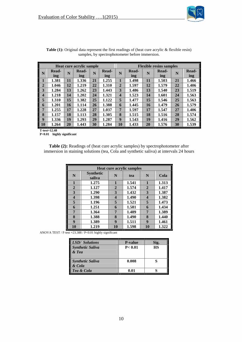
color change. Table (1) & Figure (10),
showed the first readings of (heat cure
acrylic & flexible resin) samples, by using
spectrophotometer (before immersion in any
staining solutions). From the first sight to
these numbers, the readings of flexible resin
samples are more than those of heat cure
acrylic samples, LSD showed highly
significant between them.
The results in table (2) showed the readings
of (heat cure acrylic samples) by
spectrophotometer after immersion in
staining solution (tea, Cola and synthetic
saliva as a “control") at intervals 24 hours.
The LSD appears that there are highly
significant differences between tea &
synthetic saliva, and significant differences
between Cola & synthetic saliva and
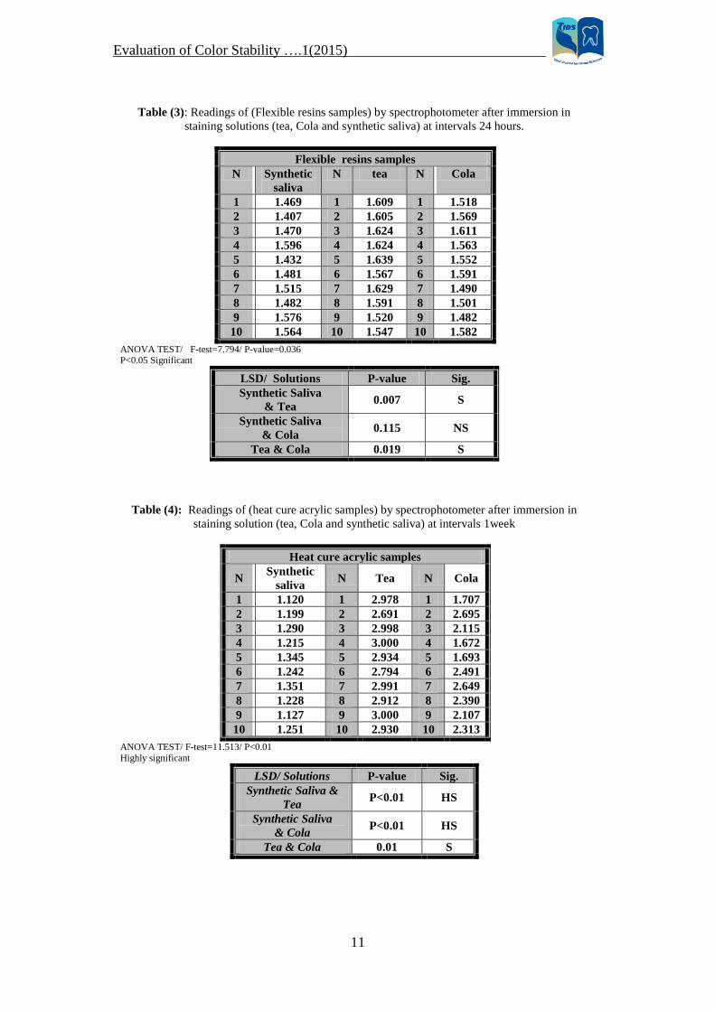
between tea & Cola. While the results
associated with (Flexible resins samples) at
the same intervals, appeared that there are
significant differences between tea &
synthetic saliva, and between tea & Cola.
There were non-significant differences
between Cola & synthetic saliva as shown in
table (3).
The results in table (4) & (5) showed the
readings of (heat cure acrylic) and
(flexible resin) samples respectively after
immersion in staining solutions for
1week. Highly significant differences
appeared between synthetic saliva & both
tea & Cola, & significant differences
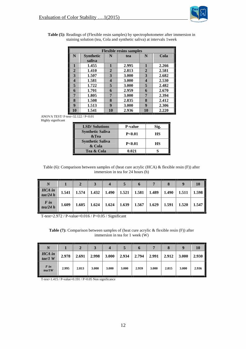
between Tea & Cola. The results in table (6), (7) showed a
compares between the two types of
denture materials used in this study, after
immersion in tea at two intervals (24
hours & 1week) respectively, While the
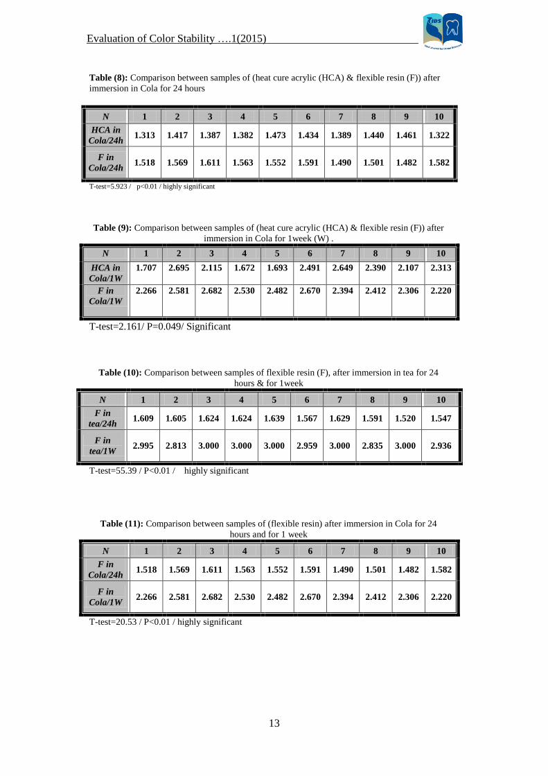
results in table (8), (9) showed a
compares between these materials after
immersion in Cola at the same intervals. Tables (10, 11, 12, and 13) showed the
effect of immersion time on the samples.
Table (10 & 11) showed the results of
flexible samples, while table (12, 13)
showed the results of heat cure acrylic
samples. In all tables there are highly
significant differences between the two
intervals.
Discussion: According to the types of denture
materials: The results showed that both types of
denture materials used in this study had
color changes after immersion in staining
solution. These are because; denture base
materials and denture teeth collect
deposits and stain in the same manner, as
doing natural teeth. Soft debris that clings
to a denture can be removed easily by
light brushing followed by rinsing. Hard
deposits and stains such as those that
occur from tea, coffee, cola and tobacco
tars are much difficult to remove. So
stains are pigmented deposits result from
the pigmentation of the colorless salivary
pellicle and may be removed by
polishing. The surface of the material
possess a certain degree of porosity and
surface roughness, and an organic mucin
and inorganic salt matrix must be developed
to increase the tenacity of the stain, which
should be indicative of these found in the
oral environment (19).
Porosity or a surface quality conducive to
the accumulation of debris & lead to a
significant discoloration (11) .This explained
the results which showed highly significant
differences between heat cure acrylic &
)5(2011…. Evaluation of Color Stability
5
flexible resin. Staining of acrylic resins
related to the chemico-physical
properties of the resin & also to the
patient habits. Fluid pigments from food,
beverage, drugs and nicotine are
deposited in the prosthetic appliances and
restorations, especially on the acrylic
resins, which are more porous than light
activated resin, reinforced resins and
hybrid resins (20).
The surface roughness of the flexible
resin is more than that of heat cure
acrylic resin. This is due to the finishing
& polishing procedures that are important
steps in the fabrication of heat cured
acrylic resin dentures (21). The role of
finishing & polishing is crucial in
reducing pigmentations (20). It would
seem that any efficient polishing
treatment should produce a smooth
surface, certainly on smooth surfaces
absorption of proteins is likely to be
reduced and cleaning would be facilitated (22).
According to the types of staining
solutions:
A- Tea solution: Maximum discoloration
was seen in (synthetic saliva & tea)
solution for both two materials. That
according to the results in table (2, 3, 4, 5),
the tea solution was the solution that cause
the most color changes for both heat cure
acrylic & flexible resin. This is duo to the
chief constituents of tea: - Inorganic
constituents (K+, Ca+2, P+3, Fe+2, Mg+2, S-2,
Al+3, Na+, Si+2, Zn+2, Cu+2 and F ), Nitrogen
compounds (a stimulant and diuretic
caffeine), and Polyphenols which are the
most important constitutions of tealeaf
that occurring in tea as derivatives of
gallic acid and cathechin. The best-
known gallic acid derivatives are the tannins (23). The stain is more likely composed of a
combination of iron and components of
the denatured salivary pellicle. So it
appeared necessary to denature the
salivary pellicle by tannic acid, organic
acids or detergents to produce stains
with iron (24). This would explain why
consumption of tannic acid would cause
discoloration. Scotti et al. in 1997 (20),
made an in vitro comparative evaluation
of the color variation of acrylic resins for
provisional prostheses. Auto-polymerized
resin-specimens were immersed in
synthetic saliva alone, synthetic saliva
with tea and synthetic saliva with coffee,
for periods of 20 and 30 days.
The resulting color changes were evaluated
using a spectrophotometer and compared to
the baseline, they concluded that all the
solutions darkened all the specimens,
darkening was found to be less in saliva
and saliva with tea solutions produced the
greatest darkening. Finally, Hanna & Al-
Ameer in 1999 (25), confirm the previous
results and concluded that tea caused more
stain of acrylic resin specimens when
compared to other solutions like saliva &
distilled water.
B- Cola Solution: The results in table (2,
3, 4, 5), showed that Cola solution is also
caused color changes for both hot cure
acrylic & flexible resin but it is less than
color changes caused by tea solution .This
is because the color or pigments is one of
the contents of Cola which are: Carbonated
water, high fructose corn syrup, caramel,
color, phosphoric acid, natural flavors, and
caffeine content (26).
Later on, Yuodelis et al. in 1980 (27),
immersed acrylic resins materials in Cola
& other types of solution for up 1month.
They reported that all materials showed
perceptible color changes after
immersion.
C- Synthetic saliva: The synthetic saliva
of Fusayama-Meyer type was the
electrolyte solution chosen for testing (17)
(18), this is to resemble the composition of
the normal saliva which is a mixture of
water, proteins, electrolytes and mineral
elements that constitute the saliva’s pH
and organic elements (mucin, Ig A) that
constitute its biological properties (28).
The total solids of saliva may vary from
0.3 % up to 1.4 %. Saliva of 0.6 % total
solids would contain about 0.4 % organic
and 0.2 % inorganic material.
The chief organic constituent is a
glycoprotein, mucin and the important
constituent is salivary amylase. The other
organic constituents include small
amounts of albumin and globulin, urea
and uric acid, and traces of thiocyanic
acid. The inorganic constituents are K+,
Na+, Ca+2, Mg+2, HCO3-, Cl-, HPO4-2 (29).
Extrinsic discoloration may be caused
by retention of colored substances in the
)5(2011…. Evaluation of Color Stability
6
salivary pellicle or by chemical alterations
of this organic integument (30).
In vitro spectrophotometric method, the
modifying effect of saliva upon the
staining of acrylic specimens by tea was
determined and showed that there was a
significant increase in the staining of
specimens by tea pretreated with saliva
when compared to specimens exposed to
tea only and it was concluded that pellicle
derived from salivary glycoprotein would
to encourage stain formation (31). These
results were confirmed by Addy & Robert (32). Hanna & Al-Ameer in 1999(25), found
that acrylic specimens soaked in saliva
for one hour twice daily then kept in tea
solution for the rest of time showed
highly significant differences from that
soaked in distilled water. This mean that
saliva caused increase in the staining of
specimens when compared to distilled
water.
According to the immersion time: Color changes of both materials were
increased with the increase of immersion
time. In this study, two intervals of
immersion were employed, 24 hours & 1
week. The immersion for 24 hours is
equivalent in time to 1 year of 4 minute
daily use as described by El-Badrawy et al. (33). And the color changes normally increase
at intervals (1 week) with increasing of
dipping time because of increasing of
exposure of the materials to the staining
solutions. Both the concentration of the
staining agents and the period of
exposure may affect the degree of
pigmentation (34) (20).
Extrinsic discoloration may be caused
by retention of colored substances in the
salivary pellicle or by chemical alterations
of this organic integument (35). It was
indicated that the salivary pellicle is a -
thin, structure less, organic layer formed
by a selective adsorption of glycoprotein
from saliva. The amount of these
discolorations was increase with the
increase of exposure of salivary pellicle to
the colored agent. (36)
Conclusions: 1- There was a highly significant
difference between heat cure acrylic &
flexible resin at baseline. Both of them,
had color changes after immersed in
staining solution and the (flexible resins
samples) were the higher value in
discoloration than (heat cure acrylic resin
samples) regardless the type of staining
solution.
2- Maximum discoloration was seen in
(synthetic saliva & tea) solution for both
two materials. Followed by (synthetic
saliva & Cola) & (synthetic saliva alone)
respectively.
3- The color changes of both materials were
increased with the increase of immersion
time.
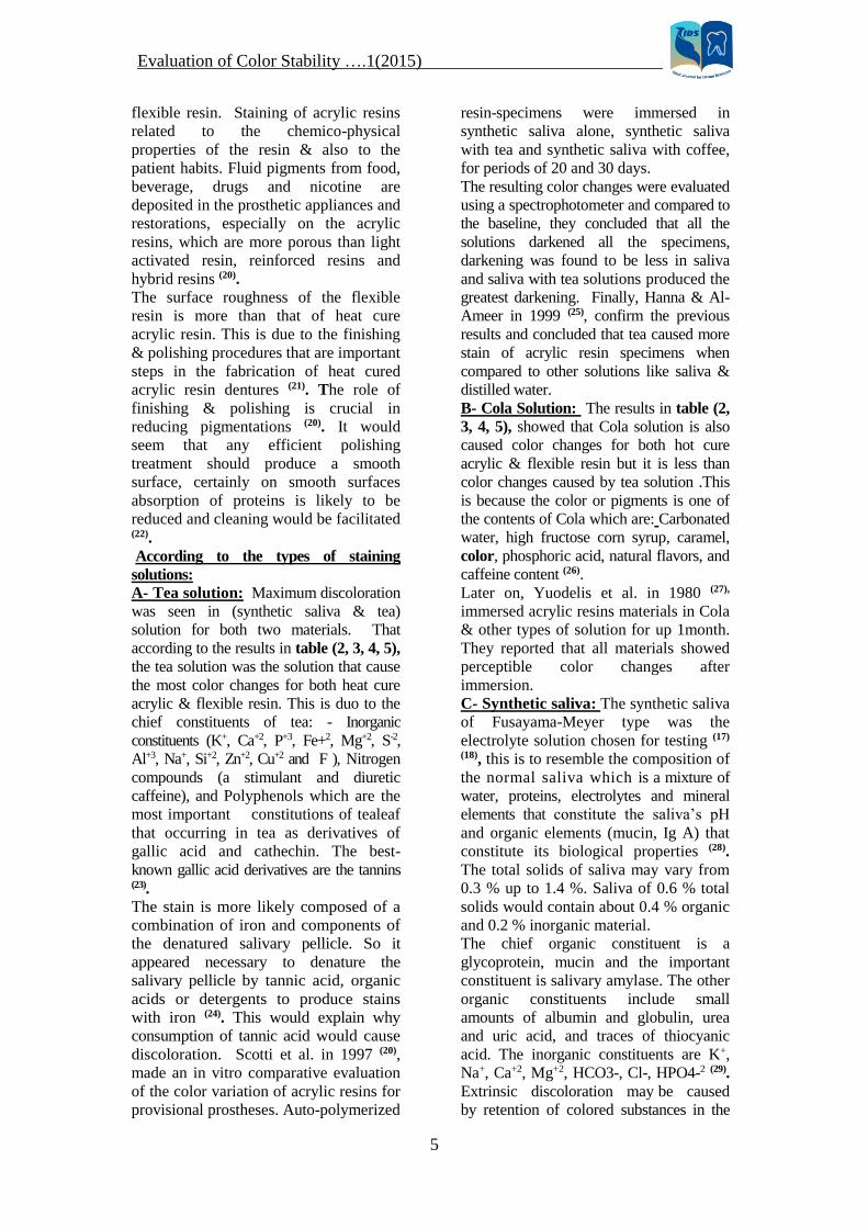
Figure (1): Samples used in this study.
A- 30 samples of heat cure acrylic resin.
B -30 samples of flexible resin.
)5(2011…. Evaluation of Color Stability
7
Figure (2): Mold used in this study Figure (3): The patterns were inserted to one half
of its depth.
Figure (4): Finishing of the
samples.
A B
Figure (5): A- A major sprue attach to the middle of specimen.
B- Two minor sprues attached to each other from one end and other end attached to the
specimen
)5(2011…. Evaluation of Color Stability
8
Figure (6): Test solution of tea & synthetic
saliva.
Figure (7): Test solution of Cola & synthetic
saliva.
Figure (8): 10 Samples of heat cured acrylic & 10 samples of flexible
resin are immersed in synthetic saliva alone as a control.
Figure (9): Spectrophotometer Devise

)5(2011…. Evaluation of Color Stability
9
1,259
1,521
0
0,2
0,4
0,6
0,8
1
1,2
1,4
1,6
Mean
Hot cure acrylic samples Flexible resins samples
1,29
1,499 1,522
1,595
1,401
1,545
0
0,2
0,4
0,6
0,8
1
1,2
1,4
1,6
Me
an
Synthetic saliva tea cola
Hot cure acrylic samples
Flexible resins samples
1,238
1,578
2,9222,956
2,184
2,48
0
0,5
1
1,5
2
2,5
3
Me
an
Synthetic saliva tea cola
Hot cure acrylic samples
Flexible resins samples
Figure (10): Histogram shows the mean of the optical density of (heat
cure acrylic & flexible resin), by using spectrophotometer (before
immersion in any staining solutions)
Figure (11): Histogram shows the mean of the optical density of
(heat cure acrylic samples &flexible resin samples) by
spectrophotometer after immersion in staining solution (tea, Cola
and synthetic saliva) at intervals 24 hours.
Figure (12): Histogram for the mean of the optical density of the
samples after immersion at intervals 1week.
)5(2011…. Evaluation of Color Stability
10
Table (1): Original data represent the first readings of (heat cure acrylic & flexible resin)
samples, by spectrophotometer before immersion.
Flexible resins samples Heat cure acrylic sample
Read-
ing N
Read-
ing N
Read-
ing N
Read-
ing N
Read-
ing N
Read-
ing N
1.466 21 1.503 11 1.498 1 1.255 21 1.336 11 1.381 1
1.406 22 1.579 12 1.597 2 1.310 22 1.219 12 1.046 2
1.519 23 1.540 13 1.486 3 1.443 23 1.262 13 1.284 3
1.563 24 1.601 14 1.523 4 1.321 24 1.202 14 1.218 4
1.563 25 1.546 15 1.477 5 1.122 25 1.382 15 1.310 5
1.579 26 1.479 16 1.445 6 1.388 26 1.114 16 1.201 6
1.406 27 1.547 17 1.597 7 1.037 27 1.228 17 1.255 7
1.574 28 1.516 18 1.515 8 1.305 28 1.113 18 1.157 8
1.562 29 1.416 19 1.543 9 1.287 29 1.293 19 1.336 9
1.539 30 1.576 20 1.433 10 1.284 30 1.443 20 1.264 10
T-test=12.48
P<0.01 highly significant
Table (2): Readings of (heat cure acrylic samples) by spectrophotometer after
immersion in staining solutions (tea, Cola and synthetic saliva) at intervals 24 hours
Heat cure acrylic samples
Cola N tea N Synthetic
saliva N
1.313 1 1.541 1 1.275 1
1.417 2 1.574 2 1.127 2
1.387 3 1.432 3 1.290 3
1.382 4 1.490 4 1.398 4
1.473 5 1.521 5 1.196 5
1.434 6 1.581 6 1.251 6
1.389 7 1.489 7 1.364 7
1.440 8 1.490 8 1.388 8
1.461 9 1.511 9 1.389 9
1.322 10 1.598 10 1.219 10
ANOVA TEST / F-test =23.388 / P<0.01 highly significant
LSD/ Solutions P-value Sig.
Synthetic Saliva
& Tea
P< 0.01 HS
Synthetic Saliva
& Cola
0.008 S
Tea & Cola 0.01 S
)5(2011…. Evaluation of Color Stability
11
Table (3): Readings of (Flexible resins samples) by spectrophotometer after immersion in
staining solutions (tea, Cola and synthetic saliva) at intervals 24 hours.
Flexible resins samples
Cola N tea N Synthetic
saliva
N
1.518 1 1.609 1 1.469 1
1.569 2 1.605 2 1.407 2
1.611 3 1.624 3 1.470 3
1.563 4 1.624 4 1.596 4
1.552 5 1.639 5 1.432 5
1.591 6 1.567 6 1.481 6
1.490 7 1.629 7 1.515 7
1.501 8 1.591 8 1.482 8
1.482 9 1.520 9 1.576 9
1.582 10 1.547 10 1.564 10
ANOVA TEST/ F-test=7.794/ P-value=0.036 P<0.05 Significant
LSD/ Solutions P-value Sig.
Synthetic Saliva
& Tea 0.007 S
Synthetic Saliva
& Cola 0.115 NS
Tea & Cola 0.019 S
Table (4): Readings of (heat cure acrylic samples) by spectrophotometer after immersion in
staining solution (tea, Cola and synthetic saliva) at intervals 1week
Heat cure acrylic samples
Cola N Tea N Synthetic
saliva N
1.707 1 2.978 1 1.120 1
2.695 2 2.691 2 1.199 2
2.115 3 2.998 3 1.290 3
1.672 4 3.000 4 1.215 4
1.693 5 2.934 5 1.345 5
2.491 6 2.794 6 1.242 6
2.649 7 2.991 7 1.351 7
2.390 8 2.912 8 1.228 8
2.107 9 3.000 9 1.127 9
2.313 10 2.930 10 1.251 10
ANOVA TEST/ F-test=11.513/ P<0.01
Highly significant
LSD/ Solutions P-value Sig.
Synthetic Saliva &
Tea P<0.01 HS
Synthetic Saliva
& Cola P<0.01 HS
Tea & Cola 0.01 S
)5(2011…. Evaluation of Color Stability
12
Table (5): Readings of (Flexible resin samples) by spectrophotometer after immersion in
staining solution (tea, Cola and synthetic saliva) at intervals 1week
Flexible resins samples
Cola N tea N Synthetic
saliva
N
2.266 1 2.995 1 1.455 1
2.581 2 2.813 2 1.410 2
2.682 3 3.000 3 1.507 3
2.530 4 3.000 4 1.581 4
2.482 5 3.000 5 1.722 5
2.670 6 2.959 6 1.701 6
2.394 7 3.000 7 1.805 7
2.412 8 2.835 8 1.508 8
2.306 9 3.000 9 1.513 9
2.220 10 2.936 10 1.541 10
ANOVA TEST/ F-test=32.122 / P<0.01
Highly significant
LSD/ Solutions P-value Sig.
Synthetic Saliva
&Tea P<0.01 HS
Synthetic Saliva
& Cola P<0.01 HS
Tea & Cola 0.021 S
Table (6): Comparison between samples of (heat cure acrylic (HCA) & flexible resin (F)) after
immersion in tea for 24 hours (h)
10 9 8 7 6 5 4 3 2 1 N
1.598 1.511 1.490 1.489 1.581 1.521 1.490 1.432 1.574 1.541 HCA in
tae/24 h
1.547 1.520 1.591 1.629 1.567 1.639 1.624 1.624 1.605 1.609 F in
tea/24 h
T-test=2.972 / P-value=0.016 / P<0.05 / Significant
Table (7): Comparison between samples of (heat cure acrylic & flexible resin (F)) after
immersion in tea for 1 week (W)
10 9 8 7 6 5 4 3 2 1 N
2.930 3.000 2.912 2.991 2.794 2.934 3.000 2.998 2.691 2.978 HCA in
tae/1 W
2.936 3.000 2.815 3.000 2.959 3.000 3.000 3.000 2.813 2.995 F in
tea/1W
T-test=1.415 / P-value=0.191 / P>0.05 Non significance
)5(2011…. Evaluation of Color Stability
13
Table (8): Comparison between samples of (heat cure acrylic (HCA) & flexible resin (F)) after
immersion in Cola for 24 hours
10 9 8 7 6 5 4 3 2 1 N
1.322 1.461 1.440 1.389 1.434 1.473 1.382 1.387 1.417 1.313 HCA in
Cola/24h
1.582 1.482 1.501 1.490 1.591 1.552 1.563 1.611 1.569 1.518 F in
Cola/24h
T-test=5.923 / p<0.01 / highly significant
Table (9): Comparison between samples of (heat cure acrylic (HCA) & flexible resin (F)) after
immersion in Cola for 1week (W) .
10 9 8 7 6 5 4 3 2 1 N
2.313 2.107 2.390 2.649 2.491 1.693 1.672 2.115 2.695 1.707 HCA in
Cola/1W
2.220 2.306 2.412 2.394 2.670 2.482 2.530 2.682 2.581 2.266 F in
Cola/1W
T-test=2.161/ P=0.049/ Significant
Table (10): Comparison between samples of flexible resin (F), after immersion in tea for 24
hours & for 1week
10 9 8 7 6 5 4 3 2 1 N
1.547 1.520 1.591 1.629 1.567 1.639 1.624 1.624 1.605 1.609 F in
tea/24h
2.936 3.000 2.835 3.000 2.959 3.000 3.000 3.000 2.813 2.995 F in
tea/1W
T-test=55.39 / P<0.01 / highly significant
Table (11): Comparison between samples of (flexible resin) after immersion in Cola for 24
hours and for 1 week
10 9 8 7 6 5 4 3 2 1 N
1.582 1.482 1.501 1.490 1.591 1.552 1.563 1.611 1.569 1.518 F in
Cola/24h
2.220 2.306 2.412 2.394 2.670 2.482 2.530 2.682 2.581 2.266 F in
Cola/1W
T-test=20.53 / P<0.01 / highly significant
)5(2011…. Evaluation of Color Stability
14
References
1- Anusavice Jk: Philips Science Of Dental
Materials.10th Philadelphia: WB Saunders CO;
1996, p: 211-235.
2- Wang X, Powers JM, Connelly ME. Color
stability of heat activated and chemically activated
fluid resin acrylics. J Prosthodont 1996; 5(2), P:
266-69.
3- Wang RL, Moore BK, Goodacre CJ, Swartz ML,
and Anders CJ., A comparison of resins for
fabricating provisional fixed restorations . INT J
Prosthodont. 1989; 2(1), P: 173-84.
4- kutsch V.K. whitehous J; schermerhorn K;
bowers R; The evolution and advancement of dental
thermoplastics. Dental town, 2003; Feb., 2:52-56.
5- Negrutiu M.; Sinescue C.R.; Daniela P.; lakatos
S. ; Thermoplastic resin for flexible framework
partial denture, Aust. Dent J; 2005; (3):269.
6- Backenstose WM & Wells JG., Side effect of
immersion type cleansers on the metal component
of dentures., J Prosthet Dent 1977; 37 (6), P: 615-
621.
7- Polyzois GL: Denture cleansing habits: A survey.
Aust Dent J 1983; 28 (3), P: 171-173.
8- Jagger DC & Harrison A: Denture cleansing-the
best approach (Abstract). Br Dent J 1995; 178 (11),
P: 413-417.
9- Lindhe J: Text-book of clinical periodontology.
Compenhagen: Munksgaard, 1996, P: 123.
10- Craig RG, O’Brein WJ and Powers JM: Dental
materials: Properties and manipulation, second ed.
St. Louis: The CV-Mosby Company, 1979, P: 142.
11- Crispin BJ, Caputo AA. Color stability of
temporary restorative materials, J Prosthet
Dent.1979; 42(4), P: 27-33.
12- P. Malik & M. Rathee: Evaluation of color
stability of temporary fixed partial denture
materials: In –Vitro study. The Internet Journals of
Dental Science, 2010, Volume 9, number 1, P: 67.
13- Abdul-Karim J.: Evaluation Of Some Technical
Properties Of Acrylic Denture Base Material
Relined With Different Denture Relines Materials.
M.Sc Thesis Collage Of Dentistry.University Of
Baghdad, 2001.
14- Abdul-Rahmann B.: Evaluation of Water
Sorption, Solubility and Bone Strength Of Some
Soft Lining, 2002.
15- Mustafa M.J. , Evaluation of Candida albicans
attachment to flexible denture base materials and
heat cure acrylic resin using different finishing and
polishing techniques ,M Sc. Thesis ,University of
Baghdad ,Iraq, .2011.
16- P. Malik & M. Rathee: Evaluation of color
stability of temporary fixed partial denture
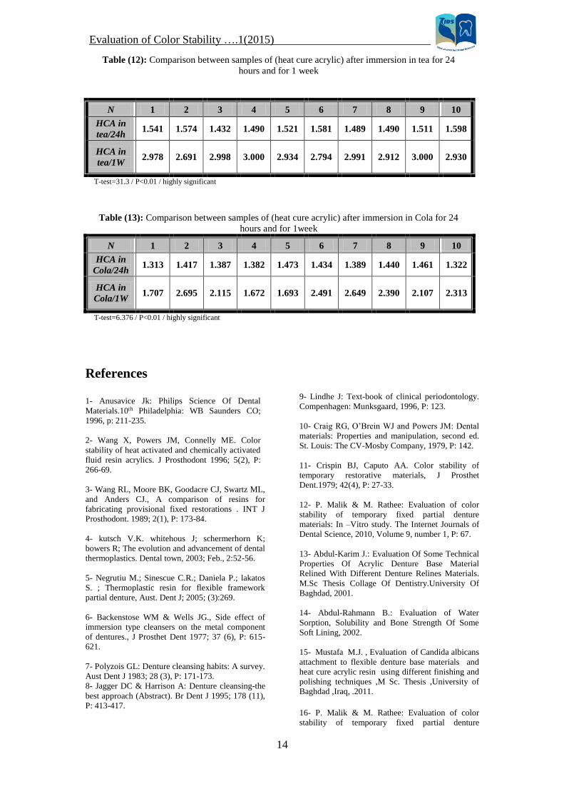
Table (12): Comparison between samples of (heat cure acrylic) after immersion in tea for 24
hours and for 1 week
10 9 8 7 6 5 4 3 2 1 N
1.598 1.511 1.490 1.489 1.581 1.521 1.490 1.432 1.574 1.541 HCA in
tea/24h
2.930 3.000 2.912 2.991 2.794 2.934 3.000 2.998 2.691 2.978 HCA in
tea/1W
T-test=31.3 / P<0.01 / highly significant
Table (13): Comparison between samples of (heat cure acrylic) after immersion in Cola for 24
hours and for 1week
10 9 8 7 6 5 4 3 2 1 N
1.322 1.461 1.440 1.389 1.434 1.473 1.382 1.387 1.417 1.313 HCA in
Cola/24h
2.313 2.107 2.390 2.649 2.491 1.693 1.672 2.115 2.695 1.707 HCA in
Cola/1W
T-test=6.376 / P<0.01 / highly significant
)5(2011…. Evaluation of Color Stability
15
materials: In –Vitro study. The Internet Journals of
dental science, 2010, Volume 9, number 1.
17- Brune D, Hultquist G And Leygrat
C,"Corrosion Resistance Of Passivated & Hoh-
Passivated Cobalt-Chromium Alloy", Scard J Pent
Res ,1984;92(3):262-67.
18- Luthy H,Marinello R And Schare R, "Corrosion
Considerations In The Braming Trepair Of Cobalt
Based Partial Dentures", J .Prosthet Dent,
1996;75:515-24.
19- Pipko Dj And El-Sadeek M: An In Vitro
Investigation Of Abrasion And Staining Of Dental
Resins, J Den Res, 1972; 15(3), P: 689-705.
20- Scotti R, Mascellani Sc and Foruiti F: The In
Vitro Color Stability of Acrylic Resin for
Provisional Restorations. Int Prosthodont 1997; 10
(2), P: 164-168.
21- Uluosoy M, Uluosoy N and Aydin Ak: An
Evaluation Of Polishing Techniques On Surface
Roughness Of Acrylic Resin. J Prosthet Dent 1986;
56(1), P: 107-112.
22- Ellis B and Faraj S: The Structure and Surface
Topography of Acrylic Denture Base Materials. J
Dent, 1980, 8(2), P: 102-108.
23- Thorpe WV, Bray HG and James SP:
Biochemistry for Medical Students. Ninth Edition,
J&A Churchill Ltd.1970.
24- Nordbo H, Attramadal A And Eriksen Hm: Iron
Discoloration Of Acrylic Resin Exposed To
Chlorhexidine Or Tannic Acid: A Model Study. J
Prosthet Dent 1983; 49(1), P: 126-129.
25- Hanna NH and Al-Ameer SH: The influence of
saliva and / or tea on the staining ability of
chlorhexidine to hot cures acrylic resin as a mouth
wash and staining effect as a disinfectant. J College
Dentistry, 1999; 4: 159-168.
26- Addy M,Prayitno S, Taylor L and Cadogan S:
An In Vitro Study Of The Role Of Dietary Factors
In The Etiology Of Acrylic Staining Associated
With The Use Of Chlorohexidine, J Perio Res,
1979; 14(5), P:403-410.
27- Yuodelis RA, Faucher R, Provisional
restorations: An integrated approach to periodontics
and restorative dentistry. Dent Clin North Am 1980;
24-285-03.
28- Fakhoury M and Poraldi C: Importance of
Saliva in the Preventation of Dental Caries. Dental
News, 1996; 3(2), P: 17-22.
29- Thorpe WV, Bray HG and James SP:
Biochemistry for Medical Students, Ninth Edition
.J&A Churchill Ltd.1970.
30- Eriksen HM and Nordbo H: Extrinsic
Discoloration of Teeth. J Clin Perio, 1978; 5; P:
229-236.
31- Prayitno S and Addy M: An in vitro study of
factors affecting the development of staing
associated with the use of chlorhexidin. J Perio Res,
1979; 14, P: 397-402.
32- Addy M and Robert WE: Comparison of the
bisbiguanide antiseptics alexidine and
chlorhexidine: Clinical and in vitro staining
properties. J Cli Perio, 1989; 8: 220-230.
33- El-Badrawy WA, McComb D & Wood RE.
Effect of home-use fluoride gels on glass ionomer
and composite resin restorations. Dent Mater, 1993;
9, P: 63-67.
34- Khokhar ZA, Razzoog ME and Yaman P:
Color Stability Of Restorative Resins, Quint Int,
1991, 22(9), P: 733-737.
35- Eriksen HM and Nordbo H: Extrinsic
Discoloration of Teeth .J Clin Perio 1998; 5; 229-
236.
36-Collins WJM, Forrest JO and Walsh TF: A
Handbook of Dental Hygeinists. Wright Bristol,
1986.


![LINEARITY OF STABILITY CONDITIONS - Brandeis Universitypeople.brandeis.edu/~igusa/Papers/Linearity1706.pdfStability Theorem in [10] gives the precise relation between semi-invariants](https://img.pdfslide.us/doc/110x75/5f89c0236233081a6279f15d/linearity-of-stability-conditions-brandeis-igusapaperslinearity1706pdf-stability.jpg)