Embed Size (px)
Citation preview

Periodontic _ Endodontic Relationship

• DEFINITION
• CLASSIFICATION
• PATHWAYS OF COMMUNICATION BETWEEN PULP
& PERIDONTAL DISEASE
• ETIOLOGY OF PULPAL DISEASE
• CLASSIFICATION OF PULPAL DISEASE
• CLASSIFICATION OF PERIODONTAL DISEASE
• CLINICAL FINDINGS OF ENDODONTIC &
PERIODONTIC LESION
• CLINICAL FINDINGS OF COMBINED ENDO- PERIO
LESION
• TREATEMENT
INDEX

DEFINITION
Simring M, Goldberg M.The pulp pocket approach:retrograde periodontitis. J periodontal 1964:35:22-48
It,s the spread of inflammation and infection from one
component to the other.
Periodontic _ Endodontic Relationship

endo
perio
Embryonic , anatomic , functional

Pulpal and Periodontal problems are responsible for more than 50% of tooth mortality.
Chen SY,Wang HI,Clickman GN. The infuence of endodontic treatment upon periodontal wound healing.
J Clin periodontal24;449-456,1997
Bender IB. Factors influencing radiographic appearance of bony lesions. J Endod 8;161-170,1982

Perio-Endo
Relationship
Apical
Foramen
Dentinal
Tubules
Lateral & Accessory
canals


Complicate Instrumentation
Progression of dental Caries
Direct Local Trauma
1
2
3
Etiologic Factors
( Bacteria or Biochemical Toxins….)
( tooth fracture....)
(perio,resto,prosth...)
Pulpal disease

Chen SY,Wang HI,Clickman GN. The infuence of endodontic treatment upon periodontal wound healing.
J Clin periodontal24;449-456,1997
Bender IB. Factors influencing radiographic appearance of bony lesions. J Endod 8;161-170,1982
Pulpal disease :the major causes of pulpal inflammation are: 1- Instrumentation during periodontal , Restorative , or Prosthestic dentistry .
2- Progression of dental caries .
3- Direct trauma such as tooth fracture .

Dental caries is the most common cause of pulpal disease . Due to the ability of micro – organisms and their products to penetrate through the dentinal tubules to the pulp and causing pulpal inflammation .
Chen SY,Wang HI,Clickman GN. The infuence of endodontic treatment upon periodontal wound healing.
J Clin periodontal24;449-456,1997
Bender IB. Factors influencing radiographic appearance of bony lesions. J Endod 8;161-170,1982

Vascular&
Lymphatic
Drainage
Bacteria,s
Virulence
Host
Responce
Effectiveness
Of Pulpal
Circulation
The dynamics of the pulpal reaction is dictated by the following :

Pulpal inflammation and necrosis are initiated by:
Dental caries, Restorative procedures, Trauma,
Chemical irritation and Severe thermal stimulation.
Pathogenesis
These inflammatory lesions cause localized edema and a
resulting increase in intra- pulpal pressure and cell death.

Periodontal lesions are initiated by deposits of plaque and
calculus: The toxins produced by these bacteria can irritate the gum tissues and
cause the body’s immune system to “turn on” (chronic inflammation) –
this inflammation can break down and destroy the tissues and bone
supporting the tooth. The gum tissues separate from the tooth, forming
pockets. As the disease progresses, the pockets deepen, destroying more
supporting tissues.
Pathogenesis

Bacteria Associated with Pulpitis
Eubacterium
Peptostreptococcus
Fusobacterium
Porphyromonas
Prevotella
Streptococcus
Lactobacillus
Wolinell
Actinomyces

Bacteria Associated with Periodontitis

Classification of Pulpal Disease
Reversible pulpitis
Irreversible pulpitis
Pulpal Necrosis

Classification of pulpal disease : 1- Reversible pulpitis : minor injury such as periodontal root planing or the conservative preparation of a tooth for a restoration may lead to pulpal damage . A transient hypersensitivity to thermal stimuli is the most common symptom noted .The response rapidly disappears after removal of the stimulus . The reversibility of inflammation and symptoms , without permanent pulpal damage ,has led to classification of this condition as reversible pulpitis .
Chen SY,Wang HI,Clickman GN. The infuence of endodontic treatment upon periodontal wound healing.
J Clin periodontal24;449-456,1997
Bender IB. Factors influencing radiographic appearance of bony lesions. J Endod 8;161-170,1982

Classification of pulpal disease : 2- Irreversible pulpitis : If the pulp is affected to the point that the inflammatory lesion cannot be resolved , even though the source of truma is eliminated , a progressive degeneration of the pulp results. This progression has been described as Irreversible pulpitis.
Chen SY,Wang HI,Clickman GN. The infuence of endodontic treatment upon periodontal wound healing.
J Clin periodontal24;449-456,1997
Bender IB. Factors influencing radiographic appearance of bony lesions. J Endod 8;161-170,1982
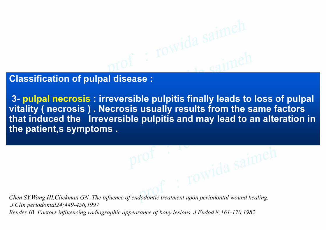
Classification of pulpal disease : 3- pulpal necrosis : irreversible pulpitis finally leads to loss of pulpal vitality ( necrosis ) . Necrosis usually results from the same factors that induced the Irreversible pulpitis and may lead to an alteration in the patient,s symptoms .
Chen SY,Wang HI,Clickman GN. The infuence of endodontic treatment upon periodontal wound healing.
J Clin periodontal24;449-456,1997
Bender IB. Factors influencing radiographic appearance of bony lesions. J Endod 8;161-170,1982

AAP Classification of Periodontal Diseases and Conditions (1999)
• Gingival Diseases
– Dental plaque-induced gingival diseases
– Non-plaque induced gingival lesions
• Chronic Periodontitis (Slight: 1-2mm CAL; moderate:
3-4mm CAL; severe: >5mm CAL)
– Localized
– Generalized (>30% of sites are involved)
• Aggressive Periodontitis (Slight: 1-2mm CAL; moderate: 3-
4mm CAL; severe: >5mm CAL)
– Localized
– Generalized

• Periodontitis as a Manifestation of
Systemic Diseases
– Associated with hematological disorders
– Associated with genetic disorders
– Not otherwise specified
• Necrotizing Periodontal Diseases
– Necrotizing ulcerative gingivitis
– Necrotizing ulcerative periodontitis
• Abscesses of the Periodontium
– Gingival abscess
– Periodontal abscess
– Pericoronal abscess
AAP Classification of Periodontal Diseases and Conditions (1999)

• Periodontitis Associated with Endodontic Lesions
– Combined periodontic-endodontic lesions
• Developmental or Acquired Deformities and Conditions
– Localized tooth-related factors that modify or predispose to plaque-
induced gingival diseases periodontitis
– Mucogingical deformities and conditions around teeth
– Mucogingival deformities and conditions on edentulous ridges
– Occlusal trauma
AAP Classification of Periodontal Diseases and Conditions (1999)

As long as the pulp remains vital, it is unlikely that
significant changes will occur in the periodontium..
Necrosis , can result in bone resorption and rediolucency
at the apex , In the furcation , or at points along the root .
Effect on the periodontium
Pulpal tissue inflamed may be little or no effect on the periodontium .

Effect on the periodontium
Dental radiographs usually document the presence of apical
or lateral lesion .The resulting lesion may be an acute apical
lesion , or abscess , a more chronic periradicular Lesion ( cyst
or granuloma ), or lesion associated with a lateral or accesory
canals . The lesion may remain small or it can expand to
destroy the attachment of the tooth and communicate with a
lesion of periodontitis .

The ability of inflammatory periodontal disease to affect the pulp is
much less certain.
Effect of the periodontitis on the dental pulp
Effect of the periodontitis on the dental pulp
It has been suggested that the presence of an intact layer of
cementum may protect the pulp from damage produced by
microbia .
Sever breakdown of the pulp does not occur until periodontitis
has reached a terminal state , when bacteria plaquehas involved
the main apical foramen .
The pulp has a good capacity for defence as long as the blood
supply through the apical foramen is intact .therefore ,retrograde
periodontitis , if it occurs ,is rare .

Effect of the periodontitis on the dental pulp
The effects of pulpal disease on the periodontium are well
documented , a clear –cut relationship between periodontitis
and pulpal involvement is less evident .One may possible that
bacteria and the inflammatory products of periodontitis could
gain access to the pulp through accessory canals , apical
foramen , or dentinal tubules .
This process , the reverse of the effects of a necrotic pulp on the
periodontum has been referred to as retrograde pulpitis .

Differences between periodontal and pulpal lesion
Signs and symptoms of periodontitis :teeth with chronic
periodontal lesions are typically free of acute symptoms,
the patient may be unware of the condition ,except for
bleeding on brushing and flossing , or bad breath .
Increased tooth mobility may occur if sufficient
attachment has been lost .
dental radiographs usually disclose the extent of
attachment loss , which should correlate with clinical
probing data .

Signs and symptoms of pulpal disease:
the teeth with pulpal inflammation ,respond normally
to percussion and palpation .
Thermal stimuli or percussion applied to teeth with
irreversible pulpitis can provoke sever pain .
This pain may be intense and is often described as
bright or throbbing .
Differences between periodontal and pulpal lesion

If the inflammatory process extends to involve the
periodontal ligament , the affected tooth can become
tender to pressure , biting ,or light tapping with an
instrument .
Dental radiograghs usally document the presence of
apical or lateral lesions .
The clinician should remember that some inflamed and
necrotic pulps are asymptomatic and that the patient is
unaware of their existence .
Differences between periodontal and pulpal lesion

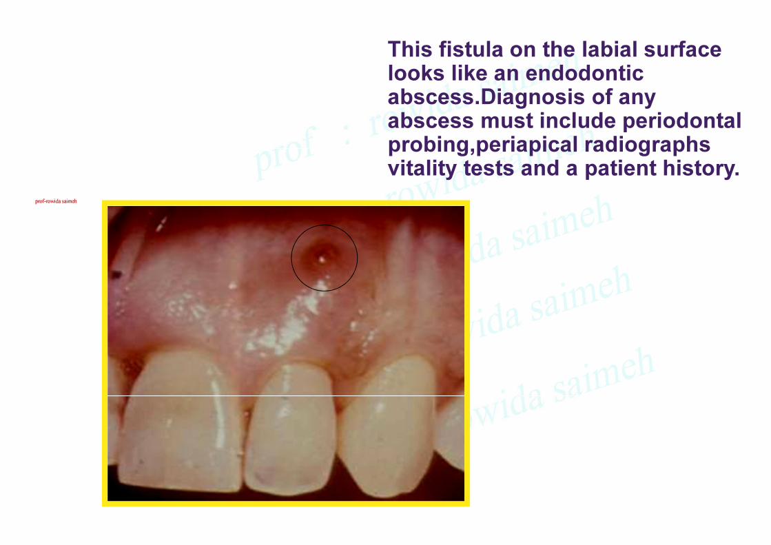
These photos are from the patient shown in the previous slide. The lateral incisor tested vital and the abscess was a periodontal abscess that was initiated with pockets starting in a cingulum groove of the palatal surface.

The path of the sinus tract can be determined
by carefully placing a fine gutta percha point
into the fistula and then making a radiograph .
The point stops within the periodontal pocket .
Careful probing confirms the presence of the
pocket , and dilation of the sulcus usually
results in drainge .
This fistula on the labial surface looks like an endodontic abscess.Diagnosis of any abscess must include periodontal probing,periapical radiographs vitality tests and a patient history.

Vertical root fracture

Vertical root fracture

This case shows a combination of periodontitis and endodontic inflammation causing bone loss at the crest and at the apex.


classification
PRIMARY ENDODONTIC LESION.
SIMON 1972
Chronic apical lesion on a tooth with a necrotic
pulp may drian coronally through the periodontal
ligament into the gingival sulcus.
Usually heal following well root canal treatment .
Pulpal infection may cause a tissue-destructive
process that proceeds from the apical region of
a tooth toward the gingival margin.
“retrograde periodontitis”
Based on the primary source of infection.
PRIMARY ENDODONTIC LESION
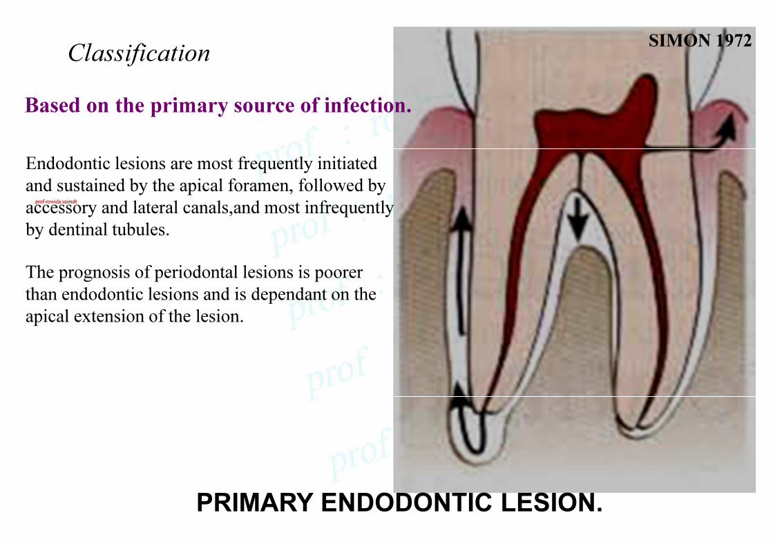
Classification
PRIMARY ENDODONTIC LESION.
SIMON 1972
Based on the primary source of infection.
Endodontic lesions are most frequently initiated
and sustained by the apical foramen, followed by
accessory and lateral canals,and most infrequently
by dentinal tubules.
The prognosis of periodontal lesions is poorer
than endodontic lesions and is dependant on the
apical extension of the lesion.

classification
PRIMARY ENDODONTIC LESION WITH SECONDARY PERIODONTAL INVOLVEMENT.
SIMON 1972
It,s untreated primary endodontic
lesion involved with secondary
periodontal breakdown. This cases
may also occurs as a result of root
perforation during root canal
treatment, or where pins and posts
may have been misplaced during
restoration of the crown. Root
fractures may also be present.
Based on the primary source of infection.

classification
PRIMARY PERIODONTIC LESION.
SIMON 1972
In this process chronic periodontitis progresses
apically along the root surface. In this cases pulpal
test indicate a clinically normal pulp reaction. There
is frequently an accumlation of plaque and the
presence of deep pockets may be detected.
Based on the primary source of infection.
PRIMARY PERIODONTAL LESION

classification
PRIMARY PERIODONTIC LESION WITH SECONDARY ENDODONTIC INVOLVEMENT.
SIMON 1972
The apical progression of a periodontal pocket
continue until the apical tissues are involved.
The pulp may become necrotic as a result of
infection entering through lateral canals or apical
foramen.
Pulpal changes resulting from periodontal disease
are more likely to occur when the apical foramen
is involved.
In molars not all roots may infected.
Based on the primary source of infection.

classification SIMON 1972
Based on the primary source of infection.
Two independent lesions, periapical and
marginal, can coexist and eventually fuse
with each other.
Combined endodontic-periodontic lesions

TREATEMENT

TREATEMENT
PRIMARY ENDODONTIC LESION
CONVENTIONAL ENDODONTIC THERAPY
Are sufficient to result in healing of the lesion .Periodontal treatment is not required in the absence of any periodontal involvement .

TREATEMENT
Appropriate treatment varies with the
presence , nature , and extent of
Involvement of the disease .

TREATEMENT
PRIMARY PERIODONTAL LESION
1-PERIODONTAL THERAPY
2-GUIDED TISSUE REGENERATION
3-ROOT AMPUTATION & HEMISECTION
4-PULP THERAPY


PRIMARY PERIODONTAL LESION
WITH SECONDARY ENDO LESION
1-PULP THERAPY
2-PERIODONTAL THERAPY
3-ROOT AMPUTATION
4-GTR
TREATEMENT

TREATEMENT
INDEPENDENT ENDODONTIC AND PERIODONTAL LESION
ENDO - PERIO THERAPY
THE PROMPT MANAGEMENT OF THE PULPAL
LESION IS THE PRIMARY CONCERN .THERAPY
FOR PERIODONTITIS MAY BE DELAYED UNTIL
THE ACUTE SYMPTOMS OF PULPAL DISEASE
ARE ALLEVIATED .

TREATEMENT
INDEPENDENT ENDODONTIC AND PERIODONTAL LESION
The involvement of the apical periodontium by a pulpal lesion may obscure the symptoms of periodontitis. In most cases the lesions are independent .The patient,s history and probing allow adetermination of the extent of each problem and the independence of the two defects .Endodontic annd periodontic therapy is required for a successfu result .

classification
TRUE COMBINED LESION.S
SIMON 1972
Occurs less frequently than other,s. It is formed
when an endodontic lesion progressing coronally
joins an infected periodontal pocket progressing
apically.
In molar teeth, root resection can be an alternative
treatment.
The prognosis of a true-combined perio-endo
lesion is often poor or even hopeless, especially
when periodontal lesions are chronic with
extensive loss of attachment.
Based on the primary source of infection.
combined endodontic-periodontic lesions

Endodontic-Periodontic lesions
Such lesion , may present with the characteristics of both
diseases , which may :

Complicate Complicate the Diagnosis
Complicate the Treatment Plan
Affect the Sequence of Care
1
2
3
Periodontic _ Endodontic Relationship

In combined endodontic-periodontic lesions, it is
generally wise to treat the Endodontic component
first, because in many cases this will lead to
complete resolution of the problem.

If The periodontitis progresses to
involve a lateral canal or apical tissues.
A secondary pulpal infection may be
induced , referred to as retrograde
pulpitis .if it exists ,it is Rare .

The pain from the loss of pulpal vitality is the most common
complaint of patients with combined lesions.

History and careful clinical and radiographic examinations are required to identify lesion and contribution of each lesion ,and
to produce an optimal treatment result .
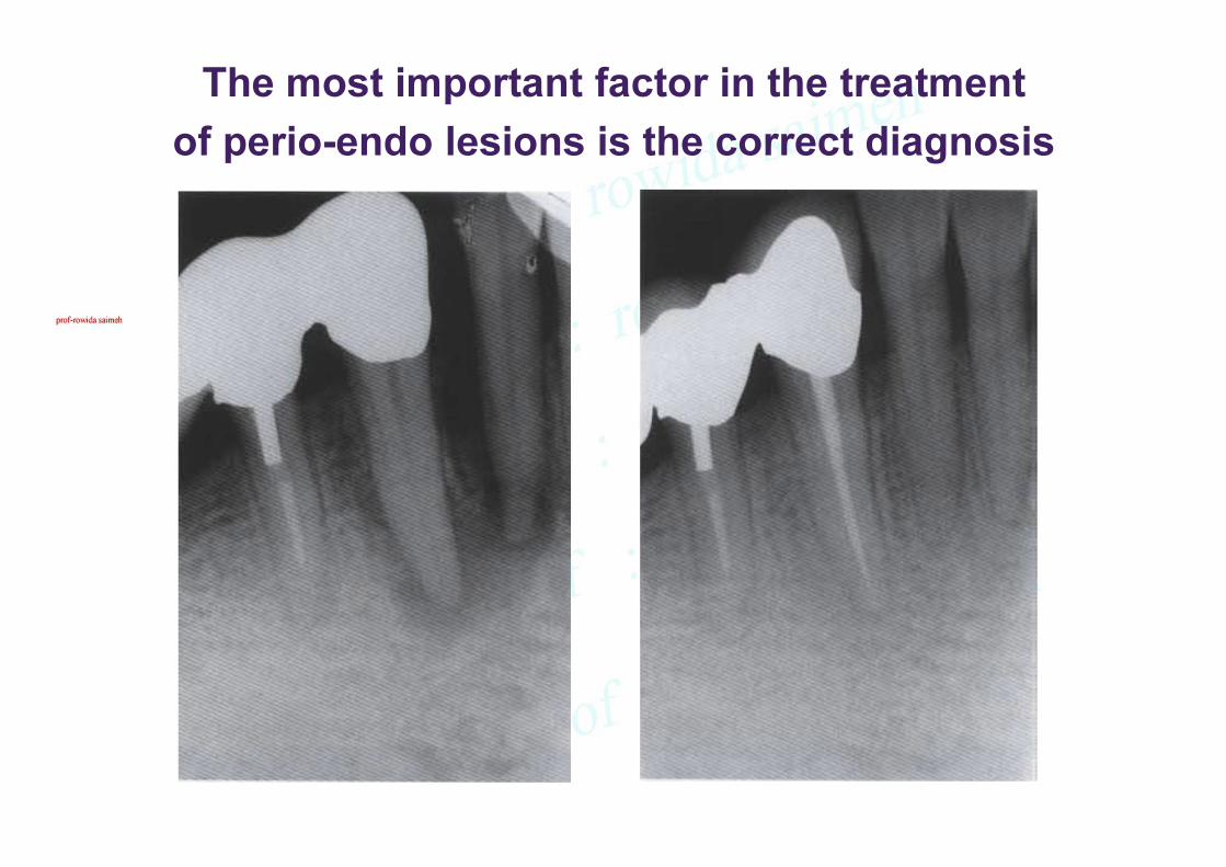
The most important factor in the treatment
of perio-endo lesions is the correct diagnosis


Removal of caries
Endodontic therapy
Furcal bone
loss resolved
after endodontic
treatment carried
out before any
periodontal care

The location, extention, severity of inflammation and the degree of
tissue involvment helps the dentist to select the proper treatment.

The residual periodontal pocket that remains can be best treated .
Thus, periodontal treatment may include scaling and root planing ,
as well as various surgical treatment . If the endodontic lesion
requires apical surgery .The surgical treatment of both lesion
performed at the same time .

The periodontal portion of the defect has plaque , calculus , or root roughness
This contaminated root and the associated osseous defect constitute the major
complication to treatment of combined lesion .once the decision to retain the
tooth is made , endodontic therapy should precede attempts at periodontal
pocket elimination .after successful endodontic treatment .

Prognosis of combind lesion :
The prognosis of periodontal lesions is
poorer than endodontic lesions ,and is
dependant on the apical extension of
the lesion.
The prognosis of a true-combined perio-
endolesion is often poor or even
hopeless, especially when periodontal
lesions are chronic with extensive loss
of attachment.

DIAGNOSIS •PAIN
•SWELLING
•MOBILITY
•SUPPURATION
•PERIODONTAL PROBING
•PRESENCE OF LOCAL DEPOSITS
•PRESENCE OF CARIES AND RESTORATION
•PALPATION
•PULP VITALITY TEST
•RADIOGRAPHIC INTERPRETATION

DIAGNOSIS
Diagnosis of primary endodontic or
periodontic disease usually easy clinically.
The pulp is vital and responsive to testing
in periodontic disease, while it,s infected
and nonvital in primary endodontic disease.

CLINICAL FINDINGS IN ENDODONTIC AND
PERIODONTIC LESIONS
CLINICAL FINDINGS
ENDO LESION PERIO LESION
PULPAL RESPONSE
ABSENT PRESENT
BONE DEFORMITY
TUBULAR ‘U’ TRIANGULAR ‘V’
PLAQUE & CALCULUS
ABSENT PRESENT
CARIES/ RESTORATION
PRESENT ABSENT
MOBILITY
ABSENT PRESENT
GEN PERIODONTITIS
ABSENT PRESENT


CLINICAL FINDINGS IN ENDODONTIC AND COMBINED
ENDODONTIC-PERIODONTIC LESIONS
CLINICAL FINDINGS ENDO LESION
COMBINED LESION
PULPAL STATUS
NECROTIC NECROTIC
PERIO STATUS
NORMAL GEN PERIODONTITIS
PROBING
NARROW POCKET WIDE POCKET
PLAQUE & CALCULUS
ABSENT PRESENT
TREATMENT
ENDODONTIC COMBINED
PROGNOSIS
GOOD DEPENDS ON PERIO

There is no shame in
making a mistake. It would
be a shame not to learn
from your mistakes.