Embed Size (px)
Citation preview

EVALUATION OF AXILLARY LYMPH NODES
AFTER NEOADJUVANT SYSTEMIC THERAPY
KIM, MIN JUNG
SEVERANCE HOSPITAL, YONSEI UNIVERSITY

Axillary lymph node metastasis is one of the most important prognostic factors
in breast cancer .
as an indicator of poor prognosis.
with the 5-year survival decreasing by approximately 28% to 40% in patients with such
a condition
Preoperative identification of LNM in the axilla
The surgical approach:
Axillary lymph node dissection
Neoadjuvant chemotherapy
AXILLARY LYMPH NODE METASTASIS
2
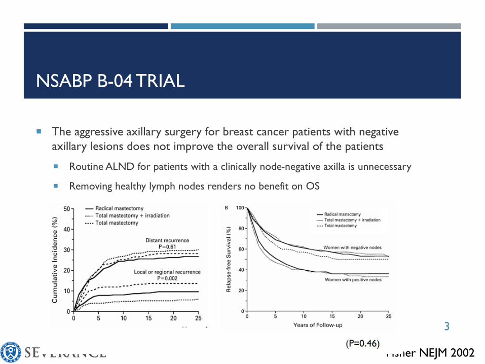
NSABP B-04 TRIAL
The aggressive axillary surgery for breast cancer patients with negative
axillary lesions does not improve the overall survival of the patients
Routine ALND for patients with a clinically node-negative axilla is unnecessary
Removing healthy lymph nodes renders no benefit on OS
Fisher NEJM 2002
3

AMERICAN COLLEGE OF SURGEONS
ONCOLOGY GROUP (ACOSOG) Z0011 TRIAL
Giuliano JAMA 2011, ACSOG Z0011
• ALND provided no survival benefit over less invasive SLN dissection during a 6-year follow-up period in a selected cohort of patients with clinical T1–T2 invasive breast cancer, who had no palpable
adenopathy and one or two biopsy-proven metastatic sentinel lymph nodes
4
Identification of an SLN (lymph node that initially harbored cancer) by
lymphatic mapping after NAC can be difficult
because obstructions occur from live or necrotic tumor emboli from the
tumor itself and/or fibrosis of lymphatic channels from chemotherapy
LYMPHATIC MAPPING AFTER NEOADJUVANT
CHEMOTHERAPY
5

NEOADJUVANT CHEMOTHERAPY (NAC)
The increasing use of neoadjuvant chemotherapy (NAC) for operable breast
cancer has raised questions about optimal local therapy for the axilla.
The poor identification rate of SLN due to possible alteration of lymphatics
as a result of NAC.
The high false negative rate because upfront chemotherapy may show
different effects on sentinel and non-sentinel nodes;
The malignancy may be eradicated in sentinel nodes, while residual disease may persist
in non-sentinel nodes.
6

SLNB AFTER NAC
IN CLINICALLY NEGATIVE AXILLA
SLNB in patients with cN0 axilla before NAC (n=980) and post-NAC (n=203).
SLN identification rate : 98% before NAC and 95% after NAC (p = 0.032)
Patients who underwent SLNB after NAC had a higher chance of having
negative SLN than those who underwent SLNB before NAC (67% vs.
54%, p=0.001) and had lesser chance of undergoing ALND (33% vs. 45%,
p=0.006).
SLNB after NAC in patients with cN0 axilla is a feasible treatment
option, and additional axillary treatment is not necessary in those with
negative SLN.
van der Heiden-van der Loo EJC 2015
7

the utility of SLNB in the treatment of patients with bx-proven node(+) disease
who underwent NAC(T0–T4,N1–N2,M0 )
phase 2 study of the clinical efficacy of substituting SLNB for ALND ; false-
negative rate (FNR) of SLNB, 10% of goal
Boughey SABCS 2012 : 31.5 % FNR with one SLN
Boughey JAMA 2013: 21.1% FNR with 2 SLN and single agent
THE ACOSOG Z1071 TRIAL
8

Studies evaluating the IDENTIFICATION RATE & FALSE-NEGATIVE RATE \
among patients with CLINICALLY NODE-POSITIVE DISEASE
undergoing SLNB following NAC
a Ultrasound performed in all patients.b False-negative rate excluding immunohistochemically detected isolated tumor cells.
Study ACOSOG Z1071 SENTINA SN FNAC
Design Single arm 4 Arms Single arm
No. of patients 756 Arm C (592) 153
Patient population T0-4, N1-2, M0 N1-2, M0 N1-2, M0
cN0 after NAC 100a 83 Unknowna
SLN identification rate (%) 92.9 80.1 87.6
False negative rate (%) 12.6 14.2 9.6
2 SLNs 21.1 18.5 4.9 (≥2 SLNs)
≥3 SLNs 9.1 7.3 -
Dual tracer technique 10.8 8.6 5.2
Inclusion of ypN0(i+) 8.7 - 8.49
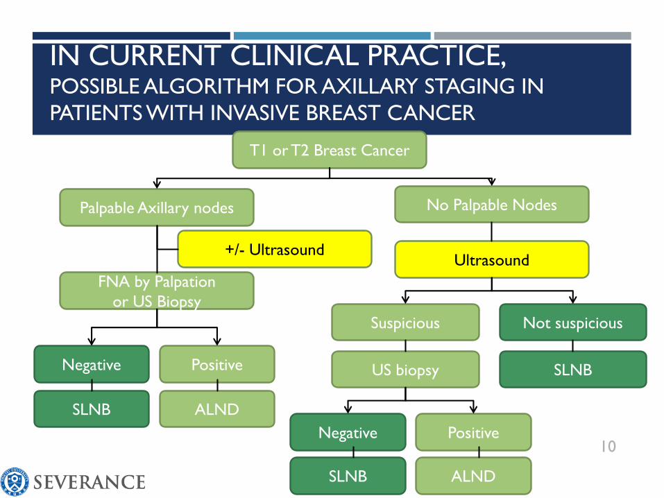
IN CURRENT CLINICAL PRACTICE, POSSIBLE ALGORITHM FOR AXILLARY STAGING IN
PATIENTS WITH INVASIVE BREAST CANCER
10
T1 or T2 Breast Cancer
Palpable Axillary nodes No Palpable Nodes
FNA by Palpation
or US Biopsy
Ultrasound
Suspicious Not suspicious
US biopsy SLNB
Negative Positive
SLNB ALND
Negative Positive
SLNB ALND
+/- Ultrasound

US FINDINGS OF AXILLARY LN
- CORRELATED WITH FINAL SURGICAL RESULTS
Surgical Results
Positive Negative PPV (%)
Cortical features
Thin 6 16 27
Thick or lobular 16 6 73
Hypoechoic 33 1 97
Hilar features
Central 9 22 29
Eccentric 15 1 94
Completely replaced 20 0 100
11Deurloo 2003 EJC
Koelliker et al Radiology 2008
Maximal cortical thickness ≥ 2.3mm

SUSPICIOUS MR FINDINGS
OF AXILLARY NODAL METASTASES
Descriptor SN (%) SP (%) PPV (%)
Irregular margin 41.2 95.2 77.8
cortex
Homogeneous 29.4 16.7 12.5
Inhomogeneous 47.1 90.5 66.7
Nodular thickening 23.5 92.9 57.1
Hilus sign 52.9 4.3 20.0
Perifocal edema 29.4 100.0 100.0
Rim sign 23.5 100.0 100.0
Asymmetry 76.7 85.7 68.4
Baltzer AJR 2011
12
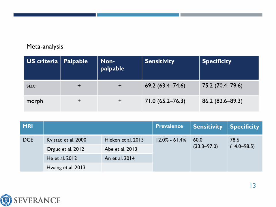
MRI Prevalence Sensitivity Specificity
DCE Kvistad et al. 2000 Hieken et al. 2013 12.0% - 61.4% 60.0
(33.3–97.0)
78.6
(14.0–98.5)Orguc et al. 2012 Abe et al. 2013
He et al. 2012 An et al. 2014
Hwang et al. 2013
US criteria Palpable Non-
palpable
Sensitivity Specificity
size + + 69.2 (63.4–74.6) 75.2 (70.4–79.6)
morph + + 71.0 (65.2–76.3) 86.2 (82.6–89.3)
13
Meta-analysis

IN CURRENT CLINICAL PRACTICE, POSSIBLE ALGORITHM FOR AXILLARY STAGING IN
PATIENTS WITH INVASIVE BREAST CANCERNo Palpable Nodes
Ultrasound/MR
Suspicious
US biopsy
Positive
ALND
SLNB: sentinel lymph node biopsy
ALND: axillary lymph node dissection
Chemotherapy
Positive
Ultrasound/MR
?14

Imaging Evaluation of Axillary Lymph Nodes after Neoadjuvant Systemic Therapy
How to improve the performance of Sentinel Node Biopsy after Neoadjuvant
Chemotherapy: Targeted axillary dissection
15

IMAGING EVALUATION OF AXILLARY
LYMPH NODES AFTER NEOADJUVANT
SYSTEMIC THERAPY
16

AXILLARY ULTRASOUND IDENTIFIES RESIDUAL
NODAL DISEASE AFTER CHEMOTHERAPY: RESULTS
FROM THE ACSOG Z1071 TRIAL (ALLIANCE)
Lymph node features on axillary ultrasound (US) images obtained after
neoadjuvant chemotherapy associated with residual nodal disease
Increased cortical thickness (mean, 3.5 mm for node-pos vs 2.5 mm for node-neg)
longer short-axis diameter, longer long-axis diameter, absence of fatty hilum
Le-Petross AJR 2018
Axillary US performed after neoadjuvant chemotherapy is useful for nodal response
assessment, with longer short-axis diameter, longer long-axis diameter, increased cortical
thickness, and absence of fatty hilum significantly associated with residual nodal disease after
neoadjuvant chemotherapy.
17
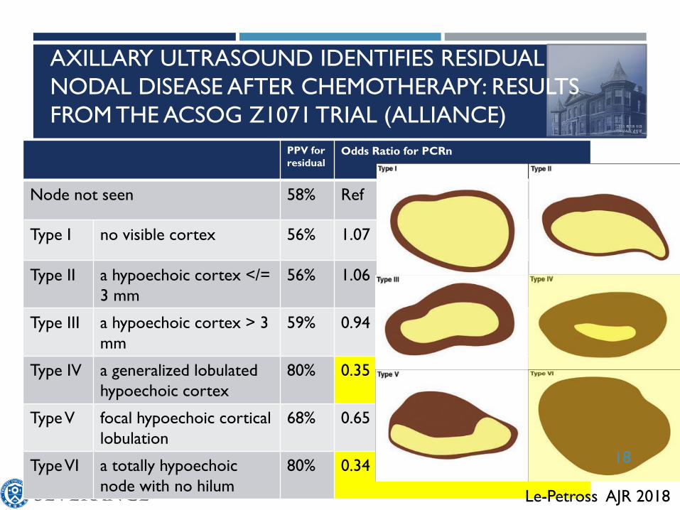
PPV for
residual Odds Ratio for PCRn
Node not seen 58% Ref
Type I no visible cortex 56% 1.07
Type II a hypoechoic cortex </=
3 mm
56% 1.06
Type III a hypoechoic cortex > 3
mm
59% 0.94
Type IV a generalized lobulated
hypoechoic cortex
80% 0.35
Type V focal hypoechoic cortical
lobulation
68% 0.65
TypeVI a totally hypoechoic
node with no hilum
80% 0.34
AXILLARY ULTRASOUND IDENTIFIES RESIDUAL
NODAL DISEASE AFTER CHEMOTHERAPY: RESULTS
FROM THE ACSOG Z1071 TRIAL (ALLIANCE)
Le-Petross AJR 2018
18

AXILLARY US AFTER NAC: IMPACT ON SLN SURGERY
Z1071
Z1071 Pts with T0-4, N1-2, M0 BC underwent AUS after NAC
The SLN FNR was not different based on AUS result
The use of SLNB only when sonographic findings were negative, potentially reduce the FNR in Z1071 patients with 12.6% to 9.8%
Preop/afterchemo AUS results are considered as part of SLN surgery.
Boughey JCO 2015
19

SENTINEL NODE BIOPSY AFTER NEOADJUVANT CHEMOTHERAPY
FOR NODE-POSITIVE BREAST CANCER:
DOES AXILLARY ULTRASOUND IMPROVE PERFORMANCE?
Question whether AUS has
anything to do with it.
Among negative AUS, the FNR of
SLN biopsy was15% (28 of 187)
Among positive AUS, the false-
positive rate of AUS was 29% (39
of 138 patients)
An abnormal AUS post-NAC is not
sufficient for proceeding to ALND.
Koslow JCO 2015
20

THE DIAGNOSTIC PERFORMANCE OF BREAST
MRI FOR AXILLARY NODAL STAGING AFTER NAC
A retrospective review using single institutional cancer registry.
135 Patients who started NAC from 2005 to 2010 with clinically node positive disease
Analysis Total MRI,
n = 135
Luminal,
n = 73
HER2þ,
n = 34
TN,
n =28
True positive, n (%) 42 (31) 18 (25) 9 (27) 15 (54)
True negative, n (%) 23 (17) 8 (11) 11 (32) 4 (14)
False positive, n (%) 3(2) 0 (0) 1 (3) 2 (7)
False negative, n (%) 67 (50) 47 (64) 13 (38) 7 (25)
Sensitivity (%) 39 28 41 68
Specificity (%) 88 100 92 67
PPV (%) 93 100 90 88
NPV (%) 26 15 46 36
Accuracy (%) 48 36 59 68
Steiman J Surg Res 2016
21

PREOPERATIVE AXILLARY LYMPH NODE EVALUATION IN BREAST
CANCER PATIENTS BY BREAST MAGNETIC RESONANCE IMAGING
(MRI): DIAGNOSTIC PERFORMANCE OF MRI ACCORDING TO NAC
Hyun ER 2016
Advance (pN2/3)
SN w/ cN1-3 80.0 (24/30) 91.7 (11/12) 83.3 (15/18 )
SN w/ cN2-3 36.7 (11/30) 33.3 (4/12) 38.9 (7/18)
NPV w/cN0 98.2 (335/341) 99.6 (256/257) 94.0 (79/84) 0.039
NPV w/cN0/1 95.3 (388/407) 97.4 (294/302) 89.5 (94/105) 0.017 22

Patients with advanced ALN metastases were more likely to have a higher
number (≥2) of positive LNs (OR, 8.06; P = 0.015) on restaging MR imaging.
No clinico-pathological factors were significantly associated with advanced ALN
metastases.
A higher number of positive ALNs on restaging MR imaging was an
independent predictor for advanced ALN metastases after NAC.
PREDICTION OF ADVANCED AXILLARY LN METASTASES
(ypN23) USING BREAST MR IMAGING AND PET/CT AFTER
NEOADJUVANT CTX IN IDC PATIENTS
Kim W, Scientific Reports 2018
Characteristics Univariate OR (95% CI) Multivariate OR (95% CI) P value
Number of positive ALNs on initial staging MR imaging*
<4 1.00 1.00
≥4 5.91 6.04 0.080
Number of positive ALNs on restaging MR imaging*
<2 1.00 1.00
≥2 12.04 8.06 0.015
Short diameter of the largest ALN on restaging MR imaging
≤7.7mm 1.00 1.00
>7.7mm 5.00 4.44 0.063
23

PREDICTION OF ADVANCED AXILLARY LN
METASTASES (ypN23) USING BREAST MR IMAGING AND
PET/CT AFTER NEOADJUVANT CTx IN IDC PATIENTS
The AUC and sensitivity of restaging MR imaging plus PET/CT was higher than that
of each restaging MR imaging and PET/CT; however, the differences were not
statistically significant (AUC, P = 0.318 and P = 0.119, respectively; sensitivity, P =
0.500 and 0.063, respectively).
Kim W, Scientific Reports 2018
24

HOW TO IMPROVE THE PERFORMANCE OF
SENTINEL NODE BIOPSY AFTER NAC
25

Targeted axillary dissection (TAD)
• Placing a clip in the positive lymph node could sole the high FNR?
• 31.5 % FNR with one SLN in Z1071 trial
• Data from Z1071: the FNR in cases with placing a clip in the positive lymph
node was lower than that using dual tracer technique (7.4% vs 10.8%)
Targeted axillary dissection
• After completion of NAC, the clipped node is
localized by using a wire or radioactive seed
• During the SLND procedure, taking both
all SLNs and clipped node with localization
Mittendorf ASO 2014

IDENTIFICATION AND RESECTION OF CLIPPED NODE
DECREASES THE FNR OF SLNB IN NAC PTS
PRESENTING WITH NODE (+) BC results from ACOSOG Z1071 (ALLIANCE)
525 patients with cN1 disease and ≧2 SLNs confirmed
170 clip in LN vs 355 no clip in LN
N Residual Disease identified
in SLNs or ALND, n (%)
FNR
(%)
95% CI
Patients with ≥ 2SLNs removed and cN1 disease
Clip in SLN 107 59 (55.1) 6.8 1.9-16.5
Clip in ALND 34 21 (61.8) 19.0 5.4-41.9
Clip location unknown 29 21 (72.4) 14.3 3.0-36.3
Clip not placed 355 209 (59.0) 13.4 9.1-18.8
Boughey AS 2016
False-negative Rate by Clip Location and Patient Group Analyzed
27

Selective Surgical Localization of Axillary Lymph Nodes Containing Metastases in Patients With Breast Cancer
• the feasibility of image-guided localization and resection of lymph nodes
containing known-metastases.
• 12 patients with ALN-meta confirmed by FNA, who had a clip placed in the
lymph node targeted for biopsy.
• US guided wire –needle localization (n=2) & radioactive iodine I 125 (n=10)
• Image-guided localization & selective removal were successful in all 12 patients.
• 80% : the clipped node = one of the SLNs
• 20% : the clipped node ≠ one of the SLNs
• Without clip-localization, the biopsy-proven metastatic axillary node with the
clip would not have been included in surgery in these 20% of patients.
Caudle JAMA 2015

Targeted axillary dissection (TAD)
Caudle JCO 2016
SLNB +/- Clip
Clinically node-positive patients with NAC N=208
No SLNB N=74
No ALND N=16
Evaluable patients (SLNB +ALND) N=118
Clipped node & SLNs negative N=1 of 74
SLN negative = 7 of 69SLN not identified =5
Pathologic node NegativeN=44 (37%)
Pathologic node PositiveN=74 (63%)
False-negative rate
Clipped node +SLNB1.4% (95% CI, 0.03 to 7.3)
SLNB alone10.1% (95% CI 4.2 to 19.8)
p=0.03
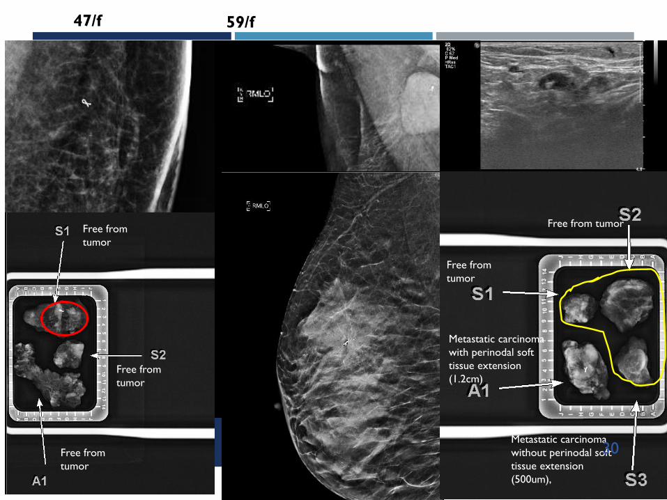
Free from tumor
Free from
tumor
Metastatic carcinoma
with perinodal soft
tissue extension
(1.2cm)
Metastatic carcinoma
without perinodal soft
tissue extension
(500um),
47/f 59/f
Free from
tumor
Free from
tumor
Free from
tumor
30

31

Charcoal Tattooing of Metastatic ALN followed by SLNB after NAC in Breast Cancers in YUHS
Park S & Kim MJ CRT epub

Tatoo node
Blue-dyed node
Park CRT epub
CHARCOAL TATTOOING OF METASTATIC ALN FOLLOWED BY
SLNB AFTER NAC IN BREAST CANCERS IN YUHS
33

SLN 1
1st Hot LN ≠ Blue colored
LN ≠ Tattoo LN
SLN 2
2nd Hot LN = Blue
colored LN ≠ Tattoo
LN
Blue-dyed
node
ALN 1
Cold LN = Tatto LN ≠ Blue
LN
Tatoo node
Metastasis , diameter 13mm
34
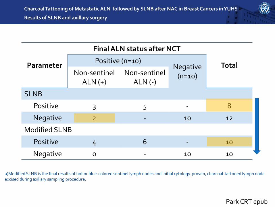
Charcoal Tattooing of Metastatic ALN followed by SLNB after NAC in Breast Cancers in YUHS
Results of SLNB and axillary surgery
Parameter
Final ALN status after NCT
TotalPositive (n=10)
Negative(n=10)Non-sentinel
ALN (+)Non-sentinel
ALN (-)
SLNB
Positive 3 5 - 8
Negative 2 - 10 12
Modified SLNB
Positive 4 6 - 10
Negative 0 - 10 10
a)Modified SLNB is the final results of hot or blue-colored sentinel lymph nodes and initial cytology-proven, charcoal-tattooed lymph node excised during axillary sampling procedure.
Park CRT epub
NAC is increasingly used for patients with operable breast cancer to
allow more limited surgery in the breast and axilla.
However, the diagnostic performance of imaging evaluation after NAC is
limited. It could be helpful to exclude advanced nodal disease, but not
sufficient to replace SLNB.
A multidisciplinary approach is needed to evaluate re-staging of axillary
lymph node after NAC, using imaging findings, tumor biology, and
localization of proven metastatic lymph nodes.
TAKE HOME MASSAGE
36

37
THANK YOU FOR YOUR ATTENTION!