Embed Size (px)
Citation preview
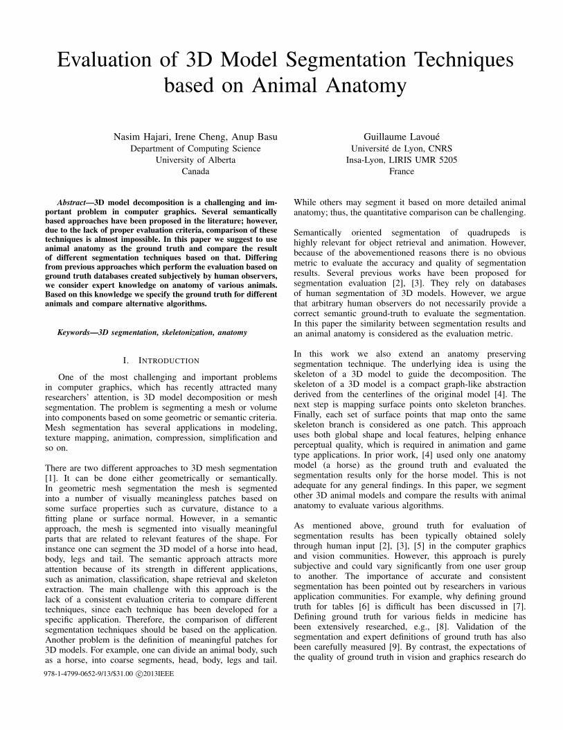
Evaluation of 3D Model Segmentation Techniquesbased on Animal Anatomy
Nasim Hajari, Irene Cheng, Anup BasuDepartment of Computing Science
University of AlbertaCanada
Guillaume LavoueUniversite de Lyon, CNRS
Insa-Lyon, LIRIS UMR 5205France
Abstract—3D model decomposition is a challenging and im-portant problem in computer graphics. Several semanticallybased approaches have been proposed in the literature; however,due to the lack of proper evaluation criteria, comparison of thesetechniques is almost impossible. In this paper we suggest to useanimal anatomy as the ground truth and compare the resultof different segmentation techniques based on that. Differingfrom previous approaches which perform the evaluation based onground truth databases created subjectively by human observers,we consider expert knowledge on anatomy of various animals.Based on this knowledge we specify the ground truth for differentanimals and compare alternative algorithms.
Keywords—3D segmentation, skeletonization, anatomy
I. INTRODUCTION
One of the most challenging and important problemsin computer graphics, which has recently attracted manyresearchers’ attention, is 3D model decomposition or meshsegmentation. The problem is segmenting a mesh or volumeinto components based on some geometric or semantic criteria.Mesh segmentation has several applications in modeling,texture mapping, animation, compression, simplification andso on.
There are two different approaches to 3D mesh segmentation[1]. It can be done either geometrically or semantically.In geometric mesh segmentation the mesh is segmentedinto a number of visually meaningless patches based onsome surface properties such as curvature, distance to afitting plane or surface normal. However, in a semanticapproach, the mesh is segmented into visually meaningfulparts that are related to relevant features of the shape. Forinstance one can segment the 3D model of a horse into head,body, legs and tail. The semantic approach attracts moreattention because of its strength in different applications,such as animation, classification, shape retrieval and skeletonextraction. The main challenge with this approach is thelack of a consistent evaluation criteria to compare differenttechniques, since each technique has been developed for aspecific application. Therefore, the comparison of differentsegmentation techniques should be based on the application.Another problem is the definition of meaningful patches for3D models. For example, one can divide an animal body, suchas a horse, into coarse segments, head, body, legs and tail.
While others may segment it based on more detailed animalanatomy; thus, the quantitative comparison can be challenging.
Semantically oriented segmentation of quadrupeds ishighly relevant for object retrieval and animation. However,because of the abovementioned reasons there is no obviousmetric to evaluate the accuracy and quality of segmentationresults. Several previous works have been proposed forsegmentation evaluation [2], [3]. They rely on databasesof human segmentation of 3D models. However, we arguethat arbitrary human observers do not necessarily provide acorrect semantic ground-truth to evaluate the segmentation.In this paper the similarity between segmentation results andan animal anatomy is considered as the evaluation metric.
In this work we also extend an anatomy preservingsegmentation technique. The underlying idea is using theskeleton of a 3D model to guide the decomposition. Theskeleton of a 3D model is a compact graph-like abstractionderived from the centerlines of the original model [4]. Thenext step is mapping surface points onto skeleton branches.Finally, each set of surface points that map onto the sameskeleton branch is considered as one patch. This approachuses both global shape and local features, helping enhanceperceptual quality, which is required in animation and gametype applications. In prior work, [4] used only one anatomymodel (a horse) as the ground truth and evaluated thesegmentation results only for the horse model. This is notadequate for any general findings. In this paper, we segmentother 3D animal models and compare the results with animalanatomy to evaluate various algorithms.
As mentioned above, ground truth for evaluation ofsegmentation results has been typically obtained solelythrough human input [2], [3], [5] in the computer graphicsand vision communities. However, this approach is purelysubjective and could vary significantly from one user groupto another. The importance of accurate and consistentsegmentation has been pointed out by researchers in variousapplication communities. For example, why defining groundtruth for tables [6] is difficult has been discussed in [7].Defining ground truth for various fields in medicine hasbeen extensively researched, e.g., [8]. Validation of thesegmentation and expert definitions of ground truth has alsobeen carefully measured [9]. By contrast, the expectations ofthe quality of ground truth in vision and graphics research do
978-1-4799-0652-9/13/$31.00 c©2013IEEE
not seem to be that high. For example, in Figure 1 in [5] theelephants are identified as one object in one segmentation,while three in others. Despite issues like this, the differentsegmentations are considered to be “highly consistent.” Itappears that application domains, and what level of accuracyand consistency are necessary in them, have been largelyignored in vision and graphics research. Thus, we proposeusing biologically defined anatomical models as a morereliable approach to ground truth definition and subsequentevaluation for a class of objects in 3D segmentation.
The remainder of this paper is organized as follows: SectionII discusses some of the semantic segmentation techniques.Section III explains our skeleton based segmentationapproach. In Section IV the evaluation criteria and theproposed anatomical ground-truth are presented. Section Vcompares the results of skeleton based segmentation with themost efficient state of the art methods. Concluding remarksare given in Section VI.
II. RELATED WORK
Semantically oriented segmentation techniques try togenerate the segmentation patches such that a cost functionis minimized based on a given criterion. The main differenceamong various algorithms in this category is the cost functionand the criterion used. In this section we will explain someof these techniques in greater detail.
The authors in [10] proposed a hierarchical decompositiontechnique using fuzzy clustering. This algorithm is based ona hierarchical tree. Each node in the tree is associated withthe mesh of a particular patch and the root is associated withthe whole input object. The higher level nodes correspond tocoarser patches while leaves and lower level nodes correspondto finer patches. The algorithm determines a suitable number(k) of patches at each node, and then computes a k-waysegmentation of this node. First, the algorithm finds themeaningful components along with the boundaries betweenthe components that are considered to be fuzzy. The nextstep is finding the exact boundaries in the fuzzy areas whichpreserve the features of the object. However, the approachrelaxes the condition that every face should belong to exactlyone patch and allows fuzzy membership, which means thateach face has probabilities associated to belonging to differentpatches. This probability is based on geodesic and angulardistances between all pairs of the faces. Another hierarchicalmethod has been proposed in [11]. This algorithm is alsobased on a hierarchical tree and proceeds from coarse tofine scale. The main advantage of this technique is that itis insensitive to pose and proportions. The approach firsttransforms the mesh vertices into a pose invariant space, thenrobustly extracts the feature points, and finally extracts thecore component of the mesh. [12] proposed an algorithmbased on the fitting primitives. Initially, each triangle of atriangular mesh corresponds to a single cluster. All the pairsof adjacent clusters are considered and compared at everyiteration, and the one that can be best approximated by oneof the primitives forms a new single cluster. The primitivesare planes, spheres and cylinders; and, an L2 metric is usedto compare each combination of the cluster with one of theseprimitives.
The Shape Diameter Function (SDF) is another 3Dsegmentation approach which has been proposed in [13].The SDF is defined as the diameter of the object in theneighborhood of each point on its surface. Given a point onthe surface mesh a set of rays is sent inside a cone centeredaround its inward-normal direction (the opposite direction ofits normal) to the other side of the mesh. The value of theSDF at the point is defined as the weighted average of allthe lengths of rays that fall within one standard deviationfrom the median of all lengths. [14] presented a segmentationtechnique based on random cuts. The idea is to generate arandom set of mesh segmentations, and then measure howoften each edge of the mesh lies on a segmentation boundaryin the randomized set. An interactive approach based onrandom walk has been proposed in [15]. The user choosessome faces as seeds, then a probability is assigned to eachof the three edges of non-seed faces. This value determineswhether or not a random walker moves across a particularedge to the corresponding face. A face belonging to the regionis grouped with the seed X if a random walker starting at thatface has a higher probability of reaching the seed X than anyother seeds.
III. SEGMENTATION BASED ON SKELETONIZATION
Since the human eyes are very sensitive to changes alongthe boundary of an object it is logical to use the structuralshape for simplification and segmentation purposes. Since theprojected 2D contour of a 3D model can vary significantlydepending on a change of view, it is more effective to use themodel skeleton in order to guide segmentation. The skeletonof a 3D model is a compact graph-like abstraction derivedfrom the centerlines of the original model [4].
It is very important to extract the skeleton as accuratelyas possible. For segmentation purposes a unit width skeleton,such as the Valance Normalized Spatial Median (VNSM) [16]is needed. However, these skeletons are very prone to localand global noise and may contain some unwanted branches.The Scale Space and Gaussian filters can be used to removethe noise and smooth the skeleton. The unwanted branchesshould be removed considering the length and topologicalposition of a branch [4].
After extracting the smooth and noise-free skeleton, thenext step is to decompose the model into different segmentsaccording to the following steps.
• Decompose the skeleton at junction points. Junctionpoints are the points that are connected to more thanone skeleton branch.
• Map surface nodes to skeleton branches. This isbased on the L2 distance between surface nodes andskeleton vertices. In other words, to map a point onthe surface to a skeleton branch node, it should beclose to the skeleton branch nodes, with skeleton-to-surface normal vector pointing in a direction similarto the surface normal vectors. Also, the neighboring
nodes should have the same label if the curvature isclose. These distances are computed for each surfacepoint or a subset of surface points against all skeletonvertices.
• The last step is labeling the neighborhood nodes. Theprevious step just labeled some selected sample points.The watershed method is used to flood the labels fromsamples.
IV. EVALUATION
The main challenge for semantically oriented segmentationis defining the evaluation criteria, which is related tothe application. However, for animation purposes andgenerating natural movements of a model, a more meaningfulsegmentation for animals is based on anatomy. Shi et al. [4]used a metric function distance for evaluation purposes. Theerror is then the Euclidean distance between registered pairof cuts plus the length of non-paired cuts. However, they justused one anatomy model, that of a horse, as the ground truthand evaluated the segmentation results only for the horsemodel. This limited evaluation is not adequate.
For evaluation purposes we used the same method as Shi etal. First, we registered different cuts to the correspondingground truth ones; then calculated the Euclidean distance foreach cut plus the length of non-paired cuts. Smaller errorsimply better automatic segmentation compared to the groundtruth. Since the ground truth is generated manually basedon animal anatomies, different experts may specify slightlydifferent ground truths. However, this does not affect thecomparison results very much, since the ground truth is notsolely based on human perception but anatomical informationis also considered in the process.
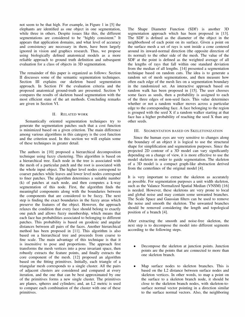
We use sheep [17], cow [17], dog [18] and giraffe [19]anatomies. There are several free 3D models of these animalsavailable on public databases [3], [20]. These anatomy modelsare very detailed and in order to use them as the groundtruth, we manually segment them at a coarse level. However,it is possible to ask the user what level of details he/sheprefers. Figure 1 shows the anatomy models for these animals.Figure 2 shows the corresponding ground-truth segmentations.Figure 3 presents the ground-truth of giraffe at a finer levelof detail.
V. RESULTS
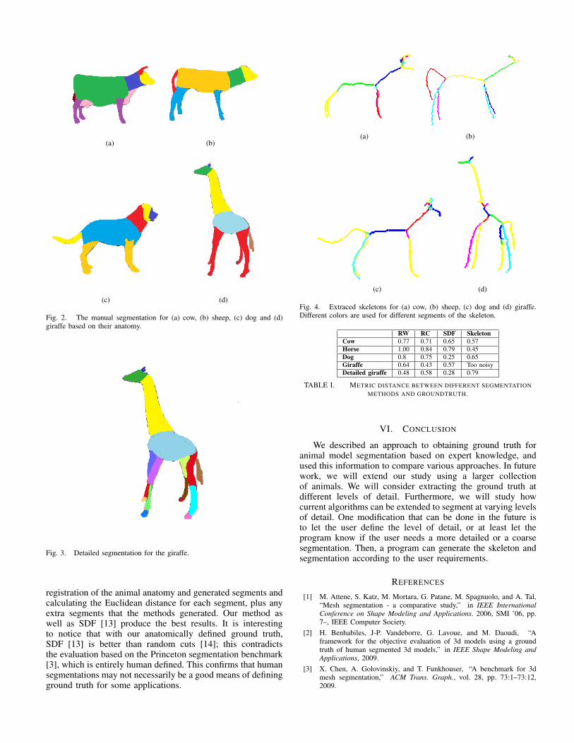
Using the method proposed in [4], we extracted the skele-tons and performed 3D segmentation for cow, sheep, dogand giraffe. Figure 4 shows the extracted skeletons. Figure 5compares the segmentation results with random walk (RW)[15], random cuts (RC) [14], shape diameter (SDF) [13] andthe ground truth. Note that the extracted skeletons may stillcontain some noise or unwanted branches which can lead topoor segmentation quality. One technique that can be usedto eliminate the noise and improve the generated skeletonsis scale space filtering. In future work we will use thismethod and other techniques to improve the results. Table Ishows the metric distance between the segmented models andthe corresponding animal anatomies. It is based on manual
(a)
(b)
(c)
(d)
Fig. 1. The anatomy model of (a) cow [17], (b) sheep [17] , (c) dog [18]and (d) giraffe .
(a) (b)
(c) (d)
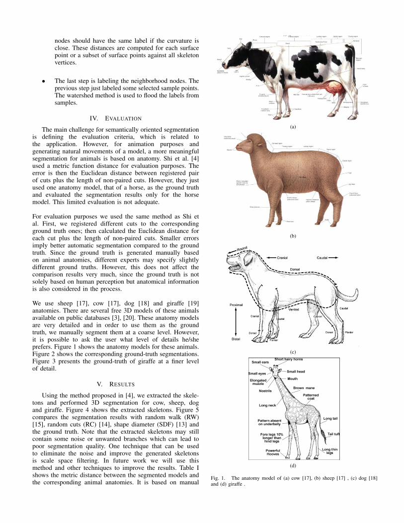
Fig. 2. The manual segmentation for (a) cow, (b) sheep, (c) dog and (d)giraffe based on their anatomy.
Fig. 3. Detailed segmentation for the giraffe.
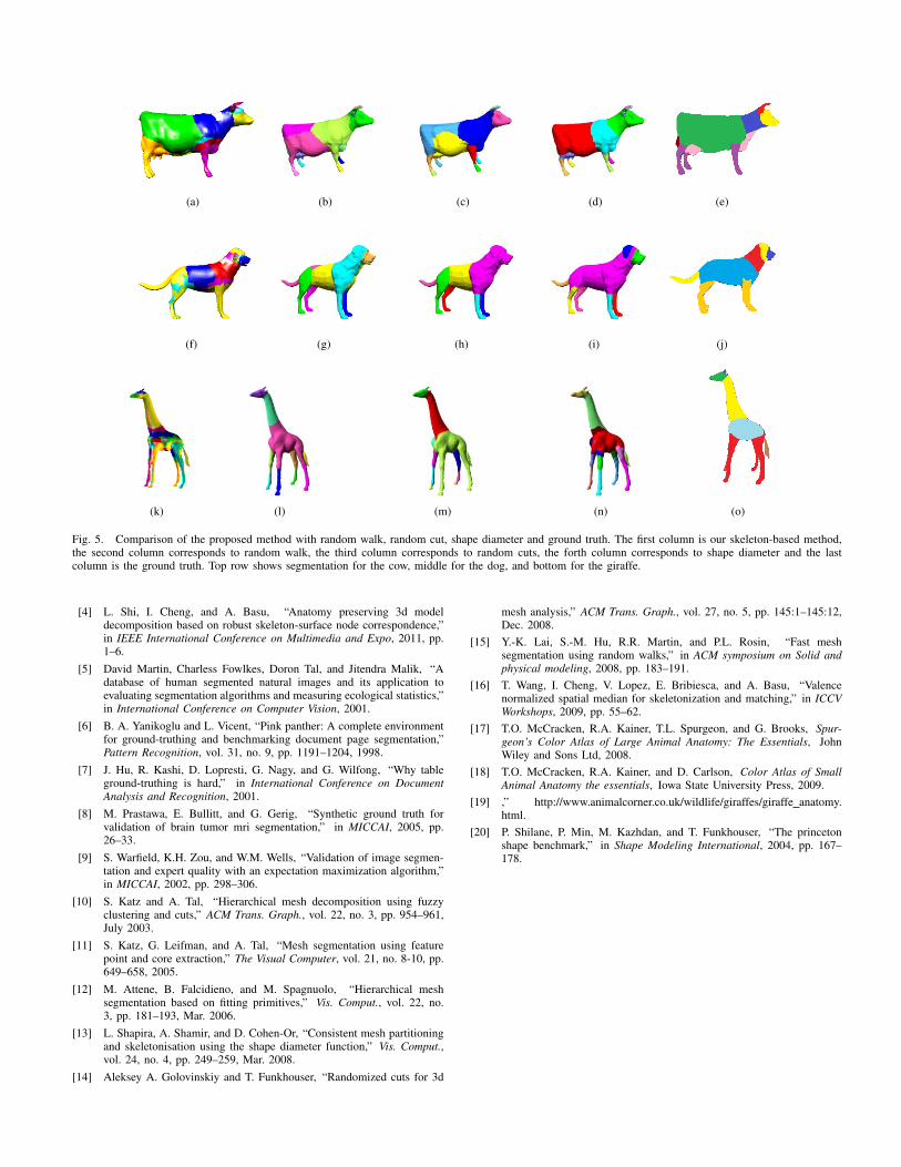
registration of the animal anatomy and generated segments andcalculating the Euclidean distance for each segment, plus anyextra segments that the methods generated. Our method aswell as SDF [13] produce the best results. It is interestingto notice that with our anatomically defined ground truth,SDF [13] is better than random cuts [14]; this contradictsthe evaluation based on the Princeton segmentation benchmark[3], which is entirely human defined. This confirms that humansegmentations may not necessarily be a good means of definingground truth for some applications.
(a) (b)
(c) (d)
Fig. 4. Extraced skeletons for (a) cow, (b) sheep, (c) dog and (d) giraffe.Different colors are used for different segments of the skeleton.
RW RC SDF SkeletonCow 0.77 0.71 0.65 0.57Horse 1.00 0.84 0.79 0.45Dog 0.8 0.75 0.25 0.65Giraffe 0.64 0.43 0.57 Too noisyDetailed giraffe 0.48 0.58 0.28 0.79
TABLE I. METRIC DISTANCE BETWEEN DIFFERENT SEGMENTATIONMETHODS AND GROUNDTRUTH.
VI. CONCLUSION
We described an approach to obtaining ground truth foranimal model segmentation based on expert knowledge, andused this information to compare various approaches. In futurework, we will extend our study using a larger collectionof animals. We will consider extracting the ground truth atdifferent levels of detail. Furthermore, we will study howcurrent algorithms can be extended to segment at varying levelsof detail. One modification that can be done in the future isto let the user define the level of detail, or at least let theprogram know if the user needs a more detailed or a coarsesegmentation. Then, a program can generate the skeleton andsegmentation according to the user requirements.
REFERENCES
[1] M. Attene, S. Katz, M. Mortara, G. Patane, M. Spagnuolo, and A. Tal,“Mesh segmentation - a comparative study,” in IEEE InternationalConference on Shape Modeling and Applications. 2006, SMI ’06, pp.7–, IEEE Computer Society.
[2] H. Benhabiles, J-P. Vandeborre, G. Lavoue, and M. Daoudi, “Aframework for the objective evaluation of 3d models using a groundtruth of human segmented 3d models,” in IEEE Shape Modeling andApplications, 2009.
[3] X. Chen, A. Golovinskiy, and T. Funkhouser, “A benchmark for 3dmesh segmentation,” ACM Trans. Graph., vol. 28, pp. 73:1–73:12,2009.
(a) (b) (c) (d) (e)
(f) (g) (h) (i) (j)
(k) (l) (m) (n) (o)
Fig. 5. Comparison of the proposed method with random walk, random cut, shape diameter and ground truth. The first column is our skeleton-based method,the second column corresponds to random walk, the third column corresponds to random cuts, the forth column corresponds to shape diameter and the lastcolumn is the ground truth. Top row shows segmentation for the cow, middle for the dog, and bottom for the giraffe.
[4] L. Shi, I. Cheng, and A. Basu, “Anatomy preserving 3d modeldecomposition based on robust skeleton-surface node correspondence,”in IEEE International Conference on Multimedia and Expo, 2011, pp.1–6.
[5] David Martin, Charless Fowlkes, Doron Tal, and Jitendra Malik, “Adatabase of human segmented natural images and its application toevaluating segmentation algorithms and measuring ecological statistics,”in International Conference on Computer Vision, 2001.
[6] B. A. Yanikoglu and L. Vicent, “Pink panther: A complete environmentfor ground-truthing and benchmarking document page segmentation,”Pattern Recognition, vol. 31, no. 9, pp. 1191–1204, 1998.
[7] J. Hu, R. Kashi, D. Lopresti, G. Nagy, and G. Wilfong, “Why tableground-truthing is hard,” in International Conference on DocumentAnalysis and Recognition, 2001.
[8] M. Prastawa, E. Bullitt, and G. Gerig, “Synthetic ground truth forvalidation of brain tumor mri segmentation,” in MICCAI, 2005, pp.26–33.
[9] S. Warfield, K.H. Zou, and W.M. Wells, “Validation of image segmen-tation and expert quality with an expectation maximization algorithm,”in MICCAI, 2002, pp. 298–306.
[10] S. Katz and A. Tal, “Hierarchical mesh decomposition using fuzzyclustering and cuts,” ACM Trans. Graph., vol. 22, no. 3, pp. 954–961,July 2003.
[11] S. Katz, G. Leifman, and A. Tal, “Mesh segmentation using featurepoint and core extraction,” The Visual Computer, vol. 21, no. 8-10, pp.649–658, 2005.
[12] M. Attene, B. Falcidieno, and M. Spagnuolo, “Hierarchical meshsegmentation based on fitting primitives,” Vis. Comput., vol. 22, no.3, pp. 181–193, Mar. 2006.
[13] L. Shapira, A. Shamir, and D. Cohen-Or, “Consistent mesh partitioningand skeletonisation using the shape diameter function,” Vis. Comput.,vol. 24, no. 4, pp. 249–259, Mar. 2008.
[14] Aleksey A. Golovinskiy and T. Funkhouser, “Randomized cuts for 3d
mesh analysis,” ACM Trans. Graph., vol. 27, no. 5, pp. 145:1–145:12,Dec. 2008.
[15] Y.-K. Lai, S.-M. Hu, R.R. Martin, and P.L. Rosin, “Fast meshsegmentation using random walks,” in ACM symposium on Solid andphysical modeling, 2008, pp. 183–191.
[16] T. Wang, I. Cheng, V. Lopez, E. Bribiesca, and A. Basu, “Valencenormalized spatial median for skeletonization and matching,” in ICCVWorkshops, 2009, pp. 55–62.
[17] T.O. McCracken, R.A. Kainer, T.L. Spurgeon, and G. Brooks, Spur-geon’s Color Atlas of Large Animal Anatomy: The Essentials, JohnWiley and Sons Ltd, 2008.
[18] T.O. McCracken, R.A. Kainer, and D. Carlson, Color Atlas of SmallAnimal Anatomy the essentials, Iowa State University Press, 2009.
[19] ,” http://www.animalcorner.co.uk/wildlife/giraffes/giraffe anatomy.html.
[20] P. Shilane, P. Min, M. Kazhdan, and T. Funkhouser, “The princetonshape benchmark,” in Shape Modeling International, 2004, pp. 167–178.