Embed Size (px)
Citation preview
20
Evaluation and Treatment of Hypotension in Premature Infants
Shoichi Ezaki and Masanori Tamura Division of Neonatal Medicine, Center for Maternal,
Fetal and Neonatal Medicine, Saitama Medical Center, Saitama Medical University,
Japan
1. Introduction
Sixteen to 98% of extremely preterm infants are treated for hypotension within the first
week of life. The enormous variation in this estimate is due to a lack of reliable evidence.
While selecting a vasoactive agent, it is necessary to consider the goals of the therapy. To
achieve those goals, the clinician must assess the mechanisms of action of the potential
therapies. This chapter details the unique characteristics of the neonatal cardiovascular
system and defines hypotension in preterm infants. It provides indications for treatment and
appropriate therapies for individual cases.
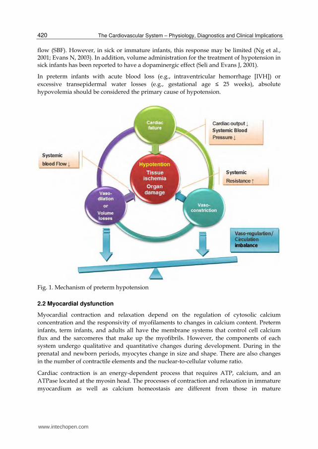
2. Characteristic pathophysiology of hypotension in preterm infants
Blood pressure increases with advancing gestational and postnatal age, which is a
developmentally regulated phenomenon (Noori and Seli, 2005). Since cardiac output (CO)
and systemic vascular resistance (SVR) both contribute to blood pressure, elevation in blood
pressure during development may be the result of increased CO, increased SVR, or both
(Fig.1)
2.1 Hypovolemia
In preterm infants, absolute hypovolemia is the most frequent cause of hypotension.
Peripheral vasodilation with or without myocardial failure is the most frequent primary
etiological factor (Seli and Evans J, 2001). Absolute hypovolemia is defined as a loss of
volume from the intravascular compartment; alternatively, relative hypovolemia is defined
as vasodilatation with an inadequate volume to fill the expanded intravascular
compartment. In both situations, the result is inadequate filling pressure (also known as
preload) in the heart. If severe enough, hypovolemia can reduce CO, resulting in inadequate
tissue perfusion and oxygenation (Fig 1).
In cases of absolute hypovolemia, the body releases corticosteroids, adrenaline, and noradrenaline, which cause vascular contraction in order to maintain blood pressure and filling pressure and cause increased heart rate and contractility to maintain systemic blood
www.intechopen.com
The Cardiovascular System – Physiology, Diagnostics and Clinical Implications
420
flow (SBF). However, in sick or immature infants, this response may be limited (Ng et al., 2001; Evans N, 2003). In addition, volume administration for the treatment of hypotension in sick infants has been reported to have a dopaminergic effect (Seli and Evans J, 2001).
In preterm infants with acute blood loss (e.g., intraventricular hemorrhage [IVH]) or
excessive transepidermal water losses (e.g., gestational age ≤ 25 weeks), absolute
hypovolemia should be considered the primary cause of hypotension.
Fig. 1. Mechanism of preterm hypotension
2.2 Myocardial dysfunction
Myocardial contraction and relaxation depend on the regulation of cytosolic calcium
concentration and the responsivity of myofilaments to changes in calcium content. Preterm
infants, term infants, and adults all have the membrane systems that control cell calcium
flux and the sarcomeres that make up the myofibrils. However, the components of each
system undergo qualitative and quantitative changes during development. During in the
prenatal and newborn periods, myocytes change in size and shape. There are also changes
in the number of contractile elements and the nuclear-to-cellular volume ratio.
Cardiac contraction is an energy-dependent process that requires ATP, calcium, and an
ATPase located at the myosin head. The processes of contraction and relaxation in immature
myocardium as well as calcium homeostasis are different from those in mature
www.intechopen.com
Evaluation and Treatment of Hypotension in Premature Infants
421
myocardium. Specifically, immature myocytes do not rely as heavily on the release and re-
uptake of calcium from the sarcoplasmic reticulum; instead, they depend more on
extracellular calcium concentration. As such, the immature myocardium of the fetus and
newborn depends on L-type calcium channels as a calcium source for contraction.
Furthermore, immature myocytes have greater cell surface area-to-volume ratios, which
may compensate for their underdeveloped T-tubule systems. The alterations in myocardial
structure and function with maturation and the developmental changes in cardiovascular
function provide the cellular and molecular bases for differences in myocardial contractility
among preterm newborns, term newborns, and older infants (Rowland and Gutgesell, 1995;
Noori and Seli, 2005).
Therefore, preterm infants with hypotension have a limited ability to increase CO in
response to inotropes or changes in volume (Teitel and Sidi, 1985). Furthermore, they have
an elevated sensitivity to increased afterload (Van Hare et al., 1990), which commonly leads
to decreased CO (Belik and Light, 1989).
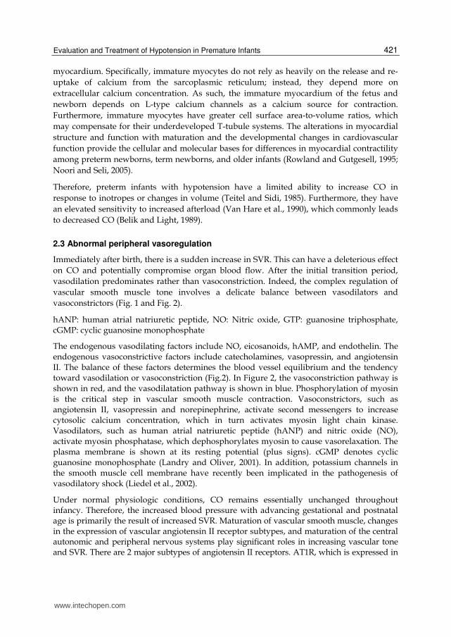
2.3 Abnormal peripheral vasoregulation
Immediately after birth, there is a sudden increase in SVR. This can have a deleterious effect
on CO and potentially compromise organ blood flow. After the initial transition period,
vasodilation predominates rather than vasoconstriction. Indeed, the complex regulation of
vascular smooth muscle tone involves a delicate balance between vasodilators and
vasoconstrictors (Fig. 1 and Fig. 2).
hANP: human atrial natriuretic peptide, NO: Nitric oxide, GTP: guanosine triphosphate, cGMP: cyclic guanosine monophosphate
The endogenous vasodilating factors include NO, eicosanoids, hAMP, and endothelin. The endogenous vasoconstrictive factors include catecholamines, vasopressin, and angiotensin II. The balance of these factors determines the blood vessel equilibrium and the tendency toward vasodilation or vasoconstriction (Fig.2). In Figure 2, the vasoconstriction pathway is shown in red, and the vasodilatation pathway is shown in blue. Phosphorylation of myosin is the critical step in vascular smooth muscle contraction. Vasoconstrictors, such as angiotensin II, vasopressin and norepinephrine, activate second messengers to increase cytosolic calcium concentration, which in turn activates myosin light chain kinase. Vasodilators, such as human atrial natriuretic peptide (hANP) and nitric oxide (NO), activate myosin phosphatase, which dephosphorylates myosin to cause vasorelaxation. The plasma membrane is shown at its resting potential (plus signs). cGMP denotes cyclic guanosine monophosphate (Landry and Oliver, 2001). In addition, potassium channels in the smooth muscle cell membrane have recently been implicated in the pathogenesis of vasodilatory shock (Liedel et al., 2002).
Under normal physiologic conditions, CO remains essentially unchanged throughout infancy. Therefore, the increased blood pressure with advancing gestational and postnatal age is primarily the result of increased SVR. Maturation of vascular smooth muscle, changes in the expression of vascular angiotensin II receptor subtypes, and maturation of the central autonomic and peripheral nervous systems play significant roles in increasing vascular tone and SVR. There are 2 major subtypes of angiotensin II receptors. AT1R, which is expressed in
www.intechopen.com

The Cardiovascular System – Physiology, Diagnostics and Clinical Implications
422
mature tissues and the umbilical artery, mediates smooth muscle contraction and regulates fluid and electrolyte balance. AT2R, which is expressed in fetal and newborn tissues, has an unknown function. The developmentally regulated transition from expression of AT2R to AT1R begins following the first 2 weeks of life and is complete by month 3 (Noori and Seli, 2005; Engle, 2001). The vasodilating factor NO increases under conditions of oxidative stress and sepsis. Because preterm infants are prone to these conditions (Ezaki et al., 2009a), their NO levels can easily increase. Together, these physiological characteristics of preterm infants make them susceptible to vasoregulatory dysfunction.
Fig. 2. Regulation of vascular smooth muscle tone
3. The significance of hypotension requiring treatment in preterm infants
3.1 Clinical outcomes
Hypotension is a common complication among preterm infants. Importantly, there is an
association between systemic hypotension and neonatal morbidities, including IVH and
neurodevelopmental disorders (Watkins et al., 1989; Goldstein et al., 1995). Unfortunately,
www.intechopen.com
Evaluation and Treatment of Hypotension in Premature Infants
423
common conditions among preterm infants, such as sepsis, renal failure, and neonatal
asphyxia, can lead to the development of clinical hypotension and confer a poor prognosis.
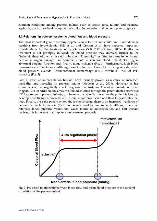
3.2 Relationship between systemic blood flow and blood pressure
The most important goal in treating hypotension is to prevent cellular and tissue damage resulting from hypovolemia. Seli et al. and Greisen et al. have reported important considerations for the treatment of hypotension (Seli, 2006; Greisen, 2005). If effective treatment is not promptly initiated, the blood pressure may decrease further to the “ischemic threshold, which is said to be about 30 mmHg,” resulting in tissue ischemia and permanent organ damage. For example, a loss of cerebral blood flow (CBF) triggers abnormal cerebral function and, finally, tissue ischemia (Fig. 3). Furthermore, high blood pressure is also deleterious. Although, exact value is not noted in existing reports, when blood pressure exceeds “intraventricular hemorrhage (IVH) threshold”, risk of IVH increases (Fig. 3).
Loss of vascular autoregulation has not been formally proven as a cause of increased morbidity and mortality in preterm infants (McLean et al., 2008). However, it has consequences that negatively affect prognosis. For instance, loss of autoregulation often triggers IVH. In addition, the amount of blood shunted through the patent ductus arteriosus (PDA), present in preterm infants, can become unstable. Furthermore, the patient is likely to develop necrotizing enterocolitis (NEC) due to compromised blood flow to gastrointestinal tract. Finally, once the patient enters the ischemic stage, there is an increased incidence of periventricular leukomalacia (PVL) and severe renal failure. As such, although the exact reference blood pressure values that cause failure of autoregulation and CBF remain unclear, it is important that hypotension be treated properly.
Fig. 3. Proposed relationship between blood flow and mean blood pressure in the cerebral circulation of the preterm infant
www.intechopen.com
The Cardiovascular System – Physiology, Diagnostics and Clinical Implications
424
4. Definitions of normotension and hypotension in the preterm infant
Most preterm infants admitted to the neonatal intensive care unit (NICU) have medical conditions, such as respiratory disorders, electrolyte abnormalities, or neonatal asphyxia. In addition, because there is a wide range of ages and body weights, it is difficult to define hypotension as a single value in preterm infants. A neonate is considered to be hypotensive if the mean blood pressure is below the fifth or tenth percentile of the normative data according to gestational and postnatal age and weight (Cunningham et al., 1999). Another definition of hypotension is a mean blood pressure less than or equal to the patient’s gestational age in weeks. Although this definition is a useful tool, it is only valid during the first 48 hours of life (Nuntnarumit et al., 1999). However, due to its simplicity, this value is a good indicator for neonatologists to suspect hypotension.
4.1.1 Definition of preterm hypotension and its relationship to low systemic perfusion
Figure 3 indicates blood pressure values that are thought result from a failure of autoregulation. Although they would be an ideal definition of hypotension, no consensus has yet been reached. Of preterm infants with a gestational age of 23–26 weeks, >90% have a mean blood pressure >30 mmHg (Nuntnarumit et al., 1999). Recent studies suggest that it may be as high as 28–30 mmHg, even among extremely low birth-weight infants (Munro et al., 2004).
4.1.2 Permissive hypotension
A recent study in very preterm neonates suggested that blood pressure below the clinically-accepted lower limit during the first postnatal days may not require intervention, as long as adequate tissue perfusion is maintained (Dempsey et al., 2009). This study suggested that although treatment must be initiated promptly, overzealous treatment may worsen the prognosis. Therefore, a diagnosis of hypotension must be based on clinical and laboratory findings.
4.2 Clinical signs
Many conditions may trigger hypotension in preterm infants (Table 1). The need for tests and treatments to prevent decreased tissue perfusion is examined.
Vasoregulation imbalance Hemorrhage: Placental hemorrhage, abruption placenta prevail, feto-maternal hemorrhage, birth trauma-subaponeurotic bleed, massive pulmonary hemorrhage. Other: Twin-to-twin transfusion, third-space losses, asphyxia, sepsis and septic shock, disseminated intravascular coagulopathy, NEC Cardiogenic shock Asphyxia, electrolyte abnormality, cardiac disease: arrhythmias, congenital heart disease, PDA, cardiomyopathy, myocarditis, air leak syndromes Endocrine Adrenal hemorrhage, adrenal insufficiency Drug induced Anesthetic drugs, sedative drugs
Table 1. Causes of hypotension in preterm infants
www.intechopen.com
Evaluation and Treatment of Hypotension in Premature Infants
425
4.3 Hemodynamic monitoring in preterm infants
An ideal method for monitoring blood pressure would be simple, reliable, non-invasive, and painless and would provide continuous measurement. However, such an ideal method has not yet been developed. As such, the only reasonable approach to obtaining meaningful hemodynamic data in preterm infants is the use of complex, multi-channel, real-time monitoring towers combined with streamlined data-acquisition systems and observation of clinical symptoms.
4.3.1 Conventional assessment
Direct invasive measurements (via umbilical or peripheral artery catheterization) allow for constant monitoring of blood pressure in hypotensive preterm infants. Although this method is controversial, in our experience, blood pressure values obtained through intra-arterial catheterization are more accurate than non-intermittent blood pressure measurements taken during times of vasoconstriction. In addition, once intermittent blood pressure measurements become necessary, the patient’s condition is often already severe, making the insertion of an arterial catheter impossible. It is important to note the risks of an indwelling catheter, including thrombus formation, hemorrhage, and infection.
In neonates admitted to the NICU, heart rate is continuously, accurately, and routinely monitored. However, factors such as anemia, drugs affecting the cardiovascular system, and infection can also affect heart rate. Therefore, heart rate monitoring has a limited role in the diagnosis of circulatory compromise.
Similarly, SpO2 measurements are performed routinely on neonates admitted to the NICU. This measures arterial oxygenation as an indicator of the arterial circulation. However, in contrast to adults, neonates have unique clinical complications. Clinical oximeters cannot detect carbon monoxide hemoglobin, methemoglobin, fetal hemoglobin, or other hemoglobin variations. Therefore, blood tests are needed for the accurate assessment a neonate’s oxygenation status (Shiao and Ou, 2007). Nevertheless, SpO2 monitors are also useful for estimating the extent of the peripheral circulation on the basis of oxygenation waveforms.
Conventional monitoring of neonatal hemodynamics was restricted to intermittent evaluation of indirect clinical and laboratory indices of perfusion, such as peripheral-to-core temperature difference, skin color, urine output, capillary refill time, acid-base balance, and serum lactate levels. There are limited data available on capillary refill time in preterm infants. In the first 24 hours, the use of a capillary refill time of ≥ 3 seconds had a 55% sensitivity and 81% specificity for detecting low superior vena cava (SVC) flow (Osborn et al., 2004). In addition, abnormalities in skin color, urine output, base excess, and serum lactate often arise in other conditions of poor tissue oxygenation. For example, anemia can cause skin color abnormalities; kidney disease can cause abnormal urine output; dehydration and late metabolic acidosis can exacerbate BE and cause abnormal lactate levels. Hence, these measurements are not specific to hypotension and must be assessed in combination with other test findings.
4.3.2 Echocardiography
Echocardiographic examination may provide useful information regarding CO, contractility,
pulmonary hemodynamics, and PDA shunting in hypotensive preterm infants. Recently,
www.intechopen.com
The Cardiovascular System – Physiology, Diagnostics and Clinical Implications
426
functional echocardiography has been increasingly used to assess CO, myocardial function,
and organ blood flow in neonates requiring intensive care (Kluckow et al., 2007).
4.3.2.1 Systolic performance
Left ventricular systolic performance can be assessed by measuring the shortening factor
(SF) and ejection fraction. Normal neonatal values for the SF are 28–40% (El-Khuffash and
McNamara, 2011). A normal neonatal value for the ejection fraction is approximately 55%
(Evans N and Kluckow, 1996).
4.3.2.2 Cardiac output
Normal left and right ventricular output ranges from 170 to 320 mL · kg-1 · min-1. Low left and
right ventricular output is defined as < 150 mL · kg-1 · min-1 (normal values range from 170 to
320 mL · kg-1 · min-1) (Evans N and Kluckow, 1996). Superior Vena Cava Flow (SVC flow) in
preterm infants is 50–110 mL · kg-1 · min-1. Low SVC flow is defined as below 30 mL · kg-1 ·
min-1 at the first 5 hours post-natally or below 46 mL · kg-1 · min-1 at the first 48 hours
postnatally (Kluckow, 2005). Approximately 35% of preterm infants of < 30 weeks gestational
age encounter a period of SVC flow below 40 mL · kg-1 · min-1 during the first 12 hours
postnatally. After this point, SVC flow typically improves (Kluckow and Evans N, 2000).
4.3.2.3 Assessment of hypovolemia
The left ventricular end-diastolic diameter (LVEDD) is used to assess hypovolemia. LVEDD is measured at the point of maximal ventricular filling. Normally, the mean LVEDD increases from 11 mm at 23–25 weeks, 12 mm at 26–28 weeks, and 13 mm at 29–31 weeks to 14 mm at 32–33 weeks (Skelton et al., 1998). However, the utility of LVEDD as an indicator of hypovolemia in infants has not been systematically examined. In addition to LVEDD, other factors can affect left ventricular load in the transitional circulation (Evans N, 2003). However, once a preterm infant has been diagnosed with hypovolemia, LVEDD is a useful measurement for evaluation.
Thus, echocardiography is the most suitable test for evaluating cardiac activity and systemic perfusion in hypotensive preterm infants. Its drawback is that it does not allow for continuous observation. Additionally, there is no evidence that its use is associated with better outcomes. Alternatively, ultrasound Doppler, which continuously monitors CO, has also been used in neonates (Meyer et al., 2009).
4.3.3 Assessment of systemic and organ blood flow
Near-infrared spectroscopy (NIRS) measures hemoglobin flow and venous saturation in the forearm to calculate oxygen delivery and consumption and fractional oxygen extraction. In a previous study, Nagdyman et al. used NIRS to measure the cerebral tissue oxygenation index (TOI), regional cerebral oxygenation index (rSO2), venous oxygen saturation SjO2, and central SvO2 from the SVC. They found an association between cerebral TOI and SjO2, between cerebral TOI and SvO2, between cerebral rSO2 and SjO2, and between rSO2 and SvO2 (Nagdyman et al., 2008).
Peripheral and mucosal blood flow can be monitored using laser Doppler (Stark et al., 2009;
Ishiguro et al., 2011), side-stream dark field imaging (Hiedl et al., 2010), and visible light T-
www.intechopen.com
Evaluation and Treatment of Hypotension in Premature Infants
427
Sta (Van Bel et al., 2008) technologies. However, these devices have only been used in
neonates for research purposes.
4.3.4 Further assessment of hypotension in preterm infants
As previously described, the diagnosis, treatment determination, and outcome evaluation of
hypotension must be based on a combination of findings rather than a single marker. If
possible, a time-course observation can improve the prognosis of hypotensive neonates.
Soleymani et al. designed a system for hemodynamic monitoring and data collection in
neonates (Soleymani et al., 2010; Cavabvab et al., 2009). The system integrated conventional
technologies (i.e., continuous monitoring of heart rate, blood pressure, SpO2, and
transcutaneous CO2) with novel technologies, including impedance IEC for continuous
assessment of CO and stroke volume and NIRS to monitor blood flow distribution to the
brain, kidney, intestine, and/or muscle.
5. Treatment/ assessment of neonatal hemodynamics during postnatal transition
The first priority in treating hypotensive preterm infants is to maintain hemodynamics
while the primary etiology is identified and its pathogenesis is addressed. Hemodynamic
therapy consists of 3 broad categories: fluid resuscitation, vasopressor therapy, and
inotropic therapy.
5.1 Fluid bolus
There is no evidence from randomized trials to support the routine use of early volume
expansion in very preterm infants with hypotension. Fluid boli are useful in treating
hypovolemia caused by twin-to-twin transfusion, third-space losses, or hemorrhage.
However, circulating blood volumes are normal in most hypotensive infants, and there is
little to no response to volume administration (Bauer et al., 1993). Moreover, preterm infants
have immature cardiac contractile systems and vascular regulation; as such, volume
management through fluid boli is not always effective.
Goldberg et al. observed an increased incidence of IVH among preterm infants receiving rapid volume expansion (Goldberg et al., 1980). Additionally, adverse neurological outcomes have been reported in preterm infants receiving colloid infusions (Greenough et al., 2002). The use of multiple fluid boli is also associated with an increased mortality in preterm infants (Ewer et al., 2003). Moreover, the administration of fluid boli has been reported be ineffective for cardiopulmonary resuscitation in cases other than at birth (Wyckoff et al., 2005).
There is insufficient evidence to determine the ideal type of volume expansion for preterm infants or for early red cell transfusions. Normal saline is equally effective as albumin in restoring blood pressure in hypotensive preterm infants. Normal saline is efficacious, safe, readily available, and inexpensive; therefore, it has become the fluid of choice for volume expansion (Oca et al., 2003). Furthermore, other crystalloids are costly and increase the risk of infection and neurodevelopmental deficits (Greenough et al., 2002).
www.intechopen.com
The Cardiovascular System – Physiology, Diagnostics and Clinical Implications
428
5.2 Vasopressors and inotropes
5.2.1 Catecholamines
5.2.1.1 Mechanisms of action of catecholamines
The term “catecholamines” encompasses dopamine (DOA), NE (norepinephrine), and epinephrine (E). Catecholamines are produced by adrenal medullary cells and by neurons, specifically sympathetic postganglionic neurons. Indeed, adrenal medullary cells can be considered a subtype of postganglionic sympathetic neurons. Secretion of catecholamines by the adrenal medulla is regulated mainly by acetylcholine released from sympathetic nerve endings.
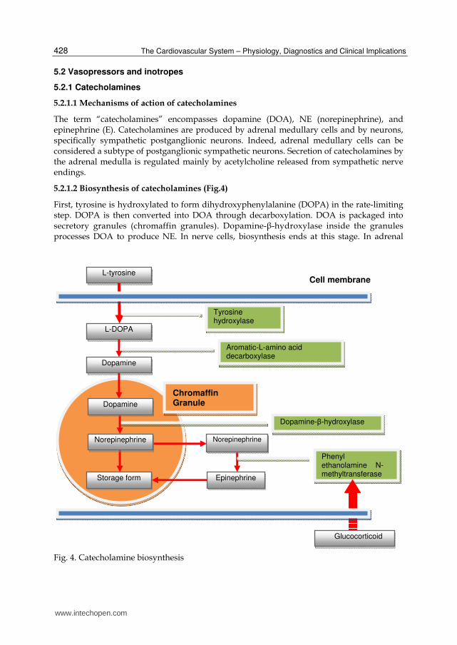
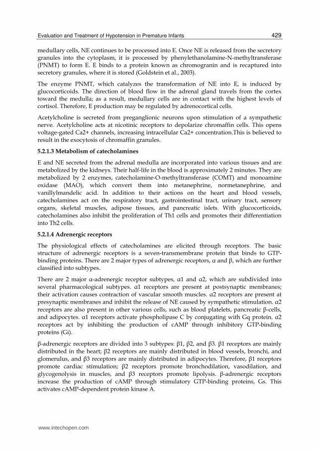
5.2.1.2 Biosynthesis of catecholamines (Fig.4)
First, tyrosine is hydroxylated to form dihydroxyphenylalanine (DOPA) in the rate-limiting step. DOPA is then converted into DOA through decarboxylation. DOA is packaged into secretory granules (chromaffin granules). Dopamine-┚-hydroxylase inside the granules processes DOA to produce NE. In nerve cells, biosynthesis ends at this stage. In adrenal
Fig. 4. Catecholamine biosynthesis
Epinephrine
Phenyl ethanolamine N-methyltransferase
Norepinephrine
L-DOPA
L-tyrosine Cell membrane
Dopamine
Tyrosine hydroxylase
Aromatic-L-amino acid decarboxylase
Glucocorticoid
Chromaffin Granule
Dopamine-β-hydroxylase
Norepinephrine
Storage form
Dopamine
www.intechopen.com
Evaluation and Treatment of Hypotension in Premature Infants
429
medullary cells, NE continues to be processed into E. Once NE is released from the secretory granules into the cytoplasm, it is processed by phenylethanolamine-N-methyltransferase (PNMT) to form E. E binds to a protein known as chromogranin and is recaptured into secretory granules, where it is stored (Goldstein et al., 2003).
The enzyme PNMT, which catalyzes the transformation of NE into E, is induced by glucocorticoids. The direction of blood flow in the adrenal gland travels from the cortex toward the medulla; as a result, medullary cells are in contact with the highest levels of cortisol. Therefore, E production may be regulated by adrenocortical cells.
Acetylcholine is secreted from preganglionic neurons upon stimulation of a sympathetic nerve. Acetylcholine acts at nicotinic receptors to depolarize chromaffin cells. This opens voltage-gated Ca2+ channels, increasing intracellular Ca2+ concentration.This is believed to result in the exocytosis of chromaffin granules.
5.2.1.3 Metabolism of catecholamines
E and NE secreted from the adrenal medulla are incorporated into various tissues and are
metabolized by the kidneys. Their half-life in the blood is approximately 2 minutes. They are
metabolized by 2 enzymes, catecholamine-O-methyltransferase (COMT) and monoamine
oxidase (MAO), which convert them into metanephrine, normetanephrine, and
vanillylmandelic acid. In addition to their actions on the heart and blood vessels,
catecholamines act on the respiratory tract, gastrointestinal tract, urinary tract, sensory
organs, skeletal muscles, adipose tissues, and pancreatic islets. With glucocorticoids,
catecholamines also inhibit the proliferation of Th1 cells and promotes their differentiation
into Th2 cells.
5.2.1.4 Adrenergic receptors
The physiological effects of catecholamines are elicited through receptors. The basic structure of adrenergic receptors is a seven-transmembrane protein that binds to GTP-binding proteins. There are 2 major types of adrenergic receptors, ┙ and ┚, which are further classified into subtypes.
There are 2 major ┙-adrenergic receptor subtypes, ┙1 and ┙2, which are subdivided into
several pharmacological subtypes. ┙1 receptors are present at postsynaptic membranes;
their activation causes contraction of vascular smooth muscles. ┙2 receptors are present at
presynaptic membranes and inhibit the release of NE caused by sympathetic stimulation. ┙2
receptors are also present in other various cells, such as blood platelets, pancreatic ┚-cells,
and adipocytes. ┙1 receptors activate phospholipase C by conjugating with Gq protein. ┙2
receptors act by inhibiting the production of cAMP through inhibitory GTP-binding
proteins (Gi).
┚-adrenergic receptors are divided into 3 subtypes: ┚1, ┚2, and ┚3. ┚1 receptors are mainly
distributed in the heart; ┚2 receptors are mainly distributed in blood vessels, bronchi, and
glomerulus, and ┚3 receptors are mainly distributed in adipocytes. Therefore, ┚1 receptors
promote cardiac stimulation; ┚2 receptors promote bronchodilation, vasodilation, and
glycogenolysis in muscles, and ┚3 receptors promote lipolysis. ┚-adrenergic receptors
increase the production of cAMP through stimulatory GTP-binding proteins, Gs. This
activates cAMP-dependent protein kinase A.
www.intechopen.com
The Cardiovascular System – Physiology, Diagnostics and Clinical Implications
430
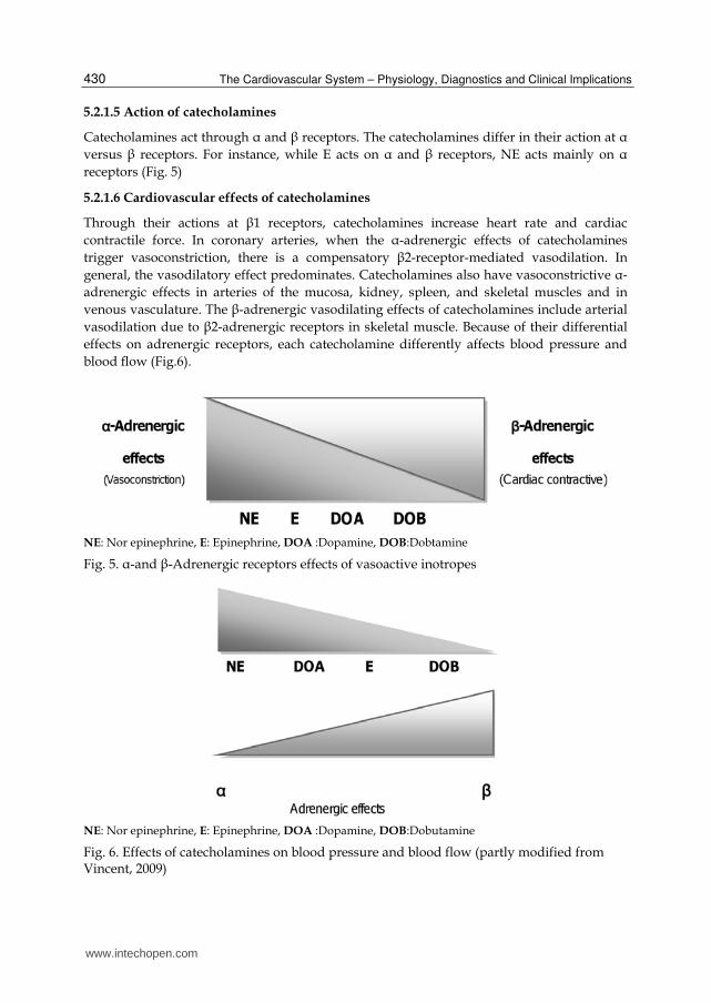
5.2.1.5 Action of catecholamines
Catecholamines act through ┙ and ┚ receptors. The catecholamines differ in their action at ┙
versus ┚ receptors. For instance, while E acts on ┙ and ┚ receptors, NE acts mainly on ┙
receptors (Fig. 5)
5.2.1.6 Cardiovascular effects of catecholamines
Through their actions at ┚1 receptors, catecholamines increase heart rate and cardiac
contractile force. In coronary arteries, when the ┙-adrenergic effects of catecholamines
trigger vasoconstriction, there is a compensatory ┚2-receptor-mediated vasodilation. In
general, the vasodilatory effect predominates. Catecholamines also have vasoconstrictive ┙-
adrenergic effects in arteries of the mucosa, kidney, spleen, and skeletal muscles and in
venous vasculature. The ┚-adrenergic vasodilating effects of catecholamines include arterial
vasodilation due to ┚2-adrenergic receptors in skeletal muscle. Because of their differential
effects on adrenergic receptors, each catecholamine differently affects blood pressure and
blood flow (Fig.6).
NE: Nor epinephrine, E: Epinephrine, DOA :Dopamine, DOB:Dobtamine
Fig. 5. ┙-and ┚-Adrenergic receptors effects of vasoactive inotropes
NE: Nor epinephrine, E: Epinephrine, DOA :Dopamine, DOB:Dobutamine
Fig. 6. Effects of catecholamines on blood pressure and blood flow (partly modified from Vincent, 2009)
www.intechopen.com
Evaluation and Treatment of Hypotension in Premature Infants
431
5.2.1.7 Downregulation of adrenergic receptors
Recently, it has been proposed that exogenous catecholamine administration downregulates adrenergic receptors and their associated second-messenger systems (Hausdorff et al., 1999; Collins et al., 1991). During receptor downregulation, adrenergic receptors undergo lysosomal destruction; therefore, reversal of this process requires new protein synthesis.
5.2.1.8 Levels of catecholamines in hypotensive preterm infants
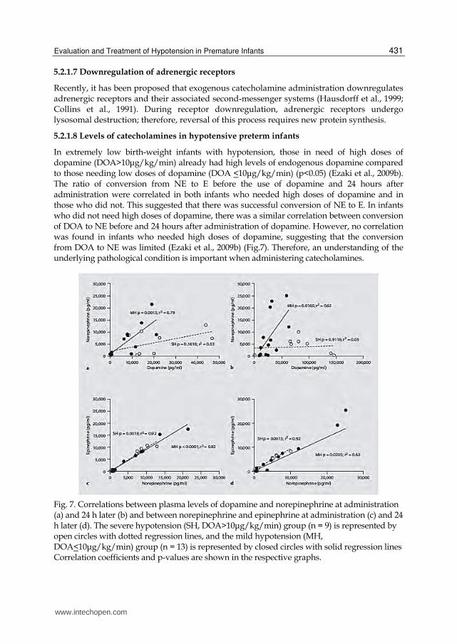
In extremely low birth-weight infants with hypotension, those in need of high doses of dopamine (DOA>10μg/kg/min) already had high levels of endogenous dopamine compared to those needing low doses of dopamine (DOA <10μg/kg/min) (p<0.05) (Ezaki et al., 2009b). The ratio of conversion from NE to E before the use of dopamine and 24 hours after administration were correlated in both infants who needed high doses of dopamine and in those who did not. This suggested that there was successful conversion of NE to E. In infants who did not need high doses of dopamine, there was a similar correlation between conversion of DOA to NE before and 24 hours after administration of dopamine. However, no correlation was found in infants who needed high doses of dopamine, suggesting that the conversion from DOA to NE was limited (Ezaki et al., 2009b) (Fig.7). Therefore, an understanding of the underlying pathological condition is important when administering catecholamines.
Fig. 7. Correlations between plasma levels of dopamine and norepinephrine at administration (a) and 24 h later (b) and between norepinephrine and epinephrine at administration (c) and 24 h later (d). The severe hypotension (SH, DOA>10μg/kg/min) group (n = 9) is represented by open circles with dotted regression lines, and the mild hypotension (MH, DOA<10μg/kg/min) group (n = 13) is represented by closed circles with solid regression lines Correlation coefficients and p-values are shown in the respective graphs.
www.intechopen.com
The Cardiovascular System – Physiology, Diagnostics and Clinical Implications
432
5.2.2 Dopamine
5.2.2.1 Treatment of dopamine in preterm hypotension
Dopamine is the most commonly used vasopressor/inotrope for the treatment of systemic hypotension in preterm infants (Seli,1996). DOA stimulates ┙-adrenergic receptors, ┚-adrenergic receptors, and dopaminergic receptors (See 5.2.1.4). DOA stimulates dopamine receptors at low doses (0.5 μg · kg-1 · min-1), mainly triggering effects in renal, mesenteric, and coronary blood vessels. At doses of 2–4 μg · kg-1 · min-1, DOA acts at ┙-adrenergic receptors, and at doses of 4–8 μg · kg-1 · min-1, DOA acts at ┚-adrenergic receptors (Seli, 2006).
With the exception of E administration, DOA administration is the most effective treatment for elevating blood pressure in preterm infants. The increase in CBF following DOA administration was found to be greater in hypotensive preterm infants compared to normotensive preterm infants, suggesting the presence of pressure-passive CBF in hypotensive neonates (Sassano et al., 2011). Therefore, we recommend the use of DOA as a first-line inotrope for the treatment of hypotension in preterm infants.
DOA is also an important neurotransmitter that affects both cerebral vasculature and neuronal activity. This is exemplified by pathological conditions caused by dopaminergic dysfunction, including abnormalities in CBF and neuronal metabolism (Edvinsson and Krause, 2002). In the mature brain, CBF is coupled to oxygen consumption (CMRO2). In contrast, CBF coupling to metabolism is strikingly different in the brains of very preterm infants, in which cerebral oxygen extraction, not CBF, sustains CMRO2. However, preterm infants receiving DOA treatment exhibit flow-metabolism coupling similar to that of the mature brain. This suggest a role for DOA in promoting flow-metabolism coupling in the preterm brain (Wong et al., 2009). In addition, we previously reported that high-dose administration of DOA can limit the conversion of NE to DOA (Ezaki et al., 2009b). Therefore, extreme caution must be taken when administering high doses of DOA.
5.2.2.2 Adverse effects of dopamine treatment
┙2-adrenergic receptors are important in endocrine regulation; as such, even low doses of systemically administered DOA have profound endocrine effects. For instance, DOA infusion reduces thyroid stimulating hormone and thyroxine levels in very low birth-weight infants (Filippi et al., 2004).
Doses of DOA should rarely exceed 20 μg · kg-1 · min-1, because there is a risk of excessive ┙-adrenergic-receptor-mediated peripheral vasoconstriction and a subsequent reduction in CO (Rozé et al., 1993). DOA failed to raise blood pressure in more than 30% of preterm infants with systemic hypotension (Pellicer et al., 2005).
5.2.3 Norepinephrine
5.2.3.1 The use of norepinephrine in the treatment of preterm hypotension
NE is a potent vasopressor with ┙-and, to a lesser extent, ┚-1 receptor agonist activity (Hollenberg et al., 2004). In the adult, NE is primarily used as a vasopressor in states of hyperdynamic shock, in which SVR is decreased and mean arterial blood pressure is low (Corley, 2004). Experimental studies in fetal lambs have shown that NE may decrease basal
www.intechopen.com
Evaluation and Treatment of Hypotension in Premature Infants
433
pulmonary vascular tone (Houfflin-Debarge et al., 2001) and elevate pulmonary blood flow through activating ┙2-adrenergic receptors and NO release (Magnenant et al., 2003).
NE can reduce damage incurred by neuroinflammatory and neurodegenerative conditions. It induces the expression of the chemokine CCL2 in astrocytes, which is neuroprotective against excitotoxic damage (Madrigal et al., 2009). Indeed, early associative somatosensory conditioning requires NE (Landers and Sulliyan, 1999).
Thus, NE plays an important role not only in the cardiovascular system, but also in neonatal
development. However, there are few studies on the use of NE in the treatment of
hypotension in preterm infants. While no studies have compared NE to other drugs, its
therapeutic effects in neonates have recently been reported (Paradisis and Osborn, 2004).
The use of NE (0.5-0.75 μg · kg-1 · min-1) is effective in the treatment of term and near-term
infants with septic shock that are resistant to DOA and dobutamine (Tourneux et al., 2008a).
In neonates with persistent pulmonary hypertension-induced cardiac dysfunction, NE can
reduce O2 requirements and normalize the systemic artery pressure (Tourneux et al., 2008b).
5.2.3.2 Adverse effects of norepinephrine treatment
In all previous reports describing the use of NE in neonates, NE was administered after
other inotropes, making it impossible to describe the side effects solely attributable to NE. In
addition, there are no reports on the long-term consequences of the use of NE in preterm
infants. In general, excessive peripheral vasoconstriction causes a decrease in the contractile
forces of the immature heart. This may result in tachycardia or decreased tissue perfusion.
Therefore, capillary refill time, lactate levels, and peripheral and organ blood flow should be
monitored.
5.2.4 Epinephrine
5.2.4.1 The use of epinephrine for the treatment of preterm hypotension
Low and moderate doses of E (0.125–0.5 μg · kg-1 · min-1) have found to be as effective as
low and moderate doses of DOA (2.5–10 μg · kg-1 · min-1) for the treatment of hypotension
in preterm infants (Valverde et al., 2006). In addition, the infusion of E increases mean
arterial blood pressure and heart rate without decreasing urine output in very low birth-
weight infants with hypotension that do not respond to dopamine infusion up to 15 μg · kg-
1 · min-1 (Heckmann et al., 2002).
5.2.4.2 Adverse effects of epinephrine
Compared DOA, E use cases temporary dysfunction of carbohydrate and lactate metabolism
(Valverde et al., 2006) and increased metabolic acidosis (Heckmann et al., 2002). E directly
affects lactate metabolism by increasing lactate production and decreasing lactate
metabolism, thus increasing serum lactate concentrations (Cheung et al., 1997). At very high
doses, E induces vasoconstriction sufficient to counteract its inotropic benefits, and CO may
fall (Barrington et al., 1995).
Pellicer et al. recently reported that the long-term prognosis of E use was the same as DOA use, and that both were safe (Pellicer et al., 2009). This important study provided an additional treatment option for preterm hypotension.
www.intechopen.com
The Cardiovascular System – Physiology, Diagnostics and Clinical Implications
434
5.3 Non-catecholamine inotropic/pressor agents
5.3.1 Dobutamine
5.3.1.1 Physiology of dobutamine in preterm hypotension
Dobutamine is a racemic mixture of 2 isomers, the D-isomer with ┙1- and ┙2-adrenergic effects and the L-isomer with ┙1- and ┙1- adrenergic effects. Dobutamine is predominantly inotropic via stimulation of ┙1 receptors and has a variable effect on blood pressure (Hollenberg, 2011). Dobutamine administration results in a variable decrease in total SVR. Unlike DOA, dobutamine increases myocardial contractility exclusively through direct stimulation of myocardial adrenergic receptors (Noori et al., 2004).
5.3.1.2 The use of dobutamine for the treatment of preterm hypotension
At a dose of 2–15 μg · kg-1 · min-1, dobutamine increases CO mainly through augmenting stroke volume (Noori et al., 2004; Roze et al., 1993; Bhatt-Mehta and Nahata, 1989).
5.3.1.3 Adverse effects of dobutamine treatment
Adverse effects of dobutamine occur at high doses and include increased heart rate. At very high doses, dobutamine may increase blood pressure and SVR (Cheung et al., 1999), likely due to stimulation of á-receptors (Fig.5 and 6). One study suggested that dobutamine’s potential benefit of increased oxygen delivery to the tissues was offset by increased tissue metabolic rate (Penny et al., 2001).
5.3.2 Vasopressin
5.3.2.1 Physiology of vasopressin in preterm infants
Vasopressin induces its physiological responses through 4 receptors, V1, V2, V3, and oxytocin receptors (OTR) (Holmes et al., 2001). When vasopressin binds to V1 receptors in vascular smooth muscle (Va1 receptors), it activates phospholipase C, triggering calcium release from intracellular calcium stores (Fig. 2). This results in vasoconstriction and a subsequent increase in blood pressure. Activation of V2 receptors in the stomach increases intracellular cyclic AMP levels through the mediation of adenylate cyclase and have an anti-diuretic effect. V3 receptors (also known as V1b receptors) are involved in vasopressin’s adrenocorticotropic hormone (ACTH)-stimulating effects. Finally, OTR receptors mediate vasopressing’s oxytocic effects on uterine contractility.
V2 receptors and OTR receptors also have vasodilating effects that are antagonistic to the effects of V1 receptors. In addition, V1 receptors and OTR receptors have diuretic effects, which are antagonistic to the anti-diuretic effects of V2 receptors. Vasopressin’s effects are most adapted to disease-induced changes.
Previous reports have indicated that blood levels of endogenous vasopressin show a two-phased response in adults with shock (Holmes et al., 2001; Landry et al., 1997; Morales et al., 1999). During the initial phase of shock, endogenous vasopressin is released in large amounts and reaches high blood levels in order to maintain tissue perfusion. However, its concentration in the blood decreases over time. As such, vasopressin may be depleted due to its initial release in large amounts. The release of vasopressin from the pituitary gland may also be inhibited by NO produced by the vascular endothelium or due to autonomic nervous system disorders (Holmes et al., 2001; Landry et.al, 1997; Morales et al., 1999).
www.intechopen.com
Evaluation and Treatment of Hypotension in Premature Infants
435
The effects of the small amounts of exogenous vasopressin may be a result of enhancing the
effects of catecholamines, inhibiting inducible NO synthase (iNOS), inhibiting increased
cGMP induced by NO and ANP, or inactivating KATP channels in vascular smooth muscles
(Fig.2) (Landry et al., 2001; Hamu et al.,1999).
In preterm infants, the levels of vasopressin were high during the first 24 hours following
birth (Ezaki et al., 2009b). The effects of these high levels of endogenous vasopressin on the
cardiovascular system are not fully understood.
5.3.2.2 The use of vasopressin for the treatment of hypotension in preterm infants
Meyer et al. reported that vasopressin (0.035–0.36 U · kg-1 · hr-1) may be a promising rescue
therapy for catecholamine-resistant shock in extremely-low-birth-weight infants with acute
renal injury (Meyer et al., 2006). Similarly, Ikegami et al. found that administration of
vasopressin (0.001–0.01 U · kg-1 · hr-1) was effective in extremely-low-birth-weight infants
resistant to treatment with catecholamines and steroids (Ikegami et al., 2010).
5.3 Adverse effects of vasopressin treatment
The side effects of vasporessin include severe cutaneous ischemia, hepatic necrosis, neurological deficits, and dysmetria (Meyer et al., 2006; Rodríguez-Nunez et al., 2006; Zeballos et al., 2006). Unlike DOA, E, and dobutamine, there are few reports on the side effects of vasopressin. Moreover, it is unclear whether its side effects are dose-dependent and what the long-term prognoses are. However, vasopressin is a pharmacological agent that can be considered for use in patients in whom other drugs are ineffective.
5.4 Lusitropes
5.4.1 Physiology of phosphodiesterase-III inhibitors in preterm hypotension
Phosphodiesterase inhibitors increase intracellular cyclic AMP and thus have inotropic
effects independent of ┙-adrenergic receptors. As such, they result in fewer chronotropic
and arrhythmogenic effects than catecholamines. However, increased cyclic AMP in
vascular smooth muscle cells can cause vasodilation, thus reducing SVR, which can
exacerbate hypotension. In addition, this can reduce pulmonary artery pressure. (Chen et
al.,1997,1998; Kato et al.,1998). Milrinone, a cyclic nucleotide phosphodiesterase-III inhibitor,
improves contractility and reduces afterload in adults and newborns with cardiac
dysfunction.
5.4.2 The use of phosphodiesterase-III inhibitors for the treatment of preterm hypotension
McNamara et al. reported that intravenous Milrinone (0.33–0.99 μg · kg-1 · min-1) administration produced early improvements in oxygenation without compromising systemic blood pressure in patients with severe persistent pulmonary hypertension (McNamara et al., 2006). One randomized clinical trial did not support the use of Milrinone (0.75 μg · kg-1 · min-1 for 3 hrs, then 0.2μ g · kg-1 · min-1 until 18 hours after birth) in the prevention of low SVC in the early transitional circulation of preterm infants (Paradisis et al., 2009).
www.intechopen.com
The Cardiovascular System – Physiology, Diagnostics and Clinical Implications
436
5.4.3 Adverse effects of Milrinone treatment
Milrinone can cause hypotension and tachycardia (Chang et al., 1995). The long-term effects of Milrinone in preterm infants have not been reported.
5.5 Corticosteroids
5.5.1 Physiology of corticosteroids
The adrenal glands are involved in the growth and maturation of fetal organs during intrauterine life. In most mammals, a cortisol surge occurs as the full gestational term approaches; this triggers increased synthesis of pulmonary surfactant, reduced sensitivity of the arteries to prostaglandins, and increased conversion of pancreatic ┚-cells from T4 to T3 in the mature liver. These changes allow the fetus to survive in the extrauterine environment. There is also a surge in catecholamines produced by the adrenal medulla during delivery. This surge also allows adaptation to the extrauterine environment by influencing the cardiovascular system, including elevating the blood pressure and increasing the heart function, and by influencing glucose metabolism, fat metabolism, and water absorption in the lungs (Fisher, 2002).
The hypothalamic-pituitary-adrenal system in fetuses and neonates has been implicated in late-onset circulatory collapse (Masumoto et al., 2008) and in the fetal programming of the cardiovascular system. Preterm infants have low adrenal function due to their low levels of 3┚-hydroxysteroid dehydrogenase (HSD) (Mesiano and Jaffe, 1997) and weak 11b-HSD2 activity (Donaldson et al.,1991).
Corticosteroids reverse neonatal hypotension by improving capillary-leak syndrome (Briegel et al., 1994), potentiating transmembrane calcium currents, increasing ┚-receptor sensitivity to catecholamines, reversing the downregulation of ┚-receptors, increasing the density of ┚-receptors, and inhibiting NO synthase expression (Prigent et al., 2004).
5.5.2 The use of corticosteroids in the treatment of preterm hypotension
Hydrocortisone administration is effective in the treatment of hypotension and vasopressor dependence in hypotensive preterm infants. Its clinical benefits include increasing blood pressure and decreasing the requirement for vasopressor administration (Higgins et al., 2010). Fernandez et al. have reviewed the use of hydrocortisone in the treatment of premature infants (Fernandez and Watterberg, 2009). Before initiating therapy with hydrocortisone in extremely preterm infants with refractory hypotension, a blood specimen should be analyzed for cortisol concentration. Pending that result, an initial dose of 1 mg/kg can be administered. If the blood pressure improves within 2 to 6 h, 0.5 mg/kg can be administered every 12 h (approximately 8–10 mg/m2 per day). This long dosing interval is used, because hydrocortisone has a longer half-life in immature infants (Watterberg et al., 2005). This dosing strategy increases serum values by an average of 5 μg/100 ml; higher doses are associated with very high serum concentrations. If the initial cortisol concentration is high (>15–20 μg/100 ml), drug administration may be discontinued, especially in the absence of a clinical response.
5.5.3 Adverse effects of corticosteroid treatment
Although corticosteroid therapy improves blood pressure and circulation, there are many potential complications, including spontaneous gut perforation, hyperglycemia, and
www.intechopen.com
Evaluation and Treatment of Hypotension in Premature Infants
437
hypertension and long-term consequences, including cerebral palsy and intellectual impairment. These complications necessitate the judicious use of corticosteroids to support blood pressure in preterm infants (Yeh et al., 2004).
Hydrocortisone therapy administered simultaneously with indomethacin or ibuprofen has been associated with acute spontaneous gastrointestinal perforation in extremely preterm infants. Therefore, care should be taken to avoid concurrent therapy (Watterberg et al., 2004; Peltoniemi et al., 2005). Infants who develop spontaneous perforation often have high endogenous cortisol concentrations (Watterberg et al., 2004; Peltoniemi et al., 2005).
Watterberg et al. reported that early, low-dose hydrocortisone treatment was not associated with an increased risk of cerebral palsy. In fact, infants treated with hydrocortisone displayed improved developmental outcomes. Together with the short-term benefits, these data support the use of hydrocortisone for the treatment of adrenal insufficiency in extremely premature infants (Watterberg et al., 2007).
6. Conclusion
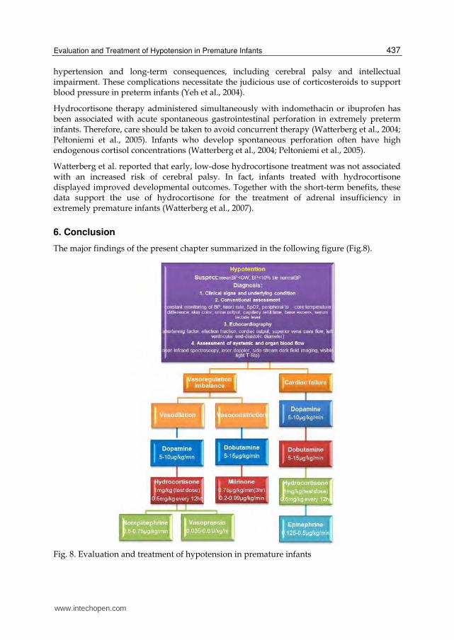
The major findings of the present chapter summarized in the following figure (Fig.8).
Fig. 8. Evaluation and treatment of hypotension in premature infants
www.intechopen.com
The Cardiovascular System – Physiology, Diagnostics and Clinical Implications
438
7. Acknowledgment
We express gratitude to the efforts of the authors whose research is cited in this article. First author (FA)’s personal research on vasoactive factors in neonates was inspired by my wife, a physician who introduced me to the use of vasopressin for the management of shock. Therefore, FA offer my wife, Yuko Ezaki, my most sincere gratitude. Finally, FA dedicate this chapter to my famiiy: Munenori, Tomi, Yuko, Yoshiko, Yukiko, and Saeka Ezaki.
FA hope that this work will promote future advances in neonatal care.
8. References
Bauer, K. & Linderkamp, O. & Versmold, HT. (1993). Systolic blood pressure and blood volume in preterm infants. Archives of disease in childhood, Vol. 69, 5 Spec No, (Nov), pp. 521-522, ISSN 0003-9888
Barrington, KJ. & Finer, NN. & Chan, WK. (1995). A blind, randomized comparison of the circulatory effects of dopamine and epinephrine infusions in the newborn piglet during normoxia and hypoxia. Critical Care Medicine, Vol. 23, No. 4, (Apr). pp. 740-748, ISSN 0090-3493
Belik, J., Light, RB. (1989). Effect of increased afterload on right ventricular function in newborn pigs. Journal of Applied Physiology, Vol. 66, No. 2, (Feb), pp. 863-869, ISSN 8750-7587
Bhatt-Mehta, V. & Nahata, MC. (1989). Dopamine and dobutamine in pediatric therapy. Pharmacotherapy, Vol. 9, No. 5, (May), pp. 304-314, ISSN 0277-0008
Briegel, J. & Kellermann, W. & Forst, H et al. (1994). Low-dose hydrocortisone infusion attenuates the systemic inflammatory response syndrome. The Phospholipase A2 Study Group. The Clinical investigator, Vol. 72, No. 10, (Oct), pp. 782-787, ISSN 0941-0198
Cayabyab, R. & McLean, CW. & Seri, I. (2009). Definition of hypotension and assessment of hemodynamics in the preterm neonate. Journal of Perinatology, Vol. 29, Suppl. 2, (May), pp. S58-S62, ISSN 0743-8346
Chang, AC. & Atz, AM. & Wernovsky, G et al. (1995). Milrinone: systemic and pulmonary hemodynamic effects in neonates after cardiac surgery. Critical care medicine, Vol. 23, No. 11, (Nov), pp. 1907-1914, ISSN 0090-3493
Cheung, PY. & Barrington, KJ. & Pearson, RJ et al. (1997). Systemic, pulmonary and mesenteric perfusion and oxygenation effects of dopamine and epinephrine. American Journal of Respiratory and Critical Care Medicine, Vol. 155, No. 1, (Jan), pp. 32-37, ISSN 1073-449X
Cheung, PY. & Barrington, KJ. & Bigam, D. (1999). The hemodynamic effects of dobutamine infusion in the chronically instrumented newborn piglet. Critical Care Medicine, Vol. 27, No. 3, (Mar), pp. 558-564, ISSN 0090-3493
Chen, EP. & Bittner, HB. & Davis, RD Jr et al. (1997). Milrinone improves pulmonary hemodynamics and right ventricular function in chronic pulmonary hypertension. The Annals of Thoracic Surgery, Vol. 63, No. 3, (Mar), pp. 814-821, ISSN 0003-4975
Chen, EP. & Bittner, HB. & Davis, RD et al. (1998), Hemodynamic and inotropic effects of milrinone after heart transplantation in the setting of recipient pulmonary
www.intechopen.com
Evaluation and Treatment of Hypotension in Premature Infants
439
hypertension. The Journal of Heart and Lung Transplantation, Vol. 17, No. 7, (Jul), pp. 669-678, ISSN 1053-2498
Collins, S. & Caron, MG. & Lefkowitz, RJ. (1991). Regulation of adrenergic receptor responsiveness through modulation of receptor gene expression. Annual Review of Physiology, Vol. 53, pp. 497-508, ISSN 0066-4278
Corley, KT. (2004). Inotropes and vasopressors in adults and foals. The Veterinary clinics of North America. Equine practice, Vol. 20, No. 1, (Apr), pp. 77-106, ISSN 0749-0739
Cunningham, S. & Symon, AG. & Elton, RA et al. (1999). Intraarterial blood pressure reference ranges, death and morbidity in very low birth weight infants during the first seven days of life. Early Human Development, Vol. 56, No. 2-3, (Dec), pp. 151-165, ISSN 0378-378256.
Dempsey, EM. & Al Hazzani, F. & Barrington, KJ. (2009). Permissive hypotension in the extremely low birthweight infant with signs of good perfusion. Archives of Disease in Childhood Fetal & Neonatal Edition, Vol. 94, No. 4, (Jul), pp. F241-F244, ISSN 1359-2998
Donaldson, A. & Nicolini, U. & Symes, EK et al. (1991). Changes in concentrations of cortisol, dehydroepiandrosterone sulphate and progesterone in fetal and maternal serum during pregnancy. Clinical endocrinology, Vol. 35, No. 5,(Nov), pp. 447-451, ISSN 0300-0664
Edvinsson, L. & Krause, D. (2002). Catecholamines, In: Cerebral blood flow and metabolism, Edvinsson, L & Krause, D, (Eds.), 191-211, Lippincott Williams & Wilkins, ISBN 978-0781722599, Philadelphia
El-Khuffash, AF. & McNamara, PJ. (2011). Neonatologist-performed functional echocardiography in the neonatal intensive care unit. Seminars in fetal & neonatal medicine, Vol. 16, No. 1, (Feb), pp. 50-60, ISSN 1744-165X
Engle, WD. (2001). Blood pressure in the very low birth weight neonates. Early Human Development, Vol. 62, No. 2, (May), pp. 97-130, ISSN 0378-3782
Evans, N. (2003). Volume expansion during neonatal intensive care: do we know what we are doing? Semin Neonatol, Vol. 8, No. 4, (Aug), pp. 315-323, ISSN1744-165X52.
Evans, N. & Kluckow, M. (1996). Early determinants of right and left ventricular output in ventilated preterm infants. Archives of disease in childhood. Fetal and neonatal edition, Vol. 74, No. 2, (Mar), pp. F88-F94, ISSN 1359-2998
Ewer, AK. & Tyler, W. & Francis, A et al. (2003). Excessive volume expansion and neonatal death in preterm infants born at 27-28 weeks gestation. Paediatric and Perinatal Epidemiology, Vol. 17, No.2, (Apr), pp. 180-186, ISSN 0269-5022
Ezaki, S. & Suzuki, K. & Kurishima, C et al. (2009a). Resuscitation of preterm infants with reduced oxygen results in less oxidative stress than resuscitation with 100% oxygen. Journal of clinical biochemistry and nutrition, Vol. 44, No.1, (Jan), pp. 111-118, ISSN 0912-0009
Ezaki, S. & Suzuki, K. & Kurishima, C et al. (2009b). Levels of catecholamines, arginine vasopressin and atrial natriuretic peptide in hypotensive extremely low birth weight infants in the first 24 hours after birth. Neonatology, Vol. 95, No. 3, (Nov 4), pp. 248-255, ISSN 1661-7800
Fernandez, EF. & Watterberg, KL. (2009). Relative adrenal insufficiency in the preterm and term infant. Journal of Perinatology, Vol. 29, Suppl 2, (May), pp. S44-S49, ISSN 0743-8346
www.intechopen.com
The Cardiovascular System – Physiology, Diagnostics and Clinical Implications
440
Fisher, D. ( 2002). Endocrinology of Fetal Development, In: Williams Textbook of Endcrinology.11th, Kronenberg, HM. & Melmed, S, & Polonsky, KS. & Larsen,
(Eds.), pp.756-776, WB Saunders, ISBN 978-1437703245, Phil ad elphia Filippi, L. & Cecchi, A. & Tronchin, M et al. (2004). Dopamine infusion and
hypothyroxinaemia in very low birth weight preterm infants. Early Human Development, Vol. 163, No. 1, (Jan), pp. 7-13, ISSN 0378-3782
Goldberg, RN. & Chung, D. & Goldman, SL et al. (1980). The association of rapid volume expansion and intraventricular hemorrhage in the preterm infant. The Journal of Pediatrics, Vol. 96, No. 6, (Jun), pp. 1060-1063, ISSN 0022-3476
Goldstein, DS. & Eisenhofer, G. & Kopin IJ. (2003). Sources and significance of plasma levels of catechols and their metabolites in humans. Journal of Pharmacology and Experimental Therapeutics, Vol. 305, No. 3, (Jun), PP. 800-811, ISSN 0022-3565
Goldstein, RF. & Thompson, RJ. & Oehler, JM et al. (1995). Influence of acidosis, hypoxemia, and hypotension on neurodevelopmental outcome in very low birth weight infants. Pediatrics, Vol. 95, No. 2, (Feb), pp. 238-243, ISSN 0031-4005
Greenough, A. & Cheesemen, P. & Kawadia, V et al. (2002). Colloid infusion in the perinatal period and abnormal neurodevelopmental outcome in very low birth weight infants. European Journal of Pediatrics, Vol. 161, No. 6, (Jun), pp. 319-323, ISSN 0340-6199
Greisen G. (2005). Autoregulation of cerebral blood flow in newborn babies. Early Human Development, Vol. 81, No. 5, (May), pp. 423-428, ISSN 0378-3782
Hausdorff, WP. & Hnatowich, M. & O’Dowd BF et al. (1990). A mutation of the beta 2-adrenergic receptor impairs agonist activation of adenylyl cyclase without affecting high affinity agonist binding. Distinct molecular determinants of the receptor are involved in physical coupling to and functional activation of Gs. The Journal of Biological Chemistry, Vol. 265, No. 3, (Jan), pp.1388-1393, ISSN 0021-9258
Hamu, Y. & Kanmura, Y. & Tsuneyoshi, I et al. (1999). The effects of vasopressin on endotoxin-induced attenuation of contractile responses in human gastroepiploic arteries in vitro. Anesthesia & Analgesia, Vol. 88, No. 3, (Mar), pp. 542-548, ISSN 0003-2999
Heckmann, M. & Trotter, A. & Pohlandt, F et al. (2002). Epinephrine treatment of hypotension in very low birthweight infants. Acta Paediatrica, Vol. 91. No. 5, (May), pp. 566-570, ISSN 0803-5253
Higgins, S. & Friedlich, P. & Seri, I. (2010). Hydrocortisone for hypotension and vasopressor dependence in preterm neonates:
a meta-analysis. Journal of Perinatology, Vol. 30, No. 6, (Jun), pp. 373-378, ISSN 0743-8346
Hiedl, S. & Schwepcke, A. & Weber, F et al. (2010). Microcirculation in preterm infants: profound effects of patent ductus arteriosus. The Journal of Pediatrics, Vol. 156, No. 2, (Feb), pp. 191-196, ISSN 0022-3476
Hollenberg, SM. & Ahrens, TS. & Annane, D et al. (2004). Practice parameters for hemodynamic support of sepsis in adult patients: 2004 update. Critical Care Medicine, Vol. 32, No. 9, (Sep), pp, 1928-1948, ISSN 0090-3493
Hollenberg, SM. (2011). Vasoactive drugs in circulatory shock. American Journal of Respiratory and Critical Care Medicine, Vol. 183, No. 7, (Apr 1), pp. 847-855, ISSN 1073-449X
Holmes, CL. & Patel, BM. & Russell, JA et al. (2001). Physiology of vasopressin relevant to management of septic shock. Chest, Vol. 120, No. 3, (Sep), pp. 989-1002, ISSN 0012-3692
www.intechopen.com
Evaluation and Treatment of Hypotension in Premature Infants
441
Ikegami, H. & Funato, M. & Tamai, H et al. (2010). Low-dose vasopressin infusion therapy for refractory hypotension in ELBW infants. Pediatrics International, Vol. 52, No. 3, (Jun), pp. 368-373, ISSN 1328-8067
Ishiguro, A. & Sekine, T. & Suzuki, K et al.(2011). Changes in skin and subcutaneous perfusion in very-low-birth-weight infants during the transitional period. Neonatology, Vol. 100, No. 2, (Mar), pp. 162-168, ISSN 1661-7800
Jaillard, S. & Houfflin-Debarge, V. & Riou Y et al. (2001). Effects of catecholamines on the pulmonary circulation in the ovine fetus. American journal of physiology. Regulatory, integrative and comparative physiology, Vol. 281, No. 2, (Aug), pp. R607-R614, ISSN 0363-6119
Kato, R. & Sato, J. & Nishino, T. (1998). Milrinone decreases both pulmonary arterial and venous resistances in the hypoxic dog. British Journal of Anaesthesia , Vol. 81, No. 6, (Dec), pp. 920-924, ISSN 0007-0912
Kluckow, M. & Evans, N. (2000). Superior vena cava flow in newborn infants: a novel marker of systemic blood flow. Archives of Disease in Childhood Fetal & Neonatal Edition, Vol. 82, No. 3, (May), pp. F182-F187, ISSN 1359-2998
Kluckow, M. & Seri, I. & Evans, N. (2007). Functional echocardiography: an emerging clinical tool for the neonatologist. The Journal of Pediatrics, Vol. 150, No. 2, (Feb), pp. 125-130, ISSN 0022-3476
Kluckow, M. (2005). Low systemic blood flow and pathophysiology of the preterm transitional circulation. Early Human Development, Vol. 81, No. 5, (May), pp. 429-437, ISSN: 0378-3782
Landry, DW. & Levin, HR. & Gallant, EM et al. (1997). Vasopressin deficiency contributes to the vasodilation of septic shock. Circulation, Vol. 95, No. 5, (Mar 4), pp. 1122-1125, ISSN 0009-7322
McNamara, PJ. & Laique, F. & Muang-In, S et al. (2006). Milrinone improves oxygenation in neonates with severe persistent pulmonary hypertension of the newborn. Journal of Critical Care, Vol. 21, No. 2, (Jun), pp. 217-222 ISSN 0883-9441
Landers, MS. & Sullivan, RM. (1999). Norepinephrine and associative conditioning in the neonatal rat somatosensory system. Brain research. Developmental brain research, Vol. 114, No.2, (May 14), pp. 261-264, ISSN 0165-3806
Landry, DW. & Oliver, JA. (2001). The pathogenesis of vasodilatory shock. The New England Journal of Medicine, Vol. 345, No. 8, (Aug 23), pp. 588-595, ISSN 0028-4793
Liedel, JL. & Meadow, W. & Nachman, J et al. (2002). Use of vasopressin in refractory hypotension in children with vasodilatory shock: five cases and a review of the literature. Pediatric Critical Care Medicine, Vol. 3, No. 1, (Jan), pp. 15-18, ISSN 1529-7535
Masumoto, K. & Kusuda, S. & Aoyagi, H et al. (2008). Comparison of serum cortisol concentrations in preterm infants with or without late-onset circulatory collapse due to adrenal insufficiency of prematurity. Pediatric Research, Vol. 63, No. 6, (Jun), pp. 686-690, ISSN 0031-3998
Magnenant, E. & Jaillard, S. & Deruelle, P et al. (2003). Role of the alpha2-adrenoceptors on the pulmonary circulation in the ovine fetus. Pediatric Research, Vol. 54, No. 1, (Jul), pp. 44-51, ISSN 0031-3998
Madrigal, JL. & Leza, JC. & Polak P et al. (2009). Astrocyte-derived MCP-1 mediates neuroprotective effects of noradrenaline. Journal of Neuroscience, Vol. 29, No. 1, (Jan 7), pp. 263-267, ISSN 0270-6474
www.intechopen.com
The Cardiovascular System – Physiology, Diagnostics and Clinical Implications
442
McLean, CW. & Cayabyab, R. & Noori, S et al. (2008). Cerebral circulation and hypotension in the premature infant- diagnosis and treatment, In: Neonatology Questions and Controversies: Neurology, Perlman, JM. (Ed.), pp. 3–26, Saunders/Elsevier, ISBN, 978-1416031574, Philadelphia
Meyer, S. & Gottschling, S. & Baghai, A et al. (2006). Arginine-vasopressin in catecholamine-refractory septic versus non-septic shock in extremely low birth weight infants with acute renal injury. Critical Care, Vol. 10, No. 3, (May 5), R71, ISSN 1364-8535
Meyer, S. & Todd, D. & Shadboldt, B. (2009). Assessment of portable continuous wave Doppler ultrasound (ultrasonic cardiac output monitor) for cardiac output measurements in neonates. Journal of Paediatrics and Child Health, Vol. 45, No. 7-8, (Jul-Aug), pp. 464-468, ISSN 1034-4810
Mesiano, S. & Jaffe, RB. (1997). Developmental and functional biology of the primate fetal adrenal cortex. Endocrine Reviews, Vol. 18, No. 3, (Jun), pp. 378-403, ISSN 0163-769X
Morales, D. & Madigan, J. & Cullinane, S et al. (1999). Reversal by vasopressin of intractable hypotension in the late phase of hemorrhagic shock. Circulation, Vol. 100, No. 3, (Jul 20), pp. 226-229, ISSN 0009-7322
Munro, MJ. & Walker, AM. & Barfield, CP. (2004). Hypotensive extremely low birth weight infants have reduced cerebral blood flow. Pediatrics, Vol. 114, No. 6, (Dec), pp. 1591-1596, ISSN 0031-4005
Nagdyman, N & Ewert, P. & Peters, B et al. (2008). Comparison of different near-infrared spectroscopic cerebral oxygenation indices with central venous and jugular venous oxygenation saturation in children. Pediatric Anesthesia, Vol. 18, No. 2, (Feb), pp. 160-166, ISSN 1155-5645
Ng, PC. & Lam, CW. & Fok, TF et al. (2001) Refractory hypotension in preterm infants with adrenocortical insufficiency. Archives of Disease in Childhood Fetal & Neonatal Edition, Vol. 84, No. 2, (Mar), pp. F122-F124, ISSN 1359-2998
Noori, S. & Seri, I. (2005). Pathophysiology of newborn hypotension outside the transitional period. Early Human Development, Vol.81, No.5, (May), pp. 399-404, ISSN 0378-3782
Noori, S. & Friedlich, P. & Seri, I. (2004). Pharmacology Review: The Use of Dobutamine in the Treatment of Neonatal Cardiovascular Compromise. NeoReviews. org, Vol. 5, No. 1, (Jan 1), pp. e22-e26
Nuntnarumit, P. & Yang, W. & Bada-Ellzey, HS. (1999). Blood pressure measurements in the newborn. Clinics in Perinatology, Vol. 26, No. 4, (Dec), pp. 981-996, ISSN 0095-5108
Oca, MJ. & Nelson, M. & Donn, SM. (2003). Randomized trial of normal saline versus 5% albumin for the treatment of neonatal hypotension. Journal of Perinatology, Vol. 23, No. 6, (Sep), pp. 473-476, ISSN 0743-8346
Osborn, DA. & Evans, N. & Kluckow, M. (2004). Clinical detection of low upper body blood flow in very premature infants using blood pressure, capillary refill time, and central-peripheral temperature difference. Archives of Disease in Childhood Fetal & Neonatal Edition, Vol. 89, No. 2, (Mar), pp. F168-F173, ISSN 1359-2998
Paradisis, M. & Evans, N. & Kluckow, M et al. (2009). Randomized trial of milrinone versus placebo for prevention of low systemic blood flow in very preterm infants. The Journal of Pediatrics, Vol. 154, No. 2, (Feb), pp. 189-195, ISSN 0022-3476
Paradisis, M. & Osborn, DA. (2004). Adrenaline for prevention of morbidity and mortality in preterm infants with cardiovascular compromise. Cochrane database of systematic reviews, No.1, CD003958, ISSN 1469-493X
www.intechopen.com
Evaluation and Treatment of Hypotension in Premature Infants
443
Pellicer, A. & Valverde, E. & Elorza, MD et al. (2005). Cardiovascular support for low birth weight infants and cerebral hemodynamics: a randomized, blinded, clinical trial. Pediatrics, Vol. 115, No. 6, (Jun), pp. 1501-1512, ISSN 0031-400568. Pellicer, A. & Bravo, MC. & Madero, R et al. (2009). Early systemic hypotension and vasopressor support in low birth weight infants: impact on neurodevelopment. Pediatrics, Vol. 123, No. 5, (May), pp. 1369-1376, ISSN 0031-4005
Penny, DJ. & Sano, T. & Smolich, JJ. (2001). Increased systemic oxygen consumption offsets improved oxygen delivery during dobutamine infusion in newborn lambs. Intensive Care Medicine, Vol. 27, No. 9, (Sep), pp. 1518-1525, ISSN 0342-4642
Peltoniemi, O. & Kari, MA. & Heinonen, K et al. (2005), Pretreatment cortisol values may predict responses to hydrocortisone administration for the prevention of bronchopulmonary dysplasia in high-risk infants. The Journal of Pediatrics, Vol. 146, No. 5, (May), pp. 632-637, ISSN 0022-3476
Prigent, H. & Maxime, V. & Annane, D. (2004). Clinical review: corticotherapy in sepsis. Critical Care, Vol. 8, No. 2, (Apr), pp.122-129, ISSN 1364-8535
Tourneux, P. & Rakza, T. & Abazine, A et al. (2008a). Noradrenaline for management of septic shock refractory to fluid loading and dopamine or dobutamine in full-term newborn infants. Acta Paediatrica, Vol. 97, No. 2, (Feb), pp. 177-180, ISSN 0803-5253
Tourneux, P. & Rakza, T. & Bouissou, A et al. (2008b). Pulmonary circulatory effects of norepinephrine in newborn infants with persistent pulmonary hypertension. The Journal of Pediatrics, Vol. 153, No. 3, (Sep), pp. 345-349, ISSN 0022-3476
Rodríguez-Núñez, A. & López-Herce, J. & Gil-Antón, J et al. (2006). Rescue treatment with terlipressin in children with refractory septic shock: a clinical study. Critical Care, Vol. 10, No. 1, (Jan 31), R20, ISSN 1364-8535
Rowland, DG. &, Gutgesell, HP. (1995). Noninvasive assessment of myocardial contractility, preload, and afterload in healthy newborn infants. American Journal of Cardiology,Vol. 75, No.12, (Apr 15), pp. 813-821, ISSN: 0002-9149
Rozé, JC. & Tohier, C. & Maingueneau C et.al. (1993). Response to dobutamine and dopamine in the hypotensive very preterm infant. Archives of disease in childhood, Vol. 69, 1 Spec No, (Jul), pp. 59-63, ISSN 0003-9888
Sassano-Higgins, S. & Friedlich, F. & Seri, I. (2011). A meta-analysis of dopamine use in hypotensive preterm infants: blood pressure and cerebral hemodynamics. Journal of Perinatology, Vol. 31, No. 10, (Oct 31), pp. 647-655, ISSN 0743-8346
Seri, I. (1995). Cardiovascular, renal, and endocrine actions of dopamine in neonates and children. The Journal of Pediatrics, Vol. 126, No. 3, (Mar), pp.333-344, ISSN 0022-3476
Seri, I. & Evans, J. (2001). Controversies in the diagnosis and management of hypotension in the newborn infant. Current Opinion in Pediatrics, Vol.13, No.2, (Apr), pp. 116-123, ISSN 1040-8703
Seri, I. (2006). Management of hypotension and low systemic blood flow in the very low birth weight neonate during the first postnatal week. Journal of Perinatology, Vol. 26, Suppl. 1, (May 26), pp. S8-S13, ISSN 0743-8346
Shiao, SY. & Ou, CN. (2007). Validation of oxygen saturation monitoring in neonates. American Journal of Critical Care, Vol. 16, No. 2, (Mar), pp. 168-178, ISSN 1062-3264
Skelton, R. & Gill, AB. & Parsons, JM. (1998). Reference ranges for cardiac dimensions and blood flow velocity in preterm infants. Heart, Vol. 80, No. 3, (Sep), pp. 281-285, ISSN 1468-201X 124, No. 1, (Jul), pp. 277-284, ISSN 0031-4005
www.intechopen.com
The Cardiovascular System – Physiology, Diagnostics and Clinical Implications
444
Soleymani, S. & Cayabyab, R. & Borzage, TM et al. (2010). Comparison between charted and continuously recorded vital signs and hemodynamic data. Annual PAS/SPR Meeting, Vancouver, Abstract.
Stark, MJ. & Clifton, VL. & Wright, IM. (2009). Carbon monoxide is a significant mediator of cardiovascular status following preterm birth. Pediatrics, Vol.
Teitel, DF. & Sidi, D. (1985). Developmental changes in myocardial contractile reserve in the lamb. Pediatric Research, Vol. 19, No. 9, (Sep), pp. 948-955, ISSN 0031-3998
Valverde, E. & Pellicer, A. & Madero, R et al. (2006). Dopamine versus epinephrine for cardiovascular support in low birth weight infants: analysis of systemic effects and neonatal clinical outcomes. Pediatrics, Vol. 117, No. 6, (Jun), pp. e1213-e1222, 1098-4275
Van Hare, GF., & Hawkins, JA., & Schmidt, KG et al. (1990). The effects of increasing mean arterial pressure on left ventricular output in newborn lambs. Circulation Research, Vol. 67, No. 1, (Jul), pp. 78-83, ISSN 0009-7330
Van Bel, F. & Lemmers, P. & Naulaers, G. (2008). Monitoring neonatal regional cerebral oxygen saturation in clinical practice: value and pitfalls. Neonatology, Vol. 94, No. 4, (Sep 119), pp. 237-244, ISSN 1661-7800
Vincent, JL. (Ed.). (2009). Critical care medicine: Churchill’s ready reference, Churchill Livingstone, pp. 12–13, ISBN 978-0080451367, Philadelphia
Watkins, AM. & West, CR. & Cooke, RW. (1989). Blood pressure and cerebral haemorrhage and ischaemia in very low birthweight infants. Early Human Development, Vol. 19, No. 2, (May), pp. 103-110, ISSN 0378-3782
Watterberg, KL. & Gerdes, JS. & Cole, CH et al. (2004). Prophylaxis of early adrenal insufficiency to prevent bronchopulmonary dysplasia: a multicenter trial. Pediatrics, Vol. 114, No.6, (Dec), pp. 1649-1657, ISSN 0031-4005
Watterberg, KL. & Shaffer, ML. & The PROPHET Study Group. (2005), Cortisol concentrations and apparent serum half-life during hydrocortisone therapy in extremely low birth weightinfants. Pediatric academic societies annual meeting, Vol 57, p. 1501
Watterberg, KL. & Shaffer, ML. & Mishefske, MJ et al. (2007). Growth and neurodevelopmental outcomes after early low-
dose hydrocortisone treatment in extremelylow birth weight infants. Pediatrics, Vol. 120, No.1, (Jul), pp. 40-48, ISSN 0031-4005
Wong, FY. & Barfield, CP. & Horne, RS et al. (2009). Dopamine therapy promotes cerebral flow-metabolism coupling in preterm infants. Intensive Care Medicine, Vol. 35, No. 10, (Oct), pp. 1777-1782, ISSN 0342-4642
Wyckoff, MH. & Perlman, JM. & Laptook AR. (2005). Use of volume expansion during delivery room resuscitation in near-term and term infants. Pediatrics, Vol. 115, No. 4, (Apr), pp. 950-955, ISSN 0031-400
Yeh, TF. & Lin, YJ. & Lin, HC et al. (2004). Outcomes at school age after postnatal dexamethasone therapy for lung
disease of prematurity. The New England Journal of Medicine, Vol. 350, No. 13, (Mar 25), pp. 1304-1313. ISSN 0028-4793.
Zeballos, G. & López-Herce, J. & Fernández, C et al. (2006). Rescue therapy with terlipressin by continuous infusion in a child with catecholamine-resistant septic shock. Resuscitation, Vol. 68, No. 1, (Jan), pp. 151-153 ISSN 0300-9572
www.intechopen.com
The Cardiovascular System - Physiology, Diagnostics and ClinicalImplicationsEdited by Dr. David Gaze
ISBN 978-953-51-0534-3Hard cover, 478 pagesPublisher InTechPublished online 25, April, 2012Published in print edition April, 2012
InTech EuropeUniversity Campus STeP Ri Slavka Krautzeka 83/A 51000 Rijeka, Croatia Phone: +385 (51) 770 447 Fax: +385 (51) 686 166www.intechopen.com
InTech ChinaUnit 405, Office Block, Hotel Equatorial Shanghai No.65, Yan An Road (West), Shanghai, 200040, China
Phone: +86-21-62489820 Fax: +86-21-62489821
The cardiovascular system includes the heart located centrally in the thorax and the vessels of the body whichcarry blood. The cardiovascular (or circulatory) system supplies oxygen from inspired air, via the lungs to thetissues around the body. It is also responsible for the removal of the waste product, carbon dioxide via airexpired from the lungs. The cardiovascular system also transports nutrients such as electrolytes, amino acids,enzymes, hormones which are integral to cellular respiration, metabolism and immunity. This book is notmeant to be an all encompassing text on cardiovascular physiology and pathology rather a selection ofchapters from experts in the field who describe recent advances in basic and clinical sciences. As such, thetext is divided into three main sections: Cardiovascular Physiology, Cardiovascular Diagnostics and lastly,Clinical Impact of Cardiovascular Physiology and Pathophysiology.
How to referenceIn order to correctly reference this scholarly work, feel free to copy and paste the following:
Shoichi Ezaki and Masanori Tamura (2012). Evaluation and Treatment of Hypotension in Premature Infants,The Cardiovascular System - Physiology, Diagnostics and Clinical Implications, Dr. David Gaze (Ed.), ISBN:978-953-51-0534-3, InTech, Available from: http://www.intechopen.com/books/the-cardiovascular-system-physiology-diagnostics-and-clinical-implications/evaluation-and-treatment-of-hypotension-in-premature-infants

© 2012 The Author(s). Licensee IntechOpen. This is an open access articledistributed under the terms of the Creative Commons Attribution 3.0License, which permits unrestricted use, distribution, and reproduction inany medium, provided the original work is properly cited.