Embed Size (px)
Citation preview

1
Evaluation and Risk
Stratification in
Patients with Acute
Chest Pain
David M. Larson, MD, FACEP
MMIC Webinar
February 26, 2014
USA Today
Oct 25, 2006
360,000 Stable Angina
1,570,000 (26%)
Acute Coronary Syndrome
910,000 Non Ischemic Cardiac
2,000,000 Non Cardiac
1,360,000 Non Cardiac
4,000,000 Suspected or Actual Cardiac
Chest Pain in the ED 6 million chest pain visits
2nd most common presenting complaint
24,000 (4%)
Missed ACS
Discharged Admitted/Observed
NCHS, ED Utilization and Hospital Discharge Data, 2002
Pope et al, NEJM, 2000

2
Malpractice Claims by Specialty Missed MI - Diagnostic Misadventure
Emergency Medicine - 19.7 %
PIAA
Malpractice Claims by Specialty Missed MI - Diagnostic Misadventure
Cardiology - 4.3 %
PIAA
Malpractice Claims by Specialty Missed MI - Diagnostic Misadventure
IM - 24.7 % FP - 42.8 %
PIAA

3
Life Threatening Causes of
Chest Pain
Acute Coronary Syndrome
Pulmonary Embolism
Acute Aortic Dissection
Tension Pneumothorax
Esophageal Rupture
Pericardial Tamponade
Stress Cardiomyopathy (Takotsubo)
Other Causes of chest pain
Pneumonia
Anxiety
Gastrointestinal
Esophageal reflux, spasm
Biliary colic
Peptic ulcer disease
Musculoskeletal
No cause determined
Diagnostic Approach to
Undifferentiated Chest Pain
History and physical exam
Electrocardiogram
Chest xray
Limited bedside echocardiography
Cardiac biomarkers
D-dimer
CBC, Cr, Liver tests, Lipase
Chest CT – pulmonary angiogram, Aorta

4
Hospitalizations in the U.S. Due to ACS
Acute Coronary
Syndromes*
1.57 Million Hospital Admissions - ACS
UA/NSTEMI† STEMI
1.24 million (75%) Admissions per
year
0.33 million (25%) Admissions per
year
*Primary and secondary diagnoses. †About 0.57 million NSTEMI and 0.67 million UA. Heart Disease and Stroke Statistics – 2007 Update. Circulation 2007; 115:69–171.
Plaque rupture
Platelet adhesion
Platelet activation
Partially occlusive arterial thrombosis & unstable angina
Microembolization & non-ST elev ation MI
Totally occlusive arterial thrombosis & ST elevation MI
Pathogenesis of ACS
Adapted from Davies MJ. Circulation. 1990; 82 (supl II):
30-46.

5
10,689 patients
> 30 yrs, sx of possible
ischemia
30 day follow-up (99%)
19/889 AMIs missed (2.1%)
22/966 UA missed (2.3%)
What Types of AMI Patients Are
More Likely to be Missed
Younger age
Atypical symptoms
Non white race
Women < 55 yrs old
Physicians with less experience
Lower volume EDs

6
The primary objective
when evaluating patients
with chest pain in the
outpatient or ED setting
is Risk Stratification
Primary Risk Stratification
History and Physical Exam
Electrocardiogram
Initial Biomarker results
Question: Home vs Admit
Secondary Risk Stratification
Serial Electrocardiogram
Serial Biomarker results
Stress testing
Imaging
Echocardiography
Nuclear
CTCA
Question: Home vs Cath Lab

7
History and Physical
Typical vs Atypical Chest Pain
First description of
ischemic chest pain: “A
painful sensation in the breast
accompanied by a strangling sensation,
anxiety and
occasional radiation of pain to the left
shoulder” Medical Transactions, 1772
William Heberden, 1768

8
Swap, CJ JAMA 2005
Risk Stratification by History
Risk
Stratification
Chest Pain History
Low Risk Pain that is pleuritic, positional, or reproducible
with palpation or is described as stabbing
Probable Low
Risk
Pain not related to exertion or occurs in a small
inframammary area of the chest wall
Probable High
Risk
Pain described as pressure, is similar that of
prior myocardial infarction or worse than prior
anginal pain, or is accompanied by nausea, vomiting or diaphoresis
High Risk Pain that radiates to one or both shoulders or
arms or is related to exertion
Does the presence of chest
wall tenderness rule out
myocardial ischemia?
9
Chest Wall Tenderness
Likelihood ratios of chest wall tenderness – 0.3
Post-test probability of AMI 4.3-6.3%
Chest wall tenderness suggests that acute
coronary syndrome is less likely but does not
effectively rule the diagnosis out.
“Although certain elements of the chest pain history are associated with increased or
decreased likelihoods of a diagnosis of ACS or AMI, none of them identify a group of
patients that can be discharged without
further diagnostic testing”

10
“Women are
significantly less likely
to report chest pain or
discomfort than men”
Acute MI without chest pain
One third of AMI patients
More common in:
Diabetics
Elderly
Women
Is the patient’s response to
sublingual nitroglycerin a useful
diagnostic test when evaluating a
patient with chest pain in the
emergency department?

11
Diercks, DB et al. Changes in the Numeric Descriptive Scale for Pain After Sublingual Nitroglycerin Do Not Predict Cardiac Etiology of Chest Pain. Ann Emerg Med. 45(6):581, June 2005
Conclusion: “The response to sublingual nitroglycerin does not appear to be a reliable predictor of the underlying etiology in patients presenting to the ED with chest pain.”
Henrikson, CA., et al. Chest Pain Relief by Nitroglycerin Does Not Predict Active Coronary Disease. Ann Intern Med. 139(12):979. December 2003.
Conclusion: “The response to nitroglycerin was a poor diagnostic and prognostic marker in patients presenting to the ED with chest pain.”
Shry, E., et al. Usefulness of the Response to Sublingual Nitroglycerin as a Predictor of Ischemic Chest Pain in the Emergency Department. Am J Card. 90:1264. December 2002.
Conclusion: “In this study, the response to nitroglycerin was an unreliable predictor of a cardiac or noncardiac etiology of the discomfort.”
Steele, R. Chest Pain in Emergency Department Patients: If the Pain is Relieved by Nitroglycerin, is it More Likely to Be Cardiac Chest Pain? Can J Emerg Med. 8(3):164, May2006.
Conclusion: “The belief that relief of chest pain with nitroglycerin is a useful diagnostic test to differentiate cardiac from non-cardiac chest pain appears to be ‘more myth than science’ “
Is a “GI Cocktail” helpful in
distinguishing cardiac from non-
cardiac chest pain?

12
“GI cocktail” given for chest pain (n=40) or dyspepsia
(n=49).
Symptomatic relief in two thirds of patients
No correlation between symptomatic relief and final
diagnosis.
2 patients who underwent angioplasty for cardiac
ischemia had complete relief of pain with “GI cocktail”
Annals Emerg Med 1995
Physical Exam
Usually normal in Chest Pain patients
Important findings indicating increased risk:
Abnormal Vital signs
Signs of heart failure
Peripheral vascular exam – BP in both arms
Cardiac Murmurs
Pericardial friction rub
Abnormal lung sounds
Point of care ultrasound – an
extension of the physical exam

13
Point of Care Ultrasound
Global assessment of LV function
Pericardial effusion
RV enlargement – Pulmonary embolism
Enlarged Aortic Root - Aortic dissection
Septal Wall - Hypertrophic cardiomyopathy
Aortic or Mitral regurgitation
Primary Risk Stratification
History and Physical
Electrocardiogram
Anatomic relation of
ECG leads to the
human heart

14
Electrocardiogram
A snapshot in time in a rapidly evolving process
ST Segment Elevation
New ST elevation at the J point in 2 contiguous
leads ≥ 1mm in 2 contiguous leads, (≥ 2mm in V2-
3)
High risk NSTEMI/UA
Ischemic ECG changes: ST depression > 0.5mm or
T-wave inversion > 2mm
T wave changes in very early LAD occlusion
T wave and ST segment changes in early LAD occlusion

15
T wave changes 24 hours after successful reperfusion of LAD
ST segment elevation
missed in 8% of
cases

16
76 yo with CP at 7:00am and
diaphoresis. ECG at 8:10am.
Troponin – neg.
Pain was relieved by a GI cocktail.
Discharged from the ED. Followup in the
clinic.
65 yo male with typical chest pain, shortness
of breath and diaphoresis

17
65 yo male with typical chest pain, shortness
of breath and diaphoresis
65 yo male with typical chest pain, shortness
of breath and diaphoresis
Had occlusion with large amount of
clot in RCA

18
Electrocardiogram in ACS
733,191 patients with AMI
4.4% - normal ECG
20.8% - non-specific ECG
Welch, R. JAMA 2001

19
“Patients who are symptomatic
during acquisition of a normal or
non-specific ECG have rates of adverse cardiovascular events
similar to those of patients
without symptoms”
Primary Risk Stratification
History and Physical
Electrocardiogram
Initial Biomarker results
Timing of Release of Various Biomarkers After
Acute Myocardial Infarction
Shapiro BP, Jaffe AS. J Am Coll Cardiol 2007;50:e1–e157, Figure 5.

20
Troponin
Current 4th generation Troponin I or T renders
CK-MB and myoglobin obsolete
Use the 99% upper limit of normal cutoff
High sensitivity Troponin will be here soon and
may allow rapid rule outs (within 2-3 hrs)
Not all patients with an elevated troponin are
having an MI

21
Universal Definition of Acute MI
Detection of a rise and /or fall of Troponin I
with at least one value ≥ 99% of the upper
reference limit, with at least one of the
following:
Symptoms of ischemia
ECG changes indicative of new ischemia (new ST-T
changes or new LBBB)
Non-ACS related Troponin Elevation (rise and/or fall may be seen)
Myopericarditis
Acute pulmonary embolism
Acute Heart Failure
Atrial Tachyarrhythmias
Severe sepsis
Aortic dissection
Stroke, Subarachnoid hemorrhage
Stress cardiomyopathy
Drug toxicity
Extreme exertion
Chronic Stable Elevations of
Troponin
Stable Coronary Artery Disease
Stable Chronic Heart Failure
Diabetes
Chronic Kidney Disease
Chronic Pulmonary Hypertension
Left Ventricular Hypertrophy
Elderly patients

22
Risk Scores
TIMI
GRACE
PURSUIT
Download Risk Calculator for PDA – www.TIMI.org

23
Are Risk Scores Useful?
Derived from ACS Clinical Trials that excluded
the highest and lowest risk patients
Not used very often in clinical practice
Chest Pain Protocols
Chest Pain Protocols
All hospitals should have chest pain protocols
that are specific for that institution
Based on risk stratification
Allows standardization and implementation of
evidenced based therapies for patients with
acute coronary syndromes

24
Primary Risk Stratification
History and Physical Exam
Past Medical History
Electrocardiogram
Initial Biomarker results
Chest Pain Risk Stratification
Level 1: STEMI
Level 2: NSTEMI and High risk UA
Level 3: Probable ACS (Intermediate risk)
Level 4: Possible ACS (low risk)
Level 1: STEMI
Symptoms < 24 hr
New ST elevation at the J point in 2 contiguous
leads ≥ 1mm in 2 contiguous leads, (≥ 2mm in
V2-3)
New LBBB?

25
Level 1: STEMI
Protocol:
Aspirin 325mg
Clopidogrel 600mg PO
Heparin 60u/kg load
Metoprolol 25mg PO
Activate and transfer to Cath lab
Level 2: NSTEMI/high risk UA
Symptoms typical for cardiac ischemia and at
least one of the following:
Ischemic ECG changes: ST depression > 0.5mm or
T-wave inversion > 2mm
Elevated troponin
New onset CHF presumed secondary to
ischemia
Hemodynamic instability
Level 2: NSTEMI/high risk UA
Protocol:
Aspirin 325 mg PO
Clopidogrel 600mg PO
Heparin 60u/kg load, 12u/kg/hr
Lopressor 25mg PO
Nitroglycerin IV as needed
Cardiac cath within 24-48 hrs or immediate if
unstable or ongoing pain

26
Level 3: Intermediate Risk
Normal or nonspecific ECG
and
Normal Troponin (<99% cutoff)
and
Typical symptoms suggesting ischemia or
3 or more CV risk factors ( FH, smoking, DM,
HTN, Chol), or
Past Hx of CAD, PAD, CVD
Age ≥ 75
Level 3: Intermediate Risk
Protocol:
Aspirin 325mg
UFH or Enoxaparin (consider)
Admit to hospital (or obs unit)for monitoring
Serial ECG
Repeat Troponins x 2
Imaging Stress test or CTCA
Level 4: Low Risk
Normal or Nonspecific ECG
Normal Troponin
No history of CAD, PAD or CVD
Age < 75

27
Level 4: Possible ACS (Low Risk)
Aspirin only
ED or Observation Unit
Repeat ECG, Baseline Troponin
Exercise Treadmill Test (ETT) or CTCA prior to discharge during daytime.
After hours, admit to Observation Unit with ETT or CTCA in AM
Negative ETT or CTCA: discharge with clinic followup
Chest Pain Pocket Card – Front
Risk Stratification
Chest Pain Pocket Card – Back
Initial Therapy
28
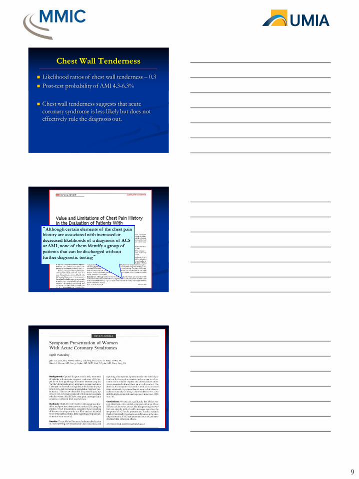
Evaluation of the Low Risk Patient
Variety of protocols based on hospital
resources
Accelerated Diagnostic Protocols
Chest Pain Units
Probability of ACS <5%
What is the Question?
Can I send this patient home?
Is today’s symptoms (chest pain) due to cardiac
ischemia? (or AD, PE)
Does this patient have coronary artery disease?
Testing Low Risk CP Patients
Functional data:
Exercise Treadmill Testing
Myocardial Perfusion Imaging (Nuclear)
Stress Echocardiography
Anatomical data:
CT Coronary Angiogram

29
UC Davis Low risk inclusion criteria
No hemodynamic instability
No arrhythmias
Normal or non-specific ECG
Prior CAD also included
Excluded:
Repolarization abnormality
Amsterdam, JACC 2002
UC Davis Protocol
One set of cardiac biomarkers
Modified Bruce protocol stress test
Negative
Non-diagnostic (<85% max predicted HR)
Positive (significant symptoms, ECG criteria for
ischemia)
If negative discharge home.
30 day follow-up.

30
No deaths in any group
Negative – 1 Non Qwave MI
Non-diagnostic – 7 revascularizations
Positive – 4 Non Qwave MI, 12
revascularizations
Accelerated 2 hr Diagnostic Protocol
JAMA Int Med. Jan, 2014
Criteria for 2 hr ADP
TIMI Risk Score of 0
No new ischemic ECG changes
Negative Troponin I at 0 and 2 hrs
Discharged with return within 72 hrs for ETT
No difference in MACE at 30 days comparing
ADP to standard care pathway

31
CT Coronary Angiography
April 12, 2012
ROMICAT-II

32
CTCA - Benefits
High sensitivity and Neg predictive value
Faster TAT than Functional tests
Reduced number of repeat evaluation for Chest
pain recidivism
Lower radiation and cost than MPI
May exclude other life threatening causes of CP
– PE, Aortic dissection
Value in risk factor modification
CTCA - Limitations
Technical contraindications (25%) – contrast
allergy, obesity, renal insufficiency, arrhythmias,
unable to tolerate Beta blockade
Elderly patients with calcified coronary arteries
Not useful in patients with known CAD
(usually)
Additional testing required in 10-20%
Higher radiation and cost compared to ETT
Diagnostic Accuracy and Clinical Utility of Noninvasive Testing for CAD
Pretest Probability
Sensitivity NPV Specificity PPV
Overall Stress test 78% 76% 77% 80%
CTCA 99% 99% 89% 91%
Low Stress test 71% 92% 83% 50%
(< 20%) CTCA 100% 100% 89% 69%
Intermediate Stress test 79% 77% 77% 79%
(20-80%) CTCA 99% 99% 93% 94%
High Stress test 79% 37% 60% 91%
(>80%) CTCA 99% 95% 72% 95%
Weustink et al. Annals Internal Med. 2010;152:630

33
Outpatient Stress Testing or CTCA
Low risk patients with the following:
No further ischemic discomfort
Normal ECG
Normal Troponin
Stress testing can be performed within 72 hrs (24 hrs
preferred) – Schedule the test. Don’t rely on the
patient to schedule after discharge
Plan to return to ED if any recurrent symptoms
Ridgeview Medical Center Emergency Department
ED Chest Pain Patients - 2013
Level 1 (44), 9.7%
Level 2 (71), 15.7%
Level 3 (93), 20.5%
Level 4 (245), 54.1%
Level 1 (44) Level 2 (71) Level 3 (93) Level 4 (245)
34
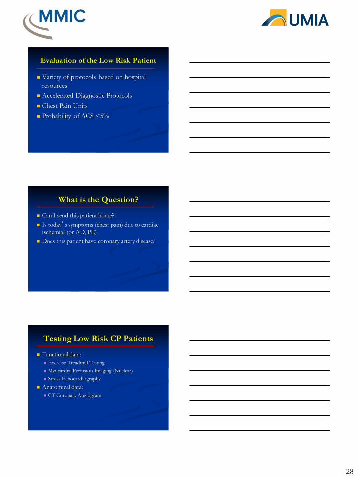
Level 4 - (low risk) Disposition
Admit (10), 4.1%
OBS (27), 11.0%
Transfer (15), 6.1%
Home (193), 78.8%
Admit (10)
OBS (27)
Transfer (15)
Home (193)
Deceased (0)
Not Provided (0)
Level 4 – (low risk) Testing
CTA (17)9%
Echo TM (51)29%
Stress TM (93)52%
Nuclear TM (3)2%
Angiogram (2)1%
Lexiscan, (Ph) Adenosine (12)
7%
CTA (17)
Echo TM (51)
Stress TM (93)
Nuclear TM (3)
Angiogram (2)
Lexiscan, (Ph) Adenosine (12)
Key Points
Risk stratify patients to determine what workup
is needed.
The history and exam is helpful but not enough
to discharge the chest pain patient without
further testing
Know your institution’s Troponin limits
Expertise in ECG interpretation is essential.
Send an image to an expert if needed
Develop and implement Chest Pain protocols in
your institution

35
Questions?