Embed Size (px)
Citation preview
Evaluation & Management of Common Shoulder and Elbow Disorders
Anil K. Koganti, M.D. Sports Medicine, Shoulder/Elbow Reconstruction
EVALUATION OF THE SHOULDER
History
• Location of Pain
• Onset
• Trauma?
• Weakness?
• Numbness or Paresthesias?
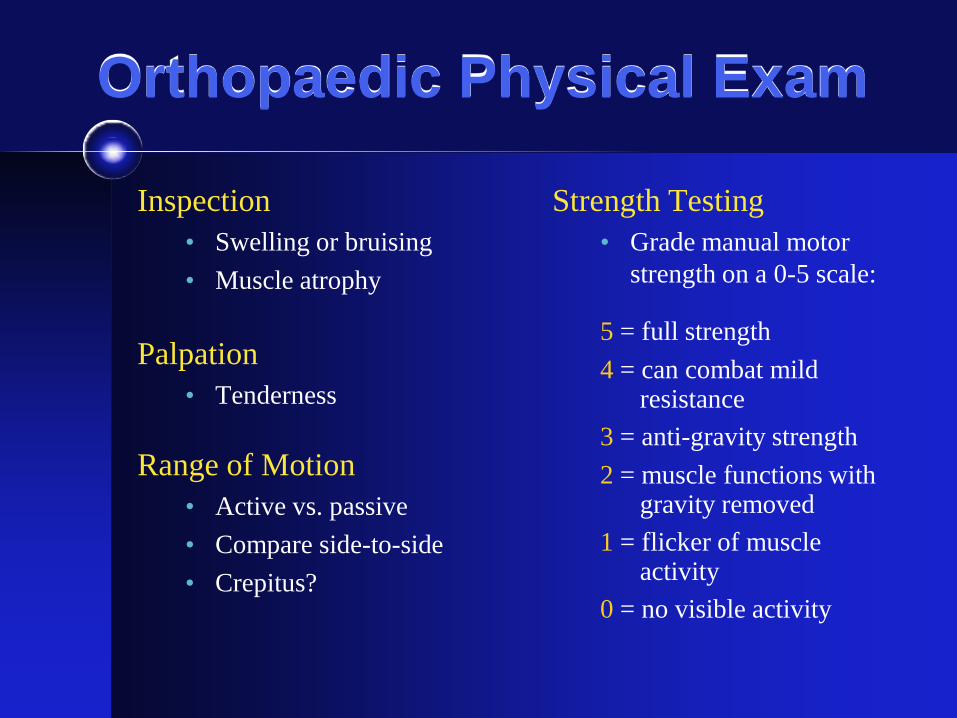
Orthopaedic Physical Exam
Inspection • Swelling or bruising • Muscle atrophy
Palpation
• Tenderness Range of Motion
• Active vs. passive • Compare side-to-side • Crepitus?
Strength Testing • Grade manual motor
strength on a 0-5 scale:
5 = full strength 4 = can combat mild resistance 3 = anti-gravity strength 2 = muscle functions with gravity removed 1 = flicker of muscle activity 0 = no visible activity
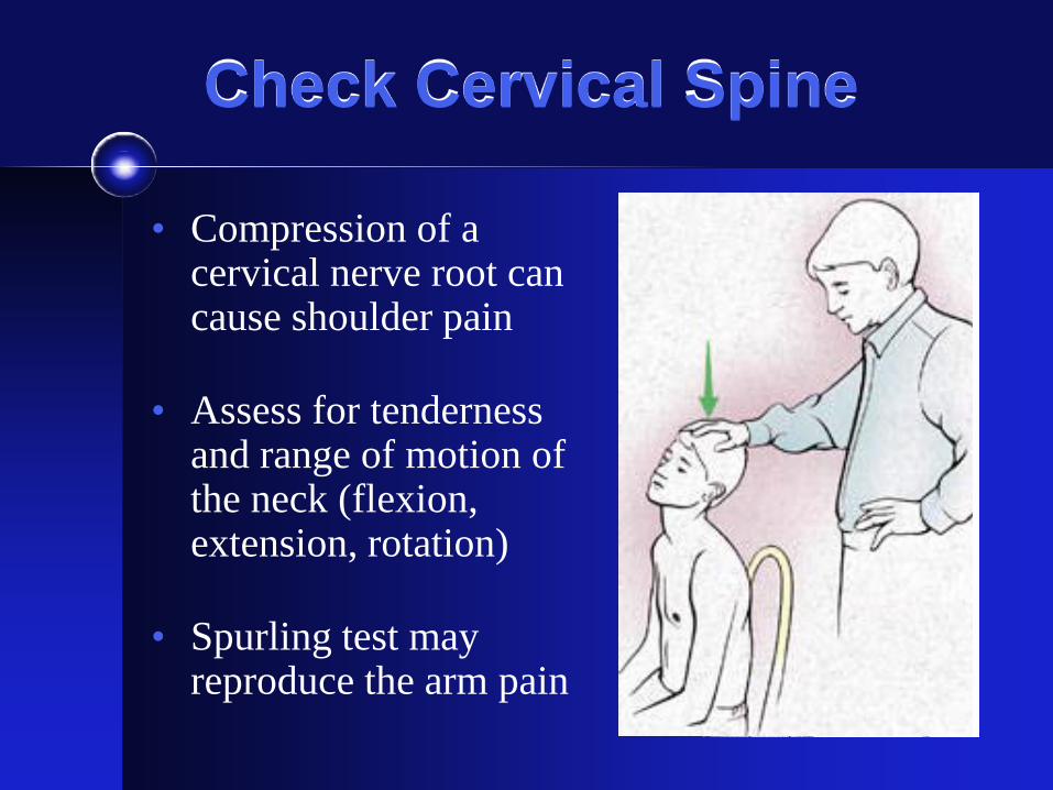
Check Cervical Spine
• Compression of a cervical nerve root can cause shoulder pain
• Assess for tenderness and range of motion of the neck (flexion, extension, rotation)
• Spurling test may reproduce the arm pain
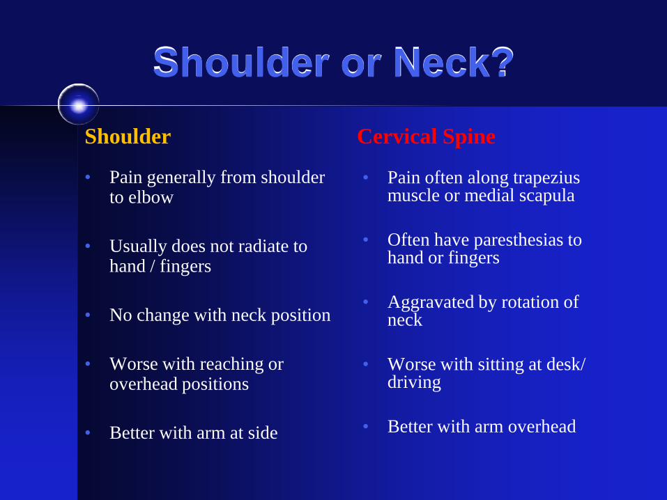
Shoulder or Neck? Shoulder
• Pain generally from shoulder to elbow
• Usually does not radiate to hand / fingers
• No change with neck position
• Worse with reaching or overhead positions
• Better with arm at side
Cervical Spine
• Pain often along trapezius muscle or medial scapula
• Often have paresthesias to hand or fingers
• Aggravated by rotation of neck
• Worse with sitting at desk/ driving
• Better with arm overhead
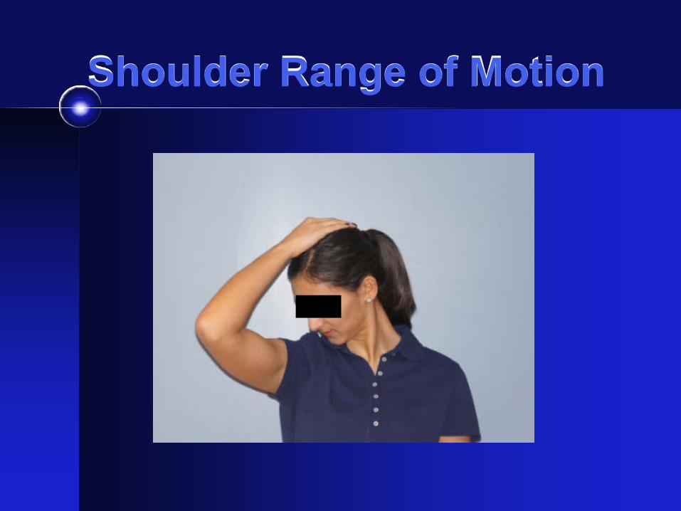
Shoulder Range of Motion
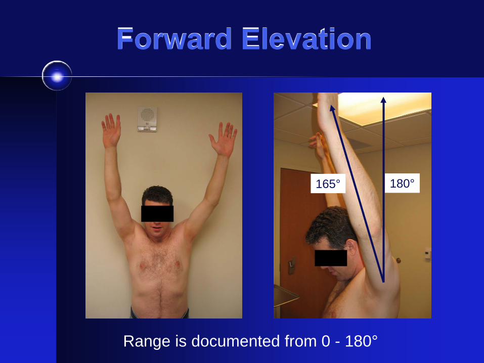
Forward Elevation
Range is documented from 0 - 180°
180° 165°
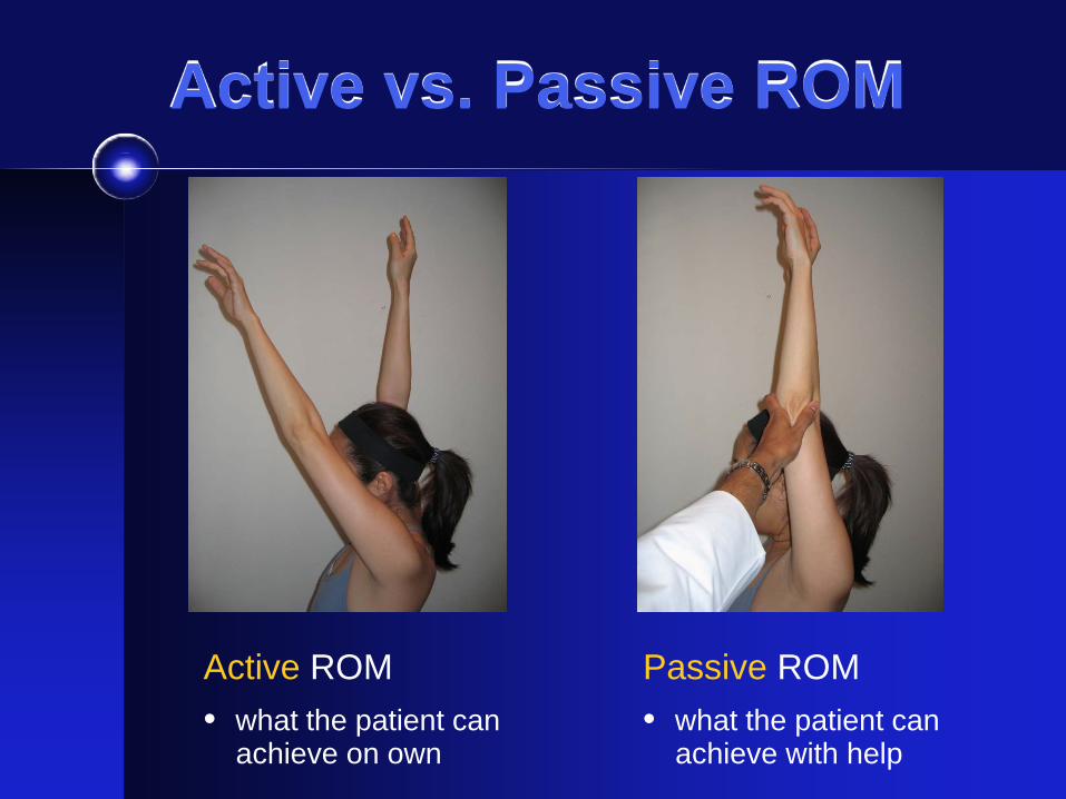
Active vs. Passive ROM
Active ROM • what the patient can achieve on own
Passive ROM • what the patient can achieve with help
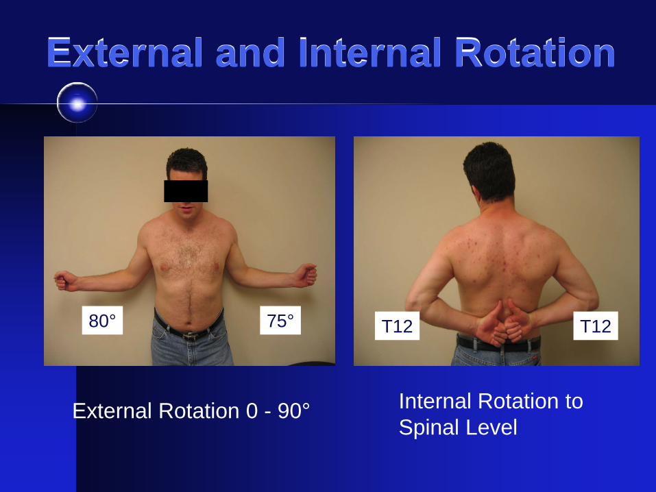
External and Internal Rotation
External Rotation 0 - 90°
80° 75°
Internal Rotation to Spinal Level
T12 T12
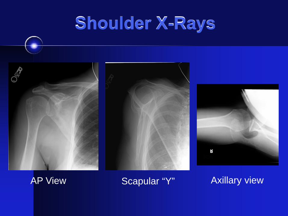
Shoulder X-Rays
AP View Scapular “Y” Axillary view
Common Shoulder Problems
1. Rotator Cuff Pathology • Tendinitis / Bursitis
• Tear
2. Adhesive Capsulitis (Frozen Shoulder)
3. Glenohumeral Arthritis
4. AC Joint Pain / Arthritis
5. Shoulder Instability
ROTATOR CUFF DISEASE
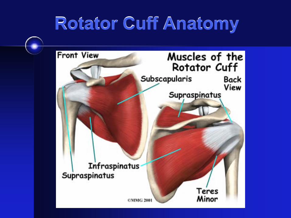
Rotator Cuff Anatomy
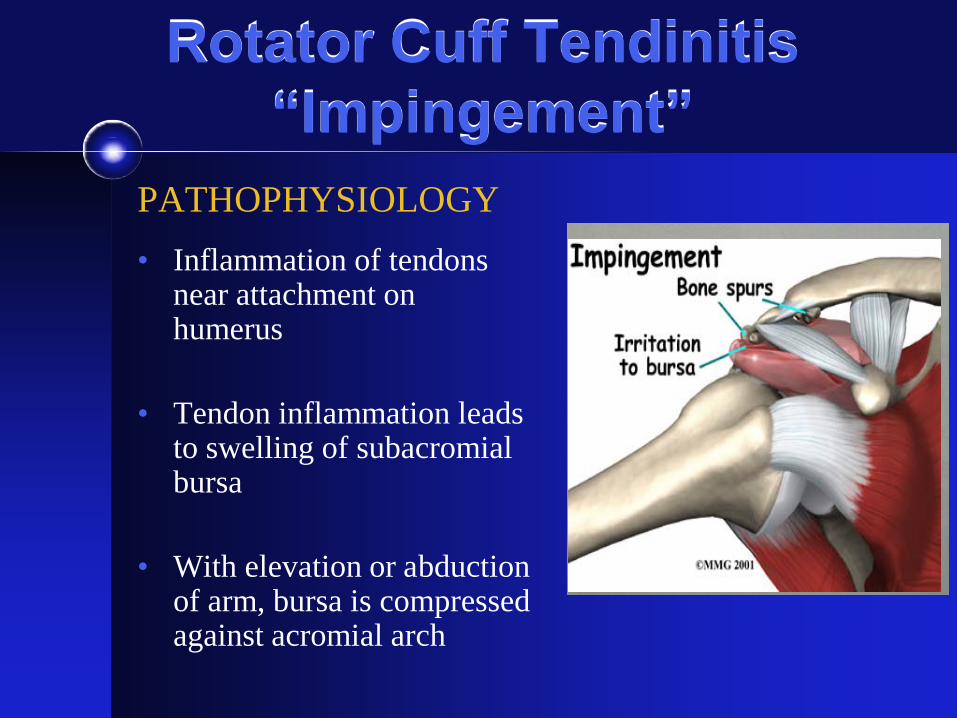
Rotator Cuff Tendinitis “Impingement”
PATHOPHYSIOLOGY • Inflammation of tendons
near attachment on humerus
• Tendon inflammation leads to swelling of subacromial bursa
• With elevation or abduction of arm, bursa is compressed against acromial arch
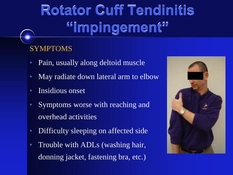
Rotator Cuff Tendinitis “Impingement”
SYMPTOMS
• Pain, usually along deltoid muscle
• May radiate down lateral arm to elbow
• Insidious onset
• Symptoms worse with reaching and overhead activities
• Difficulty sleeping on affected side
• Trouble with ADLs (washing hair, donning jacket, fastening bra, etc.)
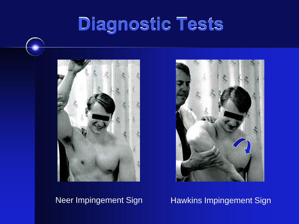
Diagnostic Tests
Neer Impingement Sign Hawkins Impingement Sign
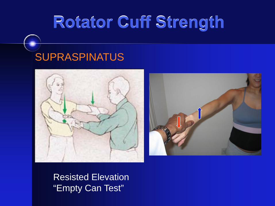
Rotator Cuff Strength
Resisted Elevation “Empty Can Test”
SUPRASPINATUS
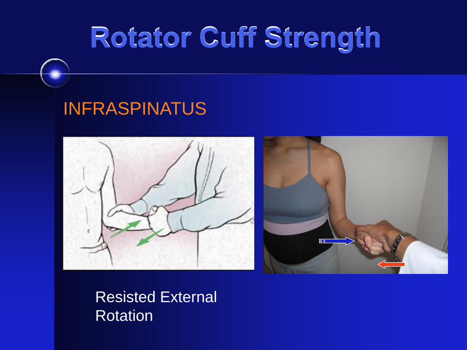
Rotator Cuff Strength
Resisted External Rotation
INFRASPINATUS
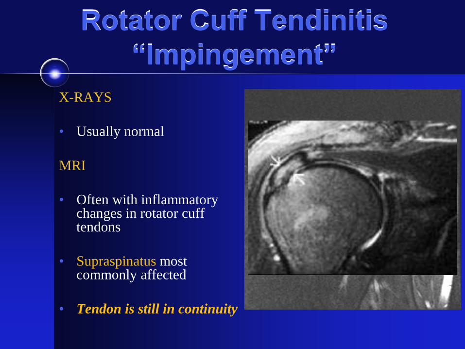
Rotator Cuff Tendinitis “Impingement”
X-RAYS
• Usually normal MRI
• Often with inflammatory
changes in rotator cuff tendons
• Supraspinatus most commonly affected
• Tendon is still in continuity
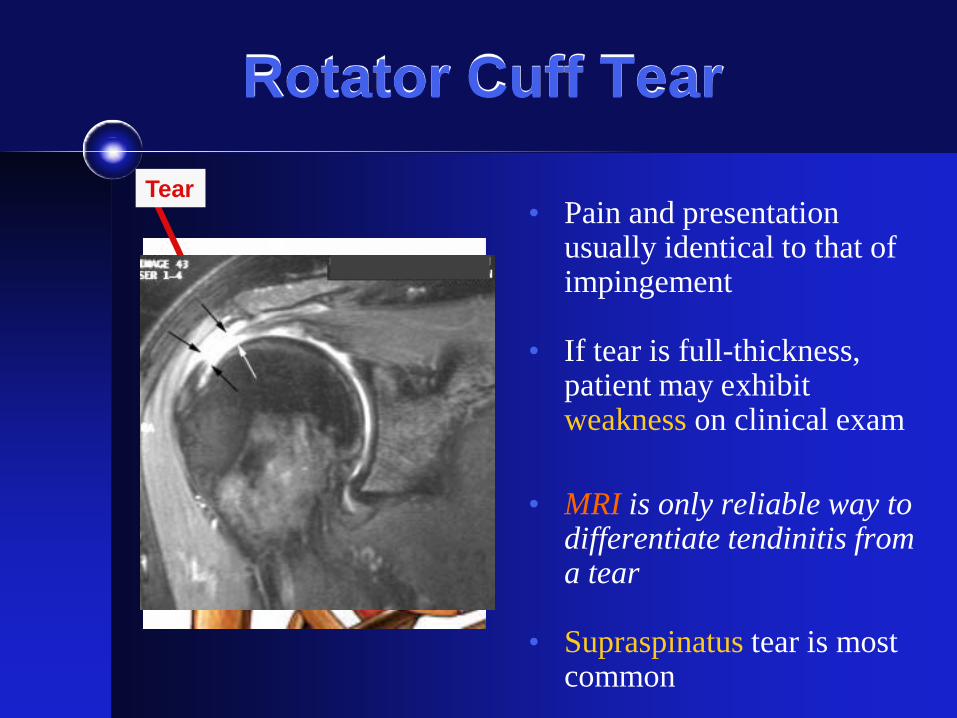
Rotator Cuff Tear
• Pain and presentation usually identical to that of impingement
• If tear is full-thickness, patient may exhibit weakness on clinical exam
• MRI is only reliable way to differentiate tendinitis from a tear
• Supraspinatus tear is most common
Tear
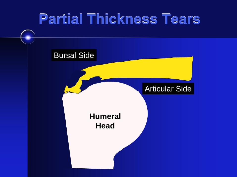
Partial Thickness Tears
Humeral Head
Bursal Side
Articular Side
Humeral Head
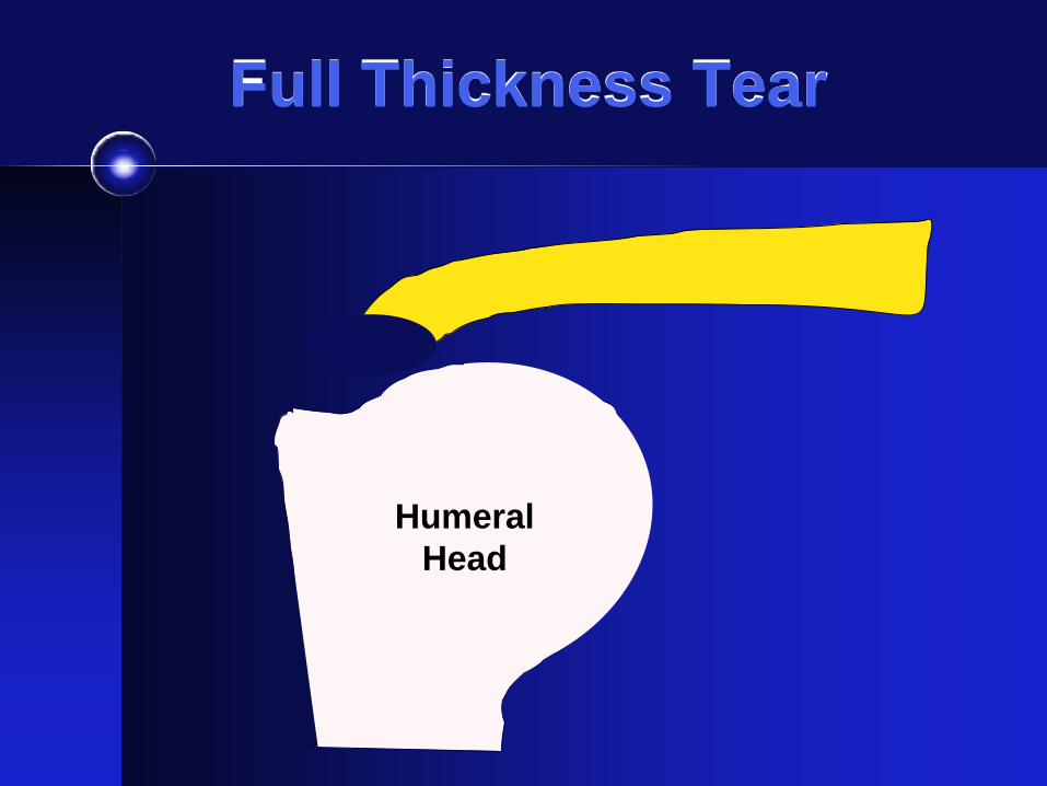
Full Thickness Tear
Humeral Head
Humeral Head
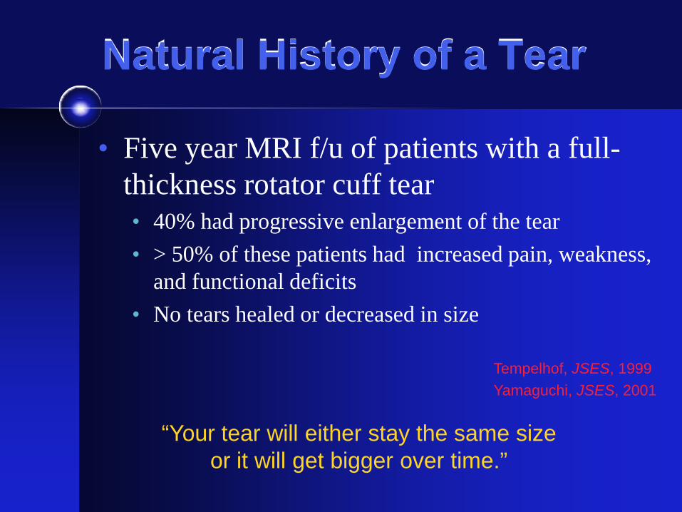
Natural History of a Tear
• Five year MRI f/u of patients with a full-thickness rotator cuff tear • 40% had progressive enlargement of the tear • > 50% of these patients had increased pain, weakness,
and functional deficits • No tears healed or decreased in size
Tempelhof, JSES, 1999 Yamaguchi, JSES, 2001
“Your tear will either stay the same size or it will get bigger over time.”
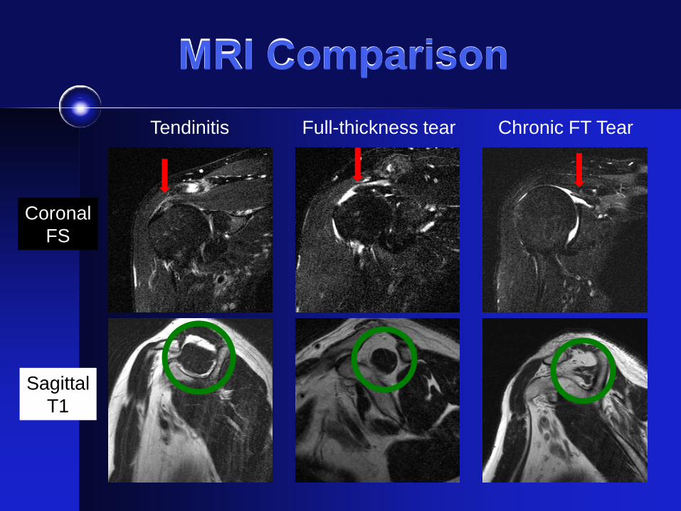
MRI Comparison
Coronal FS
Sagittal T1
Tendinitis Full-thickness tear Chronic FT Tear
Rotator Cuff Management
• Rotator Cuff Tendinitis / Bursitis and Partial Thickness Tears can be treated conservatively • Rest, activity modification • Icing, NSAIDs • Physical Therapy • Subacromial Bursal Steroid Injection
• Full Thickness Rotator Cuff Tears should be
evaluated for surgical repair • Ideally before irreversible changes occur: fixed
tendon retraction and fatty atrophy of muscle
Prolonged non-operative treatment
of a full-thickness rotator cuff tear
may lead to irreversible changes and
a progressive decline in function
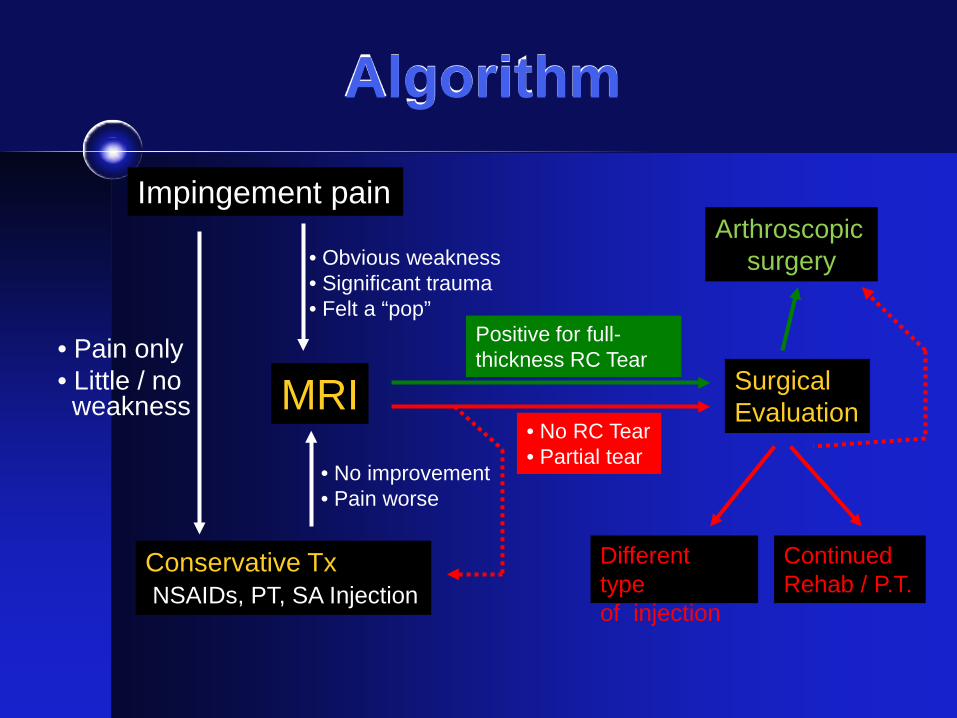
Algorithm
Impingement pain
• Pain only • Little / no weakness
Conservative Tx NSAIDs, PT, SA Injection
MRI
• Obvious weakness • Significant trauma • Felt a “pop”
• No improvement • Pain worse
• No RC Tear • Partial tear
Surgical Evaluation
Positive for full- thickness RC Tear
Arthroscopic surgery
Different type of injection
Continued Rehab / P.T.
ADHESIVE CAPSULITIS “Frozen Shoulder”
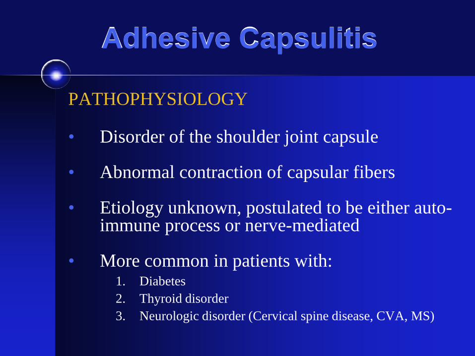
Adhesive Capsulitis
PATHOPHYSIOLOGY • Disorder of the shoulder joint capsule
• Abnormal contraction of capsular fibers
• Etiology unknown, postulated to be either auto-
immune process or nerve-mediated
• More common in patients with: 1. Diabetes 2. Thyroid disorder 3. Neurologic disorder (Cervical spine disease, CVA, MS)
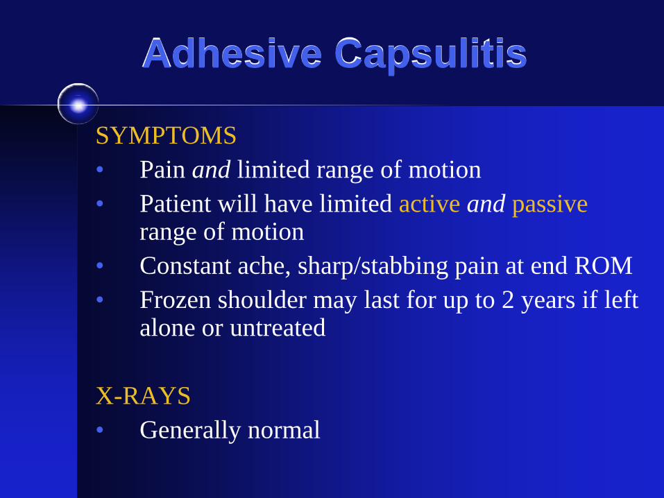
Adhesive Capsulitis
SYMPTOMS • Pain and limited range of motion • Patient will have limited active and passive
range of motion • Constant ache, sharp/stabbing pain at end ROM • Frozen shoulder may last for up to 2 years if left
alone or untreated X-RAYS • Generally normal
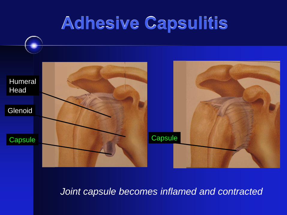
Adhesive Capsulitis
Capsule
Joint capsule becomes inflamed and contracted
Glenoid
Humeral Head
Capsule
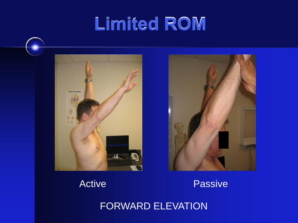
Limited ROM
FORWARD ELEVATION
Active Passive
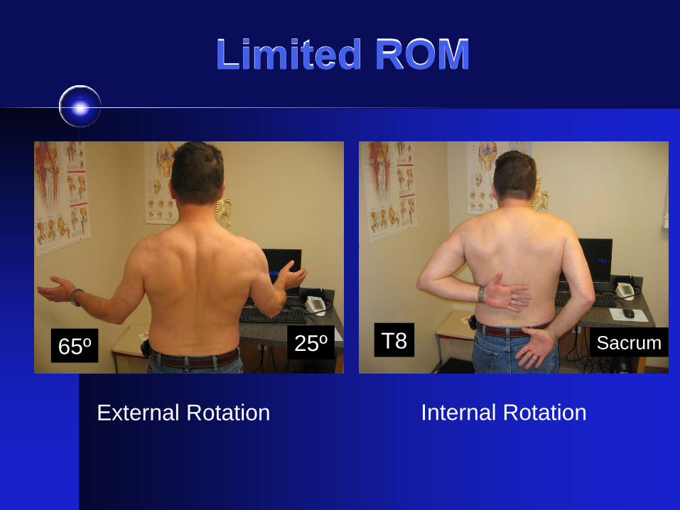
Limited ROM
External Rotation
65º 25º
Internal Rotation
T8 Sacrum
Treatment
• NSAIDs
• Physical therapy
• Corticosteroid injection intra-articular
• More physical therapy (several months)
• Manipulation under anesthesia
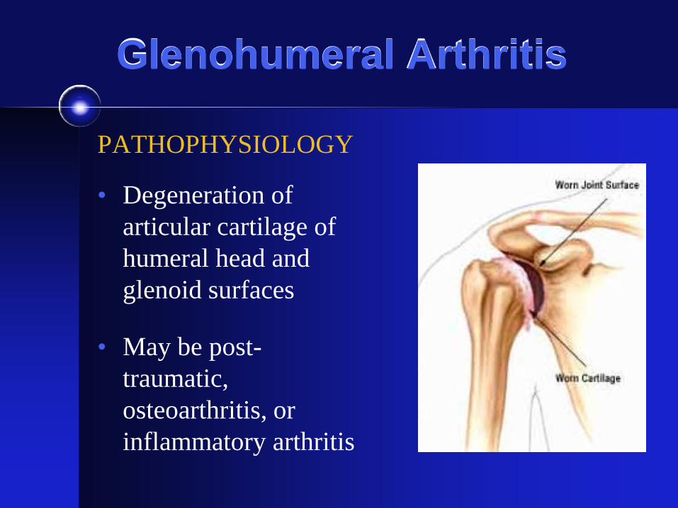
Glenohumeral Arthritis
PATHOPHYSIOLOGY • Degeneration of
articular cartilage of humeral head and glenoid surfaces
• May be post-traumatic, osteoarthritis, or inflammatory arthritis
Glenohumeral Arthritis
SYMPTOMS • Pain +/- crepitus • Limited range of motion (active and
passive) X-RAYS • Gold standard for diagnosis
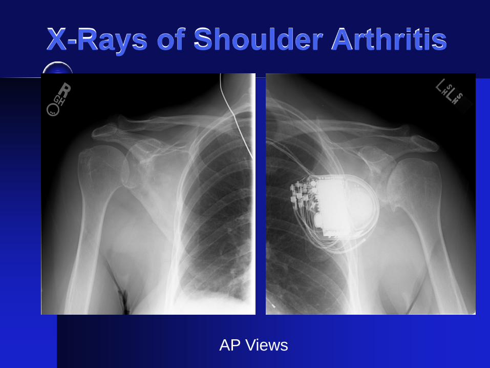
X-Rays of Shoulder Arthritis
AP Views
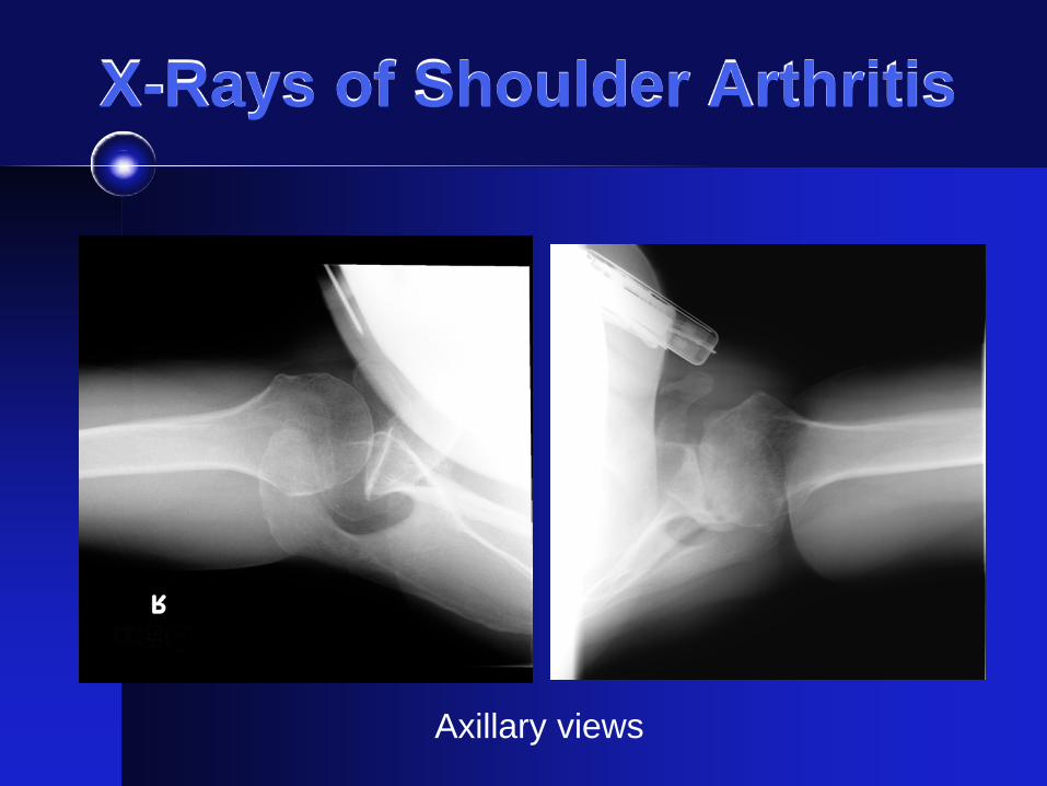
X-Rays of Shoulder Arthritis
Axillary views
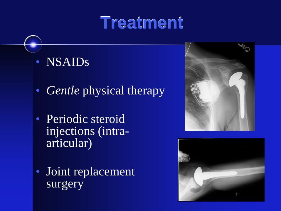
Treatment
• NSAIDs
• Gentle physical therapy
• Periodic steroid injections (intra-articular)
• Joint replacement surgery
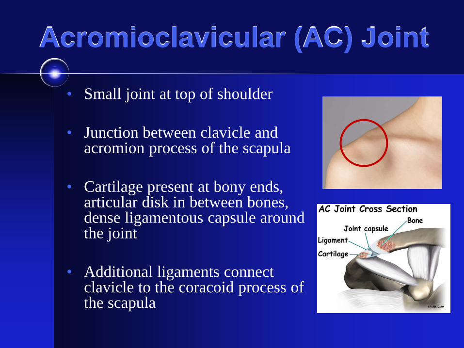
Acromioclavicular (AC) Joint
• Small joint at top of shoulder
• Junction between clavicle and acromion process of the scapula
• Cartilage present at bony ends, articular disk in between bones, dense ligamentous capsule around the joint
• Additional ligaments connect clavicle to the coracoid process of the scapula
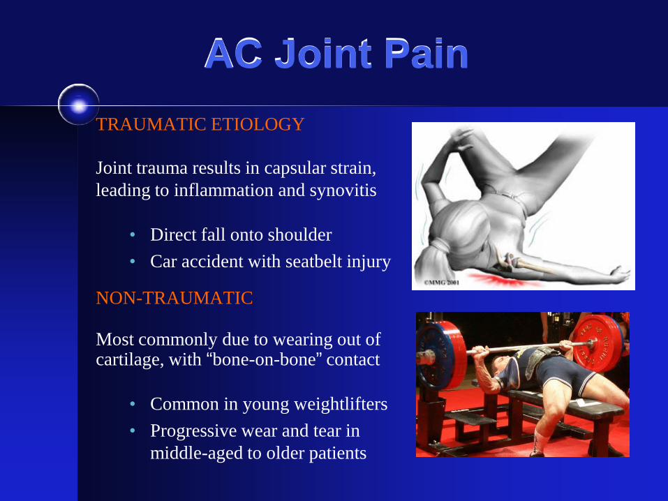
AC Joint Pain TRAUMATIC ETIOLOGY Joint trauma results in capsular strain, leading to inflammation and synovitis
• Direct fall onto shoulder • Car accident with seatbelt injury
NON-TRAUMATIC Most commonly due to wearing out of cartilage, with “bone-on-bone” contact
• Common in young weightlifters • Progressive wear and tear in
middle-aged to older patients
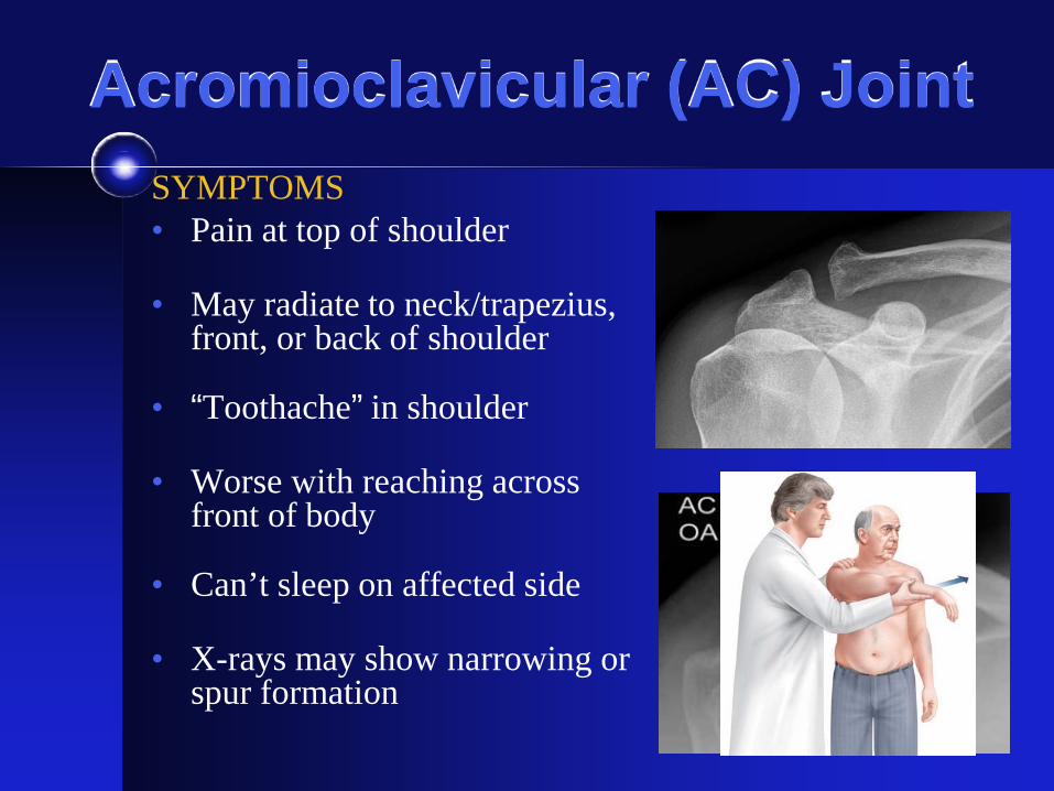
Acromioclavicular (AC) Joint SYMPTOMS • Pain at top of shoulder
• May radiate to neck/trapezius,
front, or back of shoulder
• “Toothache” in shoulder
• Worse with reaching across front of body
• Can’t sleep on affected side
• X-rays may show narrowing or spur formation
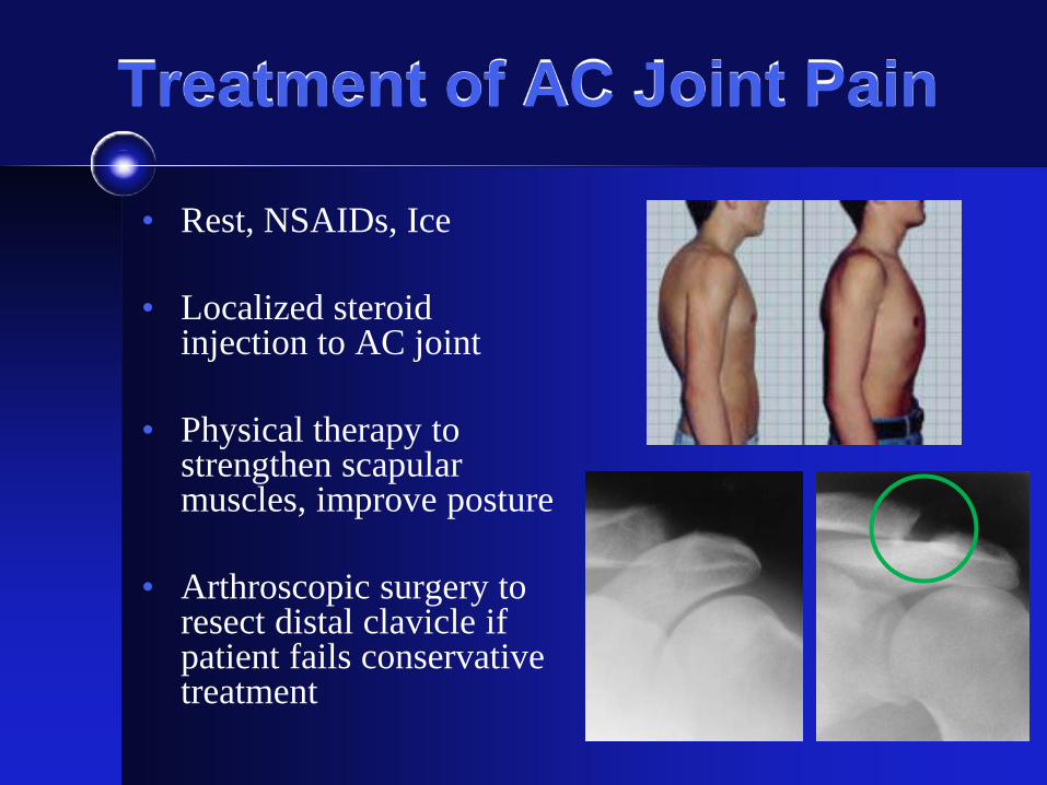
Treatment of AC Joint Pain
• Rest, NSAIDs, Ice
• Localized steroid injection to AC joint
• Physical therapy to strengthen scapular muscles, improve posture
• Arthroscopic surgery to resect distal clavicle if patient fails conservative treatment
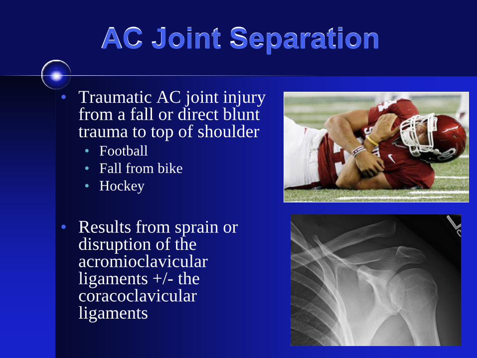
AC Joint Separation
• Traumatic AC joint injury from a fall or direct blunt trauma to top of shoulder • Football • Fall from bike • Hockey
• Results from sprain or
disruption of the acromioclavicular ligaments +/- the coracoclavicular ligaments
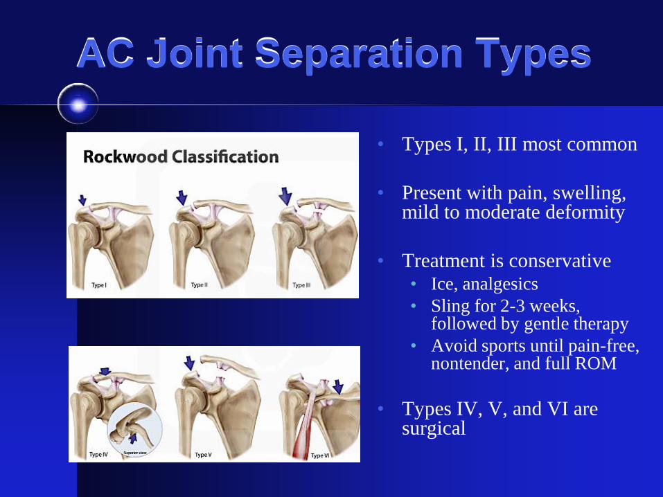
AC Joint Separation Types
• Types I, II, III most common
• Present with pain, swelling, mild to moderate deformity
• Treatment is conservative • Ice, analgesics • Sling for 2-3 weeks,
followed by gentle therapy • Avoid sports until pain-free,
nontender, and full ROM
• Types IV, V, and VI are surgical
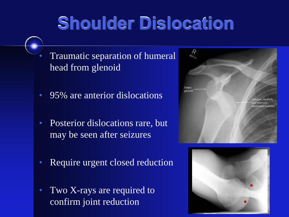
Shoulder Dislocation • Traumatic separation of humeral
head from glenoid
• 95% are anterior dislocations
• Posterior dislocations rare, but may be seen after seizures
• Require urgent closed reduction
• Two X-rays are required to confirm joint reduction
Anterior Shoulder Dislocation
• Treatment after successful reduction involves sling immobilizer for 1-2 wks, followed by progressive ROM exercises and strengthening
• Likelihood of recurrent dislocation depends on patient age at time of first dislocation • < 20 yo = > 90% or higher risk of recurrence • 20-40 yo = 40% chance of recurrence • > 40 = 14% chance of recurrence
• Though less likely to have recurrent instability,
patients over age 40 are more likely to have a rotator cuff tear following a shoulder dislocation.
Rowe, JBJS, 1962
EVALUATION OF THE ELBOW
Elbow Range of Motion
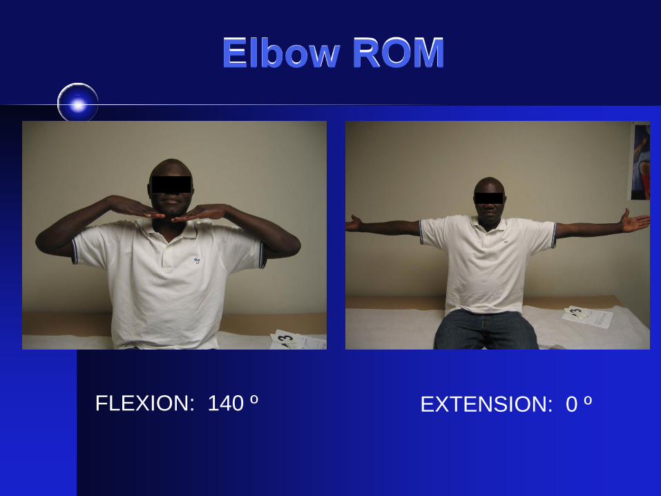
Elbow ROM
FLEXION: 140 º EXTENSION: 0 º
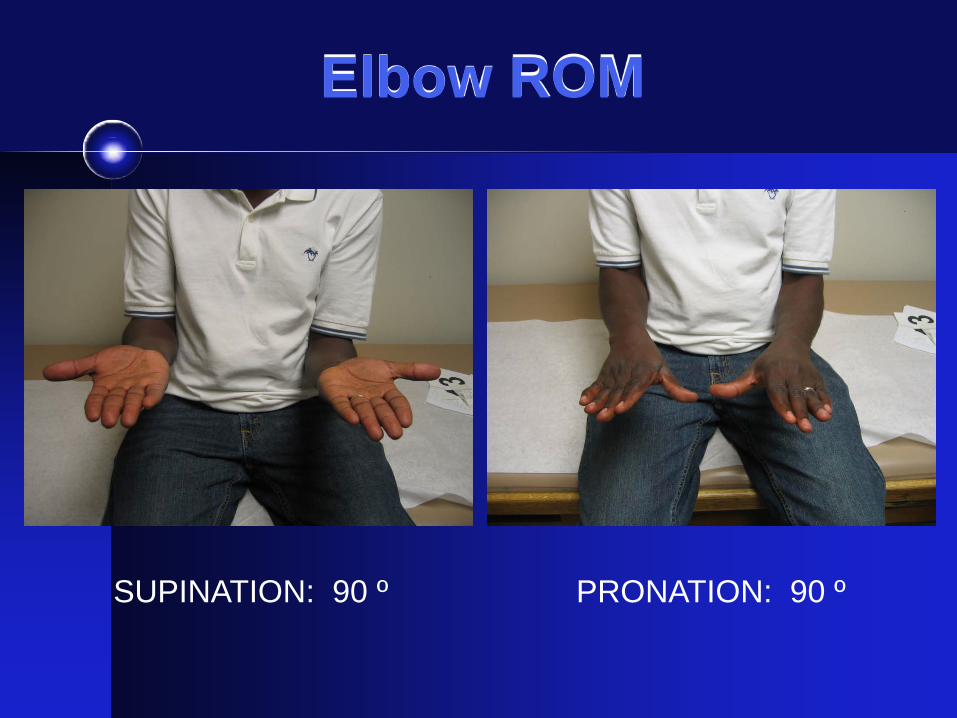
Elbow ROM
SUPINATION: 90 º PRONATION: 90 º
Common Elbow Disorders
1. Lateral Epicondylitis (Tennis Elbow)
2. Medial Epicondylitis (Golfer’s Elbow)
3. Olecranon Bursitis
4. Elbow Arthritis
5. Distal Biceps Tendon Rupture
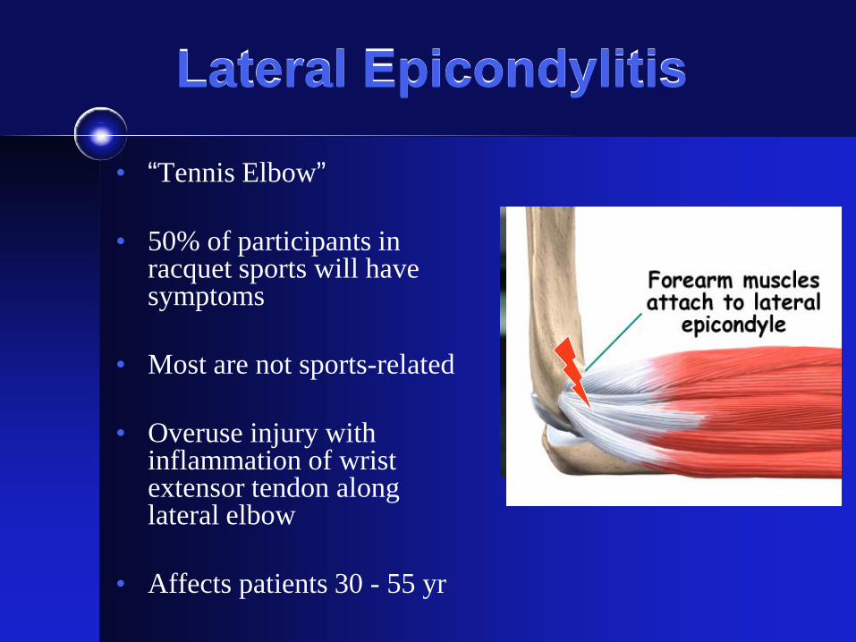
Lateral Epicondylitis
• “Tennis Elbow”
• 50% of participants in racquet sports will have symptoms
• Most are not sports-related
• Overuse injury with inflammation of wrist extensor tendon along lateral elbow
• Affects patients 30 - 55 yr
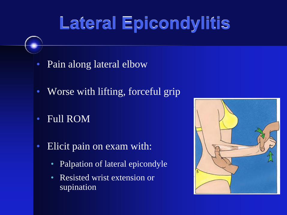
Lateral Epicondylitis
• Pain along lateral elbow
• Worse with lifting, forceful grip
• Full ROM
• Elicit pain on exam with:
• Palpation of lateral epicondyle • Resisted wrist extension or
supination
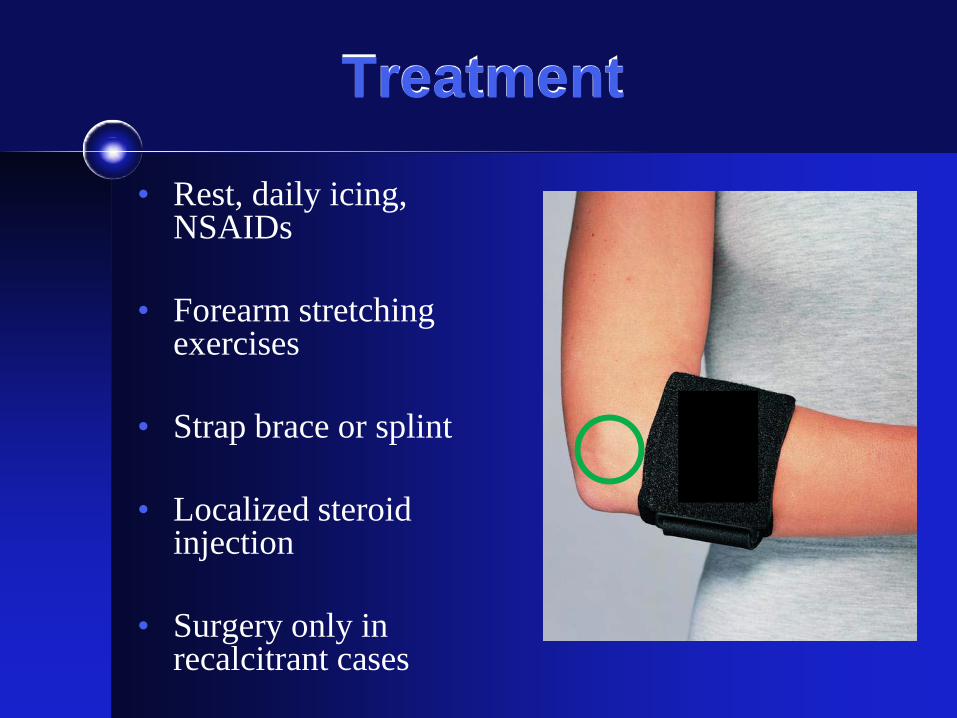
Treatment
• Rest, daily icing, NSAIDs
• Forearm stretching exercises
• Strap brace or splint
• Localized steroid injection
• Surgery only in recalcitrant cases
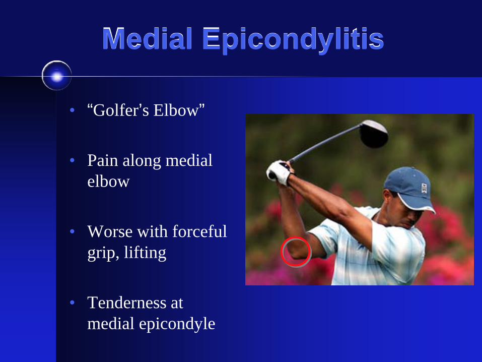
Medial Epicondylitis
• “Golfer’s Elbow”
• Pain along medial elbow
• Worse with forceful grip, lifting
• Tenderness at medial epicondyle
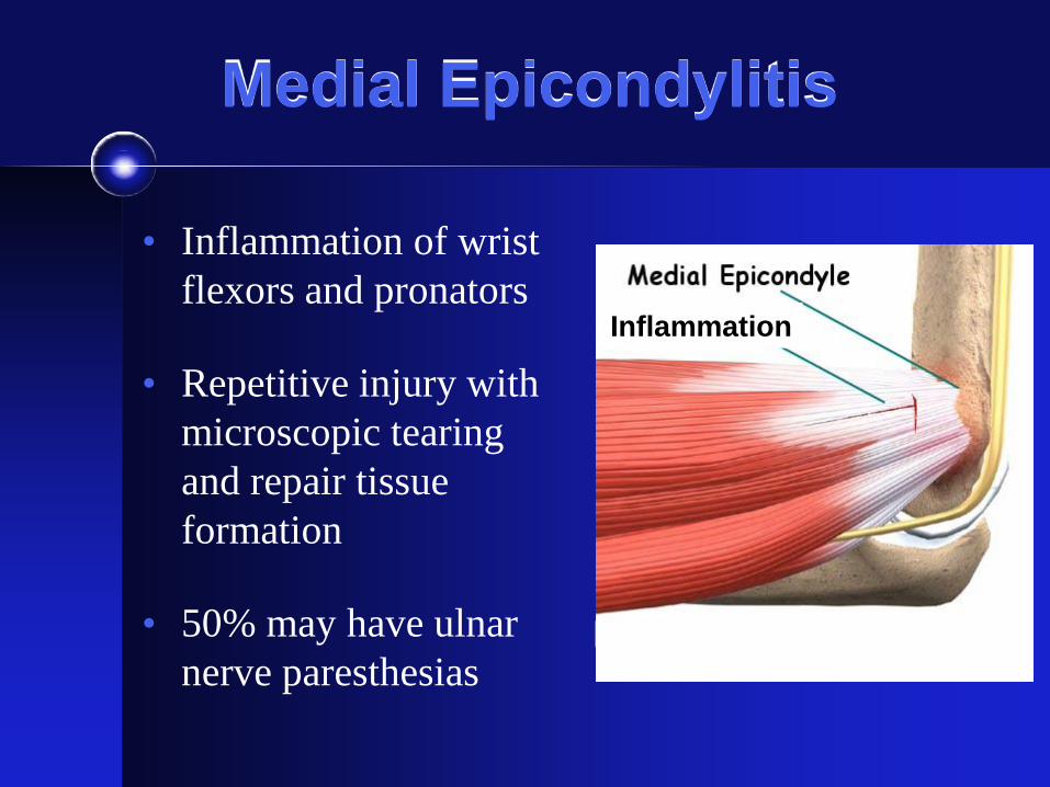
Medial Epicondylitis
• Inflammation of wrist flexors and pronators
• Repetitive injury with microscopic tearing and repair tissue formation
• 50% may have ulnar nerve paresthesias
Inflammation
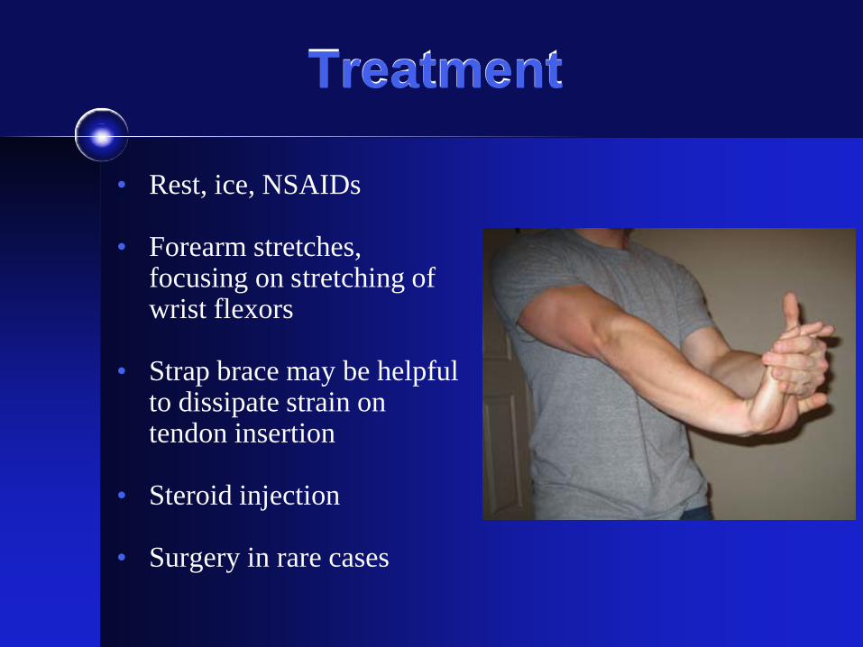
Treatment
• Rest, ice, NSAIDs
• Forearm stretches, focusing on stretching of wrist flexors
• Strap brace may be helpful to dissipate strain on tendon insertion
• Steroid injection
• Surgery in rare cases
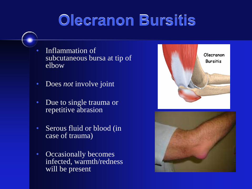
Olecranon Bursitis
• Inflammation of subcutaneous bursa at tip of elbow
• Does not involve joint
• Due to single trauma or repetitive abrasion
• Serous fluid or blood (in case of trauma)
• Occasionally becomes infected, warmth/redness will be present
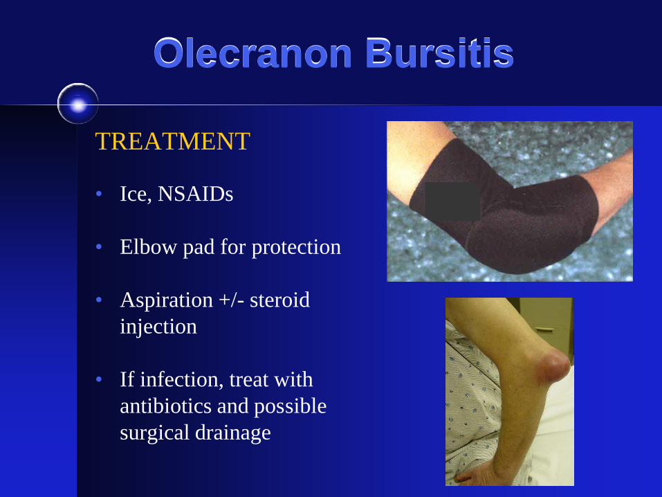
Olecranon Bursitis
TREATMENT • Ice, NSAIDs
• Elbow pad for protection
• Aspiration +/- steroid
injection
• If infection, treat with antibiotics and possible surgical drainage
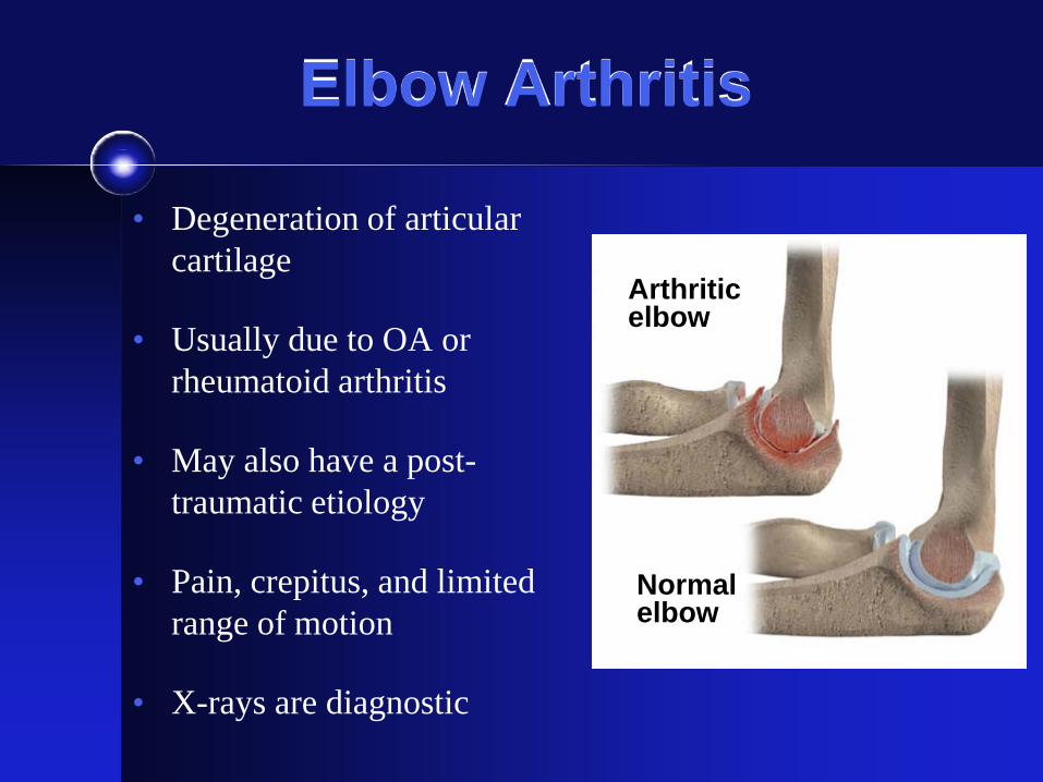
Elbow Arthritis
• Degeneration of articular cartilage
• Usually due to OA or rheumatoid arthritis
• May also have a post-traumatic etiology
• Pain, crepitus, and limited range of motion
• X-rays are diagnostic
Arthritic elbow
Normal elbow
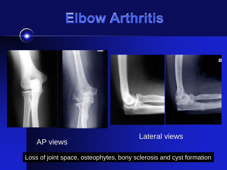
Elbow Arthritis
AP views Lateral views
Loss of joint space, osteophytes, bony sclerosis and cyst formation
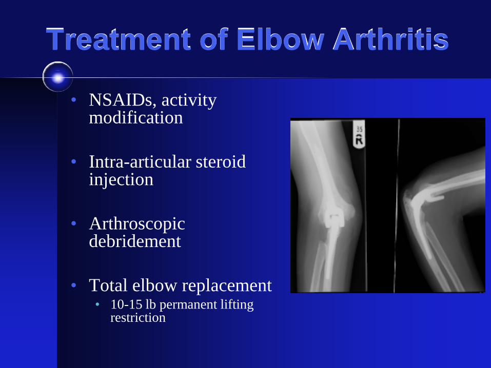
Treatment of Elbow Arthritis
• NSAIDs, activity modification
• Intra-articular steroid injection
• Arthroscopic debridement
• Total elbow replacement • 10-15 lb permanent lifting
restriction
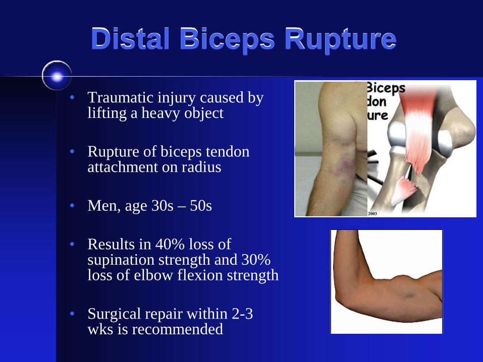
Distal Biceps Rupture
• Traumatic injury caused by lifting a heavy object
• Rupture of biceps tendon attachment on radius
• Men, age 30s – 50s
• Results in 40% loss of supination strength and 30% loss of elbow flexion strength
• Surgical repair within 2-3 wks is recommended
THANK YOU