Embed Size (px)
Citation preview
�
Abstract² The chemical, mechanical, and electrochemical
attributes of the Parylene sheath electrode (PSE) were
evaluated following a post-fabrication thermoforming process
to determine its impact on both the polymer and thin film
platinum materials. The three-dimensional conical shape of the
PSE was formed via thermal molding of a surface
micromachined Parylene C microchannel using a custom
shape-forming microwire having the desired taper at 200°C for
48 hours under vacuum. Contact angle and Fourier transform
infrared spectroscopy measurements indicated that the
thermoforming process resulted in no significant changes to the
surface and bulk chemistry of Parylene. The thermoformed
Parylene samples possessed greater <RXQJ¶V� PRGXOXV�� EXW�
retained their flexibility. Electrochemical characterization of
electrodes before and after thermoforming revealed a
decreased storage charge capacity and increased electrode
impedance, however, recording functionality was not lost as
resolvable neuronal unit activity was successfully obtained
post-implantation.
I. INTRODUCTION
Chronic implementation of intracortical prosthetic devices has been limited by multiple failure mechanisms encountered during long-term implantation. In addition to abiotic failure mechanisms (e.g. electrode corrosion, broken connectors), the decline of recording performance of electrodes has been attributed to biotic causes that occur in vivo [1]. Specifically, the inflammatory response caused by insertion trauma and the presence of a foreign body within the cortex [2, 3] forms glial cell encapsulation and neuronal retraction from the recording sites, which limit the longevity of high fidelity recordings [4]. This response is further exacerbated by the mechanical mismatch between current electrodes, made of silicon, metal, or stiffer polymers, and
*This work was sponsored by the Defense Advanced Research Projects
Agency (DARPA) MTO under the auspices of Dr. Jack Judy through the
Space and Naval Warfare Systems Center, Pacific Grant/Contract No.
N66001-11-1-4207.
B.J. Kim, S.A. Hara, J.T.W. Kuo, C. Lee, T. Hoang, and E. Meng are
with the Department of Biomedical Engineering at the University of
Southern California, Los Angeles, CA 90089 USA (corresponding author:
213-821-3949; e-mail: [email protected]).
B. Chen and M. Gupta are with the Mork Family Department of
Chemical Engineering and Materials Science at the University of Southern
California, Los Angeles, CA 90089 USA.
C. A. Gutierrez is an independent engineering consultant in Los Angeles,
CA 90089 USA.
V. Pikov is with the Huntington Medical Research Institutes (HMRI),
Pasadena, CA 91105 USA.
the surrounding neural tissue, inflicting continuous damage during brain micromotion over time [5].
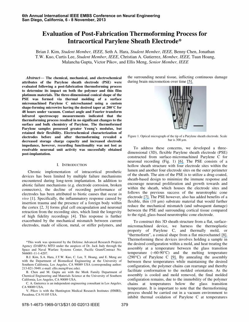
Figure 1. Optical micrograph of the tip of a Parylene sheath electrode. Scale
bar is 300 µm.
To address these concerns, we developed a three-dimensional (3D), flexible Parylene sheath electrode (PSE) constructed from surface-micromachined Parylene C for neuronal recording (Fig. 1) [6]. The PSE consists of a hollow sheath structure with four electrode sites within the lumen and another four electrode sites on the outer perimeter of the sheath. The aim of the PSE is to utilize a drug-coated, sheath-based design to minimize the immune response and encourage neuronal proliferation and growth towards and within the sheath, which houses the electrode sites and follows the previous success of the neurotrophic cone electrode [7]. The PSE however, also has added benefits of a flexible, thin (10 µm) substrate material that would further reduce the mechanical mismatch (and subsequent damage) between the PSE and surrounding cortical tissue compared to the rigid, glass-based neurotrophic cone electrode.
To construct this 3D sheath structure from a flat, surface-micromachined device, we harness the thermoplastic property of Parylene C, and thermally mold, or ³WKHUPRIRUP´, a conical shape from a flat microchannel [8]. Thermoforming these devices involves holding a sample in the desired configuration within a mold, and heat treating the assembly at a temperature between the glass transition temperature (~60-90°C) and the melting temperature (290°C) of Parylene C [9]. By annealing the assembly between these temperatures while maintaining the desired configuration, the polymer chains can rearrange and thereby facilitate conformation to the molded orientation. As the assembly is cooled and mold removed, the final molded configuration remains, due to the immobility of the polymer chains at temperatures below the glass transition temperature. It is important to note that the thermoforming process should be carried out in a vacuum environment to inhibit thermal oxidation of Parylene C at temperatures
Evaluation of Post-Fabrication Thermoforming Process for
Intracortical Parylene Sheath Electrode*
Brian J. Kim, Student Member, IEEE, Seth A. Hara, Student Member, IEEE, Benny Chen, Jonathan
T.W. Kuo, Curtis Lee, Student Member, IEEE, Christian A. Gutierrez, Member, IEEE, Tuan Hoang,
Malancha Gupta, Victor Pikov, and Ellis Meng, Senior Member, IEEE
6th Annual International IEEE EMBS Conference on Neural EngineeringSan Diego, California, 6 - 8 November, 2013
978-1-4673-1969-0/13/$31.00 ©2013 IEEE 379
>125°C [10]. The PSE was thermoformed at 200°C for 48 hours to not only achieve its 3D shape, but also to enhance the adhesion between the Parylene C layers [11]. Here we present the evaluation of the thermoforming process on the chemical and mechanical properties of Parylene C as well as the electrochemical performance of the PSE, to capture any material changes and their possible impacts on the final thermoformed device, and on the feasibility of the PSE as an implantable intracortical neural probe. We seek to assess the advantages and disadvantages of this simple post-fabrication process for the development of novel 3D structures using Parylene C for applications in neural electrode development.
II. MATERIALS AND METHODS
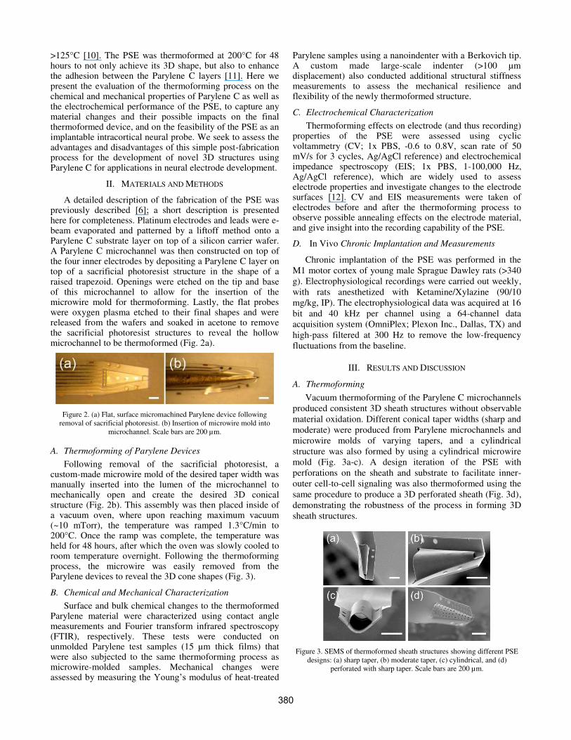
A detailed description of the fabrication of the PSE was previously described [6]; a short description is presented here for completeness. Platinum electrodes and leads were e-beam evaporated and patterned by a liftoff method onto a Parylene C substrate layer on top of a silicon carrier wafer. A Parylene C microchannel was then constructed on top of the four inner electrodes by depositing a Parylene C layer on top of a sacrificial photoresist structure in the shape of a raised trapezoid. Openings were etched on the tip and base of this microchannel to allow for the insertion of the microwire mold for thermoforming. Lastly, the flat probes were oxygen plasma etched to their final shapes and were released from the wafers and soaked in acetone to remove the sacrificial photoresist structures to reveal the hollow microchannel to be thermoformed (Fig. 2a).
Figure 2. (a) Flat, surface micromachined Parylene device following
removal of sacrificial photoresist. (b) Insertion of microwire mold into
microchannel. Scale bars are 200 µm.
A. Thermoforming of Parylene Devices
Following removal of the sacrificial photoresist, a custom-made microwire mold of the desired taper width was manually inserted into the lumen of the microchannel to mechanically open and create the desired 3D conical structure (Fig. 2b). This assembly was then placed inside of a vacuum oven, where upon reaching maximum vacuum (~10 mTorr), the temperature was ramped 1.3°C/min to 200°C. Once the ramp was complete, the temperature was held for 48 hours, after which the oven was slowly cooled to room temperature overnight. Following the thermoforming process, the microwire was easily removed from the Parylene devices to reveal the 3D cone shapes (Fig. 3).
B. Chemical and Mechanical Characterization
Surface and bulk chemical changes to the thermoformed Parylene material were characterized using contact angle measurements and Fourier transform infrared spectroscopy (FTIR), respectively. These tests were conducted on unmolded Parylene test samples (15 µm thick films) that were also subjected to the same thermoforming process as microwire-molded samples. Mechanical changes were assessed by measuring the <RXQJ¶V�PRGXOXV of heat-treated
Parylene samples using a nanoindenter with a Berkovich tip. A custom made large-scale indenter (>100 µm displacement) also conducted additional structural stiffness measurements to assess the mechanical resilience and flexibility of the newly thermoformed structure.
C. Electrochemical Characterization
Thermoforming effects on electrode (and thus recording) properties of the PSE were assessed using cyclic voltammetry (CV; 1x PBS, -0.6 to 0.8V, scan rate of 50 mV/s for 3 cycles, Ag/AgCl reference) and electrochemical impedance spectroscopy (EIS; 1x PBS, 1-100,000 Hz, Ag/AgCl reference), which are widely used to assess electrode properties and investigate changes to the electrode surfaces [12]. CV and EIS measurements were taken of electrodes before and after the thermoforming process to observe possible annealing effects on the electrode material, and give insight into the recording capability of the PSE.
D. In Vivo Chronic Implantation and Measurements
Chronic implantation of the PSE was performed in the
M1 motor cortex of young male Sprague Dawley rats (>340
g). Electrophysiological recordings were carried out weekly,
with rats anesthetized with Ketamine/Xylazine (90/10
mg/kg, IP). The electrophysiological data was acquired at 16
bit and 40 kHz per channel using a 64-channel data
acquisition system (OmniPlex; Plexon Inc., Dallas, TX) and
high-pass filtered at 300 Hz to remove the low-frequency
fluctuations from the baseline.
III. RESULTS AND DISCUSSION
A. Thermoforming
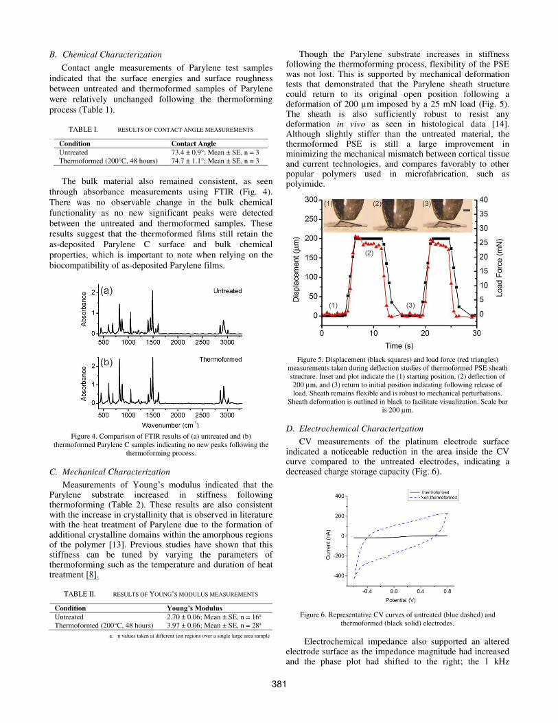
Vacuum thermoforming of the Parylene C microchannels
produced consistent 3D sheath structures without observable
material oxidation. Different conical taper widths (sharp and
moderate) were produced from Parylene microchannels and
microwire molds of varying tapers, and a cylindrical
structure was also formed by using a cylindrical microwire
mold (Fig. 3a-c). A design iteration of the PSE with
perforations on the sheath and substrate to facilitate inner-
outer cell-to-cell signaling was also thermoformed using the
same procedure to produce a 3D perforated sheath (Fig. 3d),
demonstrating the robustness of the process in forming 3D
sheath structures.
Figure 3. SEMS of thermoformed sheath structures showing different PSE
designs: (a) sharp taper, (b) moderate taper, (c) cylindrical, and (d)
perforated with sharp taper. Scale bars are 200 µm.
380
B. Chemical Characterization
Contact angle measurements of Parylene test samples
indicated that the surface energies and surface roughness
between untreated and thermoformed samples of Parylene
were relatively unchanged following the thermoforming
process (Table 1).
TABLE I. RESULTS OF CONTACT ANGLE MEASUREMENTS
Condition Contact Angle
Untreated 73.4 ± 0.9°; Mean ± SE, n = 3
Thermoformed (200°C, 48 hours) 74.7 ± 1.1°; Mean ± SE, n = 3
The bulk material also remained consistent, as seen
through absorbance measurements using FTIR (Fig. 4).
There was no observable change in the bulk chemical
functionality as no new significant peaks were detected
between the untreated and thermoformed samples. These
results suggest that the thermoformed films still retain the
as-deposited Parylene C surface and bulk chemical
properties, which is important to note when relying on the
biocompatibility of as-deposited Parylene films.
Figure 4. Comparison of FTIR results of (a) untreated and (b)
thermoformed Parylene C samples indicating no new peaks following the
thermoforming process.
C. Mechanical Characterization
Measurements of <RXQJ¶V� PRGXOXV� LQGLFDWHG� WKDW� the Parylene substrate increased in stiffness following thermoforming (Table 2). These results are also consistent with the increase in crystallinity that is observed in literature with the heat treatment of Parylene due to the formation of additional crystalline domains within the amorphous regions of the polymer [13]. Previous studies have shown that this stiffness can be tuned by varying the parameters of thermoforming such as the temperature and duration of heat treatment [8].
TABLE II. RESULTS OF YOUNG¶S MODULUS MEASUREMENTS
Condition <RXQJ¶V�0RGXOXV
Untreated 2.70 ± 0.06; Mean ± SE, n = 16a
Thermoformed (200°C, 48 hours) 3.97 ± 0.06; Mean ± SE, n = 28a
a. n values taken at different test regions over a single large area sample
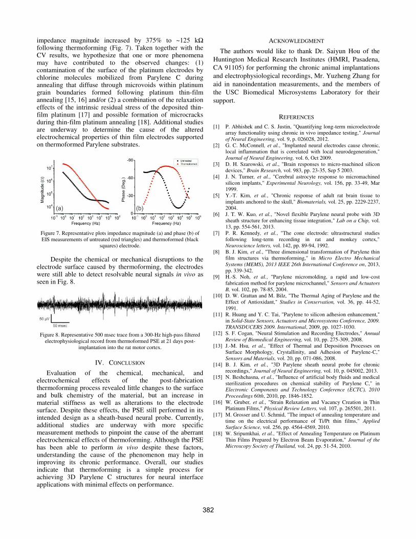
Though the Parylene substrate increases in stiffness following the thermoforming process, flexibility of the PSE was not lost. This is supported by mechanical deformation tests that demonstrated that the Parylene sheath structure could return to its original open position following a deformation of 200 µm imposed by a 25 mN load (Fig. 5). The sheath is also sufficiently robust to resist any deformation in vivo as seen in histological data [14]. Although slightly stiffer than the untreated material, the thermoformed PSE is still a large improvement in minimizing the mechanical mismatch between cortical tissue and current technologies, and compares favorably to other popular polymers used in microfabrication, such as polyimide.
Figure 5. Displacement (black squares) and load force (red triangles)
measurements taken during deflection studies of thermoformed PSE sheath
structure. Inset and plot indicate the (1) starting position, (2) deflection of
200 µm, and (3) return to initial position indicating following release of
load. Sheath remains flexible and is robust to mechanical perturbations.
Sheath deformation is outlined in black to facilitate visualization. Scale bar
is 200 µm.
D. Electrochemical Characterization
CV measurements of the platinum electrode surface indicated a noticeable reduction in the area inside the CV curve compared to the untreated electrodes, indicating a decreased charge storage capacity (Fig. 6).
Figure 6. Representative CV curves of untreated (blue dashed) and
thermoformed (black solid) electrodes.
Electrochemical impedance also supported an altered electrode surface as the impedance magnitude had increased and the phase plot had shifted to the right; the 1 kHz
381
impedance magnitude increased by 375% to ~���� N��following thermoforming (Fig. 7). Taken together with the CV results, we hypothesize that one or more phenomena may have contributed to the observed changes: (1) contamination of the surface of the platinum electrodes by chlorine molecules mobilized from Parylene C during annealing that diffuse through microvoids within platinum grain boundaries formed following platinum thin-film annealing [15, 16] and/or (2) a combination of the relaxation effects of the intrinsic residual stress of the deposited thin-film platinum [17] and possible formation of microcracks during thin-film platinum annealing [18]. Additional studies are underway to determine the cause of the altered electrochemical properties of thin film electrodes supported on thermoformed Parylene substrates.
Figure 7. Representative plots impedance magnitude (a) and phase (b) of
EIS measurements of untreated (red triangles) and thermoformed (black
squares) electrode.
Despite the chemical or mechanical disruptions to the electrode surface caused by thermoforming, the electrodes were still able to detect resolvable neural signals in vivo as seen in Fig. 8.
Figure 8. Representative 500 msec trace from a 300-Hz high-pass filtered
electrophysiological record from thermoformed PSE at 21 days post-
implantation into the rat motor cortex.
IV. CONCLUSION
Evaluation of the chemical, mechanical, and electrochemical effects of the post-fabrication thermoforming process revealed little changes to the surface and bulk chemistry of the material, but an increase in material stiffness as well as alterations to the electrode surface. Despite these effects, the PSE still performed in its intended design as a sheath-based neural probe. Currently, additional studies are underway with more specific measurement methods to pinpoint the cause of the aberrant electrochemical effects of thermoforming. Although the PSE has been able to perform in vivo despite these factors, understanding the cause of the phenomenon may help in improving its chronic performance. Overall, our studies indicate that thermoforming is a simple process for achieving 3D Parylene C structures for neural interface applications with minimal effects on performance.
ACKNOWLEDGMENT
The authors would like to thank Dr. Saiyun Hou of the
Huntington Medical Research Institutes (HMRI, Pasadena,
CA 91105) for performing the chronic animal implantations
and electrophysiological recordings, Mr. Yuzheng Zhang for
aid in nanoindentation measurements, and the members of
the USC Biomedical Microsystems Laboratory for their
support.
REFERENCES
[1] P. Abhishek and C. S. Justin, "Quantifying long-term microelectrode
array functionality using chronic in vivo impedance testing," Journal
of Neural Engineering, vol. 9, p. 026028, 2012.
[2] G. C. McConnell, et al., "Implanted neural electrodes cause chronic,
local inflammation that is correlated with local neurodegeneration,"
Journal of Neural Engineering, vol. 6, Oct 2009.
[3] D. H. Szarowski, et al., "Brain responses to micro-machined silicon
devices," Brain Research, vol. 983, pp. 23-35, Sep 5 2003.
[4] J. N. Turner, et al., "Cerebral astrocyte response to micromachined
silicon implants," Experimental Neurology, vol. 156, pp. 33-49, Mar
1999.
[5] Y.-T. Kim, et al., "Chronic response of adult rat brain tissue to
implants anchored to the skull," Biomaterials, vol. 25, pp. 2229-2237,
2004.
[6] J. T. W. Kuo, et al., "Novel flexible Parylene neural probe with 3D
sheath structure for enhancing tissue integration," Lab on a Chip, vol.
13, pp. 554-561, 2013.
[7] P. R. Kennedy, et al., "The cone electrode: ultrastructural studies
following long-term recording in rat and monkey cortex,"
Neuroscience letters, vol. 142, pp. 89-94, 1992.
[8] B. J. Kim, et al., "Three dimensional transformation of Parylene thin
film structures via thermoforming," in Micro Electro Mechanical
Systems (MEMS), 2013 IEEE 26th International Conference on, 2013,
pp. 339-342.
[9] H.-S. Noh, et al., "Parylene micromolding, a rapid and low-cost
fabrication method for parylene microchannel," Sensors and Actuators
B, vol. 102, pp. 78-85, 2004.
[10] D. W. Grattan and M. Bilz, "The Thermal Aging of Parylene and the
Effect of Antioxidant," Studies in Conservation, vol. 36, pp. 44-52,
1991.
[11] R. Huang and Y. C. Tai, "Parylene to silicon adhesion enhancement,"
in Solid-State Sensors, Actuators and Microsystems Conference, 2009.
TRANSDUCERS 2009. International, 2009, pp. 1027-1030.
[12] S. F. Cogan, "Neural Stimulation and Recording Electrodes," Annual
Review of Biomedical Engineering, vol. 10, pp. 275-309, 2008.
[13] J.-M. Hsu, et al., "Effect of Thermal and Deposition Processes on
Surface Morphology, Crystallinity, and Adhesion of Parylene-C,"
Sensors and Materials, vol. 20, pp. 071-086, 2008.
[14] B. J. Kim, et al., "3D Parylene sheath neural probe for chronic
recordings," Journal of Neural Engineering, vol. 10, p. 045002, 2013.
[15] N. Beshchasna, et al., "Influence of artificial body fluids and medical
sterilization procedures on chemical stability of Parylene C," in
Electronic Components and Technology Conference (ECTC), 2010
Proceedings 60th, 2010, pp. 1846-1852.
[16] W. Gruber, et al., "Strain Relaxation and Vacancy Creation in Thin
Platinum Films," Physical Review Letters, vol. 107, p. 265501, 2011.
[17] M. Grosser and U. Schmid, "The impact of annealing temperature and
time on the electrical performance of Ti/Pt thin films," Applied
Surface Science, vol. 256, pp. 4564-4569, 2010.
[18] W. Sripumkhai, et al., "Effect of Annealing Temperature on Platinum
Thin Films Prepared by Electron Beam Evaporation," Journal of the
Microscopy Society of Thailand, vol. 24, pp. 51-54, 2010.
382