Embed Size (px)
DESCRIPTION
An outline on Eukaryotic cell organization
Citation preview
EUKARYOTIC CELL - ORGANIZATION
INTRODUCTION
The most obvious difference between eukaryotic and prokaryotic cells is in their use of membranes.
Eucaryotic cells have membrane delimited nuclei, and membranes also play a prominent part in the structure of many other organelles.
Organelles are intracellular structures that perform specific functions in cells analogous to the functions of organs in the body.
The name organelle (little organ) was coined because biologists saw a parallel between the relationship of organelles to a cell and that of organs to the whole body.
It is not satisfactory to define organelles as membrane bound structures because this would exclude such components as ribosomes and bacterial flagella.
The complexity of eukaryotic cells is due chiefly to the use of internal membranes for several purposes.
The partitioning of the eukaryotic cell interior by membranes makes possible the placement of different biochemical and physiological functions in separate compartments so that they can more easily take place simultaneously under independent control and proper coordination.
Large membrane surfaces make possible greater respiratory and photosynthetic activity because these processes are located exclusively in membranes.
The intracytoplasmic membrane complex also serves as a transport system to move materials between different cell locations.
Thus abundant membrane systems probably are necessary in eucaryotic cells because of their large volume and the need for adequate regulation, metabolic activity, and transport.
Those organelles lying inside the plasma membrane are first described, and then components outside the membrane are discussed.
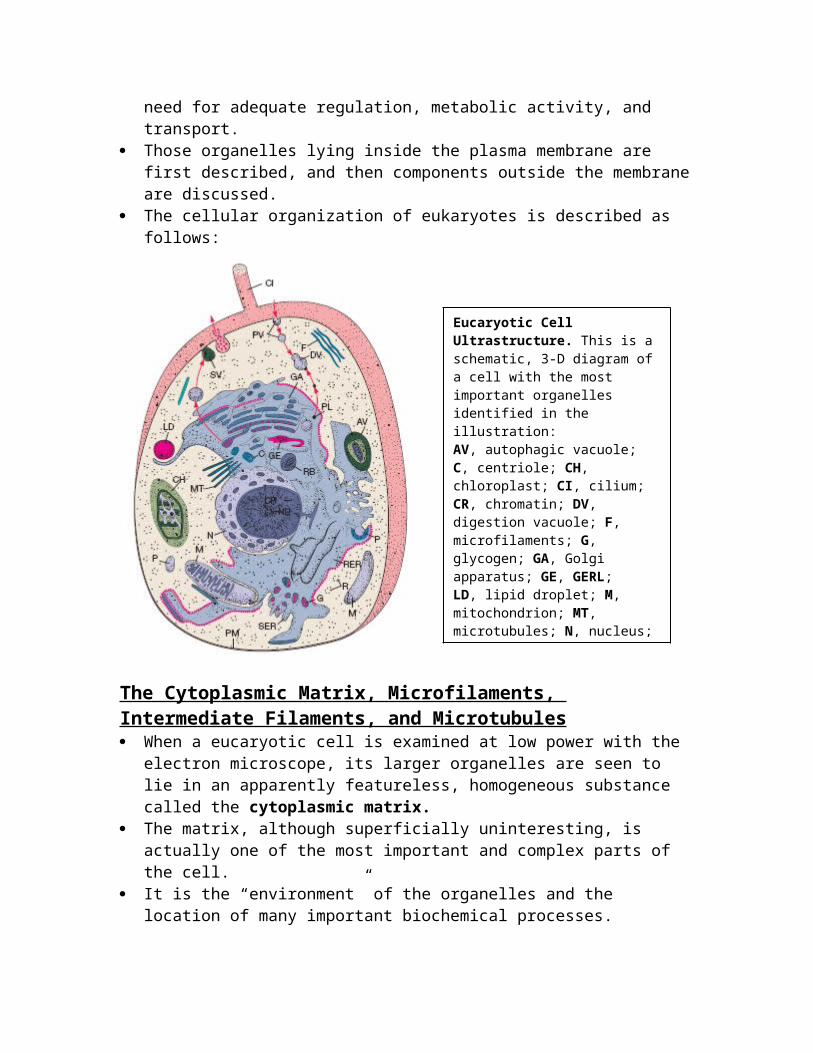
The cellular organization of eukaryotes is described as follows:
The Cytoplasmic Matrix, Microfilaments, Intermediate Filaments, and Microtubules When a eucaryotic cell is examined at low power with the electron
microscope, its larger organelles are seen to lie in an apparently featureless, homogeneous substance called the cytoplasmic matrix.
The matrix, although superficially uninteresting, is actually one of the most important and complex parts of the cell.
It is the “environment” of the organelles and the location of many important biochemical processes.
Several physical changes seen in cells—viscosity changes, cytoplasmic streaming, and others— also are due to matrix activity.
Water constitutes about 70 to 85% by weight of a eukaryotic cell. Thus a large part of the cytoplasmic matrix is water. Cellular water can exist in two different forms. Some of it is bulk or
free water; this is normal, osmotically active water. Water also can exist as bound water or water of hydration. This water is bound to the surface of proteins and other
macromolecules and is osmotically inactive and more ordered than bulk water.
There is some evidence that bound water is the site of many metabolic processes.
Eucaryotic Cell Ultrastructure. This is a schematic, 3-D diagram of a cell with the most important organelles identified in the illustration:AV, autophagic vacuole; C, centriole; CH, chloroplast; CI, cilium; CR, chromatin; DV, digestion vacuole; F, microfilaments; G, glycogen; GA, Golgi apparatus; GE, GERL;LD, lipid droplet; M, mitochondrion; MT, microtubules; N, nucleus;NU, nucleolus; P, peroxisome; PL, primary lysosome; PM, plasmamembrane; PV, pinocytotic vesicle; R, ribosomes and
The protein content of cells is so high that the cytoplasmic matrix often may be semicrystalline.
Usually matrix pH is around neutrality, about pH 6.8 to 7.1, but can vary widely.
For example, protozoan digestive vacuoles may reach pHs as low as 3 to 4.
Probably all eucaryotic cells have microfilaments, minute protein filaments, 4 to 7 nm in diameter, which may be either scattered within the cytoplasmic matrix or organized into networks and parallel arrays.
Microfilaments are involved in cell motion and shape changes. Some examples of cellular movements associated with
microfilament activity are the motion of pigment granules, amoeboid movement, and protoplasmic streaming in slime molds.
The participation of microfilaments in cell movement is suggested by electron microscopic studies showing that they frequently are found at locations appropriate for such a role.
For example, they are concentrated at the interface between stationary and flowing cytoplasm in plant cells and slime molds.
Experiments using the drug cytochalasin B have provided additional evidence.
Cytochalasin B disrupts microfilament structure and often simultaneously inhibits cell movements.
However, because the drug has additional effects in cells, a direct cause-and-effect interpretation of these experiments is sometimes difficult.
Microfilament protein has been isolated and analyzed chemically. It is an actin, very similar to the actin contractile protein of muscle
tissue. This is further indirect evidence for microfilament involvement in
cell movement. Some pathogens such as Listeria monocytogenes make use of
eucaryotic actin to move rapidly through the host cell. The ActA protein released by Listeria causes the polymerization of
actin filaments at the end of the bacterium. A tail of actin is formed and trapped in the host cytoskeleton. Its continued elongation pushes the bacterium along at rates up to
11 _m/minute. The bacterium can even be propelled through the cell surface and
into neighboring cells. A second type of small filamentous organelle in the cytoplasmic
matrix is shaped like a thin cylinder about 25 nm in diameter. Because of its tubular nature this organelle is called a
microtubule.
Microtubules are complex structures constructed of two slightly different spherical protein subunits named tubulins, each of which is approximately 4 to 5 nm in diameter.
These subunits are assembled in a helical arrangement to form a cylinder with an average of 13 subunits in one turn or circumference.
Microtubules serve at least three purposes: (1) they help maintain cell shape, (2) are involved with microfilaments in cell movements, and (3) participate in intracellular transport processes. Evidence for a structural role comes from their intracellular distribution and studies on the effects of the drug colchicine.
Long, thin cell structures requiring support such as the axopodia (long, slender, rigid pseudopodia) of protozoa contain microtubules.
When migrating embryonic nerve and heart cells are exposed to colchicine, they simultaneously lose their microtubules and their characteristic shapes.
The shapeless cells seem to wander aimlessly as if incapable of directed movement without their normal form. Their microfilaments are still intact, but due to the disruption of their microtubules by colchicine, they no longer behave normally.
Microtubules also are present in structures that participate in cell or organelle movements—the mitotic spindle, cilia, and flagella.
For example, the mitotic spindle is constructed of microtubules; when a dividing cell is treated with colchicine, the spindle is disrupted and chromosome separation blocked.
Microtubules also are essential to the movement of eucaryotic cilia and flagella.
Other kinds of filamentous components also are present in the matrix, the most important of which are the intermediate filaments (about 8 to 10 nm in diameter). The microfilaments, microtubules, and intermediate filaments are major components of a vast, intricate network of interconnected filaments called the cytoskeleton.
As mentioned previously, the cytoskeleton plays a role in both cell shape and movement. Procaryotes lack a true, organized cytoskeleton and may not possess actin like proteins.
The Endoplasmic Reticulum Besides the cytoskeleton, the cytoplasmic matrix is permeated with
an irregular network of branching and fusing membranous tubules, around 40 to 70 nm in diameter, and many flattened sacs called cisternae (s., cisterna).
This network of tubules and cisternae is the endoplasmic reticulum (ER).
The nature of the ER varies with the functional and physiological status of the cell. In cells synthesizing a great deal of protein for purposes such as secretion, a large part of the ER is studded on its outer surface with ribosomes and is called rough or granular endoplasmic reticulum (RER or GER). Other cells, such as those producing large quantities of lipids, have ER that lacks ribosomes.
This is smooth or agranular ER (SER orAER). The endoplasmic reticulum has many important functions. It transports proteins, lipids, and probably other materials through
the cell. Lipids and proteins are synthesized by ER-associated enzymes and
ribosomes. Polypeptide chains synthesized on RER bound ribosomes may be
inserted either into the ER membrane or into its lumen for transport elsewhere.
The ER is also a major site of cell membrane synthesis. New endoplasmic reticulum is produced through expansion of the
old. Many biologists think the RER synthesizes new ER proteins and
lipids. “Older” RER then loses its connected ribosomes and is modified to become SER.
The Golgi Apparatus The Golgi apparatus is a membranous organelle composed of
flattened, saclike cisternae stacked on each other. These membranes, like the smooth ER, lack bound ribosomes. There are usually around 4 to 8 cisternae or sacs in a stack,
although there may be many more. Each sac is 15 to 20 nm thick and separated from other cisternae by 20 to 30 nm.
A complex network of tubules and vesicles (20 to 100 nm in diameter) is located at the edges of the cisternae.
The stack of cisternae has a definite polarity because there are two ends or faces that are quite different from one another.
The sacs on the cis or forming face often are associated with the ER and differ from the sacs on the trans or maturing face in thickness, enzyme content, and degree of vesicle formation.
It appears that material is transported from cis to trans cisternae by vesicles that bud off the cisternal edges and move to the next sac.
The Golgi apparatus is present in most eucaryotic cells, but many fungi and ciliate protozoa may lack a well-formed structure.
Sometimes it consists of a single stack of cisternae; however, many cells may contain up to 20, and sometimes more, separate stacks.
These stacks of cisternae, often called dictyosomes, can be clustered in one region or scattered about the cell.
The Golgi apparatus packages materials and prepares them for secretion, the exact nature of its role varying with the organism.
The surface scales of some flagellated algae and radiolarian protozoa appear to be constructed within the Golgi apparatus and then transported to the surface in vesicles.
It often participates in the development of cell membranes and in the packaging of cell products.
The growth of some fungal hyphae occurs when Golgi vesicles contribute their contents to the wall at the hyphal tip.
In all these processes, materials move from the ER to the Golgi apparatus.
Most often vesicles bud off the ER, travel to the Golgi apparatus, and fuse with the cis cisternae.
Thus the Golgi apparatus is closely related to the ER in both a structural and a functional sense.
Most proteins entering the Golgi apparatus from the ER are glycoproteins containing short carbohydrate chains.
The Golgi apparatus frequently modifies proteins destined for different fates by adding specific groups and then sends the proteins on their way to the proper location (e.g., lysosomal proteins have phosphates added to their mannose sugars).
Lysosomes and Endocytosis A very important function of the Golgi apparatus and endoplasmic
reticulum is the synthesis of another organelle, the lysosome. This organelle (or a structure very much like it) is found in a variety
of microorganisms—protozoa, some algae, and fungi—as well as in plants and animals.
Lysosomes are roughly spherical and enclosed in a single membrane; they average about 500 nm in diameter, but range from 50 nm to several μm in size. They are involved in intracellular digestion and contain the enzymes needed to digest all types of macromolecules.
These enzymes, called hydrolases, catalyze the hydrolysis of molecules and function best under slightly acid conditions (usually around pH 3.5 to 5.0).
Lysosomes maintain an acidic environment by pumping protons into their interior.
Digestive enzymes are manufactured by the RER and packaged to form lysosomes by the Golgi apparatus.
A segment of smooth ER near the Golgi apparatus also may bud off lysosomes.
Lysosomes are particularly important in those cells that obtain nutrients through endocytosis.
In this process a cell takes up solutes or particles by enclosing them in vacuoles and vesicles pinched off from its plasma membrane.
Vacuoles and vesicles are membrane delimited cavities that contain fluid, and often solid material.
Larger cavities will be called vacuoles, and smaller cavities, vesicles.
There are two major forms of endocytosis: phagocytosis and pinocytosis.
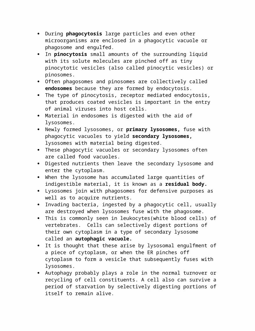
During phagocytosis large particles and even other microorganisms are enclosed in a phagocytic vacuole or phagosome and engulfed.
In pinocytosis small amounts of the surrounding liquid with its solute molecules are pinched off as tiny pinocytotic vesicles (also called pinocytic vesicles) or pinosomes.
Often phagosomes and pinosomes are collectively called endosomes because they are formed by endocytosis.
The type of pinocytosis, receptor mediated endocytosis, that produces coated vesicles is important in the entry of animal viruses into host cells.
Material in endosomes is digested with the aid of lysosomes. Newly formed lysosomes, or primary lysosomes, fuse with
phagocytic vacuoles to yield secondary lysosomes, lysosomes with material being digested.
These phagocytic vacuoles or secondary lysosomes often are called food vacuoles.
Digested nutrients then leave the secondary lysosome and enter the cytoplasm.
When the lysosome has accumulated large quantities of indigestible material, it is known as a residual body.
Lysosomes join with phagosomes for defensive purposes as well as to acquire nutrients.
Invading bacteria, ingested by a phagocytic cell, usually are destroyed when lysosomes fuse with the phagosome.
This is commonly seen in leukocytes(white blood cells) of vertebrates. Cells can selectively digest portions of their own cytoplasm in a type of secondary lysosome called an autophagic vacuole.
It is thought that these arise by lysosomal engulfment of a piece of cytoplasm, or when the ER pinches off cytoplasm to form a vesicle that subsequently fuses with lysosomes.
Autophagy probably plays a role in the normal turnover or recycling of cell constituents. A cell also can survive a period of starvation by selectively digesting portions of itself to remain alive.
Following cell death, lysosomes aid in digestion and removal of cell debris.
A most remarkable thing about lysosomes is that they accomplish all these tasks without releasing their digestive enzymes into the cytoplasmic matrix, a catastrophe that would destroy the cell.
The lysosomal membrane retains digestive enzymes and other macromolecules while allowing small digestion products to leave.
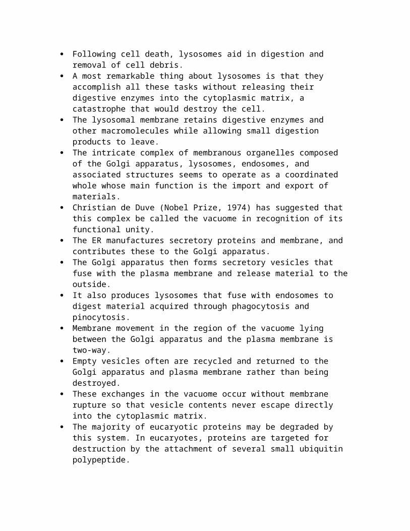
The intricate complex of membranous organelles composed of the Golgi apparatus, lysosomes, endosomes, and associated structures seems to operate as a coordinated whole whose main function is the import and export of materials.
Christian de Duve (Nobel Prize, 1974) has suggested that this complex be called the vacuome in recognition of its functional unity.
The ER manufactures secretory proteins and membrane, and contributes these to the Golgi apparatus.
The Golgi apparatus then forms secretory vesicles that fuse with the plasma membrane and release material to the outside.
It also produces lysosomes that fuse with endosomes to digest material acquired through phagocytosis and pinocytosis.
Membrane movement in the region of the vacuome lying between the Golgi apparatus and the plasma membrane is two-way.
Empty vesicles often are recycled and returned to the Golgi apparatus and plasma membrane rather than being destroyed.
These exchanges in the vacuome occur without membrane rupture so that vesicle contents never escape directly into the cytoplasmic matrix.
The majority of eucaryotic proteins may be degraded by this system. In eucaryotes, proteins are targeted for destruction by the attachment of several small ubiquitin polypeptide.
The marked protein then enters a huge cylindrical complex called a 26S proteasome, where it is degraded to peptides in an ATP-dependent process and the ubiquitins are released.
The peptides may be hydrolyzed to amino acids. In this case the system is being used to recycle proteins.
The proteosome also is involved in producing peptides for antigen presentation during many immunological responses.
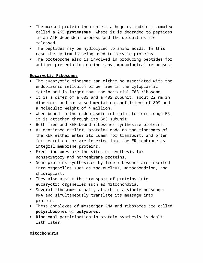
Eucaryotic Ribosomes The eucaryotic ribosome can either be associated with the
endoplasmic reticulum or be free in the cytoplasmic matrix and is larger than the bacterial 70S ribosome.
It is a dimer of a 60S and a 40S subunit, about 22 nm in diameter, and has a sedimentation coefficient of 80S and a molecular weight of 4 million.
When bound to the endoplasmic reticulum to form rough ER, it is attached through its 60S subunit.
Both free and RER-bound ribosomes synthesize proteins. As mentioned earlier, proteins made on the ribosomes of the RER
either enter its lumen for transport, and often for secretion, or are inserted into the ER membrane as integral membrane proteins.
Free ribosomes are the sites of synthesis for nonsecretory and nonmembrane proteins.
Some proteins synthesized by free ribosomes are inserted into organelles such as the nucleus, mitochondrion, and chloroplast.
They also assist the transport of proteins into eucaryotic organelles such as mitochondria.
Several ribosomes usually attach to a single messenger RNA and simultaneously translate its message into protein.
These complexes of messenger RNA and ribosomes are called polyribosomes or polysomes.
Ribosomal participation in protein synthesis is dealt with later.
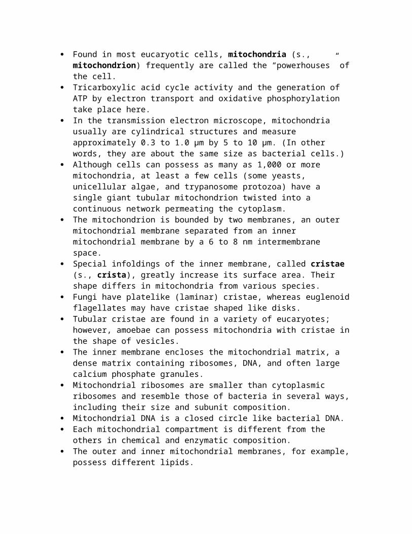
Mitochondria Found in most eucaryotic cells, mitochondria (s., mitochondrion)
frequently are called the “powerhouses” of the cell. Tricarboxylic acid cycle activity and the generation of ATP by
electron transport and oxidative phosphorylation take place here. In the transmission electron microscope, mitochondria usually are
cylindrical structures and measure approximately 0.3 to 1.0 μm by 5 to 10 μm. (In other words, they are about the same size as bacterial cells.)
Although cells can possess as many as 1,000 or more mitochondria, at least a few cells (some yeasts, unicellular algae, and trypanosome protozoa) have a single giant tubular mitochondrion twisted into a continuous network permeating the cytoplasm.
The mitochondrion is bounded by two membranes, an outer mitochondrial membrane separated from an inner mitochondrial membrane by a 6 to 8 nm intermembrane space.
Special infoldings of the inner membrane, called cristae (s., crista), greatly increase its surface area. Their shape differs in mitochondria from various species.
Fungi have platelike (laminar) cristae, whereas euglenoid flagellates may have cristae shaped like disks.
Tubular cristae are found in a variety of eucaryotes; however, amoebae can possess mitochondria with cristae in the shape of vesicles.
The inner membrane encloses the mitochondrial matrix, a dense matrix containing ribosomes, DNA, and often large calcium phosphate granules.
Mitochondrial ribosomes are smaller than cytoplasmic ribosomes and resemble those of bacteria in several ways, including their size and subunit composition.
Mitochondrial DNA is a closed circle like bacterial DNA. Each mitochondrial compartment is different from the others in
chemical and enzymatic composition. The outer and inner mitochondrial membranes, for example,
possess different lipids.
Enzymes and electron carriers involved in electron transport and oxidative phosphorylation (the formation of ATP as a consequence of electron transport) are located only in the inner membrane.
The enzymes of the tricarboxylic acid cycle and the beta-oxidation pathway for fatty acids are located in the matrix.
The inner membrane of the mitochondrion has another distinctive structural feature related to its function.
Many small spheres, about 8.5 nm diameter, are attached by stalks to its inner surface.
The spheres are called F1 particles and synthesize ATP during cellular respiration.
The mitochondrion uses its DNA and ribosomes to synthesize some of its own proteins. In fact, mutations in mitochondrial DNA often lead to serious diseases in humans.
Most mitochondrial proteins, however, are manufactured under the direction of the nucleus.
Mitochondria reproduce by binary fission. Chloroplasts show similar partial independence and reproduction by
binary fission. Because both organelles resemble bacteria to some extent, it has
been suggested that these organelles arose from symbiotic associations between bacteria and larger cells.
Chloroplasts Plastids are cytoplasmic organelles of algae and higher plants that
often possess pigments such as chlorophylls and carotenoids, and are the sites of synthesis and storage of food reserves.
The most important type of plastid is the chloroplast. Chloroplasts contain chlorophyll and use light energy to convert
CO2 and water to carbohydrates and O2. Although chloroplasts are quite variable in size and shape, they
share many structural features. Most often they are oval with dimensions of 2 to 4 μm by 5 to 10
μm, but some algae possess one huge chloroplast that fills much of the cell.
Like mitochondria, chloroplasts are encompassed by two membranes.
A matrix, the stroma, lies within the inner membrane. It contains DNA, ribosomes, lipid droplets, starch granules, and a
complex internal membrane system whose most prominent components are flattened, membrane-delimited sacs, the thylakoids.
Clusters of two or more thylakoids are dispersed within the stroma of most algal chloroplasts.
In some groups of algae, several disklike thylakoids are stacked on each other like coins to form grana (s., granum).
Photosynthetic reactions are separated structurally in the chloroplast just as electron transport and the tricarboxylic acid cycle are in the mitochondrion.
The formation of carbohydrate from CO2 and water, the dark reaction, takes place in the stroma.
The trapping of light energy to generate ATP, NADPH, and O2, the light reaction, is located in the thylakoid membranes, where chlorophyll and electron transport components are also found.
The chloroplasts of many algae contain a pyrenoid, a dense region of protein surrounded by starch or another polysaccharide.
Pyrenoids participate in polysaccharide synthesis.
The Nucleus and Cell Division The cell nucleus is by far the most visually prominent organelle. It was discovered early in the study of cell structure and was shown
by Robert Brown in 1831 to be a constant feature of eukaryotic cells.
The nucleus is the repository for the cell’s genetic information and is its control center.
Nuclear Structure Nuclei are membrane-delimited spherical bodies about 5 to 7 μm in
diameter. Dense fibrous material called chromatin can be seen within the
nucleoplasm of the nucleus of a stained cell. This is the DNA-containing part of the nucleus.
In non-dividing cells, chromatin exists in a dispersed condition, but condenses during mitosis to become visible as chromosomes.
Some nuclear chromatin, the euchromatin, is loosely organized and contains those genes that are expressing themselves actively.
Heterochromatin is coiled more tightly, appears darker in the electron microscope, and is not genetically active most of the time.
The nucleus is bounded by the nuclear envelope, a complex structure consisting of inner and outer membranes separated by a 15 to 75 nm perinuclear space.
The envelope is continuous with the ER at several points and its outer membrane is covered with ribosomes.
A network of intermediate filaments, called the nuclear lamina, lies against the inner surface of the envelope and supports it.
Chromatin usually is associated with the inner membrane. Many nuclear pores penetrate the envelope, each pore formed by
a fusion of the outer and inner membranes. Pores are about 70 nm in diameter and collectively occupy about 10
to 25% of the nuclear surface. A complex ringlike arrangement of granular and fibrous material
called the annulus is located at the edge of each pore.
The nuclear pores serve as a transport route between the nucleus and surrounding cytoplasm. Particles have been observed moving into the nucleus through the pores.
Although the function of the annulus is not understood, it may either regulate or aid the movement of material through the pores.
Substances also move directly through the nuclear envelope by unknown mechanisms.
The Nucleolus Often the most noticeable structure within the nucleus is the
nucleolus. A nucleus may contain from one to many nucleoli. Although the nucleolus is not membrane-enclosed, it is a complex
organelle with separate granular and fibrillar regions. It is present in nondividing cells, but frequently disappears during
mitosis. After mitosis the nucleolus reforms around the nucleolar organizer, a particular part of a specific chromosome.
The nucleolus plays a major role in ribosome synthesis. The nucleolar organizer DNA directs the production of ribosomal
RNA (rRNA). This RNA is synthesized in a single long piece that then is cut to form the final rRNA molecules.
The processed rRNAs next combine with ribosomal proteins (which have been synthesized in the cytoplasmic matrix) to form partially completed ribosomal subunits.
The granules seen in the nucleolus are probably these subunits. Immature ribosomal subunits then leave the nucleus, presumably
by way of the nuclear envelope pores and mature in the cytoplasm.
External Cell Coverings Eucaryotic microorganisms differ greatly from procaryotes in the
supporting or protective structures they have external to the plasma membrane.
In contrast with most bacteria, many eucaryotes lack an external cell wall. Eg: amoeba
Eucaryotic cell membranes, unlike most procaryotic membranes, contain sterols such as cholesterol in their lipid bilayers, and this may make them mechanically stronger, thus reducing the need for external support.
Of course many eukaryotes do have a rigid external cell wall. Algal cell walls usually have a layered appearance and contain
large quantities of polysaccharides such as cellulose and pectin. In addition, inorganic substances like silica (in diatoms) or calcium carbonate (some red algae) may be present.
Fungal cell walls normally are rigid. Their exact composition varies with the organism; but usually, cellulose, chitin, or glucan (a glucose polymer different from cellulose) are present.
Despite their nature the rigid materials in eucaryotic walls are chemically simpler than procaryotic peptidoglycan.
Many protozoa and some algae have a different external structure, the pellicle.
This is a relatively rigid layer of components just beneath the plasma membrane (sometimes the plasma membrane is also considered part of the pellicle).
The pellicle may be fairly simple in structure. For example, Euglena has a series of overlapping strips with a ridge at the edge of each strip fitting into a groove on the adjacent one. In contrast, ciliate protozoan pellicles are exceptionally complex with two membranes and a variety of associated structures. Although pellicles are not as strong and rigid as cell walls, they do give their possessors a characteristic shape.
Cilia and Flagella Cilia (s., cilium) and flagella (s., flagellum) are the most
prominent organelles associated with motility. Although both are whiplike and beat to move the microorganism
along, they differ from one another in two ways. First, cilia are typically only 5 to 20 μm in length, whereas flagella
are 100 to 200 μm long. Second, their patterns of movement are usually distinctive.
Flagella move in an undulating fashion and generate planar or helical waves originating at either the base or the tip. If the wave moves from base to tip, the cell is pushed along; a beat traveling from the tip toward the base pulls the cell through the water.
Sometimes the flagellum will have lateral hairs called flimmer filaments (thicker, stiffer hairs are called mastigonemes).
These filaments change flagellar action so that a wave moving down the filament toward the tip pulls the cell along instead of pushing it. Such a flagellum often is called a tinsel flagellum, whereas the naked flagellum is referred to as a whiplash flagellum.
Cilia, on the other hand, normally have a beat with two distinctive phases.
In the effective stroke, the cilium strokes through the surrounding fluid like an oar, thereby propelling the organism along in the water.
The cilium next bends along its length while it is pulled forward during the recovery stroke in preparation for another effective stroke.
A ciliated microorganism actually coordinates the beats so that some of its cilia are in the recovery phase while others are carrying out their effective stroke.
This coordination allows the organism to move smoothly through the water.
Despite their differences, cilia and flagella are very similar in ultrastructure. They are membrane-bound cylinders about 0.2 μm in diameter.
Located in the matrix of the organelle is a complex, the axoneme, consisting of nine pairs of microtubule doublets arranged in a circle around two central tubules.
This is called the 9+2 pattern of microtubules. Each doublet also has pairs of arms projecting from subtubule A (the complete microtubule) toward a neighboring doublet.
A radial spoke extends from subtubule A toward the internal pair of microtubules with their central sheath. These microtubules are similar to those found in the cytoplasm.
Each is constructed of two types of tubulin subunits, α- and β-tubulins, that resemble the contractile protein actin in their composition
A basal body lies in the cytoplasm at the base of each cilium or flagellum.
It is a short cylinder with nine microtubule triplets around its periphery (a 9+0 pattern) and is separated from the rest of the organelle by a basal plate.
The basal body directs the construction of these organelles. Cilia and flagella appear to grow through the addition of preformed microtubule subunits at their tips.
Cilia and flagella bend because adjacent microtubule doublets slide along one another while maintaining their individual lengths. The doublet arms, about 15 nm long, are made of the protein dynein.
ATP powers the movement of cilia and flagella, and isolated dynein hydrolyzes ATP.
It appears that dynein arms interact with the B subtubules of adjacent doublets to cause the sliding.
The radial spokes also participate in this sliding motion. Cilia and flagella beat at a rate of about 10 to 40 strokes or waves per second and propel microorganisms rapidly.
The record holder is the flagellate Monas stigmatica, which swims at a rate of 260 μm/second (approximately 40 cell lengths per second); the common euglenoid flagellate, Euglena gracilis, travels at around 170 μm or 3 cell lengths per second. The ciliate protozoan Paramecium caudatum swims at about 2,700 μm/second (12 lengths per second).