Embed Size (px)
Citation preview

Various drug delivery approaches have been explored for successful delivery ofdrugs to the target site. However, the oral route of administration is considered to be the
529
Acta Pharm. 62 (2012) 529–545 Original research paper
DOI: 10.2478/v10007-012-0034-x
Eudragit S-100 coated sodium alginate microspheresof naproxen sodium: Formulation, optimization and
in vitro evaluation
ANUJ CHAWLA
POOJA SHARMA
PRAVIN PAWAR
Chitkara College of PharmacyChandigarh-PatialaNational Highway, Rajpura-140401Patiala, Punjab, India
Accepted September 4, 2012
The aim of the study was to prepare site specific drug deliveryof naproxen sodium using sodium alginate and Eudragit S-100as a mucoadhesive and pH-sensitive polymer, respectively. Coremicrospheres of alginate were prepared by a modified emulsifi-cation method followed by cross-linking with CaCl2, which wasfurther coated with the pH dependent polymer Eudragit S-100(2.5 or 5 %) to prevent drug release in the upper gastrointestinalenvironment. Microspheres were characterized by FT-IR spectro-scopy, X-ray diffraction, differential scanning calorimetry andevaluated by scanning electron microscopy, particle size analy-sis, drug loading efficiency, in vitro mucoadhesive time studyand in vitro drug release study in different simulated gastric flu-ids. Stability studies of the optimized formulation were carriedout for 6 months. SEM images revealed that the surface morpho-logy was rough and smooth for core and coated microspheres,respectively. Core microspheres showed better mucoadhesioncompared to coated microspheres when applied to the mucosalsurface of freshly excised goat colon. The optimized batch of co-re microspheres and coated microspheres exhibited 98.42 ± 0.96and 95.58 ± 0.74 % drug release, respectively. Drug release fromall sodium alginate microsphere formulations followed Higuchikinetics. Moreover, drug release from Eudragit S-100 coated mi-crospheres followed the Korsmeyer-Peppas equation with aFickian kinetics mechanism. Stability study suggested that thedegradation rate constant of microspheres was minimal, indicat-ing 2 years shelf life of the formulation.
Keywords: colon specific, Eudragit S-100, microspheres, napro-xen sodium, sodium alginate
* Correspondence; e-mail: [email protected]

most convenient and preferred route for a sustained as well as controlled drug deliverysystem (1).
Various drug delivery strategies have been employed to trigger the release of drugsto the large intestine; however, they do not reach the site of action in appropriate con-centrations. Thus, to ensure an effective and safe therapy for the large bowel diseases,the colon specific drug delivery system is considered to be the preferable approach (2).
Targeting of drugs to the colon offers several potential therapeutic advantages, likeincreasing the systemic absorption of poorly absorbed drugs and effective treatment of thecolonic diseases such as amoebiasis, ulcerative colitis, Crohn’s disease, colorectal cancer,etc. This delivery system can be also used in certain conditions where drugs should be de-livered after a lag time, like in chronopharmacotherapy of diseases showing Circadianrhythms in their pathophysiology (3). Several available approaches for targeting the drugselectively to the colon include pH sensitive polymers, time dependent dosage forms, useof carriers degraded by enzymes produced by colonic bacteria, prodrug based ap-proaches, bioadhesive and osmotically controlled drug delivery systems (4). Colon spe-cific drug delivery (CDDS) microspheres are advantageous owing to the fact that, in com-parison with conventional dosage forms, CDDS microspheres provide a more consistentand reproducible transit through the gastrointestinal tract (GIT). Moreover, they also pro-vide more uniform drug dispersion in the GI tract, which results in more homogeneousdrug absorption. This helps in predicting gastric emptying, increases colon residence time,decreases local irritation (5).
The third most common deadly cancer in the world is colorectal cancer. Epidemio-logical investigations and clinical trials done in patients with hereditary colon cancerand familial adenomatous polyposis (FAP) highlight the importance of nonsteroidalanti-inflammatory drugs (NSAIDs), which act as COX inhibitors and hence, due to theirmode of action similar to that of anticancer drugs, halt the development of colon cancer-ous cell growth (6).
Naproxen [(S)-2-(6-methoxynaphthalen-2-yl)propanoic acid], is a non selective COXinhibitor widely used as an analgesic in the treatment of rheumatoid arthritis and colitis.It can be used in FAP and in colon cancers due to its inhibitory action on COX-2 en-zymes (7, 8). Moreover, because of the same mode of action, it shows synergistic actionwith that of anticancer drugs. Sodium alginate is a sodium salt of alginic acid and is anatural polysaccharide derived from brown seaweeds. It is composed of b-D-mannuro-nic acid and a-L-guluronic acid. It is biocompatible, biodegradable, non-toxic and showsmucoadhesive properties as well. Due to its mucoadhesion nature, its residence time inthe colon can be increased, which subsequently results in maximum bioavailability (9).Eudragit belongs to another class of biocompatible polymers. Eudragit S-100 is a pH--sensitive anionic copolymer consisting of methacrylic acid and methacrylate in the ratio1:2. It does not degrade below pH 7. Eudragit S-100 has been used to prevent drug re-lease from microspheres in the small intestine (10).
Taking the above information into account, a study was designed for the prepara-tion and characterization of naproxen sodium, as a non-selective COX inhibitor, incorpo-rated in sodium alginate and Eudragit S-100 coated microspheres for the colon drug de-livery system.
530
A. Chawla et al.: Eudragit S-100 coated sodium alginate microspheres of naproxen sodium: Formulation, optimization and in vitroevaluation, Acta Pharm. 62 (2012) 529–545.

EXPERIMENTAL
Materials
Naproxen sodium and Eudragit S-100 were procured as gift samples from MicroLabs Pvt. Ltd. (Bangalore, India) and Evonik Industries (Mumbai, India), respectively.Sodium alginate and Span 80 were received from Loba Chemie Pvt. Ltd. (Mumbai, In-dia). All polymers and chemicals used were of analytical grade.
Methods
Preparation of Eudragit S-100 coated sodium alginate microspheres involved twosteps, i.e., step I and step II. In step I, sodium alginate microspheres were prepared andin step II, optimized formulations from step I were coated with Eudragit S-100 polymerto prevent the release of drug content in the stomach and small intestine. Procedures ofstep I and step II were as follows (11, 12).
Preparation of sodium alginate microspheres
An emulsification method was used for the preparation of alginate microspheres,followed by cross-linking with calcium chloride (5 %, m/V, in IPA). Core microsphereswere prepared with different drug polymer ratios (NM1 to NM7), as shown in Table I.A weighed amount of sodium alginate was dissolved in warm distilled water and thedrug was then dispersed in this aqueous solution. Subsequently, the dispersion wasemulsified in light liquid paraffin containing Span 80 (2 %, V/V) with the help of a me-
531
A. Chawla et al.: Eudragit S-100 coated sodium alginate microspheres of naproxen sodium: Formulation, optimization and in vitroevaluation, Acta Pharm. 62 (2012) 529–545.
Table I. Composition, particle size and drug loading efficiency of naproxen sodium microspheresa
Formulation codeDrug/polymer
ratioParticle size
(µm)Drug loadingefficiency (%)
NM1 1 : 2 314.68 72.3 ± 1.07
NM2 1 : 3 317.09 75.9 ± 1.51b
NM3 1 : 4 426.32 76.6 ± 1.02b
NM4 1 : 5 444.08 78.1 ± 1.30c
NM5 1 : 6 450.41 87.6 ± 1.22c
NM6 1 : 7 478.65 74.9 ± 2.38
NM7 1 : 8 484.35 76.2 ± 1.35b
Coated formulations core : coat ratio – –
NM8 1 : 2.5 454.26 92.4 ± 1.16c
NM9 1 : 5 459.28 86.8 ± 1.07c
a Data are expressed as mean ± SD (n = 3).b Statistically significant difference at p < 0.05.c Statistically significant difference at p < 0.001 from control (NM 1) as determined by one-way
ANOVA, followed by Dunnett’s test.

chanical stirrer (propeller type) (Remi Instrument Ltd, Mumbai, India) at 400 rpm for 1 h.A solution of calcium chloride (5 %, m/V in IPA) was added dropwise to the emulsion ata rate of 1 mL min–1 to harden the formed microspheres and stirring was continued foranother 20 min to ensure efficient cross-linking. Microspheres were collected by filtra-tion and washed three times with petroleum ether to remove the residual liquid paraf-fin. Microspheres were frozen for 10 h and then kept in vacuum desiccators for 12 h.
Preparation of Eudragit S-100 coated microspheres
The optimized batch (NM5) of sodium alginate microspheres was coated with twodifferent concentrations of Eudragit S-100, as shown in Table I. Core microspheres weredispersed in an Eudragit S-100 solution (5 %, m/V) in acetone and isopropyl alcohol so-lution at room temperature, followed by emulsification in light liquid paraffin contain-ing Span 80 (2 %, V/V) in a beaker with the help of a mechanical stirrer (propeller type)at 400 rpm. The system was agitated for 3 h at room temperature to allow solvent evapo-ration. Finally, encapsulated microspheres (NM8, NM9) were filtered and washed withpetroleum ether to remove the traces of oil and dried in a vacuum desiccator for 24 h.
Characterization of naproxen sodium microspheres
FT-IR spectroscopy. – The FT-IR spectra of pure drug (naproxen sodium), sodiumalginate, Eudragit S-100 and coated microsphere were recorded with an FTIR spectro-photometer (Mode spectrum RX 1, Perkin Elmer, England) using the potassium bromidedisk method, in the range of 4000–400 cm–1.
X-ray diffraction analysis (XRD). – XRD analysis investigates the effect of microen-capsulation on the crystallinity of the drug as well as drug-loaded microspheres. X-raydiffractograms of naproxen sodium, sodium alginate, Eudragit S-100, physical mixture(naproxen sodium and sodium alginate), core microspheres, physical mixture (naproxensodium, sodium alginate and Eudragit S-100) and coated microsphere were recordedwith an X-ray diffractometer (XPERT-PRO, PAN analytical, The Netherlands) using thePRS measurement program using Ni-filtered, CuKa radiation with a voltage of 45 kVand a current of 40 mA. The instrument was operated at continuous scanning speedover 2q range of 5 to 49°.
Differential scanning calorimetry (DSC). – Thermal analyses of naproxen sodium, so-dium alginate, Eudragit S-100, mixture of drug and sodium alginate, core microspheres,physical mixture (naproxen sodium and sodium alginate and Eudragit S-100) and coa-ted microspheres were performed using a differential scanning calorimeter (Shimadzu,DSC 60, Japan) to study the thermal behaviour of samples. All samples were heated inhermetically sealed aluminium pans at a constant scanning rate of 10 °C min–1 from 40to 260 °C under air atmosphere (50 mL min–1) by applying the minimum possible pres-sure. An empty aluminium pan was used as reference.
Surface morphology. – Shape and surface morphology of both core and coated micro-spheres were observed using scanning electron microscopy (JSM 6100 Jeol, Japan). Sam-ples mounted on an aluminium stub were sputter coated with gold under reduced pres-sure and a thick gold coat was applied using a JFC 1100 (Japan) sputter coater. The
532
A. Chawla et al.: Eudragit S-100 coated sodium alginate microspheres of naproxen sodium: Formulation, optimization and in vitroevaluation, Acta Pharm. 62 (2012) 529–545.

sample assembly was placed in the microscope and vacuum was applied. The micro-spheres were observed under SEM.
Particle size analysis. – The particle size distribution of core as well as coated micro-spheres was determined. Freeze-dried microspheres were dispersed in 20 mL isopropylalcohol and sonicated for 5 min to bring about disaggregation of the microspheres. Mi-crospheres were sized using a particle size analyser (Malvern Instruments, Mastersizer2000, UK).
Drug loading and drug loading efficiency. – To determine the drug content in micro-spheres, an accurately weighed quantity of microspheres equivalent to 20 mg of the drugwas crushed and dissolved in 100 mL phosphate buffer pH 7.4 in a volumetric flask andstirred for 12 h (13, 14). After stirring, the solution was filtered through Whatman filterpaper, the filtrate was diluted using phosphate buffer pH 7.4 and absorbance was mea-sured for the determination of unentrapped drug at 272 nm using a UV/Visible spec-trophotometer (Systronics, Mumbai, India). Observations were taken in triplicate to cal-culate drug loading and drug loading efficiency.
In vitro drug release study
Core microspheres. – Microspheres equivalent to 2 mg of naproxen sodium wereweighed accurately and suspended in 20 mL of phosphate buffer pH 7.4 containing 1 %(m/V) sodium dodecyl sulphate (SDS) to maintain the sink condition for the drug. Themixture was stirred at 37 °C using a magnetic stirrer at a stirring speed of 50 rpm for 3 h.At specified time intervals, samples were withdrawn (2 mL) and replaced with the samevolume of fresh media. The withdrawn samples were centrifuged at 3000 rpm for 10 minand were then filtered and diluted with phosphate buffer pH 7.4. The drug content wasmeasured by taking supernatant absorbance using a UV/Visible spectrophotometer(Systronics, Mumbai, India).
Coated microspheres. – Microspheres equivalent to 2 mg of naproxen sodium wereweighed accurately and suspended in 20 mL of 0.01 mol L–1 HCl. The mixture was stir-red on a magnetic stirrer at 37 °C at a stirring speed of 50 rpm for 2 h. Samples werewithdrawn at specified intervals and an equivalent amount of fresh medium was added.Collected samples were centrifuged, filtered through a membrane filter (0.45 µm) andanalysed for drug content using a UV/Visible spectrophotometer.
The coated microspheres equivalent to 2 mg of the drug were placed in 20 mL ofphosphate buffer pH 6.8 containing 1 % (m/V) SDS (to maintain the sink condition) andstirred magnetically at 50 rpm. The pH of the medium was maintained at 6.8 for 2 h andthen it was slowly increased to 7.4 by the addition of disodium hydrogen phosphate.Two millilitres of aliquots was withdrawn at predetermined intervals with replacementof the same volume of fresh medium. The samples were centrifuged, filtered and ana-lysed for drug content at 272 nm using spectrophotometry.
Drug release kinetics. – The in vitro drug release patterns were fitted to various re-lease kinetic models, zero order, first order, Higuchi model and Korsmeyer-Peppas po-wer law equation.
533
A. Chawla et al.: Eudragit S-100 coated sodium alginate microspheres of naproxen sodium: Formulation, optimization and in vitroevaluation, Acta Pharm. 62 (2012) 529–545.

Statistical analysis
The in vitro drug release profiles from the microspheres were compared by statisti-cal analysis using one-way ANOVA, followed by Dunnett’s test. A difference was con-sidered statistically significant at a p-value less than 0.05.
In vitro mucoadhesion study
The in vitro mucoadhesion study of the microspheres was carried out using the invitro wash-off test reported by Lehr et al. (15). Proximal portion of a freshly slaughteredgoat’s large intestine was cut to expose the mucosal surface and washed with distilledwater and phosphate buffer pH 7.4. A 2×2 cm serosal side was attached via a thread ontoa glass slide. Coated microspheres (5 mg) were spread over the exposed mucosal surfaceand rinsed with phosphate buffer pH 7.4. The assembly was then kept in a humiditychamber (Thermotech, India, Model TH-7004) at 37 °C/90 % RH for 30 min. In the abovepretreatment, Eudragit S-100 coat got dissolved, exposing sodium alginate coremicrospheres. Mucoadhesiveness of the microspheres was measured by mounting thecomplete assembly onto a disintegration apparatus (EI Products, Panchkula, India) withthe help of a clamp and a thread. The apparatus was operated in such a manner that thetissue was allowed to move in reciprocating motion at a frequency of 28–32 cycles perminute while immersed in phosphate buffer pH 7.4 contained in a 1000 mL beaker. Thetime taken by the tissue to completely wash off the microsphere was considered the mu-coadhesion time.
Stability studies
Three different batches of all formulations were subjected to stability studies accor-ding to the International Conference of Harmonization (ICH) guidelines. Uncoated andcoated microspheres were put ino hard gelatin capsules wrapped in aluminium foil lami-nated on the inside with polyethylene. The samples were kept at room temperature andunder accelerated conditions in a stability chamber (Stability Oven, Nirmal Instruments,India). Real time stability studies were performed by periodical testing of the drug con-tent at intervals of 0, 30, 90 and 180 days during 6 months.
RESULTS AND DISCUSSION
FT-IR spectra of naproxen sodium, sodium alginate, Eudragit S-100, and coated mi-crospheres are depicted in Fig. 1. The drug sample showed characteristic functional grouppeaks at 1252 (acid C-O), 1583 (COO–), 1631 (C-C aromatic), 2840 cm–1 (C-H aliphatic).Coated microspheres showed characteristic peaks at 1607 (sodium alginate), 3504 and2953 (naproxen sodium), 1606 (sodium alginate), 1731 (Eudragit S-100), 1586 and 1244cm–1 (naproxen sodium). Finally, the FT-IR study concluded that major characteristicpeaks of naproxen sodium were found in entire coated microspheres, which confirmsthe presence of the drug in the polymer without any interaction.
534
A. Chawla et al.: Eudragit S-100 coated sodium alginate microspheres of naproxen sodium: Formulation, optimization and in vitroevaluation, Acta Pharm. 62 (2012) 529–545.

The XRD study suggested that the X-ray diffractogram (Fig. 2) of naproxen sodiumindicated the presence of crystalline material with sharp principle peaks at 13.50 and17.45°, whereas both polymers, sodium alginate and Eudragit S-100, were found to beamorphous in nature. The X-ray diffractogram of core and coated microspheres of na-proxen sodium showed an amorphous material devoid of any crystallinity due to the di-lution effect of the amorphous polymers.
535
A. Chawla et al.: Eudragit S-100 coated sodium alginate microspheres of naproxen sodium: Formulation, optimization and in vitroevaluation, Acta Pharm. 62 (2012) 529–545.
Fig. 1. Fourier transform infrared spectra of naproxen sodium, sodium alginate, Eudragit S-100, andcoated microspheres (NM9).

According to the differential scanning calorimetry thermograms (Fig. 3), naproxensodium showed a sharp endothermic peak at 257.50 °C corresponding to its meltingpoint in the crystalline form. DSC scans of sodium alginate under air depict a wide en-dothermic peak at 100.00 °C, which was attributed to water evaporation. A maximumexothermic peak was shown at 263.66 °C (16, 17). Eudragit S-100 exhibited two endo-thermic peaks at 82.51 and 230.01 °C. DSC scan of the uncoated microsphere of sodium
536
A. Chawla et al.: Eudragit S-100 coated sodium alginate microspheres of naproxen sodium: Formulation, optimization and in vitroevaluation, Acta Pharm. 62 (2012) 529–545.
Fig. 2. X-ray diffraction spectra of: a) naproxen sodium, b) sodium alginate powder, c) EudragitS-100, d) physical mixture of drug and sodium alginate, e) core microspheres, f) physical mixture(drug, sodium alginate and Eudragit S-100) and g) coated microsphere.

alginate showed a sharp endothermic peak around 252.00 °C and a broad exothermicpeak at 119.00 °C. In the physical mixture, the presence of Eudragit S-100, interaction be-tween Eudragit S-100 and sodium alginate (disappearance of sodium alginate exother-mic peak at 283.00 °C) can be proposed. The endothermic peak of naproxen sodium at257.00 shifted to 249.00 °C in the physical mixture with Eudragit S-100 and peak inten-sity was also reduced due to its low percentage in the physical mixture (2 %, m/m). How-ever, thermograms of coated microspheres suggest that a depressed, broad endothermicpeak at 93.00 °C could be a contribution of the dilution effect of amorphous polymer.
537
A. Chawla et al.: Eudragit S-100 coated sodium alginate microspheres of naproxen sodium: Formulation, optimization and in vitroevaluation, Acta Pharm. 62 (2012) 529–545.
Fig. 3. DSC thermograms of: a) naproxen sodium, b) sodium alginate, c) Eudragit S100, d) physicalmixture (drug and sodium alginate), e) core microspheres, f) physical mixture (drug, sodiumalginate and Eudragit S-100) and g) coated microspheres.

On the basis of scanning electron microscopy (Fig. 4) of core microspheres, it wasconcluded that the microspheres formed were spherical in shape for both core and coa-ted microspheres due to surface attached crystals of the drug. In the case of core micro-spheres, they were discrete, having a rough surface with an increased number of porescausing rapid release of the drug in the medium. On the other hand, SEM images of coa-ted naproxen sodium microspheres showed them to have a smooth surface and a smal-ler number of pores due to coating, which led to a decrease in the drug release rate frommicrospheres, as shown in Fig. 6.
The effect of polymer concentration on the particle size of microspheres was studiedand is given in Table I. As the concentration of polymer increases in core microspheres,it leads to an increase in viscosity and thus to an increase in the emulsion droplet size,and finally to higher microsphere size. In the case of coated microspheres, particle sizeagain increases with increased concentration (Eudragit S-100) and the coating thicknessover the microspheres increases, which results in larger size.
The drug loading efficiency values for all formulations are given in Table I. The per-centage drug entrapment was found to be in the range from 75.95 to 87.65 % for sodiumalginate microspheres (core microspheres). The highest drug loading efficiency wasfound to be 87.6 % for NM5 at the drug/polymer ratio 1:6. ANOVA results reveal that asignificant (p < 0.01, Table 1) effect on drug entrapment efficiency (%) of core microsphe-res was observed at varying polymer concentrations, except for formulation NM6. How-ever, a significant effect (p < 0.001) on entrapment efficiency of coated microspheres(NM8 and NM9) in comparison with core microspheres (NM1) was also shown. The re-
538
A. Chawla et al.: Eudragit S-100 coated sodium alginate microspheres of naproxen sodium: Formulation, optimization and in vitroevaluation, Acta Pharm. 62 (2012) 529–545.
Fig. 4. SEM micrographs of a) core microspheres of formulation NM3 and b) Eudragit S-100coated microspheres of batch NM8.
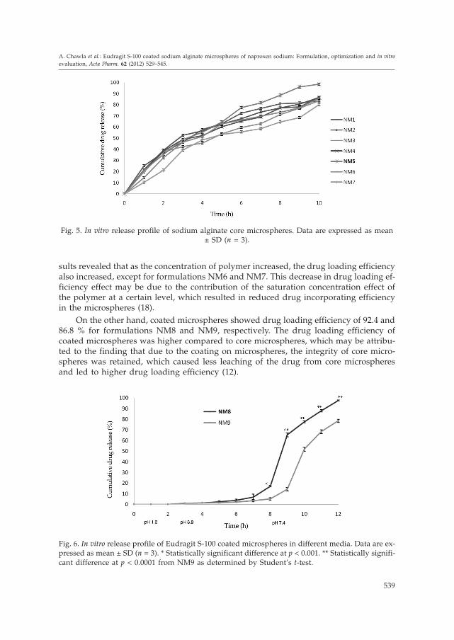
sults revealed that as the concentration of polymer increased, the drug loading efficiencyalso increased, except for formulations NM6 and NM7. This decrease in drug loading ef-ficiency effect may be due to the contribution of the saturation concentration effect ofthe polymer at a certain level, which resulted in reduced drug incorporating efficiencyin the microspheres (18).
On the other hand, coated microspheres showed drug loading efficiency of 92.4 and86.8 % for formulations NM8 and NM9, respectively. The drug loading efficiency ofcoated microspheres was higher compared to core microspheres, which may be attribu-ted to the finding that due to the coating on microspheres, the integrity of core micro-spheres was retained, which caused less leaching of the drug from core microspheresand led to higher drug loading efficiency (12).
539
A. Chawla et al.: Eudragit S-100 coated sodium alginate microspheres of naproxen sodium: Formulation, optimization and in vitroevaluation, Acta Pharm. 62 (2012) 529–545.
Fig. 5. In vitro release profile of sodium alginate core microspheres. Data are expressed as mean± SD (n = 3).
Fig. 6. In vitro release profile of Eudragit S-100 coated microspheres in different media. Data are ex-pressed as mean ± SD (n = 3). * Statistically significant difference at p < 0.001. ** Statistically signifi-cant difference at p < 0.0001 from NM9 as determined by Student’s t-test.

The in vitro release study of naproxen sodium tablets in different dissolution media,i.e., pH 1.2, phosphate buffer, pH 6.8 and phosphate buffer pH 7.4, revealed that drug re-lease from the tablet was observed faster in pH 1.2, followed by pH 6.8 and pH 7.4. Thenaproxen sodium tablet showed total release within 7 h. However, the release from so-dium alginate microspheres was slow and constant compared to naproxen sodium tab-lets. Optimized formulation of sodium alginate microspheres showed maximal drug re-lease of 98.42 % in 10 h. It also resulted in lower release in pH 1.2 and pH 6.8 comparedto the naproxen sodium tablet. Therefore, these findings suggest that sodium alginateshowed complete drug release within 10 h, which is insufficient for targeting the drug tothe colon. Hence, this indicates that there is a need for enteric coating, i.e., Eudragit S-100coating, on sodium alginate microspheres to target drug release to the colon.
The in vitro drug release profile of different core microsphere formulations contain-ing different drug/sodium alginate ratios is shown in Fig. 5. With an increase in poly-mer concentration, the drug release rate of microspheres was found to decrease, whichmight be due to the formation of a rigid polymer matrix at higher polymer concentra-tion in the microspheres. This result may be due to the increase in sodium alginate con-centration leading to the formation of relatively strong walls of microspheres, andthereby retardation of drug release. Further, the increase in polymer chain entanglementand subsequent low dissolution of the polymer might be responsible for the retardationof drug release from the microspheres (19). Core microsphere formulation NM5 showedmaximal drug release at pH 7.4 (98.42 %) compared to other core microsphere formula-tions. On the basis of drug release, NM5 (drug/polymer ratio 1:6) was selected for thepreparation of Eudragit S-100 coated microspheres.
The in vitro release study (Fig. 6) of Eudragit S-100 coated microspheres revealedthat drug release could be successfully retarded in an acidic environment, i.e., pH 1.2.Drug release at pH 6.8 was found to be negligible due to Eudragit S-100 (as enteric co-polymer methacrylic acid-methyl methacrylate) soluble at pH > 7. In release mediumpH 7.4, the NM9 microsphere showed about 4 % drug release over a period of 2 h com-pared to 7 % drug release of NM8. The amount of coating in NM9 was larger, resultingin more time to dissolve and release the drug from the microsphere. Formulation NM8showed a statistically significant (p < 0.001) difference in drug release characteristicscompared to formulation NM9, as determined by Student’s t-test. However, formulationNM8 having a lower concentration of Eudragit S-100 coating (2.5 %, m/V) showedhigher drug release, i.e., 95.6 % within a period of 5 h at pH 7.4, whereas formulationNM9 showed lower drug release (89.5 %) over 5 h at pH 7.4 due to thicker coating. Itwas observed in a previous study that the in vitro drug release of naproxen coated withEudragit L-100 showed approximately 90 % drug release, which is lower compared tothe present study (20).
The percentage drug release profiles obtained from in vitro release experiments weresubjected to different kinetics models to find the drug release mechanism and kinetics.The zero order (cumulative amount of drug release vs. time), first order (log cumulativepercentage of drug remaining vs. time) (21), and Higuchi kinetics (cumulative percent-age of drug release vs. square root of time) (22) were used as the kinetics model. Also,the Korsmeyer-Peppas equation (log cumulative percentage of drug released vs. log ti-me) (23) was used for the first 60 % of drug released from the microsphere. Regressioncoefficients (R2) for all microsphere formulations using different kinetics equations are
540
A. Chawla et al.: Eudragit S-100 coated sodium alginate microspheres of naproxen sodium: Formulation, optimization and in vitroevaluation, Acta Pharm. 62 (2012) 529–545.

listed in Table II. The table data reveal that in vitro release from the core microsphere (so-dium alginate microsphere) was better explained by the Higuchi equation, since theplots provide the highest linearity. For all sodium alginate microspheres, the n value asper Korsmeyer-Peppas model was found to be between 0.45 and 0.89, indicating anoma-lous release behaviour of the drug, i.e., both diffusion and dissolution of the hydratedpolymer matrix might be responsible for drug release from the microsphere (24, 25).Coated microspheres followed Fickian kinetics with the value n < 0.45 as per the Kors-meyer-Peppas model. This could be due to relaxation of the polymer matrix, followedby the diffusion matrix.
Determinations of in vitro mucoadhesion suggest that complete detachment of themicrosphere from mucosal tissue took 90 min, indicating good mucoadhesive propertiesof sodium alginate. To provide better mucoadhesion, the polymer should show the pres-ence of a strong hydrogen bonding group like –OH, –COOH and strong anionic chargesspreading on the mucus (26).
Sodium alginate is in the first-generation mucoadhesive polymer group, which con-tains higher hydrogen bond forming polymers (27). The presence of a large number ofhydroxyl groups, which form a complex of hydrogen bonds with the hydroxyl grouppresent in mucin, will result in mucoadhesion. However, it has been already reportedthat the time required to completely wash off the NSAID agents like valdecoxib contain-ing sodium alginate microspheres is 95 min compared to similarly prepared microsphe-res of naproxen sodium containing alginate (90 min).
All the formulations under storage were analysed for drug content and no signi-ficant differences in drug content were observed. All the formulations showed lessthan 90 % drug content, except formulation NM8 which showed 92.18 % drug contentunder both accelerated and room temperature storage conditions (Fig. 7). The degrada-
541
A. Chawla et al.: Eudragit S-100 coated sodium alginate microspheres of naproxen sodium: Formulation, optimization and in vitroevaluation, Acta Pharm. 62 (2012) 529–545.
Fig. 7. Stability of naproxen sodium microspheres under accelerated conditions and at room tem-perature (mean ± SD, n = 3). AT – accelerated temperature (40 °C), RT – room temperature, D – days,M – months.

tion rate constants (kcal) and shelf life (t90) of NM8 at room temperature were 2.46 day–1
and 795.96 days, respectively, which indicates that formulation NM8 has a shelf life ofmore than 2 years.
CONCLUSIONS
Data obtained from this study demonstrate that sodium alginate microspheres con-taining naproxen sodium were successfully prepared, followed by coating with the pH--sensitive polymer Eudragit S-100. The study also concluded that naproxen sodium tab-lets showed total release in 7 h in comparison with sodium alginate microspheres (coremicrospheres) that showed complete drug release within 10 h. This suggests that bothnaproxen sodium tables and sodium alginate microspheres were unable to target drugrelease into the colonic region. Hence, this indicates that there is a need for enteric coat-ing, i.e., Eudragit S100 coating, on sodium alginate microspheres to target drug releaseto the colon. Coated microspheres showed a longer residence time in the colon after re-moving the Eudragit S100 coating due to better mucoadhesion properties of sodiumalginate. The in vitro release profile revealed that microspheres retard drug release in theupper part of GIT due to the pH-sensitive polymer coating. Hence, Eudragit S-100 sho-wed promising drug delivery to the colon.
Acknowledgements. – The authors thank Micro Labs Pvt. Ltd., Bangalore, India, and Evonik In-dustries Mumbai, India, for providing naproxen sodium and Eudragit S-100, respectively. The au-thors are also grateful to Dr. Madhu Chitkara, Vice Chancellor, Chitkara University, Rajpura, Pun-jab, for financial and infrastructure support to the project.
542
A. Chawla et al.: Eudragit S-100 coated sodium alginate microspheres of naproxen sodium: Formulation, optimization and in vitroevaluation, Acta Pharm. 62 (2012) 529–545.
Table II. Comparison of different dissolution kinetics models
Formulationcode
Zero-order First-order Higuchi Korsmeyer-Peppas
R2 R2 R2 R2 N
NM 1 0.892 0.986 0.990 0.979 0.50
NM 2 0.896 0.986 0.990 0.976 0.71
NM 3 0.950 0.884 0.988 0.978 0.56
NM 4 0.916 0.976 0.993 0.970 0.67
NM 5 0.951 0.956 0.987 0.984 0.54
NM 6 0.919 0.977 0.980 0.975 0.89
NM 7 0.934 0.957 0.959 0.951 0.92
NM 8 0.949 0.930 0.949 0.958 0.36
NM 9 0.954 0.955 0.956 0.953 0.39
REFERENCES
1. P. R. Veerareddy and R. P. Manthri, Formulation and evaluation of compression coated piro-xicam tablets for colon specific drug delivery, Acta Pharm. Sci. 52 (2010) 281–294.
2. G. V. Mooter, Colon drug delivery, Exp. Opin. Drug Deliv. 3 (2006) 111–125; DOI: 10.1517/17425247.3.1.111.
3. K. V. V. Kumar, T. Sivakumar and T. T. Mani, Colon targeting drug delivery system: A reviewon recent approaches, Int. J. Pharm. Biomed. Sci. 2 (2011) 11–19.
4. M. K. Chourasia and S. K. Jain, Pharmaceutical approaches to colon targeted drug delivery sys-tems, J. Pharm. Pharm. Sci. 6 (2003) 33–66.
5. S. Jose, M. T. Prema A. J. Chacko, A. C. Thomas and E. B. Souto, Colon specific chitosan micro-sphere for chronotherapy of chronic stable angina, Colloids Surf. B Biointerfaces 83 (2011) 277–283; DOI: 10.1016/j.colsurfb.2010.11.033.
6. P. Ricchi, R. Zarrilli, A. Palma and A. Acquaviva, Nonsteroidal anti-inflammatory drugs in co-lorectal cancer: from prevention to therapy, Br. J. Cancer 88 (2003) 803–807; DOI: 10.1038/sj.bjc.6600829
7. M. J. Thun, J. Henley and C. Patrono, Nonsteroidal anti-inflammatory drugs as anticanceragents: mechanistic, pharmacologic, and clinical issues, J. Natl. Cancer Inst. 94 (2002) 252–266;DOI: 10.1093/jnci/94.4.252.
8. H. Berkel, R. F. Holcombe, M. Middlebrooks and K. Kannan, Nonsteroidal antiinflammatorydrugs and colorectal cancer, Epidemiol. Rev. 18 (1996) 205–217.
9. M. George and T. E. Abraham, Polyionic hydrocolloids for the intestinal delivery of proteindrugs: alginate and chitosan-a review, J. Control. Release 114 (2006) 1–14; DOI: org/10.1016/j.jconrel.2006.04.017.
10. W. M Obeidat and J. C Price, Preparation and evaluation of Eudragit S 100 microspheres aspH-sensitive release preparations for piroxicam and theophylline using the emulsion-solventevaporation method, J. Microencapsul. 23 (2006) 195–202; DOI: 10.1080/02652040500435337.
11. N. K. Thakral, A. R. Ray, D. B. Shalom, A. H. Eriksson and D. K. Majumdar, The quest for tar-geted delivery in colon cancer: mucoadhesive valdecoxib microspheres, Int. J. Nanomed. 6 (2011)1057–1068; DOI: 10.2147/IJN.S19561.
12. Z. Rahman, K. Kohli, R. K. Khar, M. Ali, N. A. Charoo and A. A. A. Shamsher, Characterizationof 5-fluorouracil microsphere for colonic delivery, AAPS PharmSciTech. 7 (2006) E1-9; DOI: 10.1208/pt070247.
13. K. Ganguly, T. M. Aminabhavi and A. R. Kulkarni, Colon targeting of 5-fluorouracil using poly-ethylene glycol cross-linked chitosan microspheres enteric coated with cellulose acetate phtha-late, Ind. Eng. Chem. Res. 50 (2011) 11797–11807; DOI: 10.1021/ie201623d.
14. S. K. Umadevi, R. Thiruganesha, S. Suresha and K. B. Reddya, Formulation and evaluation ofchitosan microspheres of aceclofenac for colon-targeted drug delivery, Biopharm. Drug Dispos.31 (2010) 407–427; DOI: 10.1002/bdd.722.
15. C. M. Lehr, J. M. Bowstra, J. J. Tukker and H. E. Junginger, Intestinal transit of bioadhesive mi-crospheres in an in situ loop in the rat, J. Control. Release 13 (1990) 51–62; DOI: org/10.1016/0168-3659(90)90074-4.
16. J. P. Soarse, J. E. Santos, J. O. Chierice and E. T. G. Cavalheiro, Thermal behavior of alginic acidand its sodium salt, Ecl. Quím. Sao Paulo 29 (2004) 53–56.
17. M. S. Crcarevska, M. G. Dodov and K. Goracinova, Chitosan coated Ca–alginate microparticlesloaded with budesonide for delivery to the inflamed colonic mucosa, Eur. J. Pharm. Biopharm. 68(2008) 565–578; DOI: org/10.1016/j.ejpb.2007.06.007.
543
A. Chawla et al.: Eudragit S-100 coated sodium alginate microspheres of naproxen sodium: Formulation, optimization and in vitroevaluation, Acta Pharm. 62 (2012) 529–545.

18. A. Vaidya, A. Jain, P. Khare, R. K. Agrawal and S. K. Jain, Metronidazole loaded pectin micro-spheres for colon targeting, J. Pharm. Sci. 98 (2009) 4229–4236; DOI: 10.1002/jps.21742.
19. P. Kumar and I. Singh, Formulation and characterization of tramadol loaded IPN microgels ofalginate and gelatin: optimization using response surface methodology, Acta Pharm. 60 (2010)295–310; DOI: 10.2478/v10007-010-0021-z.
20. M. Maghsoodi, Physicomechanical properties of naproxen-loaded microparticles preparedfrom eudragit L100, AAPS PharmSciTech. 10 (2009) 120–128; DOI: 10.1208/s12249-009-9186-5.
21. J. G. Wagner, Interpretation of percent dissolved-time plots derived from in vitro testing of con-ventional tablets and capsules, J. Pharm. Sci. 58 (1969) 1253–1257; DOI: 10.1002/jps.2600581021.
22. T. Higuchi, Mechanism of sustained-action medication; Theoretical analysis of rate of release ofsolid drugs dispersed in solid matrices, J. Pharm. Sci. 52 (1963) 1145–1149; DOI: 10.1002/jps.2600521210.
23. P. Costa and J. M. S. Lobo, Modeling and comparison of dissolution profiles, Eur. J. Pharm. Bio-pharm. 13 (2001) 123–133.
24. N. A. Peppas, Analysis of fickian and non-fickian drug release from polymers, Pharm. Acta Helv.60 (1985) 110–111.
25. H. R. Chueh, H. Zia and C. P. Rhodes, Optimization of sotalol floating and bioadhesive exten-ded release tablet formulations, Drug Dev. Ind. Pharm. 21 (1995) 1725–1747; DOI: 10.3109/03639049509069261.
26. N. A. Peppas and P. A. Burim, Surface, interfacial and molecular aspects of polymer bioadhe-sion on soft tissues, J. Control. Release 2 (1985) 257–275; DOI: 10.1016/0168-3659(85)90050-1.
27. J. D. Smart, I. W Kellaway and H. E. C. Worthington, An in-vitro investigation of mucosa-adhe-sive materials for use in controlled drug delivery, J. Pharm. Pharmacol. 36 (1984) 295–299; DOI:10.1111/j.2042-7158.1984.tb04377.
S A @ E T A K
Alginatne mikrosfere naproksen natrija oblo`ene Eudragitom S-100:Priprava, optimizacija i in vitro vrednovanje
ANUJ CHAWLA, POOJA SHARMA i PRAVIN PAWAR
Cilj istra`ivanja bila je ciljana isporuka naproksen natrija koriste}i natrijev alginat iEudragit S-100 kao mukoadhezivne, odnosno pH-osjetljive polimere. Jezgra mikrosferaod alginata pripravljena je modificiranom metodom emulgiranja te umre`avanjem po-mo}u otopine CaCl2. Sljede}i korak u pripravi mikrosfera bilo je oblaganje s pH osjetlji-vim polimerom Eudragit S-100 (2,5 ili 5 %) ~ime je sprije~eno osloba|anje lijeka u gornjemdijelu gastrointestinalnog trakta. Mikrosfere su okarakterizirane FT-IR spektroskopijom,difrakcijom rendgenskih zraka, diferencijalnom pretra`nom kalorimetrijom i pretra`nomelektronskom mikroskopijom. Nadalje, analizirana je veli~ina ~estica, koli~ina ukloplje-nog lijeka, mukoadhezivna svojstva in vitro te osloba|anje lijeka in vitro u razli~itim si-muliranim gastri~nim fluidima. Testovi stabilnosti optimiziranih pripravaka pra}eni sutijekom 6 mjeseci prema smjernicama ICH. SEM snimke otkrile su da je povr{ina jezgremikro~estica neravna, dok je povr{ina oblo`enih mikrosfera glatka. Jezgre mikrosfera imalesu ja~e izra`ena mukoadhezivna svojstva nego oblo`ene mikrosfere na testovima prove-
544
A. Chawla et al.: Eudragit S-100 coated sodium alginate microspheres of naproxen sodium: Formulation, optimization and in vitroevaluation, Acta Pharm. 62 (2012) 529–545.

denim na svje`e izrezanim dijelovima debelog crijeva koze. Iz optimiziranih neoblo`enih ioblo`enih mikrosfera osloba|a se 98.42 ± 0.96, odnosno 95.58 ± 0.74 % lijeka. Osloba|anjelijeka iz svih formulacija slijedilo je kinetiku po Higuchiju. Osloba|anje iz oblo`enih mi-krosfera slijedilo je Korsmeyer-Peppasovu jednad`bu i kinetiku po Ficku. Studije stabil-nosti pokazale su minimalnu razgradnju, te prihvatljivu stabilnost tijekom dvogodi{njegskladi{tenja.
Keywords: specifi~na isporuka u kolonu, Eudragit S-100, mikrosfere, naproksen natrij, natrijev alginat
Chitkara College of Pharmacy, Chandigarh-Patiala National Highway, Rajpura-140401, Patiala, PunjabIndia
545
A. Chawla et al.: Eudragit S-100 coated sodium alginate microspheres of naproxen sodium: Formulation, optimization and in vitroevaluation, Acta Pharm. 62 (2012) 529–545.

546
A. Chawla et al.: Eudragit S-100 coated sodium alginate microspheres of naproxen sodium: Formulation, optimization and in vitroevaluation, Acta Pharm. 62 (2012) 529–545.